malignant connective tissue tumors
TRANSCRIPT
Cleidocranial dysplasia

Etiology
a rare congenital disorder of bone with
an autosomal dominant hereditary
mode of inheritance with complete
penetrance, but variable expressivity

caused by mutation in the CBFA1gene
mapped on chromosome 6p21
encoding transcription factor RUNX2
responsible for osteoblast
differentiation

Clinical features
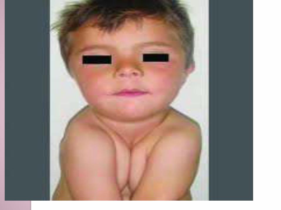
Moderately short stature, and
They tend to have a short head from
front to back (brachycephaly) and
a prominent forehead (frontal bossing).
delayed closure of fontanels, and some
adults with CCD have open fontanels.
The eyes are widely spaced, and
the nasal bridge is often flat.
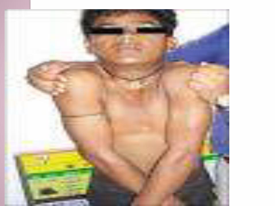
one or both clavicles are frequently
partially or in 10% cases completely
absent.
The neck appears long, and the
shoulders are narrow and down-
sloping
hypermobility of shoulders with
tendency to approximate shoulders
anteriorly

Oral manifestations
prolonged retention of deciduous
dentition and delayed eruption of
permanent teeth.
Adults have mixed dentition in their
oral cavities.
show a large number of unerupted
supernumerary teeth, often mimicking
a premolar.
Delayed and imperfect ossification of
the cranium,
Maxilla is also underdeveloped along
with ill-formed paranasal sinuses.
Skeletal Class III tendency /
mandibular prognathism in CCD->
uninterfered growth due to hypoplastic
maxilla and upward and forward
mandibular rotation.

Treatment
No specific treatment suggested


Cherubism
Etiology
SH3-binding protein SH3BP2 mapped
to 4p16.3, an important molecule in
cell signaling
There is bone loss followed by the
accumulation of fibrous tissue that
causes facial swelling especially
around the cheeks, hence the
disease's name.
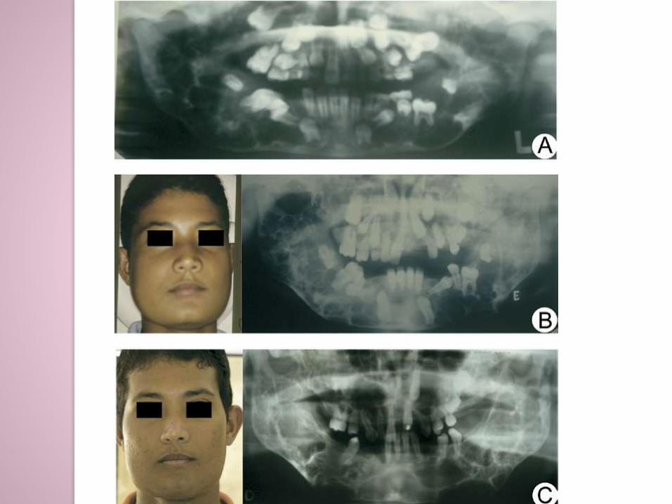
Clinical features
Age: in children
Sex: M=F
Site: Mand>max

severe bone degradation of both the
upper and lower jaws beginning at
about age three.
In adult life, complete involution of the
lesions

In some cases, the enlargement of the
floor of the orbit (the bones
surrounding the eye socket) causes
the eyeball to tip upward.
The name derived from cherub
(angelic looking, as depicted in
Renaissance paintings)

Oral manifestations
Abnormal patterns of teeth eruption
teeth agenesis and the presence of
ectopic or retained teeth.
X-ray
multilocular radiolucencies->near the
angle of the mandible and spreading
to the mandibular ramus and body.

HP
The histology is limited for diagnosis, showing fibrous hyperplasia and multinucleated giant cells.
similar to those other bone diseases such as
brown tumor of hyperparathyroidism,
giant cell tumor, and
central and peripheral giant cell granuloma
Lab diagnosis
the bone markers
phosphorous,
serum calcium, and
alkaline phosphatase are usually
at normal levels with respect to age.
cherubism may be associated with
other genetic diseases, such as
Noonan’s syndrome and Ramon
syndrome.
Treatment
multinucleated cells in cherubic
lesions ->osteoclasts from a structural
and biochemical standpoint so
calcitonin is the treatment of choice.
Pagets disease of bone
Pathogenesis
Paget disease occurs when there is a disturbance in the bone remodeling
that characteristically begins with unwarranted bone resorption followed by an increase in bone formation.
This osteoclastic hyper activity followed by substituted osteoblastic activity leads to the formation of a structurally disordered mosaic of bone which is still a woven bone,
and which is mechanically larger, weaker, more vascular, less compact, and more prone to fracture than normal adult lamellar bone
Slow virus:
paramyxovirus
respiratory synctial virus
Sequestrome 1 gene on chromosome
5.

Clinical features
Age: occurs in the aging skeleton
Sex:M=F
Site skull, spine, pelvis, and long
bones of the lower extremity.

Presentation
The bone pain is dull, constant,
boring, and deep below the soft
tissues.
It may persist or exacerbate during the
night.
Pathologic fractures commonly result
from weakened pagetic bone.
Nonspecific headaches,
impaired hearing, and tinnitus commonly result from skull involvement and compression of the 8th cranial nerve
The patient's hat size may increase (or, less commonly, decrease) as a result of skull enlargement or deformity.
These may manifest as nausea, dizziness, syncope, ataxia, incontinence, gait disturbances, or dementia.

3 phases:
Lytic
Mixed
Sclerotic
Oral manifestations
Facial disfigurement and
malocclusion may be observed
following enlargement of the maxilla or
mandible.
Tooth loss may occur with progressive
root resorption.

X-Ray
Absent periodontal membranes and
lamina dura are associated with
hypercementosis.
Both osteolysis (seen as radiolucency) and excessive bone formation(radiopacity) and a mixed state occurs.
There are specific X-ray features of Paget's disease that include:◦ A classical V-shaped pattern between healthy
and diseased long bones known as 'the blade of grass' lesion.
◦ The 'cotton wool' pattern in the skull that is also characteristic (multifocal sclerotic patches).-osteitis circumscripta
HP
woven bone and irregular broad trabeculae with disorganized cement lines in a mosaic pattern. Woven bone seperated by reversal lines
fibrous vascular tissue interspersed between trabeculae
profound bone resorption - numerous large osteoclasts with multiple nuclei per cell◦ virus-like inclusion bodies in osteoclasts
◦ Paget's osteoclasts larger, more nuclei than typical osteoclasts
Multinucleated giant cells-3-30 as opposed to 1-3 in normal osteoclasts
Treatment
NSAIDs
Bisphophonates
Calcitonin
Surgery

Lab investigations
Bone-specific alkaline phosphatase
(BSAP) levels are raised.
Urine hydroxyproline ↑.
Serum calcium, phosphorus, and
parathyroid hormone levels are
usually normal but immobilization may
lead to hypercalcaemia.