malaysian journal of - dermatology.org.my jul 2015.pdf · malaysian journal of dermatology mjd 2015...
TRANSCRIPT


Malaysian Journal of Dermatology
MJD 2015 Jul Vol 34
The Malaysian Journal of Dermatology welcomes manuscripts on all aspects of cutaneous medicine and surgery in the form of original articles, research papers, case reports and correspondence. Contributions are accepted for publication on condition that they are submitted exclusively to the Malaysian Journal of Dermatology. The Publisher and Editors cannot be held responsible for errors or any consequences arising from the use of information contained in this journal; the views and opinions expressed do not necessarily reflect those of the publisher and Editors, neither does the publication of advertisements constitute any endorsement by the publisher.
Manuscripts should be submitted via email:[email protected]
Questions regarding the Malaysian Journal of Dermatology can be sent to me at:[email protected]
Contributions should be written for one of the following categories:
Case Report*A report of 400-600 words, illustrated by no more than three illustrations. This category offers a means for rapid communication about a single subject.
Clinical TrialAn article of 700-1200 words concerning a drug evaluation. This category provides rapid publications and is meant to be a succinct presentation with a minimum of graphs and tables.
Commentary*An editorial 700-1200 words in length with approximately five references. The author may express his or her opinion without complete documentation.
Clinicopathological ChallengeA photographic essay that includes both clinical and pathological photographs in color. The diagnosis and legends for the photographs should be listed after the references in the article. The article should be no more than 2-3 pages in length.
Correspondence*Letters to the editor and short notes. Contributions should not exceed 600 words, two figures, and 10 references.
Dermatological Surgery An article relating to the surgical aspects of treatment. Article types may include Review, Report or Case Report Format.
Original ArticleAn original article including, whenever possible, an Introduction, Materials and Methods , Results, Comment and References. A Structured Abstract of not more than 240 words must be included. It should consist of four paragraphs, labelled Background, Methods, Results, and Conclusions. It should describe the problem studies, how the study was performed, the main results, and what the author(s) concluded from the results.
ReviewBy invitation only. A major didactic article that clarifies and summarizes the existing knowledge in a particular field. It should not be an exhaustive review of the literature, and references should not exceed 100 in number. Tables, diagrams, and selected figures are often helpful. The length is left to the judgment of the author, although it generally should not exceed 5000 words. Topics may include updates in clinically relevant basic science and cutaneous biology.
*No abstract required
Manuscripts should include a title page bearing the title of the paper, the author(s)’ name(s), degrees, and affiliation(s), the category of the article, the number of figures and tables, and three key words for indexing purposes. The name and full postal address (including a street address), phone and fax numbers and an email address of the corresponding author who will be responsible for reading the proofs must also be given on the title page. The author(s) must also declare any affiliation or significant financial involvement in any organizations or entity with a direct financial interest in the subject matter or materials discussed in the manuscript on this page.
All measurements should be according to the metric system. If confusion could result, please include other measurement systems in parentheses.
Refer to patients by number or letters; names or initials should not be used.
References must be listed in the order in which they appear in the manuscript. References from journals should include: (1) name(s) followed by the initials of the author(s), up to four authors: if more than four authors, include the first three authors followed by et al.; (2) title of paper; (3) title of the journal as abbreviated in the Index Medicus; (4) year of publication; (5) volume number; (6) first and final page numbers of the article.
For example:Ambrose D, Gangaram HB, Hussein SH. Sporotrichosis: A Hospital Kuala Lumpur experience. M J Dermatol 2006;19:52-55.
References to books should include: (1) author(s) or editor(s); (2) chapter (if any) book titles; (3) edition, volume, etc.; (4) place of publication; (5) publisher; (6) year; (7) page(s) referred to.
For example: Foong HBB. Transcontinental Dermatology: Virtual Grand Rounds. In: Wootton R and Oakley A, editors. Teledermatology. London. Royal Society of Medicine 2002. p.127-134.
The author is responsible for the accuracy and completeness of all references; incomplete references may result in a delay to publication.
Tables should be typed, double-spaced with a heading, each on a separate sheet, and should only include essential information. Drawings, graphs, and formulas should be submitted on separate pages.
Send illustrations as tiff or jpeg files. In the case of photomicrographs, the stain type and original magnification should be stated. Each figure should bear a reference number corresponding to a similar number in the text.
To minimise the publication time of your manuscript it is important that all electronic artwork is supplied to the Editorial Office in the correct format and resolution.
DisclaimerThe Publisher and Editors cannot be held responsible for errors or any consequences arising from the use of information contained in this journal; the views and opinions expressed do not necessarily reflect those of the publisher and Editors, neither does the publication of advertisements constitute any endorsement by the publisher and Editors of the products advertised.
Notice to Authors

Malaysian Journal of Dermatology
MJD 2015 Jul Vol 34
GENERAL DERMATOLOGY
ORIGINAL ARTICLE2 Epidemiology and clinical features of paediatric patients with psoriasis in Malaysia: Evidence from the Malaysian psoriasis registry (2007-2012) Azura MA, Fatimah AA, Asmah J, Roshidah B
QUIZ17 Multiple vesicular-like papules and plaques Yap FBB
CASE REPORT11 Adherence to acne medication and its original relation to acne severity and quality of life Tan CL, Yang SS, Toh MPH, Aw DC
20 Epidermodysplasia verruciformis in a pair of siblings Ling HN, Tagal JM, Lee BR, Leong KF
23 Verrucous haemangioma in a 10-year-old girl Lee S, Mohd Shariman MS, Teoh TZ, Choon SE
26 Hypohydrotic ectodermal dysplasia: A case series Visuvanathan VV, Najeeb AMS
29 Gorlin syndrome: A case report with clinical and radiological correlation Azura MA, Izzaty D
ContentsM A L A Y S I A N J O U R N A L O F D E R M A T O L O G Y
Malaysian Journal of Dermatology
1MJD 2015 Jul Vol 34
Editor-in-Chief Associate Professor Dr Felix Yap Boon Bin MRCP Adv MDermUniversiti Tunku Abdul Rahman
Founding EditorDr Steven Chow Kim WengFRCPIKuala Lumpur
Editorial OfficeMalaysian Dermatological Society Rumah DermatolgyG1, Medical Academics of Malaysia210, Jalan Tun Razak50400 Kuala Lumpur, Malaysia
Editorial BoardDr Henry Foong Boon Bee FRCPIpoh, Perak
Dr Chan Lee Chin MMed, Penang
Dr Ng Ting Guan MRCP AdvMDermKlang, Selangor
Dr Adawiyah Jamil MMed AdvMDermKuala Lumpur
Dr Tang Jyh Jong MRCP AdvMDermIpoh, Perak
Dr Tarita Taib AdvMDermSelayang, Selangor
Dermatological Society of Malaysia | Persatuan Dermatologi Malaysia
Executive StaffHenry Foong Boon Bee, FRCP - PresidentNajeed Ahmad Safdar, MRCP - Past PresidentAgnes Heng Yoke Hui, MRCP - Vice PresidentRohna Ridzwan, MRCP - SecretaryNoor Zalmy Azizan, AdvMDerm - TreasurerChan Lee Chin, MMedKhor Guat Ee, MRCPSabeera Begum, MMedTan Wooi Chiang, AdvMDerm
Dermatological Society of MalaysiaRumah DermatolgyG1, Medical Academics of Malaysia210, Jalan Tun Razak50400 Kuala Lumpur, Malaysia
Published by Dermatological Society of Malaysia twice a year from year 2009 (July and December issues)
Printed by Percetakan Sri Jaya, No.27, Jalan Emas SD 5/1A, Bandar Sri Damansara, 52200 Kuala LumpurTel : 03-6276 4082 Fax : 03-6275 9514
®2014 Persatuan Dermatologi Malaysia. All rights reserved.No part of this journal can be reproduced without the written permission from editorial board.

Malaysian Journal of Dermatology
2 MJD 2015 Jul Vol 34
GENERAL DERMATOLOGY - Original Article
EPIDEMIOLOGY AND CLINICAL FEATURES OF PAEDIATRIC PATIENTS WITH PSORIASIS IN MALAYSIA: EVIDENCE FROM THE MALAYSIAN PSORIASIS REGISTRY (2007-2012)Azura MA1, Fatimah AA1, Asmah J1, Roshidah B2
Abstract
Background: Psoriasis is a common dermatological condition affecting both adults and children. It causes significant physical and psychological burden on patients and adversely affect their quality of life.
Aim: To evaluate the clinical characteristics of paediatric patients with psoriasis in Malaysia.
Materials & Methods: Data were obtained from the Malaysian Psoriasis Registry (MPR). All paediatric patients aged <18 years notified to the registry from July 2007 to December 2012 were included in this study.
Results: A total of 677 patients were notified from 18 participating centres. There was a slight female preponderance (ratio 1.3:1). Malay accounted for 70.6%, followed by Chinese (8.9%), Indian (12.3%) and others (8.1%). Mean age of onset was 9.8 ± 4.4 years. Positive family history was noted in 19.1%. Plaque psoriasis was the commonest type of psoriasis (79.6%), followed by guttate psoriasis (7.4%), pustular psoriasis (1.6%), erythrodermic (1.2%) and flexural psoriasis (1.2%). Psoriatic arthropathy was reported in only 2.2% of patients. Nail involvement is common, affecting 38.1%. Pitting was the commonest (89.9%). Topical treatment remains the most popular choice of treatment and was given in 95.1% of our patients. Topical steroid was the commonest prescribed (81.4%), followed by tar preparations (78.7%) and emollients (51.6%). Only 1.2% of our patients received phototherapy. Of the patients who had phototherapy, narrowband UVB (NBUVB) was the commonest used (87.5%). Systemic therapy was given in 5.3% of paediatric patients. The most frequently used systemic therapy was methotrexate (50%) and acitretin (27.8%). The mean CDLQI score for paediatric patients with psoriasis was 7.7 ± 5.5.
Conclusion: Data from the Malaysian Psoriasis Registry highlights the clinical features of paediatric patients with psoriasis in Malaysia. We hope to get more participation from other centres in the future, especially from private sectors, so that our results can represent the Malaysian data more accurately.
Keywords: psoriasis, paediatric, epidemiology
Corresponding Author and Reprint Request Dr Azura Mohd Affandi, MBChB (UK), MRCP (UK), Adv M Derm (UKM)Department of Dermatology, Hospital Kuala Lumpur, Jalan Pahang, 50586 Kuala Lumpur, Malaysia Email: [email protected]
1 Department of Dermatology, Hospital Kuala Lumpur2 Department of Dermatology, Hospital Melaka
IntroductionPsoriasis is a genetically determined chronic inflammatory disorder affecting the skin, nails and joints. It is characterized by well demarcated, erythematous, scaly plaques. It is common, affecting 1 - 3% of the general population and can affect both adult and children.1 Both genetic and environmental factors play an important role in triggering psoriasis.

Malaysian Journal of Dermatology
3MJD 2015 Jul Vol 34
Little information is available on the prevalence of psoriasis in children. Previous studies found prevalence estimates of paediatric psoriasis ranging from 0.5% to 1.4%.2 Onset during the first 2 decades of life is reported in 31% to 45% of affected adults.3 Although a recent study suggested that childhood onset of psoriasis is not associated with disease severity, early onset may result in longer exposure to a chronic inflammatory condition and, thus, may affect the morbidity and mortality risk.4 It carries a significant physical and psychological burden on patients and adversely affect their quality of life.
Thus, we aim to evaluate the clinical characteristics of paediatric patients with psoriasis in Malaysia.
MethologyThis was a multicenter study involving 18 dermatology out-patient clinics participating in the Malaysian Psoriasis Registry (MPR). The MPR is a prospective, ongoing, systematic collection of data on patients with psoriasis in Malaysia. Confirmation of diagnosis by histopathologic examination is optional. All paediatric patients aged <18 years notified to the registry from July 2007 to December 2012 were included in this study.
Data were collected on the patient’s first visit and every 6 months during follow-up visits. The impact of psoriasis on the quality of life of paediatric patients was determined by using the 10-item Children’s Dermatology Life Quality Index (CDLQI). This CDLQI was designed for the paediatric patients from age 5 to 16 years old. Patients above the age of 16 were assessed using the Dermatology Life Quality Index (DLQI). The CDLQI contains 10 questions which measure how much the skin problem has affected the patients’ life over the last week. Each question has five possible answers (very much, a lot, a little, not at all or not relevant) with scores of 3, 2, 1 or 0 respectively. The total score ranges between 0 and 30. A score of 0-1 means no effect on QoL, 2-5 small effect, 6-10 moderate effect, 11-20 very large effect and 21-30 extremely large effect.
Collected data was tabulated using SPSS. Categorical data was presented as number and percentages whereas continuous data was presented as mean and standard deviation.
Results
Clinical FeaturesThere were a total of 677 paediatric patients notified to the registry between July 2007 and December 2012. Hospital Tengku Ampuan Rahimah, Klang notified the highest number of paediatric patients, followed by Hospital Sultanah Bahiyah and Hospital Kuala Lumpur (Table 1). Majority of the paediatric patients (83.5%) were new cases and 16.5% were follow-up cases. All patients were Malaysians. Malay accounted for 70.6% of the patients, followed by Indian (12.3%), Chinese (8.9%) and other ethnic groups (8.1%). Slightly more than half of the patients were female (56.9%). There was a slight female preponderance, with male-to-female ratio of 1:1.3.
Psoriasis may first appear at any age. The mean age of onset in our cohort of patients was 9.8 ± 4.4 years. Figure 1 illustrates the onset of psoriasis according to different age groups. The mean age at which psoriasis was first diagnosed by clinician was 11.2 ± 4.3 years. Psoriasis is a skin disorder with a polygenic mode of inheritance. In our registry, about one-fifth (19.1%) of patients had at least one family member with psoriasis. Of those with a positive family history, 34.9% had either parents affected and 16.3% had positive family history in their siblings.
At least one or multiple factors caused aggravation of psoriasis in 38.1% of paediatric patients with psoriasis. Stress was the commonest aggravating factor (57.0%), followed by sunlight (45.0%), infection (20.5%) and trauma (9.3%). Drugs aggravating psoriasis were less common and reported in only 1.2% of the patients. Analyzing the subgroup of patients who reported infection as an aggravating factor, upper respiratory tract infection (66.7%) appeared to be the commonest infective trigger. Patients with psoriasis can have a number of other concomitant diseases and co-morbidities. In children and adolescents aged below 18 years with psoriasis, the most prevalent comorbidity was overweight or obesity (BMI ≥ 85th centile), in 27.0 % of patients. Other comorbid conditions were less common.

Malaysian Journal of Dermatology
4 MJD 2015 Jul Vol 34
Table 1. Number of paediatric patients with psoriasis notified from each participating centres.
Figure 1. Age of onset of paediatric patients with psoriasis. Figure 2. Age of onset of paediatric patients with psoriasis.
No
1 Hospital Tengku Ampuan Rahimah
2 Hospital Sultanah Bahiyah
3 Hospital Kuala Lumpur
4 Hospital Tengku Ampuan Afzan
5 Hospital Umum Sarawak
6 Hospital Queen Elizabeth
7 Hospital Melaka
8 Hospital Raja Permaisuri Bainun
9 Hospital Pulau Pinang
10 Hospital Sultanah Fatimah
11 Hospital Sultanah Aminah
12 Hospital Tuanku Fauziah
13 Hospital Tuanku Jaafar
14 Hospital Sungai Buloh
15 Gleneagles Medical Centre
16 UM Medical Centre
17 UKM Medical Centre
18 Hospital Raja Perempuan Zainab II
TOTAL
2007
0
9
10
0
1
1
0
4
0
2
0
1
0
3
0
0
0
0
31
No. of paediatric patients notified
2009
18
15
17
10
12
17
6
11
13
0
10
4
0
1
0
0
0
0
134
2011
17
10
9
20
7
8
19
5
4
7
4
5
8
0
0
2
0
0
125
2008
10
30
21
4
15
8
0
3
8
6
2
8
5
5
4
0
0
0
129
2010
34
11
11
14
9
12
14
2
6
3
5
7
6
0
0
0
1
0
135
2012
10
9
13
17
17
9
13
12
0
12
7
3
0
0
0
0
0
1
123
Total
89
84
81
65
61
55
52
37
31
30
28
28
19
9
4
2
1
1
677
Age
350300250200150100
500
0-5 6-10 11-15 above15
No. o
f pat
ient
Plague
Guttate
Pustular
Reythrodermic
Flexural/inverse
Palmoplantar non-pustular
0% 7% 2%1%1%2%
7%
80%
Malaysian Journal of Dermatology
5MJD 2015 Jul Vol 34
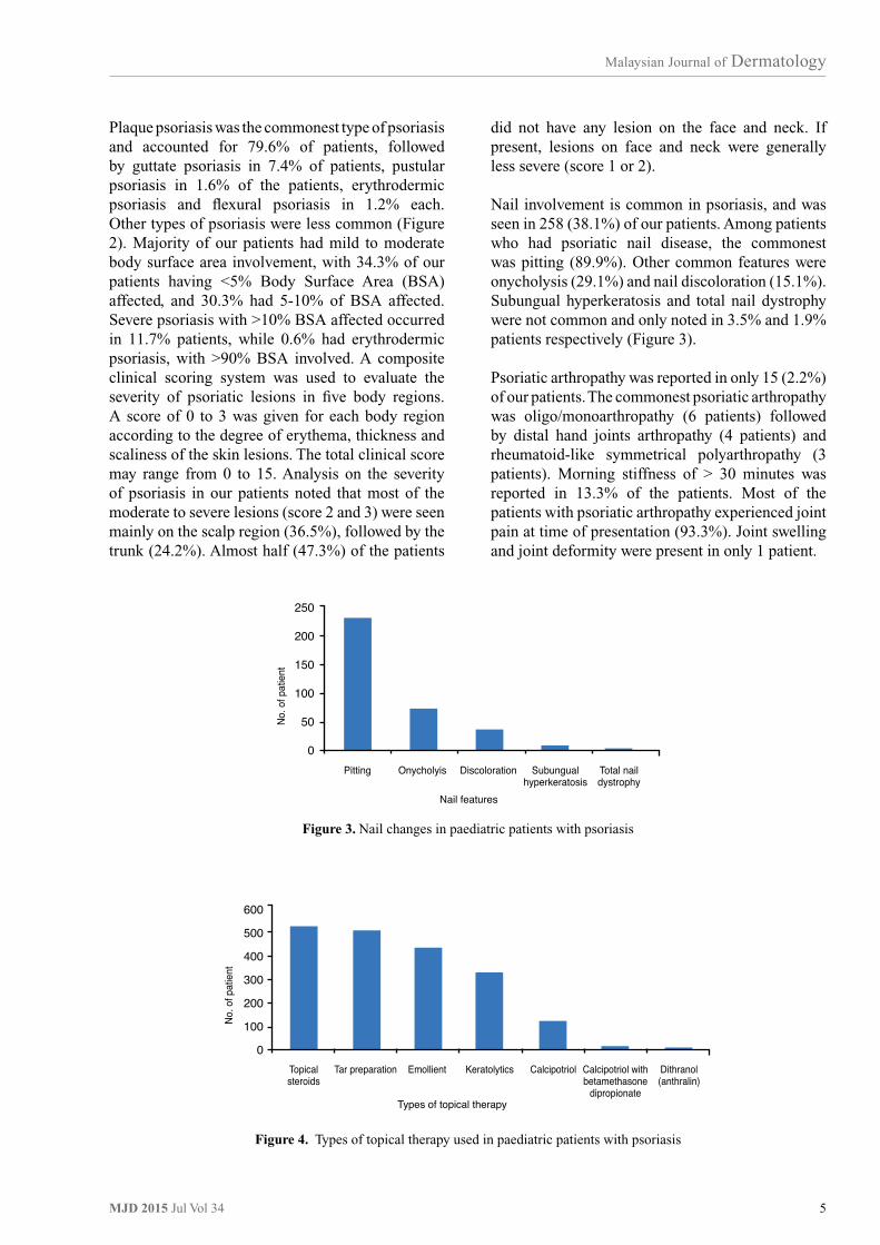
Figure 3. Nail changes in paediatric patients with psoriasis
Figure 4. Types of topical therapy used in paediatric patients with psoriasis
Plaque psoriasis was the commonest type of psoriasis and accounted for 79.6% of patients, followed by guttate psoriasis in 7.4% of patients, pustular psoriasis in 1.6% of the patients, erythrodermic psoriasis and flexural psoriasis in 1.2% each. Other types of psoriasis were less common (Figure 2). Majority of our patients had mild to moderate body surface area involvement, with 34.3% of our patients having <5% Body Surface Area (BSA) affected, and 30.3% had 5-10% of BSA affected. Severe psoriasis with >10% BSA affected occurred in 11.7% patients, while 0.6% had erythrodermic psoriasis, with >90% BSA involved. A composite clinical scoring system was used to evaluate the severity of psoriatic lesions in five body regions. A score of 0 to 3 was given for each body region according to the degree of erythema, thickness and scaliness of the skin lesions. The total clinical score may range from 0 to 15. Analysis on the severity of psoriasis in our patients noted that most of the moderate to severe lesions (score 2 and 3) were seen mainly on the scalp region (36.5%), followed by the trunk (24.2%). Almost half (47.3%) of the patients
did not have any lesion on the face and neck. If present, lesions on face and neck were generally less severe (score 1 or 2).
Nail involvement is common in psoriasis, and was seen in 258 (38.1%) of our patients. Among patients who had psoriatic nail disease, the commonest was pitting (89.9%). Other common features were onycholysis (29.1%) and nail discoloration (15.1%). Subungual hyperkeratosis and total nail dystrophy were not common and only noted in 3.5% and 1.9% patients respectively (Figure 3).
Psoriatic arthropathy was reported in only 15 (2.2%) of our patients. The commonest psoriatic arthropathy was oligo/monoarthropathy (6 patients) followed by distal hand joints arthropathy (4 patients) and rheumatoid-like symmetrical polyarthropathy (3 patients). Morning stiffness of > 30 minutes was reported in 13.3% of the patients. Most of the patients with psoriatic arthropathy experienced joint pain at time of presentation (93.3%). Joint swelling and joint deformity were present in only 1 patient.
250
200
150
100
50
0Pitting Onycholyis Discoloration Subungual
hyperkeratosisTotal naildystrophy
No. o
f pat
ient
Nail features
600
500
400
300
200
100
0Topical steroids
Tar preparation Emollient Keratolytics Calcipotriol Dithranol(anthralin)
Calcipotriol withbetamethasone
dipropionate
No. o
f pat
ient
Types of topical therapy

Malaysian Journal of Dermatology
6 MJD 2015 Jul Vol 34
TreatmentMajority of the patients (95.1%) were on topical treatment. Topical steroid was the commonest treatment prescribed (81.4%), closely followed by tar preparations (78.7%). Both emollients and keratolytics were prescribed in 51.6% and vitamin D analogue, such as calcipotriol were prescribed in 25.9% of patients. Calcipotriol with betamethasone dipropionate and dithranol were least favoured and used in 3.1% and 1.9% % of patients, respectively (Figure 4). In the last six months prior to notification, 1.2% of paediatric patients received phototherapy. Of the patients who had phototherapy, 87.5% had narrowband UVB (NBUVB) and 12.5% had topical PUVA. Systemic therapy was given in 5.3% of the patients. The most frequently used systemic therapy was methotrexate (50%), followed by acitretin (27.8%). Systemic corticosteroids were used in 8.3% patients. Other systemic agents such as suphasalazine, cyclosporine, hydroxyurea and biologics were not prescribed in our paediatric patients.
Quality of lifePsoriasis can have a major psychological impact on the patients and affect their quality of life. Out of 677 paediatric patients with psoriasis, 260 patients were investigated for their quality of life assessment with the validated questionnaire, Children’s Dermatology Life Quality Index (CDLQI). The mean CDLQI score for our patients was 7.7 ± 5.5. A CDLQI of more than 10, indicating very large or extremely large effect on their quality of life (QoL)
was reported in 18.4% of patients, and 4.6% of the patients had CDLQI of more than 20, reflecting extremely large effect on their QoL. On the other hand, 11.9% paediatric patients reported no effect at all on their QoL (Figure 5).
DiscussionPsoriasis is a common inflammatory skin condition, affecting between 1-3% of the population.1 Despite being so common, there are sparse data regarding the incidence of psoriasis in children. A Turkish study estimated the prevalence in children as high as 3.8%.4 A population case study in Minnesotta, USA found the overall age- and sex adjusted annual incidence of pediatric psoriasis to be 40.8 per 100,000, which was considerably lower than the adult incidence of 78.9 per 100,000 population.5-7 There was a slight female preponderance in our patients, with male to female ratio of 1:1.3. This concur with other studies which demonstrated a higher incidence of psoriasis in girls compared to boys.4,8 However, Tollefson et al found that boys and girls were equally affected during childhood.6
Several prevalence studies have demonstrated that approximately one third of patients with psoriasis develop their symptoms sometime during childhood, although some of these may not be diagnosed until adulthood.9 The mean age of onset of psoriasis in our cohort of patients was 9.8 ± 4.4 years. This was lower compared to other studies which reported mean age of onset of psoriasis between 10.6 - 11 years old.6,8
Figure 5. Quality of life in paediatric patients with psoriasis.
No effect at all (0-1)
Small effect (2-6)
Moderate effect (7-12)
Very large effect (13-18)
Extremely large effect (19-30)
cDLQ
I
0 4010 5020 60 8030 70 90 100
Number of patients
Dermatology Life Quality Index (DLQI) in Children with PsoriasisN=260

Malaysian Journal of Dermatology
7MJD 2015 Jul Vol 34
Psoriasis can be aggravated by several factors, including medications such as antimalarials, stress and infections such as streptococcal throat infection.10,11 More than one third of our patients (38.1%) reported one or multiple factors aggravating their psoriasis, in which stress was the commonest, followed by sunlight and upper respiratory tract infection. Drugs aggravating psoriasis were less common and reported in only 1.2% of the patients. Obesity and being overweight has recently been described as a risk factor for psoriasis in the adult population and it is likely that it plays a significant role in children as well.12 Our cohort reported 27% of the children were obese (BMI at or above 85th centile). Several prevalence studies have also demonstrated that paediatric patients with psoriasis may be associated with significant comorbidities such as obesity, diabetes mellitus, hyperlipidemia, hypertension, cardiovascular disease, rheumatoid arthritis and Crohn’s disease.13
Most of the children in our study have chronic plaque psoriasis (79.6%), followed by guttate psoriasis (7.4%). Pustular psoriasis was not very common and was found in only 1.6% of the patients. This is in concordance with other studies in children which found rates of plaque psoriasis in children ranging between 60.6% and 74%.6,8,14 Scalp and face were the most frequently affected sites in paediatric population, followed by extensor surfaces of the knees and elbows, trunk and groin.14 Guttate psoriasis is often the next most common type of childhood psoriasis with proportions
ranging from 9.7% to 28.9%, and is often linked to an infectious trigger, particularly streptococcal infection.6,8,14 Nail involvement are observed in up to 40% of children who have psoriasis.14 Most common nail changes are pitting, but other types of nail involvement such as discoloration, onycholysis, subungual hyperkeratosis and onychodystrophy can be observed. These concur with our results, which reported 38.1% of the paediatric patients with nail involvement, in which pitting was the commonest (89.9%). Although psoriatic arthropathy was reported in 8 - 20% of paediatric patients with psoriasis, our data showed that only 2.2% of our patients were affected.15
Psoriasis in paediatric group is generally mild and easy to control, but in a few cases the disease might be challenging.16 It is important to tailor the treatment to reflect the patient’s age, severity and location of the condition. In younger patients, parental involvement is required for compliance. Disease control is a more realistic objective than clearance for many children. Topical treatment is the most favoured treatment in children and is usually well tolerated. Our findings showed that topical treatment was the most frequently used agent in treating paediatric patients with psoriasis. Topical steroid was the commonest treatment prescribed (81.4%), followed by tar preparations in 78.7%, emollients in 51.6%, keratolytics in 51.6% and vitamin D analogue such as calcipotriol in 25.9% of the patients.
Figure 5. QoL impairment in paediatric patients with psoriasis based on category of DLQI.
Symptoms &feelings
Leisure School orholidays
Personalrelationships
Sleep Treatment
Perc
enta
ge
Category od cDLDI
100%90%80%70%60%50%40%30%20%10%0%
Not at allA littleA lotVery much

Malaysian Journal of Dermatology
8 MJD 2015 Jul Vol 34
In children, phototherapy is reserved for those with severe widespread plaque or guttate psoriasis that is not responding to topical therapy. There is concern about the long-term effects of repeated courses of phototherapy because of photocarcinogenesis and photoaging.17 Parents and children should therefore be fully informed of the potential risks. There are also the practical considerations of supervising young children in phototherapy cabins. Only 1.2% of our patients received phototherapy. Of the patients who had phototherapy, 87.5% had narrowband UVB (NBUVB) and 12.5% had topical PUVA. The low number could be due to under-reporting, as the notification to the registry is done every 6 months, and patients would have completed the phototherapy during this period. A systematic review on the efficacy and safety of treatments for childhood psoriasis by de Jager et al. concluded that NBUVB should not be used in toddlers and infants. In adolescents, it should be used carefully, especially if they have fair skin.18
Treatment with systemic agents, such as methotrexate, acitretin and cyclosporin is usually reserved for more severe cases, such as pustular psoriasis, erythrodermic psoriasis, psoriatic arthropathy or extensive plaque psoriasis, refractory to other treatment modalities.16,18 Methotrexate is an effective treatment option in moderate to severe childhood psoriasis, and is the commonest systemic agent used in our paediatric patients (50%). Retinoid is another systemic agent that can be used in severe psoriasis. Retinoid is an effective treatment for pustular and erythrodermic psoriasis. However, side effects are frequently seen. Acitretin is the second commonest systemic agent used in our patients and accounted for 27.8% of cases. Other systemic agents such as suphasalazine, cyclosporin, hydroxyurea and biologics were not prescribed in our patients.
Studies have shown that psoriasis may affect the quality of life of children.19,20 They may be absent from school due to clinic visits or hospitalization. They may also suffer from embarrassment due to
the clinical appearance of the disease. The mean CDLQI score for our patients was 7.7. This was higher than other studies which reported a mean CDLQI of 5.4-7.5.19,20 18.4% of our patients reported a CDLQI of more than 10 indicating very large or extremely large effect on their quality of life (QoL), and 4.6% of the patients had CDLQI of more than 20, reflecting extremely large effect on their QoL. The category of CDLQI most affected was “symptoms and feelings”. 39.0% of our patients reported that psoriasis affected very much or a lot in the “symptoms and feelings” domain. This was similar to other study which reported itch and pain to be the most bothersome symptoms in children.21
ConclusionData from the Malaysian Psoriasis Registry reported a slight female preponderance among paediatric patients with psoriasis in Malaysia. Plaque psoriasis is the commonest type of psoriasis and only a small percentage of the patients had psoriatic arthropathy. Topical therapy, which is safer, with less side effects, remains the treatment of choice in our patients. It is important to note the moderate impairment in the quality of life in paediatric patients with psoriasis. We hope to get more participation from other dermatology centres in Malaysia in the future, especially from private sectors, so that our results can represent the Malaysian data more accurately.
Conflict of interest The Malaysian Psoriasis Registry received funding from the Dermatological Society of Malaysia, Abbvie Malaysia and LeoPharma Malaysia.
Acknowledgement We would like to thank the Director General of Health, Malaysia for permission to publish this paper. We would also like to thank the doctors, allied health personnel and clerical staff from the participating dermatology centres for their contribution of data to the Malaysian Psoriasis Registry. We also appreciate the support by the Clinical Research Centre, Malaysia.

Malaysian Journal of Dermatology
9MJD 2015 Jul Vol 34
References 1. Gelfand JM, Weinstein R, Porter SB, et al. Prevalence
and treatment of psoriasis in the United Kingdom: a population-based study. Arch Dermatol 2005;141: 1537-41.
2. Augustin M, Glaeske G, Radtke MA, et al. Epidemiology and comorbidity of psoriasis in children. Br J Dermatol 2010; 162: 633-6.
3. Raychaudhuri SP, Gross J. A comparative study of pediatric onset psoriasis with adult onset psoriasis. Pediatr Dermatol 2000; 17: 174-8.
4. Wu JJ, Black MH, Smith N, et al. Low prevalence of psoriasis among children and adolescents in a large multiethnic cohort in southern California. J Am Acad Dermatol 2011; 65: 957-64.
5. Seyhan M, Coskun BK, Saglam H, et al. Psoriasis in childhood and adolescence: evaluation of demographic and clinical features. Pediatr Int 2006; 48: 525-30.
6. Tollefson MM, Crowson CS, McEvoy MT, Kremers HM. Incidence of psoriasis in children: A population-based study. J Am Acad Dermatol. 2010. 62(6): 979-987.
7. Icen M, Crowson CS, McEvoy MT, et al. Trends in incidence of adult-onset psoriasis over three decades: a population-based study. J Am Acad Dermatol 2009; 60: 394-401.
8. Fan X, Xiao FL, Yanq S, et al. Childhood psoriasis: A study from China. J Eur Acad Dermatol. 2007; 6: 762-765.
9. Raychaudhuri SP, Gross J. A comparative study of pediatric onset psoriasis with adult onset psoriasis. Pediatr Dermatol 2000; 17: 174-8.
10. Tsankov N, Angelova I, Kazandjieva J. Drug-induced psoriasis: recognition and management. Am J Clin Dermatol 2000; 1: 159-65.
11. Picardi A, Mazzotti E, Gaetano P, et al. Stress, social support, emotional regulation, and exacerbation of diffuse plaque psoriasis. Psychosomatics 2005; 46: 556-64.
12. Murray ML, Bergstresser PR, Adams-Huet B, Cohen JB. Relationship of psoriasis severity to obesity using same-gender siblings as controls for obesity. Clin Exp Dermatol 2009; 34: 140-4.
13. Augustin, M., Glaeske, G., Radtke, MA, et al. Epidemiology and comorbidity of psoriasis in children. Br J Dermatol 2010; 162: 633–636.
14. Benoit S, Hamm H. Childhood psoriasis. Clin Dermatol 2007; 25: 555–562.
15. Southwood TR, Petty RE, Malleson PN, et al. Psoriatic arthritis in children. Arthritis Rheum 1989;32:1007-13.
16. Siddha SK and Burden AD. Recognition and treatment of psoriasis in children. Paediatrics and Child Health 2007;17:10:390-394.
17. Jury CS, McHenry P, Burden AD, et al. Narrowband ultraviolet B phototherapy in children. Clin Exp Dermatol 2006; 31: 196–9.
18. de Jager ME, De Jong EG, Van de Kerkhof PC, Seyger MMB. Efficacy and safety of treatments for childhood psoriasis: A systematic literature review. J Am Acad Dermatol 2010; 62: 1013-30.
19. Oostveen AM, de Jager ME, Van de Kerkhof, et al. The influence of treatments in daily clinical practice on the Children’s Dermatology Life Quality Index in juvenile psoriasis: A longitudinal study from the Child-CAPTURE patient registry. Br J Dermatol 2012; 167(1): 145-9.
20. Lewis-Jones MS, Finlay AY. The Children’s Dermatology Life Quality Index (CDLQI): Initial validation and practical use. Br J Dermatol 1995; 132: 942-949.
21. Lin VW. Tough-skinned kids: Identifying psychosocial effects of psoriasis and helping pediatric patients and families cope. J Pediatr Nursing 2012; 27(5): 563-72.

Malaysian Journal of Dermatology
10 MJD 2015 Jul Vol 34
LEARNING POINTS FROM THIS STUDY
1. It is noted in this study that predominant ethnic group affected is Malay followed by Indian and Chinese. This likely correspond to the clinic attendance among the participating centres, mostly comprising of Dermatology Clinics in government hospitals. This composition might be different in the private setting and thus it is crucial to have data from both the public and private settings.
2. The onset of psoriasis among the Malaysian children is approximately 9.8 years. Thus, it is essential for clinicians to consider psoriasis for older children presenting with skin lesions to their clinics. Finding characteristics plaques on the extensor surfaces of the limbs, scalp, lower back and umbilicus points to the diagnosis. Although eczema is more common in childhood, psoriasis must always be considered in older children.
3. Only a fifth of the children have family history of psoriasis. Hence, clinicians should not rely on this pointer to diagnose psoriasis in the paediatric population.
4. Stress is the most common aggravating factor in psoriasis. This is the case not only in children but also in adult. Determining stress level in children especially the younger ones is a challenge. Thus, stress as an aggravating factor might only apply for older children and more specifically adolescents.
5. It is not surprising that this study found overweight children as a comorbidity in psoriasis. Studies in South East Asia shows that Malaysia is the most obese country in the region. Thus, it is essential for clinicians managing psoriasis to address the issue of obesity as to reduce the cardiovascular risks when these children grow up.
6. Joint disease is uncommon in children, accounting for 2.2% only. Nevertheless, development of arthropathy needs to be frequently checked as to treat the disease early to prevent future complications.
7. Malaysian children with psoriasis have moderate impairment in their quality of life. A CDLQI score of 7.7 points that these children are embarrassed and dismayed by their condition. Thus, optimal treatment of psoriasis is important to address this quality of life issue.
Yap FBBMD MRCP AdvMDermEditor-in-Chief, MJD

Malaysian Journal of Dermatology
11MJD 2015 Jul Vol 34
GENERAL DERMATOLOGY - Original Article
ADHERENCE TO ACNE MEDICATION AND ITS RELATIONTO ACNE SEVERITY AND QUALITY OF LIFETan CL1, Yang SS1, Toh MPH2,3, Aw DC1
Abstract
Background: Acne vulgaris is a chronic condition which commonly affects adolescents and exerts a psychological burden on its sufferers. Non-adherence to acne treatment is believed to be a major factor contributing to treatment failure. In this study, we characterize the profile of a non-adherent Asian acne patient, and evaluate the relationship between treatment adherence and acne severity and quality of life.
Methods: A total of 53 acne patients were recruited from the Dermatology outpatient clinic of National University Hospital, Singapore, and followed up over a 3 month period in this prospective observational study. The Elaboration d’un outil d’evaluation de l’observance (ECOB) adherence assessment tool was used to assess adherence to acne treatment, and acne severity was evaluated using the US Food and Drug Administration Center 5-point Acne Severity Score (ASS).
Results: Of the 53 study participants, 29 (54.7%) were non-adherent to acne treatment. There was no significant difference in gender, educational level or acne severity at time of presentation between adherent and non-adherent patients. Adherent patients had a significantly larger improvement in acne severity scores compared to non-adherent patients (change in ASS: -1.33 ± 0.64 vs -0.76 ± 0.83, p = 0.008), but this did not translate to a significant improvement in quality of life.
Conclusion: Adherence to acne treatment was not associated with demographic characteristics or acne severity. Factors contributing to adherence to acne treatment are complex and multi-faceted, and individualized motivation and education of each patient may be the method of choice in encouraging treatment adherence.
Keywords: SAcne vulgaris, adherence, severity, quality of life, Asia
Corresponding Author and Reprint Request Sam Shiyao Yang, MBBS, MRCP (UK)6 East Coast Avenue, Singapore 459176Email: [email protected]
1 Department of General Medicine, University Medicine Cluster, National University Health System, Singapore2 Information Management, Regional Health, National Healthcare Group, Singapore3 Saw Swee Hock School of Public Health, National University of Singapore, Singapore
IntroductionAcne vulgaris is a common skin condition that begins in adolescence and has a far-ranging impact. It affects up to 91% of male and 79% of female teenagers.1 Our local data from a community-based study in 2007 reported a prevalence of 88% (919 out of 1045) in teenagers 13 to 19 years of age, with acne.2
This represents a significant psychological burden of acne on society especially in adolescents3, who constitute the largest proportion of patients with acne. Whilst at the age of psychosocial development, their social, vocational and academic functioning are further compromised by acne.4

At the 3-month follow-up visit, demographic data and data on social history were collected by means of a questionnaire.
An adaptation of the Elaboration d’un outil d’evaluation de l’observance (ECOB) adherence assessment tool was utilized to assess if a patient was adherent or not to the prescribed treatment regime over the past 3 months6. Two questions were asked on adherence. Firstly, whether the patient had forgotten to take his medications at any time during the treatment period, and secondly, whether the patient ever stopped taking these medications because he thought it would do more harm than good. If the patient answered “yes” to either of these questions, he would be classified into the non-adherent group.
The Cardiff Acne Disability Index (Table 2) by Motley and Finlay, 1992, was utilized to assess a patient’s quality of life.
For each of the 5 questions, an answer of (a) was awarded 3 points, and an answer of (d) was awarded 0 points. The responses for each patient were totalled.
Malaysian Journal of Dermatology
12 MJD 2015 Jul Vol 34
This psychological effect compounds the issue of acne treatment adherence3, a universal challenge for dermatologists. In general, clinicians tend to believe that treatment failure is largely the result of non-adherence to treatment, patients failing to understand the nature of the condition and the treatment regime, or having unmet expectations5.
Our study aims to characterize the profile of a patient who is non-adherent to acne medications, in the Asian setting. We also aim to determine whether adherence to therapy impacts the clinical severity of acne, and if it affects quality of life.
MethodologyThis was a prospective observational study conducted on patients with acne who presented to the Dermatology clinic in National University Hospital Singapore in the period of May 2012 to Oct 2012 for their first visit. All patients who were at least 18 years of age and returned at 3 months for a follow-up visit were included.
Acne severity of each patient was evaluated by a dermatologist using the US Food and Drug Administration Center 5-point Acne Severity Score (Table 1) at the first visit and 3-month follow-up visit.
Table 1. Acne Global Severity Scale (ASS).
Rating
0
1
2
3
4
5
Description
Normal, clear skin with no evidence of acne vulgaris
Skin is almost clear. Rare non-inflammatory lesions present, with non-inflamed papules.
Some non-inflammatory lesions are present, with few inflammatory lesions. Papules and pustules only with no nodulocystic lesions.
Non-inflammatory lesions predominate, with multiple inflammatory lesions evident. Several to many comedones and papules/pustules, and up to one small nodulo-cystic lesion.
Inflammatory lesions are more apparent: many comedones and papules/pustules; up to a few nodulo-cystic lesions
Highly inflammatory lesions predominate: variable number of comedones, many papules/pustules nodulo-cystic lesions
Table 2. Cardiff Acne Disability Index (CADI).
As a result of having acne, during the last month have you been aggressive, frustrated or embarrassed?
Do you think that having acne during the last month interfered with your daily social life, social events or relationships with members of the opposite sex?
During the last month, have you avoided public changing facilities or wearing swimming costumes because of your acne?
How would you describe your feelings about the appearance of your skin over the last month?
Please indicate how bad you think your acne is now:
(a) Very much indeed (b) A lot (c) A little (d) Not at all
(a) Severely, affecting all activities (b) Moderately, in most activities (c) Occasionally or in only some activities(d) Not at all
(a) All of the time (b) Most of the time(c) Occasionally(d) Not at all
(a) Very depressed and miserable (b) Usually concerned(c) Occasionally concerned (d) Not bothered
(a) The worst it could possibly be (b) A major problem (c) A minor problem (d) Not a problem

Malaysian Journal of Dermatology
13MJD 2015 Jul Vol 34
Significance testing of proportions was carried using Fisher’s exact test, where a probability (p) of <0.05 was considered statistically significant.
ResultsOf the 110 patients with acne vulgaris identified at the first visit, 75 patients returned for the 3-month follow-up visit. Of these, 53 patients completed the questionnaire.
Patient characteristicsDemographic Data Demographic data of the study participants is summarized in Table 3. Of the 53 study participants, the mean age was 23.4 years, with a similar gender ratio. Chinese participants formed the majority (84.9%). In terms of educational background, 37.7% of the participants had a tertiary-level education.
Treatment regimeA larger proportion of the participants received combinations of oral and topical treatment (66%) whilst the rest received either topical treatment or oral treatment alone.
Of the 46 patients who were on topical medication, slightly less than half (47.8%) were prescribed a single agent, whereas the rest were given two or more agents. For the 40 patients who had oral medications, almost all were prescribed a single oral agent (95%).
Patient adherenceOf the 53 study participants, 24 (45.3%) were adherent to their prescribed acne therapy and 29 (54.7%) were non-adherent. Demographic and treatment data of the adherent and non-adherent groups are presented in Table 4.
No significant difference in gender, educational level, smoking status and alcohol intake was found between adherent and non-adherent. There was also no significant association between treatment regime (oral, topical, or both) and adherence. A higher percentage of non-adherent patients were on combination (both topical and oral) therapies as compared to adherent patients, however this difference also did not reach significance.
Patient characteristic
Age
Gender Male Female
Ethnicity Chinese Malay Indian Eurasian/Other ethnicity
Educational level Tertiary-level Junior college/Polytechnic GCSE O-level and below
Treatment route Topical only Oral only Topical and oral Topical and oral and chemical peel
Mean (± SD)
23.4 ± 5.7
Number (%)N = 53
24 (45.3) 29 (54.7)
45 (84.9) 1 (1.9) 4 (7.5) 3 (5.7)
2 0 (37.7) 24 (45.3) 9 (17.0)
9 (17.0) 7 (13.2) 35 (66.0) 2 (3.8)
Table 3. Patient demographics and treatment regimens.
Demographics
Treatment regime

Malaysian Journal of Dermatology
14 MJD 2015 Jul Vol 34
Patient characteristic
Gender - Female
Educational level Tertiary Junior college/Polytechnic GCSE O-level and below
Smoking status Smokers Non and Ex-smokers
Alcohol-drinking status Drinkers Ex-drinkers and teetotallers
Treatment regime Topical only Oral only Topical and oral
Adherent patients (%)
n = 24
14 (58.3)
7 ( 29.2)11 (45.8)6 (25.0)
3 (12.5)21 (87.5)
9 (37.5)15 (62.5)
5 (20.8)4 (16.7)15 (62.5)
Non-adherent patientsn = 29
15 (51.7)
13 (44.8)13 (44.8)3 (10.4)
8 (27.6)21 (72.4)
19 (60.5)10 (34.5)
4 (13.8)3 (10.3)22 (75.9)
p value
0.78
0.60
0.47
0.13
0.64
Table 4. Comparison of patient characteristics and treatment regimens between adherent and non-adherent patients.
Acne Severity Score
Initial Visit
Improvement of ASS after 3 months
CADI
Adherent patients (%)
n = 24
3.63 ± 0.82
-1.33 ± 0.64
6.92 ± 3.19
Non-adherent patientsn = 29
3.31 ± 0.85
-0.76 ± 0.83
6.62 ± 3.840
p value
0.18
0.008
0.23
Table 5. Comparison of ASS and CADI scores between adherent and non-adherent patients.
Table 6. Comparison of ASS and CADI scores.
CADI
High (6-15)
Low (0-5)
Clear & Almost Clear
4 (7.5%)
4 (7.5%)
Mild to Severe
30 (56.6%)
15 (28.3%)
Total
34
19
P =0.144
Acne Severity Score

Malaysian Journal of Dermatology
15MJD 2015 Jul Vol 34
Comparison of Acne Severity Scale Scores and Quality of Life ScoresAmongst the 53 participants, we found that there was no correlation between acne severity at the point of presentation and adherence to medication (Table 5). This was observed in spite of the fact that improvement of acne severity scores was significantly better for the adherent patients as compared to the non-adherent ones.
There was also no significant difference of the Cardiff Acne Disability Index between the adherent and non-adherent groups (Table 5). This implies that adherence to treatment does not correlate to how much a patient’s quality of life is affected by his skin condition.
The 53 participants were subdivided into two groups, based on intensity of CADI (Table 6). Of note, there was lack of correlation between acne severity (ASS) and quality of life (CADI).
DiscussionTreatment for acne is characterized by a period of latency - usually 6 to 8 weeks - until the appearance of definite clinical improvement2, which often progresses slowly. The frequency of relapses also contributes to poor adherence. In addition, the psychological effects of anger, sadness and social avoidance is amplified by treatment failure and relapses, which in turn results in a vicious cycle of costlier treatment or high doses of medications. This also leads to frustration and cessation of treatment - with the resultant psychological effects of under-treated acne.7 A review on medical adherence amongst patients with acne by Jones-Caballero et al. reported adherence rates of 38% to 57%8.
Our study reproduces similar low acne treatment adherence rates in Singapore as well.
Many studies on factors associated with poor adherence to acne treatment have proposed widely varying factors. These include the use of complex regimes, the presence of side-effects, medication shelf-life, and preference for oral medications rather than topical applications.7
We failed to demonstrate an influence of gender, smoking history, drinking habits, educational level and treatment complexity on therapeutic adherence. This is an important reminder to clinicians not to cast presumptions on likely adherence to therapy merely based on these factors.
We also noted that acne severity at presentation does not predict adherence to treatment. We believe this may be explained by a lack of correlation between acne severity and quality of life. This important finding debunks an assumption that many physicians would make - that patients who are more affected by acne would naturally be more adherent to medication than someone who is not. Thus, we postulate that the most likely factor that determines adherence to acne treatment lies in the specific individual who is correctly motivated to comply with the treatment regime and has the correct expectations of treatment efficacy.
Whilst there is an undeniable need for clinicians to provide patients with as simple a regime as their acne grade allows, our data also shows that simpler regimes do not correlate with increased adherence - and is also not the answer to therapeutic adherence.Therefore, further elaboration on individualized education regarding acne treatment and the necessity of adherence must be emphasized. This may indeed be the most important factor in the management of our patients with acne.
Non-adherent patients should require closer follow-ups and counseling. Published data of strategies to improve acne treatment adherence include text message reminders, parental reminders, phone reminders from physicians and nurses as well as skilled counseling of patients during clinic visits.9
Study LimitationsOur study is conducted amongst patients attending a tertiary care centre and is not reflective of patients who receive treatment in the primary care setting. Its limitations include patient recall bias and a lack of accurate objective measurements make assessment of adherence difficult. We also did not compare or assess how well-informed our patients were in terms of knowledge about acne therapy.
ConclusionAcne treatment adherence comprises multi-faceted overlapping factors, however, the motivation and education of each individual patient is likely to be the most effective method of encouraging treatment adherence.

Malaysian Journal of Dermatology
16 MJD 2015 Jul Vol 34
References 1. Lello J, Pearl A, Arroll B et al. Prevalence of acne vulgaris
in Auckland senior high school students. N Z Med J 1995; 108:287–9.
2. Tan HH, Tan AW, Barkham T et al. Community-based study of acne vulgaris in adolescents in Singapore. Br J Dermatol 2007; 157:547–51.
3. Fried RG, Wechsler A. Psychological problems in the acne patient. Dermatol Ther 2006; 19:237-40.
4. Motley RJ, Finlay AY. How much disability is caused by acne? Clin Exp Dermatol 1989; 14:194–8.
5. Katsambas AD. Why and when the treatment of acne fails.
What to do. Dermatology 1998; 196:158–61.6. Dréno B, Thiboutot D, Gollnick H et al. Large-scale
worldwide observational study of adherence with acne therapy. Int J Dermatol 2010; 49:448-56.
7. Lott R, Taylor SL, O’Neill J et al. Medication adherence among acne patients: a review. J Cosmet Dermatol 2010; 9:160-6.
8. Jones-Caballero M, Pedrosa E, Peñas PF. Self-reported adherence to treatment and quality of life in mild to moderate acne. Dermatology 2008; 217:309–14.
9. Baldwin HE. Tricks for improving compliance with acne therapy. Dermatol Ther 2006; 19:224-36.
LEARNING POINTS FROM THIS STUDY
1. Non adherence to treatment is common among patients with acne vulgaris. In this study, the non adherence rate was noted to be 54.7%. This is especailly common among adolescents who do not consider acne as a problem but consulted clinicians as a result of coercion form their caregivers.
2. The rate of non adherence was higher in patients treated with combination of oral and topical treatment (59.5%) compared to those who are on topical (44.4%) and oral (42.9%) alone. This might be attributed by a few causes. Non compliance might be due to complicated regimen of oral and topical medications. It might also be due to costlier medications. Alternatively, it might also be due to more severe disease at the outset and slow response to treatment leading to anger, frustration and anxiety.
3. In this study, it was noted that compliance to treatment leads to objective clinical improvement. However, this improvement did not translate to improvement in quality of life. Thus, the issue of adherence to treatment is not dependent on clinical improvement and severity of acne but it is multifaceted.
4. The authors concluded that patient’s education might be the single most important factor to ensure compliance to treatment.
Yap FBBEditor-in-Chief, MJD
Malaysian Journal of Dermatology
17MJD 2015 Jul Vol 34
QUIZ
MULTIPLE VESICULAR-LIKE PAPULES AND PLAQUES
Questions1. What is the most likely diagnosis? a. Sweet’s syndrome b. Subcutaneous fungal infection c. Pyoderma gangrenosum d. Behcet’s disease e. Leukemia cutis
2. What is the expected histological appearance in a classical case? a. Dense diffuse neutrophilic infiltrates in the reticular dermis with leukocytoclasia b. Leukocytoclastic vasculitis c. Non-caesating granulomas in the dermis d. Infiltration of the dermis with leukemic cells e. Epidermal necrosis and ulceration with dense neutrophilic infiltrates
3. The causes of this condition includes the following except a. Myelodysplasia b. Multiple myeloma c. Yersinia enterocolitica infection d. Granulocyte - colony stimulating factors (G-CSF) administration e. Trauma
4. The treatment of this condition includes the following except a. Corticosteroid b. Colchicine c. Antifungal d. Etanercept e. Dapsone
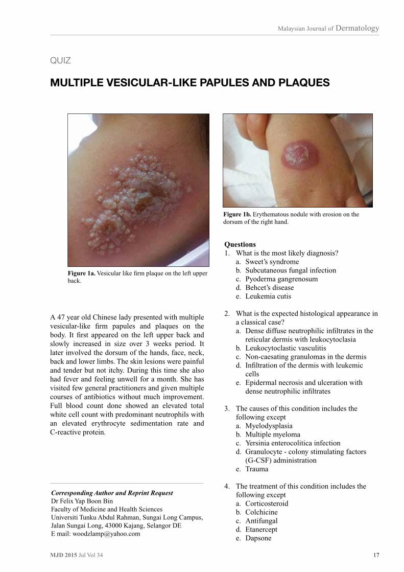
Figure 1b. Erythematous nodule with erosion on the dorsum of the right hand.
Figure 1a. Vesicular like firm plaque on the left upper back.
A 47 year old Chinese lady presented with multiple vesicular-like firm papules and plaques on the body. It first appeared on the left upper back and slowly increased in size over 3 weeks period. It later involved the dorsum of the hands, face, neck, back and lower limbs. The skin lesions were painful and tender but not itchy. During this time she also had fever and feeling unwell for a month. She has visited few general practitioners and given multiple courses of antibiotics without much improvement. Full blood count done showed an elevated total white cell count with predominant neutrophils with an elevated erythrocyte sedimentation rate and C-reactive protein.
Corresponding Author and Reprint Request Dr Felix Yap Boon BinFaculty of Medicine and Health SciencesUniversiti Tunku Abdul Rahman, Sungai Long Campus, Jalan Sungai Long, 43000 Kajang, Selangor DEE mail: [email protected]

Malaysian Journal of Dermatology
18 MJD 2015 Jul Vol 34
DiscussionSweet’s syndrome was first described by Robert Sweet in 1964 as an acute febrile neutrophilic dermatosis.1 It is characterized by fever, tender erythematous skin lesions, neutrophilia, high serum inflammatory markers, and diffure mature neutrophils infiltration of the dermis.2 Table 1 outline the diagnostic criteria for Sweet’s syndrome.2 Patients must have 2 major and 3 minor criteria to fulfil the diagnosis.
Sweet’s syndrome is seen from newborn to elderly patients. The mean age of reported cases is around 50 years. Majority of patients are females with no specific racial predilection.2 The syndrome is divided into 4 clinical types ie.
1. Classical or idiopathic Sweet’s syndrome Usually seen in women in their reproductive age and associated with infection (upper respiratory or gastrointestinal), inflammatory bowel disease or pregnancy.2,3,4
2. Drug induced Sweet’s syndrome Many drugs are implicated. The most commonly implicated drug is the G-CSF.2,3,4 Other implicated drugs include antibiotics, anti-epileptics, anti-hypertensives, oral contraceptives and retinoids.3. Malignancy associated Sweet’s syndrome Seen in 85% of hematologic malignancies, most commonly acute myeloblastic leukemia (AML) and 15% of solid neoplasia.2,3,4
4. Neutrophilic dermatosis of the hands
Localized form and the most recently described mainly in women.2
Patients with Sweet’s syndrome classically present with fever and characteristic skin lesions. The fever usually precedes the skin lesions by several days to weeks. Other symptoms include arthralgia, malaise, lethargy, headache and myalgia. The skin lesions is typically red or purple-red papules or nodules that have a strong tendency to coalesce forming irregular plaques.4 These skin lesions are firm but have transparent, vesicle like appearance due to pronounced upper dermal edema.4 The lesions are painful and tender with tendency to enlarge over weeks and heal after weeks and months without scarring. They are usually seen on the upper limbs, face and neck. Cutaneous pathergy might be present. Oral involvement is uncommon in classical type but more common with Sweet’s syndrome associated with malignancy.4 Extracutaneous manifestations can also be seen and include involvement of the bone, central nervous system, eyes, kidneys, liver, intestines, muscles, heart and lungs.3,4
The pathogenesis of Sweet’s syndrome remains elusive. Inappropriate regulation of the cytokines leading to hypersensitivity reaction to infection, tumour and drugs is the most acceptable hypothesis.3
The classic histologic features are dense diffuse mature neutrophils infiltrate in the superficial dermis and dermal edema. Leukocytoclasia or fragmented neutrophil nuclei are common. However, leukocytoclastic vasculitis is absent in Sweet’s syndrome. Occasionally, leukemic cells can be seen in patients with hematologic malignancies.3,4 There is no significant changes in the epidermis but rarely there will be neutrophilic infiltration of the subcutis in patients with underlying malignancies.3
Sweet’s syndrome must be differentiated with other neutrophilic dermatoses, infections and inflammatory dermatoses. Clinically, erythema multiforme and erythema nodosum resemble skin lesions of early Sweet’s syndrome. The vesicular like lesions can be confused with herpes simplex and zoster. Pyoderma gangrenosum can clinically and histologically resembles Sweet’s syndrome. Histologically, Sweet’s syndrome needs to be differentiated from other neutrophilic dermatoses like pyoderma gangrenosum, neutrophilic eccrine hidradenitis and granuloma faciale. Leukocytoclastic vasculitis must be excluded.
Table 1. Diagnostic criteria for Sweet’s syndrome.
Major Criteria
Minor criteria
1. Rapid onset of characteristic skin lesions which are tender erythematous plaques and nodules2. Typical histological features: dense neutrophils infiltration without leukocytoclastic vasculitis
1. Fever (> 38ºC), history of upper respiratory or gastrointestinal infection or immunization, the history of haematologic or solid neoplasia, inflammatory disorder, pregnancy, very good response to corticosteroids or potassium iodid2. ESR > 20mm/h3. WBC > 8 X 10⁹/L 4. Neutrophil > 70%5. High CRP

Malaysian Journal of Dermatology
19MJD 2015 Jul Vol 34
References 1. Sweet RD. An acute febrile neutrophilic dermatosis. Br J
Dermatol 1964; 74: 349-56.2. Paydas S. Sweet’s syndrome: a revisit for haematologists
and oncologists. Critical Rev Oncol Hematol 2013; 86: 85-95.
3. Cohen PR. Sweet’s syndrome- a comprehensive review of an acute febrile neutrophilic dermatosis. Orphanet J Rare Dis 2007; 34: 1-28.
4. Cohen PR, Kurzrock R. Sweet’s syndrome: a neutrophilic dermatosis classically associated with acute onset and fever. Clin Dermatol 2000; 18: 265-82.
5. Cohen PR, Kurzrock R. Sweet’s syndrome and cancer. Clin Dermatol 1993; 11: 149-157.
Corticosteroid is the gold standard treatment.4 High dose systemic steroid leads to dramatic response of the fever and skin lesions within hours. Other first line treatment includes colchicine and potassium iodide.3 Second line treatment include indomethacin, clofazimine, dapsone and cyclosporine. Other medications that can be used are thalidomide, cyclophosphamide, antimicrobials, etretinate, etanercept and infliximab.4
The recurrence rate for classical Sweet’s syndrome is 30%. Drug induced Sweet’s syndrome has a recurrence rate of 67%, hematologic malignancy associated 69% and solid tumour associated 41%.5

Malaysian Journal of Dermatology
20 MJD 2015 Jul Vol 34
GENERAL DERMATOLOGY - Short Case
EPIDERMODYSPLASIA VERRUCIFORMIS IN A PAIROF SIBLINGS Ling HN¹, Tagal JM², Lee BR³, Leong KF4
IntroductionEpidermodysplasia verruciformis (EV) described by Lewandowski & Lutz is a rare genodematosis, inherited mostly via autosomal recessive or X-linked. It is characterized by increased susceptibility to infection by specific human papillomavirus (HPV) genotypes. There are more than 20 known EV-HPV types, including 3, 5, 8, 9, 10, 12, 14, 15, 17, 19-25, 28, 29, 36, 46, 47, 49, and 50.¹ Classically, this viral infection leads to the development of tinea versicolor like macules on the trunk, neck, arms, and face during childhood.² In EV patients, these
Corresponding Author and Reprint Request Dr Ling Hee Ninh, MD MRCPDepartment of DermatologySarawak General Hospital, Jalan Hospital,93586 Kuching, SarawakEmail: [email protected]
¹ Department of Dermatology &² Opthalmology, Sarawak General Hospital, Jalan Hospital, 93586 Kuching, Sarawak³ Department of Pathology &4 Paediatrics, Hospital Kuala Lumpur, Jalan Pahang, 50586 Kuala Lumpur
HPV types have oncogenic potential, and over time, 30% to 60% of affected individuals will develop squamous cell carcinoma (SCC).³ This malignant transformation is a slow process, with malignancies first appearing on sun exposed skin in the fourth decade of life, usually 20 to 30 years after onset of the disease. The term ‘’acquired epidermodysplasia verruciformis’’ was introduced to describe patients with impaired cell mediated immunity acquiring susceptibility to the EV-HPV types that are innocuous for the general population. Herein, we report a case of a 10 year old boy with acquired EV.
Case ReportA 10 years old boy presented with chronic cough & asymptomatic widespread truncal hyperpigmented and hypopigmented macules with pityriasiform scales associated with pedunculated warty lesions on eyelids and chin for 1 year duration (Figure 1-B1 and B2).
Figure 1. Presence of warty lesions in both siblings before (A1 & B1) and after treatment (A2 & B2).

Malaysian Journal of Dermatology
21MJD 2015 Jul Vol 34
Immunology workup showed persistent leucopenia with persistently low T-cells (CD4/CD8) and B cells. He was later diagnosed to have primary immunodeficiency, Medellian Susceptibility to Mycobacterial Disease (MSMD) and bronchiectasis secondary to pulmonary tuberculosis. There was no family history of consanguinity.
Similar cutaneous lesions were seen in his older sister who was his only sibling (Figure 1-A1 and A2), but not in the father. His mother had passed away 8 year earlier from colon cancer. Skin biopsies taken from patient’s chin and torso confirmed the diagnosis of viral wart. Histopathological examination showed enlarged cells in the granular and spinous layer with bluish gray cytoplasm suggesting HPV) infection. Addition features were enlarged keratohyaline granules and koilocytes (Figure 2). HPV typing on the skin biopsy specimens embedded in paraffin were not available in our setting.
Lesions in both siblings were well controlled with regular cryotherapy and oral isotretinoin (Figure 1-A3 and B3). Sunscreen usage and sun avoidance was counseled to avoid malignant transformation.
DiscussionIn EV patients, HPV infections have oncogenic potential. This malignant transformation is a slow process, with malignancies first appearing on sun exposed skin in the fourth decade of life, usually 20 to 30 years after onset of the disease. In EV, HPV infections is necessary but not sufficient for malignant transformation. Ultraviolet radiation (UVR) plays an important role in the induction of SCC in EV patients. The majority of skin cancers in EV patients develop on sun-exposed sites. The oncogenic nature of EV-HPV and how it works
synergistically with UVR to induce carcinogenesis remain unclear. However, it is well known that UVR, specifically UVB, damages keratinocyte DNA and suppresses the skin’s immune system. UVB-specific mutations in the p53 tumor suppressor gene, including formation of pyrimidine dimer photoproducts, are seen in the majority of SCC.4
A wide range of therapies have been tried with variable success in the treatment of congenital EV and acquired EV. The treatments of common verrucae, including electrodesiccation, cryotherapy, topical retinoids, contact sensitization, imiquimod, 5-fluorouracil, podophyllotoxin and topical cidofovir have all been found to be ineffective in the treatment of the lesions. In a case report of a patient with EV treated with systemic retinoids (etretinate at 1 mg/kg daily for 4 months), there was a temporary decrease in the number of lesions¹.The combination of oral retinoids (acitretin 50 mg/day) and recombinant interferon alfa-2a (subcutaneously at 3 million units 3 days/wk), led to improvement in one case of EV reported after 9 months of treatment. At 3 months post treatment, there was a recurrence of lesions on the hands, but at 1 year the face remained clear.5
In our patients, the warts were controlled with combination of isotretinoin and cryotherapy. It must also be stressed that patient education regarding photoprotection and frequent skin surveillance is of utmost importance for early cancer detection.
Conclusion EV should be included in the differential diagnosis of any generalized warty lesions and work up for causes of cell mediated immunodeficiency should be performed in those with no obvious family history.
Figure 2. Presence of keratohyaline granules and koilocytes suggesting HPV infection (H & E, magnification X4, X10 and X40).

Malaysian Journal of Dermatology
22 MJD 2015 Jul Vol 34
References 1. Lutzer M, Blacnchet-Bardon C, Orth G. Clinical
observations, virologic studies, treatment trials in patients with epidermodysplasia verruciformis, a disease induced by specific human papillomaviruses. J Invest Dermatol 1984; 83: 18-25.
2. Heather DR, Jennifer LM, Kristin MN, et al. Acquired epidermodysplasia verruciformis. J Am Acad Dermatol 2009; 60: 315-20.
3. Acquired epidermodysplasia verruciformis associated with transplant related immunosuppression. J Am Acad Dermatol 2011; 2: 2503
4. Brash DE, Ponten J. Skin precancer. Cancer Surv 1998; 32: 69-113.
5. Anadolu R, Oskay T, Erdem C, et al. Treatment of epidermodysplasia verruciformis with a combination of acitretin and interferon alfa-2a. J Am Acad Dermatol 2001; 45: 296-9.
Acknowledgement The authors would like to thank the Director General of Health Malaysia for permission to publish this paper.

Malaysian Journal of Dermatology
23MJD 2015 Jul Vol 34
GENERAL DERMATOLOGY - Short Case
VERRUCOUS HAEMANGIOMA IN A 10-YEAR-OLD GIRLLee S1, Mohd Shariman MS2, Teoh TZ3, Choon SE3
IntroductionVerrucous haemangioma (VH) is a rare vascular anomaly which may be mistaken as infantile haemangioma or angiokeratoma. The characteristic clinicopathologic features of VH were first elaborated by Imperial and Helwig in 1967.1 However, its distinctive clinical and histopathologic features may not be apparent at initial manifestation and only emerges with disease evolution. We describe a case of VH, which was initially diagnosed as infantile haemangioma, to highlight the clinical histopathological features that distinguish it from other vascular anomalies.
Corresponding Author and Reprint Request Dr. Siew Eng ChoonDepartment of Dermatology Hospital Sultanah Aminah Johor Bahru80100, Johor, MalaysiaEmail: [email protected]
1 Medical student, School of Medicine and Health Sciences, University of Monash, Victoria, Australia3 Department of Dermatology and2 Pathology, Hospital Sultanah Aminah Johor Bahru, Johor, Malaysia
Case ReportA 10-year-old girl presented with multiple purplish-red verrucous plaques on her right foot and ankle. These lesions started as erythematous patches at birth on the dorsum of her right foot and gradually became larger and more warty with new lesions appearing and spreading up her right leg. Bleeding occurred occasionally with minor trauma. Although not painful, the lesions affected her shoe-wearing. Physical examination revealed multiple boggy swellings ranging from 1cm by 2cm to 5cm by 7 cm with overlying erythematous or verrucous purplish- red plaques on the dorsum of her right foot, ankle and leg (Figs 1- 2).
The lower limbs were equal in length although the swelling on the right foot resulted in asymmetry of her feet. The rest of her skin was clear. No significant lymphadenopathy or organomegaly was detected. A 4mm-punch biopsy revealed hyperplastic epidermis with hyperkeratosis, papillomatosis and acanthosis overlying vascular proliferations in both the dermis and subcutaneous fat (Fig. 3a). The papillary dermis showed dilated thin-walled vascular channels lined by flat endothelial cells (Fig. 3b). Foci of thick-walled, round capillary-sized vessels lined by single layer of plump protruding endothelial cells were conspicuous in the reticular dermis and subcutis (Fig. 3c, 3d).
Figure 2. Boggy mass on lateral malleolus of right leg surmounted by purplish red verrucous plaque.
Figure 1. Multiple verrucous purplish- red plaques on the dorsum of her right foot, ankle and leg.

Malaysian Journal of Dermatology
24 MJD 2015 Jul Vol 34
DiscussionVH is a rare vascular anomaly that classically manifests at birth as a pinkish macule/patch that grows proportionately with the child.1-4 Unilateral localization to a lower limb is characteristic but lesions had been reported on other sites. New lesions appear gradually with time. Initial pinkish lesions evolve into reddish or purplish-red verrucous plaques with underlying soft masses. The lesions are often distributed in a linear or serpiginous pattern. Bleeding and infection may supervene. VH does not involute spontaneously. Histologically, it is characterized by irregular acanthosis, papillomatosis and hyperkeratosis overlying vascular proliferations in both dermis and subcutis. The vessels in the papillary dermis are diffusely distributed and composed of thin-walled dilated blood vessels lined by flat endothelial cells whereas vascular proliferations in the deep dermis and subcutis consist of aggregates of round thick-walled vessels with plump endothelial cells and multi-lamellated basement membranes.
The nosologic status of VH is still unclear. The International Society for the Study of Vascular Anomalies (ISSVA) classified vascular anomalies into vascular tumours and vascular malformations.5
Vascular tumours grow by endothelial hyperplasia whereas malformations are due to defects of vascular morphogenesis. Vascular tumours may
involute spontaneously or persist depending on their type but vascular malformations persist for life. Vascular malformations usually present at birth and have commensurate growth during childhood. It is important to differentiate vascular tumors from malformations because management is different. Immunohistochemical marker Wilms tumor 1 (WT1) is useful in distinguishing between vascular tumours and malformations, being positive in the former and negative in the latter.6,7
Although, VH behaves like a vascular malformation, being present at birth, exhibits proportionate growth without regression, it is WT1-positive.8
VH also expresses GLUT1 (Erythrocyte-type glucose transporter protein-1), another immunohistochemical marker, not found in vascular malformations.8 GLUT1 is a marker of infantile haemangioma which distinguishes it from other vascular tumours such as congenital haemangioma, tufted angioma, pyogenic granuloma and kaposiform hemangioendothelioma.8 Hence, ISSVA parked VH under the category of “provisionally unclassified vascular anomalies”. Both WT1 and GLUT1 are not available in our laboratory.
A close clinicopathologic correlation is essential to diagnose VH as its clinical features may mimic other vascular anomalies especially angiokeratoma circumscriptum. Like VH, angiokeratoma
Figure 3. (a) Hyperplastic epidermis with hyperkeratosis, papillomatosis and acanthosis overlying vascular proliferations in both the dermis and subcutaneous fat; (b) Dilated thin-walled vascular channels lined by flat endothelial cells in the papillary dermis with thicker walled vessels in reticular dermis; (c,d) Clusters of thick-walled, round capillary-sized vessels lined by single layer of plump protruding endothelial cells in reticular dermis and subcutis. Haemotoxylin and eosin, original magnification (a) X 2; (b, c, d) X 10

Malaysian Journal of Dermatology
25MJD 2015 Jul Vol 34
circumscriptum usually presents at birth with a cluster of small reddish papules which are commonly located on lower limbs in a segmental distribution. With time, existing papules become more warty while more papules develop. These papules may coalesce to form a persistent hyperkeratotic vascular plaque which resembles verrucous haemangioma clinically. Histologically, hyperplastic epidermis is also seen in angiokeratoma circumscriptum but the confinement of dilated vessels to the papillary dermis distinguishes it from VH which displays vascular proliferations in both papillary and deep dermis as well as the subcutis. Differentiating VH from angiokeratoma circumscriptum is important because angiokeratoma is easily treated with electrocoagulation, cryotherapy or laser whereas VH requires deep excision with wide margins and frequently recur due to incomplete excision.2,4,9 Prompt diagnosis and complete excision of an early lesion provide a better prognosis.10 Staged excisions may be necessary for multiple lesions.9 Other treatment options include cryotherapy, cautery, laser therapy and a combination of laser with surgery. Although laser therapy is mainly palliative, a recent study showed that laser therapy using carbon dioxide in combination with dual pulsed dye and ND-YAG lasers may be a viable treatment option. In this study, 7 out of 8 patients treated had at least a 50% improvement in skin lesions.11 More importantly, all treated patients were satisfied with the treatment with a satisfaction score (from 1= poor to 10= excellent) of 8 to 10. A recurrence rate of 30% following surgical excision had been documented.9 Recurrence rate is high with
other treatment modalities but reliable estimates are not available due to paucity of reports. Our patient chose to try laser therapy first and was referred to Kuala Lumpur for the treatment.
VH may be mistaken as infantile haemangioma or port wine stain during its early phase when it is still flat and non-keratotic. However, infantile haemangioma is a vascular tumour which presents on the head and neck, with only 15% reported on the extremities.10 Histologically, infantile haemangioma resembles early stage of VH and diagnosis is often only made with disease progression.7 Unlike VH which persists indefinitely, infantile haemangioma exhibits an initial rapid growth phase followed by involution.2,10 Treatment options for infantile haemangioma include pharmacological treatment, laser therapy and surgery.2,10 Port wine stain is a capillary malformation that presents at birth on the head and neck as pinkish patches and persists throughout life. As it matures, the erythematous patches become violaceous and thicker with the appearance of angiomatous nodules or vascular blebs. In its advanced stage, the vascular plaque tends to appear nodular as opposed to verrucous.2 Histologically, it is characterized by ectatic vessels of variable size in the dermis. Port wine stain is best treated early with pulsed dye laser.2
In conclusion, we describe a case of VH, diagnosed at 10 years of age, due to unfamiliarity with this rare condition. Early diagnosis and treatment is important to attain a better cosmetic result.
References 1. Imperial R, Helwig EB. Verrucous hemangioma; a
clinicopathologic study of 21 cases. Arch Dermatol 1967;96:247-253
2. Moss C, Shahidullah H. Naevi and other developmental defects. In: Textbook of Dermatology, In: Burns T, Breathnach S, Cox N, Griffiths C, editors. 8th ed. Oxford: Blackwell Science; 2010 chapter18 p.40-74
3. Tennant LB, Mulliken JB, Perez-Atayde AR, et al. Verrucous hemangioma revisited. Pediatr Dermatol 2006;23:208-215
4. Clairwood MQ, Bruckner AL, Dadras SS. Verrucous hemangioma: a report of two cases and review of the literature. J Cutan Pathol 2011;38: 740-746.
5. International Society for the Study of Vascular Anomalies. ISSVA classification for vascular anomalies. http://issva.org/content.aspx?page_id=22&club_id=298433&module_id=152904 Last accessed March 12, 2015.
6. Trindade F, Tellechea O, Torrelo A, et al. Wilms tumor 1 expression in vascular neoplasms and vascular malformation. Am J Dermatopathol 2011; 33: 569
7. Al Dhaybi R, Powell J, McCuaig C, et al. Differentiation of vascular tumors from vascular malformations by expression of Wilms tumor 1 gene: evaluation of 126 cases. J Am Acad Dermatol 2010; 63: 1052
8. Trindade F, Torrelo A, Requena L, et al. An immunohistochemical study of verrucous hemangiomas. J Cutan Pathol 2013; 40: 472-476
9. Mankani MH, Dufresne CR. Verrucous malformation: their presentation and management. AnnPlast Surg 2000; 45: 31-36
10. Koc M, Kavala M, Kocatürk E, et al. An unusual vascular tumor: Verrucous hemangioma. Dermatol Online J 2009; 15 (11): 7
11. Segura Palacios JM, Boixeda P, Rocha J, et al. Laser treatment for verrucous hemangioma. Lasers Med Sci 2012; 27(3): 681-684
12. Zimmermann AP, Wiegand S, Werner JA, et al. Propranolol therapy for infantile haemangiomas: review of the literature. Int J Paediatr Otorhinolaryngol 2010; 74:338-342

Malaysian Journal of Dermatology
26 MJD 2015 Jul Vol 34
GENERAL DERMATOLOGY - Short Case
HYPOHYDROTIC ECTODERMAL DYSPLASIA: A CASE SERIESVisuvanathan VV1, Najeeb AMS2
IntroductionThe ectodermal dysplasias (ED) are a rare group of inherited disorders characterised by hypohidrosis (reduced ability to sweat), hypotrichosis (sparseness of hair), hypodontia (decreased tooth development) and onychodysplasia (nail abnormalities).1 Clinically, ectodermal dysplasia can be broadly classified into hypohidrotic ectodermal dysplasia (HED) and the hidrotic ectodermal dysplasia. X-linked hypohidrotic ectodermal dysplasia (XL-HED), which is also known as Christ-Siemens-Touraine, is the most classic subtype. Nguyen-Nielsen and Skovbo found that in Denmark, the prevalence of XL-HED was 21.9 per 100,000 overall and 1.6 per 100,000 when restricted to molecularly-confirmed XLHED cases.2 Herein, we report a case series of 3 family members with XL-HED.
Corresponding Author and Reprint Request Dr Vaani Valerie VisuvanathanInternational Medical University,Jalan Rasah, 70300 Seremban,Negeri Sembilan Darul Khusus, Malaysia.Email: [email protected]
1 International Medical University, Jalan Rasah, 70300 Seremban2 Department of Dermatology, Hospital Tuanku Jaafar, Jalan Rasah, 70300 Seremban
Case Report
Patient AA is a 6-month old child, who presented with perioral pruritic erythematous, papules for 3 weeks (Figure 1). His parents had noted that the infant had sparse hair and did not sweat even during exceptionally warm weather. Thus far, the child has not had any respiratory illness.
Clinical examination revealed erythematous, scaly, papules and macules over the perioral region. The infant was edentulous and had a saddle-shaped nose.He was prescribed a mild topical steroid (Clobetasone Butyrate) and emollients, which led to minimal residual papules one week later. Dental referral was suggested.
Patient BB, a 21-year-old male, is the younger maternal uncle of patient A. He had been noted to have abnormal dentition with sparse hair since birth, as illustrated in Figure 2. He claims to have minimal sweat but no problems with salivation and dry eyes. The abnormal dentition did not cause any problems nor limit his diet. Occasionally, he did require water to aid the swallowing of certain types of food. He had recurrent pruritic, erythematous papules on his face, especially at the periorbital and perioral region. He had a total of eight teeth, which were all peg-shaped with significant gap between the teeth. He was prescribed some mild topical steroids and advised to keep cool during warm weather by having cold showers and increasing fluid intake.
Patient CThe elder brother of Patient B, C, has a similar facies i.e. depressed nasal bridge, prominent lips, sparse hair, oligodontia with increased gaps between the teeth. He has minimal sweat but denied dry eyes or frequent respiratory illness as a child. He had been seen by a dentist who suggested dentures, but he is not keen to wear them. He had no signs of eczema nor hyperpigmentation.
Figure 1.

Malaysian Journal of Dermatology
27MJD 2015 Jul Vol 34
Figure 3. Saddle nose, sparse hair, loss of eyebrow, oligodontia and peg shaped teeth.
Figure 2. Sparse hair and eyebrow, saddle nose, oligodontia, peg shaped teeth and erythematous periorbital papules.
DiscussionAs illustrated in the case series above, patients with HED present with a myriad of symptoms. However, some patients are asymptomatic and troubled by their appearance. On the other extreme, some patients present with heat stroke and seizures due to thermo dysregulation. Other problems that may be encountered by a child with HED are feeding difficulties (deficiency of saliva), difficulty in articulation, recurrent respiratory infections (due to deficient mucus production by the respiratory epithelia in the trachea and bronchi) and dry eyes. Atopic eczema and hyperpigmentation would
bring them to the attention of the dermatologist. A thorough history and astute clinical skills would be required to suspect the diagnosis in a young patient who presents with atopic eczema.
Making an early diagnosis has been found to improve the outcome of boys with HED.4 Early fitting of artificial dentures, before the child attends school, helps with speech, facial appearance and nutrition. Management of HED involves a multidisciplinary team and should focus on thermoregulation, treatment and prevention of infections, optimising feeding, treatment of bronchial asthma and eczema, monitoring of speech and genetic counselling.
Figure 4. Genogram of the 3 patients illustrating the X-linked dominant genetic linkage.

Malaysian Journal of Dermatology
28 MJD 2015 Jul Vol 34
References 1. Lamartine, J. Towards a new classification of ectodermal
dysplasias. Clin Exp Dermatol 2003; 28: 351–355.2. Nguyen-Nielsen M, Skovbo S, Svaneby D, et al. The
prevalence of X-linked hypohidrotic ectodermal dysplasia (XLHED) in Denmark. 1995-2010. Eur J Med Genet 2013; 56(5): 236-42.
3. Pinheiro M, Freire- Maia N. Ectodermal Dysplasias: a clinical classification and causal review. Am. J. Med Genet 1994; 53: 153-162.
4. Prasun, Pankaj, Karmarkar, et al. Unusual physical features and heat stroke presentation for hypohydrotic ectodermal dysplasia. Clin Dysmorphology 2012; 21(1): 24-26.

Malaysian Journal of Dermatology
29MJD 2015 Jul Vol 34
GENERAL DERMATOLOGY - Short Case
GORLIN SYNDROME: A CASE REPORT WITH CLINICALAND RADIOLOGICAL CORRELATIONAzura MA, Izzaty D
IntroductionGorlin syndrome is a condition characterized by a wide range of developmental abnormalities and a predisposition to skin cancer, namely basal cell carcinoma (BCC). It is inherited as an autosomal dominant trait with complete penetrance and variable expressivity, and caused by mutations in
Corresponding Author and Reprint Request Dr Azura Mohd Affandi MBChB(UK), MRCP(UK), Adv M Derm (UKM)Department of Dermatology, Hospital Kuala LumpurJalan Pahang, 50586 Kuala Lumpur, MalaysiaEmail: [email protected]
the PTCH1 (Patched1) gene which is mapped to the long arm of chromosome 9q22.3-q31.1 The syndrome is also known as naevoid basal cell carcinoma syndrome, Gorlin-Goltz syndrome, basal cell naevus syndrome, fifth phacomatosis and multiple basilioma syndrome.2 Diagnosis of Gorlin syndrome is made by having two major criteria or
one major & two minor criteria.3 We report a case of Gorlin syndrome who presented with multiple basal cell carcinoma, with clinical and radiological correlations.
Case ReportA 60 year old Malay lady with underlying diabetes mellitus type II, hypertension and hypercholesterolemia, presented to our clinic in 2003 with multiple hyperpigmented papules and plaques on the trunk, upper and lower limbs (Fig. 1a-c). The lesions started to appear when she was 46 years old and gradually increased in size and number. She is of medium built, with frontal bossing and high arched palate. There were also palmo-plantar pits (Fig. 1d-e).
Figure 1a. Multiple pigmented basal cell carcinomas on the chest.
Figure 1b. Multiple pigmented basal cell carcinomas on the back.
Figure 1c. Multiple pigmented basal cell carcinomas on the lower limbs.

Malaysian Journal of Dermatology
30 MJD 2015 Jul Vol 34
Dermoscopy examination of the pigmented plaque showed maple-leaf pattern, large ovoid nests and arborizing telangectasias (Fig. 2), suggestive of basal cell carcinoma. Excision biopsies over 3 areas (left thigh, upper back and lower back) in 2003 confirmed basal cell carcinoma. Throughout her follow-up, she had multiple excisions of basal cell carcinoma over different locations: left infra-mammary region and face (2011), right chest, left shoulder , left flank and lower back (2012), chest, right and left arm (2013). The other smaller, multiple, superficial basal cell carcinoma were treated with multiple sessions of
liquid nitrogen cryotherapy. She denied any family history of skin malignancy and did not have history of prolonged exposure to sunlight.
Radiological examination revealed macrocephaly, calcification of the falx cerebri and two cysts in the mandible (Fig. 3a & 3b). There was also sprengel deformity of the left scapula (elevation of the scapula characterised by medial rotation of the distal pole of the scapula) and left 4th bifid rib evident from the chest radiography.
Figure 2. Dermoscopy showing maple leaf pattern, large ovoid nests and arborizing telangiectasia.
Figure 3b. X-ray showing multiple mandibular cysts.
Figure 3a. Skull X-ray showing macrocephaly and calcification of the falx cerebri.

Malaysian Journal of Dermatology
31MJD 2015 Jul Vol 34
She was started on acitretin 25mg daily since May 2013 to reduce the risk from getting recurrent non-melanoma skin cancers. She is tolerating the medication well. The last reported recurrence of basal cell carcinoma was in September 2014, and there was no recurrence of basal cell carcinoma since then.
DiscussionGorlin syndrome is a rare autosomal dominant syndrome which is caused by mutations in the PCTH1 gene on chromosome 9q22.3-q31.1 The syndrome is also known as naevoid basal cell carcinoma syndrome, Gorlin-Goltz syndrome, basal cell naevus syndrome, fifth phacomatosis and multiple basilioma syndrome.2 It was first reported by Jarish in 1894 who described a patient with multiple basal cell carcinomas, scoliosis and learning disability.2 In the 1960s, Gorlin and Goltz described them as a triad of disorders including multiple basal cell carcinoma, numerous keratocysts in the jaws and skeletal abnormalities, which gave rise to the Gorlin-Goltz syndrome.2,3
Although its occurrence among family members is an important diagnosing criteria, it has been found that between 20% and 40% of cases result from a de novo mutation of the PTCH1 gene.4 Our patient also did not have a positive family history of Gorlin syndrome. The prevalence range from 1:57,000 (in England) to 1:164,000, and even 1:256,000 (in Italy).3 There is equal predisposition in male and female, and it can occur in all ethnic groups, although the Caucasians are most often affected by it.5
Skin manifestations in Gorlin SyndromeMultiple basal cell carcinoma of the skin constitutes the most characteristic feature of the syndrome. The highest incidence rate is observed in people between puberty and age 35.6 In general, the mean age of onset is about 25 years of age, although it was also observed in children ages 3 to 4 years.7 The number of BCC varies from several to multiple, and their diameter ranges from 1 mm to 10 mm, and they may have various forms from skin-coloured nodules or papules, pigmented lesions to ulcerating plaques.8 They are usually located on the face, back and chest, but they may also be found on skin not exposed to the sun.8 Aggressive forms of basal cell carcinomas, which infiltrate the facial bones, hardly ever occur. The histopathology of naevoid BCC cannot be differentiated from that of ordinary sporadic BCC.
Between 30% and 65% of patients with the syndrome have small asymmetric palmo-plantar pits by the age of 10 years.2,3 This percentage rises to 80% of patients by the age of 15, and 85% of patients over the age of 20 years have palmo-plantar pits. They are small, with a diameter ranging from 2 to 3 mm and depth from 1 to 3mm. They increase in number with age, are permanent, and are a strong diagnostic indicator whenever found in a child. They are found more commonly on the palms (77%) than on the soles (50%).2,3 They become more visible after the palms have been held in warm water for about 10 minutes.
Multiple naevi are present in 30-50% of patients under 20 years of age, rising to 70% in patients over the age of 20 years. The naevi are flesh colored, reddish brown or pearly. Some of them grow rapidly for a few days (to a few weeks), then mostly remain static. Other skin signs are nodular or patch lesions, and benign dermal cysts. Small keratin-filled cysts (milia) can be found on the face in 30% of cases, most commonly in the area below the eyes, but they can also occur on the forehead.2 Epidermoid cysts occur on the limbs and the trunk in over 50% of cases. They are usually 1-2 cm in diameter and are particularly common around the knee.
Keratocystic odontogenic tumour (KCOT)The most important manifestations of Gorlin syndrome within the oral cavity are recurrent multiple jaw tumours called keratocysts. The lesions occur in as many as 90% of patients above age 40.2 They are most frequently located in the mandible - 44% are found in the mandibular angles and 18% in the zones adjacent to incisor and canine teeth.2 In the maxilla, they accompany canines and incisors (15%), as well as molars (14%). In spite of their less frequent occurrence as compared to in the mandible, they are more aggressive than those in the lower jaw area.
The KCOTs are divided into parakeratotic, orthokeratotic, and (rarely) mixed and solid lesions, and they are differentiated based on the histological image of the cells lining them.3 The cavity of the tumour is filled with thick keratinous material or a straw-coloured fluid. The tumours are usually diagnosed incidentally during routine x-ray examinations performed in the course of a regular dental treatment. Other defects of the oral cavity are malocclusion, impacted teeth, mandibular prognathism, dental ectopy or heterotopy, and dental agenesis. Cleft palate and lip are rare, occurring in 5% of patients.9

Malaysian Journal of Dermatology
32 MJD 2015 Jul Vol 34
Central nervous systemCalcifications of the central nervous system have been reported: lamellar calcification of the falx cerebri (in 70-85% of patients) and tentorium cerebelli (in 20-40% of patients).10 The calcification of the falx cerebri can appear very early in life, and is often strikingly apparent from late childhood. Other signs reported are cysts of the choroid plexus, intraparenchymal brain cysts, communicating hydrocephalus, meningioma and medulloblastoma.11,12 Seizures have occasionally been noted and mental retardation occurs in about 5% of cases.13
Musculo-skeletal systemPatients with Gorlin syndrome have various skeletal manifestations. They are usually much taller than the other family members, although in our case, she is of an average size. An abnormal skull configuration (characterized by frontal, biparetial or temporal bossing, and large calvaria) is frequent (70%).10
Macrocephaly has been reported in 50% of cases. As a result of the abnormal growth of the skull, about 70% of patients affected by NBCCS have eyes which appear wider apart than usual. In our patient, although she is of normal stature, she had frontal bossing and macrocephaly. Other skeletal signs are scoliosis (40%), and abnormalities such as bifid, wide, fused, partially missing or underdeveloped ribs (30-60%).10 Rib abnormalities may give rise to a prominent or depressed sternum in about 30% - 40% of patients. Spina bifida occulta of the cervical or thoracic vertebrae is found in 60% of cases. Bifid or fused vertebra and Sprengel anomaly (elevation of the scapula with rotation toward the spine with scoliosis) are observed in 10 to 40% of patients. Our patient had an evidence of bifid rib and sprengel deformity on the chest radiograph. Extra digits on the hands or feet can occur.
Other systems involvementPatients with Gorlin syndrome may also have involvement of the other systems, such as the ocular, genito-urinary, cardiovascular, respiratory, auditory and gastro-enteric systems (Table 1). Apart from getting recurrent basal cell carcinoma, they are also at risk of developing other tumours such as medulloblastoma,14 fibroma15 and rhabdomyosarcoma.16
DiagnosisDiagnosis of Gorlin syndrome can be made by having two major criteria or one major & two minor criteria, as shown in Table 2.1-3,6 Our patient fulfilled
five out of the six major criteria, namely multiple occurrence of basal cell carcinoma, odontogenic keratocysts, palmo-plantar pits, calcification of the falx cerebri and bifid ribs. She also had three minor criteria, which are macrocephaly, frontal bossing and sprengel deformity. Direct mutation analysis of the gene may be helpful. It was not done in our case due to limited resources. It is particularly helpful to follow a specific clinical protocol in the examination of patients with suspected Gorlin syndrome (Table 3). At least annual examination of the skin since puberty is recommended to detect the occurrence of basal cell carcinoma.
The syndrome is a hereditary condition, thus referral to a geneticist for counseling is mandatory. Gorlin syndrome is caused when one copy of the PTCH1 gene pair contains a fault; this means that every child of a person with the syndrome has a 1 in 2 (50:50) chance of inheriting the faulty gene. The definitive diagnostic test is to demonstrate a mutation in the PTCH1 gene, although this is labor-intensive as there are 24 exons.
ManagementPatients with Gorlin syndrome are best managed by multi-disciplinary team, depending on the organs affected. Keratocysts can be treated by surgical removal. This requires exposure of the lesion by making an osteotomy in the jawbone under local or general anaesthesia, finding the wall of the cyst and removing this completely.
With regards to BCC, only a few of these tumours become invasive and the very rare cases of death result from spread to the brain or lung. Results from several epidemiologic studies have indicated that the risks of BCCs show a strong positive correlation with ultraviolet (UV) exposure to UV. Thus, the patients with Gorlin syndrome are advised to avoid excessive sun exposure, to use sunscreen (SPF30+), and to use UV protective sunglasses.
Treatment of BCC can be divided into surgical and non-surgical therapy. Surgical excision is the typical approach for a patient with BCC if the number of lesions is limited. Curretage and electrodessication is a simple and fast method, and can be used to treat small BCC. Cryosurgery and CO2 laser17 can also be used to treat multiple, small, superficial BCC. Mohs micrographic surgery allows radical excision of the lesions while minimizing the damage of healthy tissue. However, due to its cost, it is mainly reserved for BCC on the face where normal tissue preservation is imperative.

Malaysian Journal of Dermatology
33MJD 2015 Jul Vol 34
Another therapeutic option for BCC is photodynamic therapy (PDT), a cancer treatment involving use of a photosensitizing agent (given intravenously or topically) that preferentially accumulates within malignant cells. BCCs are then treated with red light that leads to dye activation and, eventually to death of these cells.18
Topical treatment for BCC includes 5% imiquimod cream or topical 5-fluorouracil. 5% imiquimod cream appears to be an effective treatment for nodular basal cell carcinoma alone or if combined with curettage prior to its application.19 5% imiquiomod cream is applied five times a week for a total of six weeks, and provides clinical and histological remission in all cases.19 5-fluorouracil cream is usually applied twice a day for a period of 6-12 weeks. The cure rate ranges from 80% to 95%.20 However, it is effective only in the case of superficial BCC.20
Oral retinoid (Isotretinoin) has been used as a chemoprevention or delaying the development of recurrent BCC. Some authors indicated that doses of 0.5-1.0 mg/kg/day cause regression of lesions of less than 1.0 cm and prevent new lesions formation.21 However, a study of high-dose oral isotretinoin (mean daily dose: 3 mg/kg/day) found that only 8% of BCCs underwent complete clinical and histological regression, while all patients developed moderate-to-severe acute toxicity.22 Our patient was started on acitretin 25mg daily, and she seems to be tolerating the medication well, and had no recurrence of BCC since September 2014.
The hedgehog signaling pathway is a key regulator of cell growth and differentiation.23 Somatic mutations in this pathway are present in the vast majority of sporadic BCCs and Gorlin syndrome. In
January 2012, vismodegib became the first selective inhibitor of the Hedgehog signaling pathway to be approved by the US Food and Drug Administration for the treatment of locally advanced and metastatic basal cell carcinoma. The drug selectively binds to Smoothened, a transmembrane receptor, thereby suppressing tumor proliferation and growth.23 In a phase II study on vismodegib in 42 Gorlin syndrome patients, the patients were randomized to two groups, one group received vismodegib and the other group received placebo only. The results showed reduction in the number of BCC (2 vs 29) and less surgery (0.31 vs 4.4) in the vismodegib group, compared to the placebo group.24
ConclusionThis case highlights the multitude of clinical findings that can be encountered in patients with Gorlin syndrome. Diagnosis is challenging, and the geneticist and radiologist can play a role in diagnosis, by detecting chromosomal abnormalities and radiological features associated with Gorlin syndrome. Multidisciplinary team, comprising of dermatologist, dentist, radiologist, neurologist and ophthalmologist are essential in managing this challenging syndrome. Patients should be taught on how to examine their skin and to report any changes to the doctor.
AcknowledgementWe acknowledge the support of the pathologist, plastic surgeon and radiologist in Hospital Kuala Lumpur in the diagnosis and management of this challenging case. We would also like to thank the Director General of Health, Malaysia for permission to publish this report.
References
1. Mohan R.V.S, Vemanna N.S, Narayanappa S.M, et.al. Gorlin-Goltz syndrome: A clinico radiological illustrative case report. Ind J Dentistry 2013; 4: 165-9.
2. Lo Muzio L. Nevoid basal cell carcinoma syndrome (Gorlin syndrome). Orphanet J Rare Dis 2008; 3: 32.
3. Kiwilsza M, Sporniak-Tutak K. Gorlin-Goltz syndrome – a medical condition requiring a multidisciplinary approach. Med Sci Monit 2012; 18(9): RA145-153.
4. Huang YF, Chen YJ, Yang HW. Nevoid basal cell carcinoma syndrome – case report and genetic study. J Dent Sciences 2010; 5: 166–70.
5. Manfredi M, Vescovi P, Bonanini M, Porter S. Nevoid basal cell carcinoma syndrome: a review of the literature. Int J Oral and Maxillofac Surg 2004; 33: 117–24.
6. Gorlin R.J. Nevoid basal cell carcinoma syndrome. Dermatol Clin 1995; 13: 113-125.
7. Shanley S, Ratcliffe J, Hockey A, et.al. Nevoid basal cell carcinoma syndrome: review of 118 affected individuals. Am J Med Genet 1994; 50: 282-290.
8. Pratt MD, Jackson R. Nevoid basal cell carcinoma syndrome. A 15-year follow-up of cases in Ottawa and the Ottawa Valley. J Am Acad Dermatol 1987; 16: 964-970.
9. Ruprecht A, Austermann KH, Umstadt H. Cleft lip and palate, seldom seen features of the Gorlin-Goltz syndrome. Dentomaxillofac Radiol 1987; 16: 99-103.
10. Lo Muzio L, Nocini PF, Savoia A, et.al. Nevoid basal cell carcinoma syndrome. Clinical findings in 37 Italian affected individuals. Clin Genet 1999; 55: 34-40.
11. Fukushima Y, Oka H, Utsuki S, et.al. Nevoid Basal

Malaysian Journal of Dermatology
34 MJD 2015 Jul Vol 34
12. Mortimer PS, Geaney DP, Liddell K, Dawber RP. Basal cell naevus syndrome and intracranial meningioma. J Neurol Neurosurg Psychiatry 1984; 47: 210-212.
13. Hogan RE, Tress B, Gonzales MF, et.al. Epilepsy in the nevoid basal-cell carcinoma syndrome (Gorlin syndrome): report of a case due to a focal neuronal heterotopia. Neurology 1996, 46: 574-576.
14. Amlashi SF, Riffaud L, Brassier G, Morandi X. Nevoid basal cell carcinoma syndrome: relation with desmoplastic medulloblastoma in infancy. A population-based study and review of the literature. Cancer 2003; 98:618-624.
15. Pirschner F, Bastos P.M, Contarato G.L et.al. Gorlin syndrome and bilateral ovarian fibroma. Int J Surg Case Reports 2012; 3: 477– 480.
16. Cajaiba MM, Bale AE, Alvarez-Franco M, et.al. Rhabdomyosarcoma, Wilms tumor, and deletion of the patched gene in Gorlin syndrome. Nat Clin Pract Oncol 2006; 3:575-580.
17. Doctoroff A, Oberlender SA, Purcell SM. Full-face carbon dioxide laser resurfacing in the management of a patient with the nevoid basal cell carcinoma syndrome. Dermatol Surg 2003; 29:1236-1240.
18. Madan V, Loncaster JA, Allan D et al. Nodular basal cell carcinoma in Gorlin’s syndrome treated with systemic photodynamic therapy and interstitial optical fiber diffuser laser. J Am Acad Dermatol 2006; 55: S86-89.
19. Neville JA, Williford PM, Jorizzo JL. Pilot study using topical imiquimod 5% cream in the treatment of nodular basal cell carcinoma after initial treatment with curettage. J Drugs Dermatol 2007; 6: 910-914.
20. Walter AW: Gorlin Syndrome. www.emedicine.com/PED/topic890.html.
21. Sanchez-Conejo-Mir J, Camacho F. Nevoid basal cell carcinoma syndrome: combined etretinate and surgical treatment. J Dermatol Surg Oncol 1989; 15: 868-871.
22. Peck GL, DiGiovanna JJ, Sarnoff DS, et.al. Treatment and prevention of basal cell carcinoma with oral isotretinoin. J Am Acad Dermatol 1988; 19:176-185.
23. V. Ruiz-Salas, M. Alegre, A. López-Ferrer, J.R. Garcés. Vismodegib. A Review. Actas Dermosifiliogr 2014; 105(8): 744-751.
24. Tang JY, Mackay-Wiggan J, Aszterbaum M, et al. Inhibiting the Hedgehog pathway in patients with the basal cell nevus syndrome. N Engl J Med 2012; 366: 2180-8.

Malaysian Journal of Dermatology
35MJD 2014 Dec Vol 33
