malaysia: a cross sectional retrospective analysis
TRANSCRIPT
Page 1/24
Determinants of prolonged hospitalization and mortality among leptospirosispatients attending tertiary care hospitals in northeastern state in PeninsularMalaysia: A cross sectional retrospective analysisYassin K. Al Hariri ( [email protected] )
Ajman UniversitySyed A.S. Sulaiman
Advanced Medical and Dental Institute (IPTT), USMAmer Hayat Khan
Universiti Sains MalaysiaAzreen S. Adnan
Management Science University (MSU) Medical CentreSundos Q. Al-Ebrahem
Ajman University
Research Article
Keywords: leptospirosis, prolonged hospitalization predictors, mortality, Malaysia
Posted Date: April 5th, 2021
DOI: https://doi.org/10.21203/rs.3.rs-355356/v1
License: This work is licensed under a Creative Commons Attribution 4.0 International License. Read Full License

Page 2/24
AbstractObjectives Leptospirosis is the most common anthropozoonosis worldwide and imposes a major public health problem in many tropical countries. It is aleading cause of disease burden in form of mortality, morbidity and hospital admission. Identifying patients at high risk for mortality or for prolongedhospitalization may save lives and preserve economy. The aim of the current study was to identify signi�cant factors associated with disease mortality andprolonged hospitalization.
Design Cress-sectional retrospective study
Settings Tertiary care teaching hospitals in Kelantan, Peninsular Malaysia
Participants Adult patients proven to have leptospirosis depending on IgM ELISA were classi�ed into two classes depending on prolonged hospitalization (>7days or ≤ 7 days) and mortality (fatal cases or non-fatal cases). Patients’ clinico-laboratory data were compared according to these two outcomes using theappropriate statistical test accordingly.
Results Of the 525 patients enrolled, 136 (25.9%) had prolonged hospitalization. The mean length of stay was 6.77 ± 5.68 days. Logistic regression analysisidenti�ed acute kidney injury (AKI) (OR 2.3), Jaundice (OR 2.7), elevated alanine aminotransferase (ALT) (OR 2), and prolonged prothrombin time (PT) (OR 1.9)independently associated with prolonged hospitalization. Case fatality rate was 6.48% and around one third of fatal cases had prolonged hospitalization ofmore than seven days. Factors associated with leptospirosis mortality included age > 40 years (p<0.001), patients presented with tachypnea (p=0.002),pulmonary in�ltrate (p<0.001), T-wave changes (p<0.001), atrial �brillation (p=0.013), conducting abnormality (p<0.001), chronic kidney diseases (p<0.001),multiple organ dysfunctions (p<0.0010), respiratory failure (p<0.001), pneumonia (p<0.001), sepsis (p=0.004), low venous PH (p=0.042), AKI (P<0.001),elevated AST (p<0.001) or ALT (p=0.004), hypoalbuminemia (p<0.001), rhabdomyolysis (p<0.001), severe thrombocytopenia (p=0.042), prolonged PT(p<0.001) or prolonged aPTT (p<0.017).
Conclusions Signi�cant proportion of leptospirosis patients (25.9%) had prolonged hospital stay and less proportion died (6.48%). Early identifying patientswith factors associated with prolonged hospitalization and death will positively impact practitioners’ decisions regarding the proper and fast course ofmanagement including ICU admission
IntroductionLeptospirosis, known as rat-urine fever in some countries [1] is the most common anthropozoonosis worldwide. Nowadays, the disease in not only restricted tothe rural setting but also hits the urban areas particularly the outbreaks after the rainy season [2] and it had escaped from its homeland in tropics to causeurban epidemics in the poor communities of the developed and developing nations [3]. Moreover, WHO expects increasing the importance of the disease as aresult of the global climate changes [4], and the rise in global travel and eco-tourism particularly for recreational activities and military expeditions whichparticularly exposes individuals from the developed world to the disease, as outbreaks show [5, 6].
An estimate for the global burden of leptospirosis showed more than 1 million cases and around 59 deaths annually which led to around 3 million disability-adjusted life years DALYs [7]. Moreover, Eighty percent of this burden is disproportionately affected young male patients in the tropical region of the globeleading to substantial economic burden which makes the disease as leading cause of disease burden amongst zoonotic agents
The main mode of disease transmission is now changing from being occupational to recreational exposure [8]. Leptospires are mainly transmitted through theurine of the infected animals and unfortunately, the urine is still contagious as long as it remains moist [1] and bacteria can survive for weeks to months in theurine contaminated soil or water [9]
The spectrum of human disease caused by leptospires is extremely wide, ranging from subclinical infection to a severe syndrome of multiorgan failure withhigh mortality. However, there is often difference in the prevalence of the different clinical symptoms, severity or complications of the disease in differentregions of the globe [2, 10, 11]. The disease may appear as one of four broad clinical categories including mild, in�uenza-like illness; Weil’s syndromecharacterized by jaundice and renal failure; meningitis/meningoencephalitis; and pulmonary hemorrhage with respiratory failure [12, 13]
As economic impact of the disease is essential feature which may trap patients in poverty in addition to disease and its spillover to other family members[14], the burden of leptospirosis although being huge in form of mortality and morbidity, should not ignore the economic burden in form of the cost of careincluding hospitalization charges [15]. However, the hospitalization period is not commonly reported in the epidemiological or clinico-laboratory studies andthere is scarcity in the studies investigating the factors associated with prolonged hospitalization in tropical diseases as a whole.
The incidence of leptospirosis is increased after the outbreaks and was reported to exceed 100 per 100000 in west paci�c region [16]. Moreover, in endemicareas particularly in areas with poor housing and sanitation conditions, outbreaks commonly happen after heavy rain fall or �ooding [9]. The case fatality ratefor leptospirosis is approximately 5%–15% among patients with severe illness and can exceed 50% with severe pulmonary hemorrhagic syndrome [9] and thecost of hospitalization from leptospirosis was found to be higher than that of other infections [17].
In Malaysia, leptospirosis was recognized around one century ago (by Fletcher in 1925) [18] but considered as noti�able disease in 2010 [19] and wasrecognized as the third deadliest after dengue and malaria in 2020 [20]. It is considered as reemerging zoonosis in the country, with favorite weatherconditions of high humidity and warm temperature allow for the long survival of the pathogens in the environment. Moreover, the disease is still endemic inMalaysia [21, 22] as with the entire South East Asia sub-region, with highest prevalence in most states of Peninsular Malaysia with Kelantan, Perak, Selangorand Pahang had the highest rate [23]. Unfortunately, the Malaysian data regarding the clinical epidemiology and factors associated with leptospirosis

Page 3/24
mortality is lacking despite the endemicity of the disease in the country and the expected increased burden of the disease in the future [22] and the disease isunderestimated due to its underreporting and misdiagnosis [24].
Malaysia, being one of the most preferred tourist destinations with an abundance of water and forest resources is presently facing a lot of challenges due toLeptospira infection [23] as the country is endemic in some regions like in many west paci�c countries which share the same risk factors for epidemiology ofthe disease and despite the aggressive maneuver to reverse the increasing trend of the disease in the country, leptospirosis is still imposing substantialeconomic and disease burden on both patients and health care system. There is lack in investigating the clinical pro�le and the risk factors for the diseasemortality, and its prolonged hospitalization. In this context, it is imperative to evaluate clinico-laboratory features of the disease and factors associated with itsnegative consequences including mortality and prolonged hospitalization.
Identi�cation of early clinical markers associated with prolonged hospitalization or mortality will enable clinicians to promptly start the proper treatmentincluding antibiotic initiation, starting dialysis or even ICU admission which is expected to decreases the risk for unfavorable outcome and hopefully will resultin better consequences and reducing hospitalization duration and costs [25]. It is the best of knowledge that this study is the �rst large scale study toinvestigate the factors associated with prolonged hospitalization and mortality in Malaysian population.
Materials And MethodsStudy location and population
A cross sectional retrospective study was conducted in two tertiary care level teaching hospitals in the state of Kelantan; an agrarian state in the northeasternPeninsular Malaysia which had the highest incidence and mortality rates of leptospirosis in Malaysia in 2015 [26]. The Malay ethnic group forms the majority(95%) of the population, while Chinese constitute 4% of state population. These two hospitals are Hospital University Sains Malaysia (HUSM) and Hospital Perempuan Raja Zainab II (HPRZII) which are considered as main referral centers for the entire state of Kelantan and nearby states [27]. Medical records ofpatients with leptospirosis admitted to the selected hospitals during 7-year period (from mid of 2010 to mid of 2017) were retrospectively reviewed andalthough all leptospirosis cases were initially considered, but only con�rmed adult cases with enough data and hospital stay for two or more days wereincluded in the �nal analysis. Patients with co-infections of malaria, typhus, rickettsia, yellow fever, viral hepatitis, rocky mountain spotted fever and arenavirusinfections were excluded from the study. The process of patients’ selection and inclusion and exclusion criteria are discussed in �gure 1.
Diagnosis of leptospirosis and classi�cation of patients
Leptospirosis suspected case was de�ned as a case of acute febrile illness with history of exposure to water and/or environment possibly contaminated withinfected animal urine with any of the following symptoms: headache, myalgia particularly associated with the calf muscles and lumbar region, arthralgia,conjunctival suffusion, meningeal irritation, anuria or oliguria and/or proteinuria, jaundice, hemorrhages (from the intestines and lungs), cardiac arrhythmia orfailure, skin rash, gastrointestinal symptoms such as nausea, vomiting, abdominal pain, diarrhea [28]. Only suspected cases which were serologicallycon�rmed using IgM-speci�c ELISA irrespective of disease severity were included in the �nal analysis. To identify possible predictors of prolongedhospitalization, cases were classi�ed into cases with prolonged hospitalization (> 7 days) and without prolonged hospitalization (≤ 7 days). In a similarmanner, all cases were classi�ed into fatal or survivors and the demographics and clinical characters were compared.
Data collection and management
Data collection form was speci�cally devised and revised for collecting the related patients’ data. The data collection form was approved by the hospitals’ethics committees. After targeted patients had been identi�ed, they were given numeral codes to be used as identi�ers during the data analysis. Patient’sdemographics and clinical presentations were recorded on the day of admission, while laboratory �ndings were recorded on daily basis during hospitalizationuntil discharge or death, whichever occurred �rst. Baseline data represents the data collected on the day of admission or �rst available laboratory data if notavailable for the day of admission. Comorbidities were considered at baseline if they were mentioned in the patient’s �le. Both patient’s �le hard copy andhospital database system were used to collect the data required like the patient’s different history subsets, medication history and laboratory data.
Because of the retrospective design of the study, patient’s informed consent was waived by the abovementioned ethics committees and all data were analyzedanonymously. The hospital central computerized record system was used to identify patients with their registration numbers (RN) and data were retrievedaccordingly. Numeral codes were given to each case before starting the data analysis.
De�nitions
For the purpose of the current study, terms used are de�ned as follows.
Severe leptospirosis: “Patients with any of the following were categorized as having severe leptospirosis; jaundice (bilirubin>51.3 µmol/L), renal insu�ciency(oliguria with a urine output less than 400 ml per day or creatinine > 133 µmol/L or blood urea > 25.5 mmol/L), and other indicators of poor outcome (intensivecare unit stay, initiation of dialysis, hospital stay greater than 10 days and multi-organ dysfunction” [29]. Shock: a systolic blood pressure below 90 mmHg ormean arterial pressure below 70 mmHg and requirement for vasopressors. Respiratory failure: respiratory insu�ciency needed mechanical ventilation.Rhabdomyolysis: presence of myalgias with elevation in the CK enzyme > �ve times the upper limit normal (ULN). acute kidney injury (AKI) (Acute KidneyInjury Network (AKIN) criterion); stages of AKI based on serum creatinine values (AKIN-I, AKINII, AKIN-III). Thrombocytopenia: platelet counts less than 100 ×103 per μL (100 × 109 per L). Leukocytosis: elevated white blood cells (WBCs) count greater than 11,000 per mm3 (11.0 × 10⁹ per L). Multiple organdysfunctions (MODs): refers to dysfunction of two or more organs. Late or delayed hospitalization: hospital admission after four days of onset of symptoms.Sepsis: presence of systemic in�ammatory response syndrome SIRS criteria accompanied by infection. Low SpO2: SpO2 < 95%. Low venous PH: PH < 7.33.

Page 4/24
Hypokalemia: Potassium level < 3.5 mmol/L. Hyponatremia: Sodium level < 135 mmol/L. Elevated ALT: Elevation of the enzyme ALT > 2 x times the ULN.Elevated AST: Elevation of the enzyme AST > 2 x times the ULN. Elevated ALP: Elevation of the enzyme ALP > 2 x times the ULN. Prolonged PT: PT > 15second. Prolonged aPPT: APTT > 40 second. Hospital stay is de�ned by ≥1 day bed occupancy in hospital. Prolonged hospitalization: hospital stay longerthan seven days. Mortality: death occurred during hospital admission which is attributed to leptospirosis complications. leptospirosis risk group: the factorsassociated with occurrence of leptospirosis in the current study and include direct contact with rodents, contact with natural collection of free water, directcontact with water or mud during work, history of travel to an endemic area of leptospirosis, living near endemic area of leptospirosis, presence of domesticanimal at home/work and handling animal excreta by bare hand.
Statistical analysis
Data were analyzed using Statistical Package for Social Sciences program version 24 (SPSS Inc., Chicago, IL, USA). Based on presence or absence of eachoutcome (mortality or prolonged hospital stay), we divided patients into two groups. Measures of central tendency and dispersion were calculated forquantitative variables, and comparison of these variables were done using independent Student’s T-test (for normally distributed variables) or Mann-Whitneytest (when variables are not normally distributed). Qualitative variables were presented as frequencies and proportions and were compared using χ2 test (if atleast 80% of cells have expected frequencies of �ve or more), or Fisher’s exact test (if <80% of cells have expected frequencies of �ve or more).
To identify the independent risk factors for prolonged hospitalization in leptospirosis patients, we performed a logistic regression analysis. The potentialpredictors (risk factors) were chosen based on biological plausibility, clinical relevance and on the statistical signi�cance in group comparison. Co-linearitydiagnostics was performed on variables selected for regression analysis. We considered variables with P < 0.250 in the univariate analysis as candidates forinclusion in the multivariate analysis and this is advantageous to identify more variables to be included in the multivariate analysis compared to traditionalvalue of (P=0.05) which may ignore important clinical parameters [30, 31]. Odds ratios (OR) and 95% con�dence intervals (CI) were calculated. To predictaccuracy of the model, we used the ROC curve analysis to determine the area under the curve. P < 0.05 was considered as statistically signi�cant. Model �twas assessed by Hosmer-Lameshow test. The two-sided statistical signi�cance level was set at 0.05 for all inferential analyses in this study.
ResultsOut of more than one thousand leptospirosis patients admitted to the hospitals included in the study, only 525 patients were included in the �nal analysis afterapplying the inclusion and exclusion criteria. According to the severity classi�cation, severe form of the disease was observed in 57.7% (303/525) accordingto the severity de�nition used in the current study, while non-severe form was observed in 42.3% (222/525) of cases. The mean age of studied participantswas 38.1 ± 16.8 years with superiority of male gender (as commonly encountered in leptospirosis literature), and male to female ratio: 65.5%/34.5%. Themajority of patients were residing in urban settings (74.1%). Ethnic Malay was predominant with 94.8% followed by Chinese 3.5%, Indians 0.6%, Thais 0.4%,and all others 0.7%.
The mean length of hospital stay (LOS) was 6.77±5.68 days (median 5, IQR 4, range 2 – 50 days). Prolonged hospitalization (as de�ned by hospital stay >7days) was observed in 25.9% (n= 136/525) patients, while LOS was ≤ 7 days among 74.1% (n=389/525) of studied participants. Table 1 shows thecomparison of the demographics and other clinical �ndings between patients with and without prolonged hospitalization. The male gender was insigni�cantlyassociated with prolonged hospitalization, whereas, old age, rural residency, presence of pneumonia, MODs, longer period before admission or before startingthe antibiotic therapy were signi�cantly associated with the prolonged hospitalization in leptospirosis patients. Death cases were observed to spendinsigni�cantly longer LOS comparing to survivors.
Interestingly, patients with higher temperature on admission signi�cantly spent shorter LOS in the current study comparing to those who showed lowertemperature at admission. Also, interestingly, the majority of pregnancy cases (15/17) were observed to spend < 7 days. Moreover, patients with “leptospirosisrisk group” and patients with previous leptospirosis infection were more likely to spend < 7 days. All the comorbidities studied in the current study (except CHF)were insigni�cantly more profound among patients with prolonged hospitalization and unexpectedly, presence of multiple comorbidities was insigni�cantlyassociated with prolonged hospitalization as well.
Table 1: Comparison of Demographics and Clinical Features (at baseline) between Patients with and without prolonged hospitalization (>7 days)

Page 5/24
LOS in hospital
Overall Cases (N=525) ≤7 days (n=389) >7 days (n=136) P Value*
Age (Years)
Age>40
Age>60
38.10±16.84
216 (41.1)
62 (11.8)
36.12±15.946
140 (36.0)
35 (9.0)
43.79±18.05
76 (55.9)
27 (19.9)
<0.001
<0.001
0.001
Male Gender 344 (65.5) 248 (63.8) 96 (70.6 0.149
Rural residence 136 (25.9) 81 (20.8) 55 (40.4 <0.001
Leptospirosis Risk Group 252 (48.0) 195 (50.1) 57 (41.9 0.099
Severe leptospirosis
Non-severe leptospirosis
303 (57.7)
222 (42.3)
188 (48.3)
201 (51.7)
115 (84.6)
21 (15.4)
<0.001
Smoking 117 (22.3) 85 (21.9 32 (23.5 0.686
Temperature (◦c) 37.84±0.99 37.892±1.03 37.677±0.86 0.031
Fever > 38◦c
Fever >39 ◦c
160 (30.5)
67 (12.8)
128 (32.9
57 (14.7
32 (23.5
10 (7.4
0.041
0.028
Pulse Rate (BPM) 99.71 ± 21.81 99.20±21.75 101.17±22 0.368
Tachycardia 240 (45.7) 173 (44.5 67 (49.3 0.334
Bradycardia 7 (1.3) 3 (0.8 4 (2.9 0.055
RR 22.07±4.60 21.92±4.23 22.52±5.61 0.212
Tachypnea RR>22 141 (26.9) 103 (26.5 38 (27.9 0.740
RR>28 44 (8.4) 29 (7.5 15 (11.0 0.195
SBP mmHg 121.16±21.66 122.30±22.20 117.88±19.74 0.040
DBP mmHg 72.82±13.648 73.41±13.82 71.13±13.046 0.103
Hypotension 72 (13.7) 50 (12.9 22 (16.2 0.332
Shock 49 (9.3) 32 (8.2) 17 (12.5) 0.145
Chest X Ray abnormality
Cardiomegaly 43 (8.2) 26 (6.7 12 (8.8 0.133
Pleural Effusion 26 (5) 14 (3.6 12 (8.8 0.057
Pulmonary Edema 6 (1.1) 3 (0.8 3 (2.2 0.271
Pulmonary In�ltrate 112 (21.3) 81 (20.8 31 (22.8 0.062
Respiratory failure 13 (2.5%) 10 (2.6) 3 (2.2) 1.00
ECG Abnormality
T wave changes 32 (6.1) 22 (5.7 10 (7.4 0.930
Atrial �brillation 14 (2.7) 10 (2.6 4 (2.9 0.868
Conduction abnormality 13 (2.5) 9 (2.3 4 (2.9 0.987
Obesity 4 (0.8) 3 (0.8 1 (0.7) 0.97
Pregnancy 17 (3.2) 15 (3.9 2 (1.5 0.176
Co-morbidities
CKD 23 (4.4) 16 (4.1 7 (5.1 0.612
DM 70 (13.3) 49 (12.6 21 (15.4 0.40
HTN 86 (16.4 60 (15.4 26 (19.1 0.316
IHD 10 (1.9) 7 (1.8 3 (2.2 0.757
CHF 6 (1.1 5 (1.3 1 (0.7) 0.608
HPL 14 (2.7) 8 (2.1 6 (4.4 0.138

Page 6/24
Asthma 11 (2.1) 8 (2.1 3 (2.2 0.91
Multiple comorbidities >1 60 (11.4) 39 (10.0 21 (15.4 0.088
Multiple comorbidities >2 17 (3.2) 12 (3.1 5 (3.7 0.737
MODs >1 153 (29.1) 88 (22.6 65 (47.8 <0.001
MODs >2 53 (10.1) 29 (7.5 24 (17.6 0.001
Previous leptospirosis infection 9 (1.7) 8 (2.1 1 (0.7) 0.303
Concomitant Dengue 37 (7) 28 (7.2 9 (6.6 0.820
Pneumonia 85 (16.2) 49 (12.6 36 (26.5 <0.001
Sepsis 44 (8.4) 23 (5.9 21 (15.4 0.001
Days before hospitalization 5.18±4.2 4.90±3.58 5.98±5.42 0.017
Late hospitalization >4 d 258 (49.1) 185 (47.6 73 (53.7 0.219
LOS (days) 6.77±5.68 4.46±1.525 13.34±7.698 <0.001
Duration before AB use 6.13±5.18 5.67±3.55 7.43±8.10 0.003
Delayed AB use >2 d 464 (88.4) 339 (87.1 125 (91.9 0.136
Delayed AB use >3 days 370 (70.5) 262 (67.4 108 (79.4 0.008
ICU admission 83 (15.8%) 53 (13.7) 30 (22.1) <0.001
Death 34 (6.5) 24 (6.2 10 (7.4 0.629
RR: respiratory rate, SBP: systolic blood pressure, DBP: diastolic blood pressure, CKD: chronic kidney disease, DM: diabetes mellitus, HTN: hypertension, IHD:ischemic heart disease, CHF: congestive heart failure, HPL: hyperlipidemia, LOS: length of stay, MODs: multiple organ dysfunctions
*P values were calculated between patients with and without prolonged hospitalization
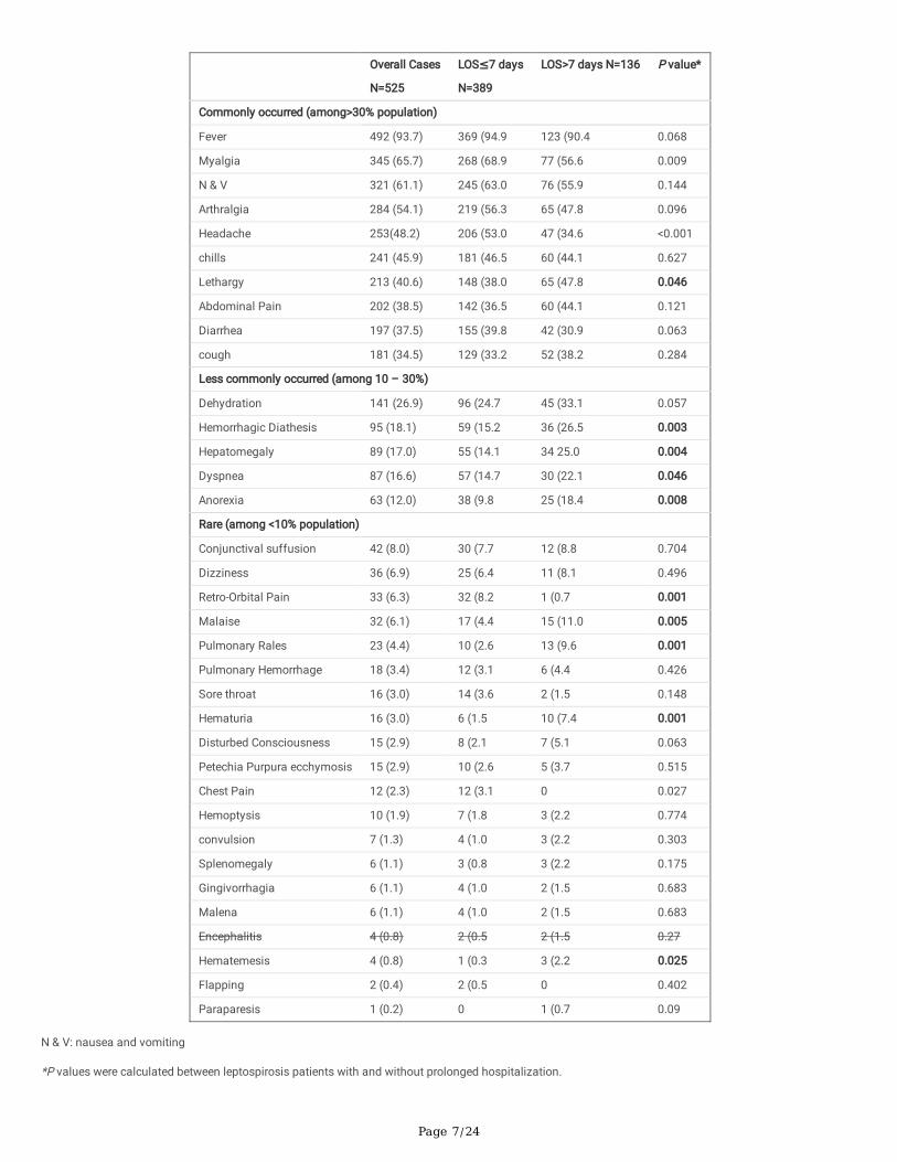
Baseline symptoms were categorized depending on their prevalence as in table 2 and , the typical symptoms of leptospirosis infection were commonlyencountered in the sample studied and included fever, myalgia, arthralgia, headache and chills and these symptoms interestingly were more profound in thepatients without prolonged hospitalization. Among the commonly occurred symptoms in the current study, lethargy was the only symptom with signi�cantassociation with prolonged hospitalization. Among the less commonly occurred symptoms, dehydration was (marginally) signi�cant associated withprolonged hospitalization, whereas all other less commonly occurred symptoms (hemorrhagic diathesis, hepatomegaly, dyspnea, and anorexia) weresigni�cantly associated with spending more than seven days in hospital. Rarely occurred symptoms were more profound in the patients with prolongedhospitalization. However, two of them (retro-orbital pain and chest pain) were signi�cantly associated with spending less than seven days, whereas sore throatwas insigni�cantly more common in patients without prolonged hospitalization.
Table 2: Comparison of Clinical manifestation (at baseline) between leptospirosis patients with and without prolonged hospitalization
Page 7/24
Overall Cases
N=525
LOS≤7 days
N=389
LOS>7 days N=136 P value*
Commonly occurred (among>30% population)
Fever 492 (93.7) 369 (94.9 123 (90.4 0.068
Myalgia 345 (65.7) 268 (68.9 77 (56.6 0.009
N & V 321 (61.1) 245 (63.0 76 (55.9 0.144
Arthralgia 284 (54.1) 219 (56.3 65 (47.8 0.096
Headache 253(48.2) 206 (53.0 47 (34.6 <0.001
chills 241 (45.9) 181 (46.5 60 (44.1 0.627
Lethargy 213 (40.6) 148 (38.0 65 (47.8 0.046
Abdominal Pain 202 (38.5) 142 (36.5 60 (44.1 0.121
Diarrhea 197 (37.5) 155 (39.8 42 (30.9 0.063
cough 181 (34.5) 129 (33.2 52 (38.2 0.284
Less commonly occurred (among 10 – 30%)
Dehydration 141 (26.9) 96 (24.7 45 (33.1 0.057
Hemorrhagic Diathesis 95 (18.1) 59 (15.2 36 (26.5 0.003
Hepatomegaly 89 (17.0) 55 (14.1 34 25.0 0.004
Dyspnea 87 (16.6) 57 (14.7 30 (22.1 0.046
Anorexia 63 (12.0) 38 (9.8 25 (18.4 0.008
Rare (among <10% population)
Conjunctival suffusion 42 (8.0) 30 (7.7 12 (8.8 0.704
Dizziness 36 (6.9) 25 (6.4 11 (8.1 0.496
Retro-Orbital Pain 33 (6.3) 32 (8.2 1 (0.7 0.001
Malaise 32 (6.1) 17 (4.4 15 (11.0 0.005
Pulmonary Rales 23 (4.4) 10 (2.6 13 (9.6 0.001
Pulmonary Hemorrhage 18 (3.4) 12 (3.1 6 (4.4 0.426
Sore throat 16 (3.0) 14 (3.6 2 (1.5 0.148
Hematuria 16 (3.0) 6 (1.5 10 (7.4 0.001
Disturbed Consciousness 15 (2.9) 8 (2.1 7 (5.1 0.063
Petechia Purpura ecchymosis 15 (2.9) 10 (2.6 5 (3.7 0.515
Chest Pain 12 (2.3) 12 (3.1 0 0.027
Hemoptysis 10 (1.9) 7 (1.8 3 (2.2 0.774
convulsion 7 (1.3) 4 (1.0 3 (2.2 0.303
Splenomegaly 6 (1.1) 3 (0.8 3 (2.2 0.175
Gingivorrhagia 6 (1.1) 4 (1.0 2 (1.5 0.683
Malena 6 (1.1) 4 (1.0 2 (1.5 0.683
Encephalitis 4 (0.8) 2 (0.5 2 (1.5 0.27
Hematemesis 4 (0.8) 1 (0.3 3 (2.2 0.025
Flapping 2 (0.4) 2 (0.5 0 0.402
Paraparesis 1 (0.2) 0 1 (0.7 0.09
N & V: nausea and vomiting
*P values were calculated between leptospirosis patients with and without prolonged hospitalization.

Page 8/24
In comparison to patients without prolonged hospitalization, those with prolonged hospitalization had signi�cantly higher mean values of potassium, urea,Scr, uric acid, liver enzymes (AST and ALP but not ALT) and bilirubin. Moreover, they have signi�cantly lower mean values of total protein, albumin, RBCs,platelets, hemoglobin and hematocrit. Clinico-laboratory characters which showed signi�cant association with prolonged hospitalization includedhyponatremia, hyperkalemia, hyperuricemia, AKI (using AKIN system), elevated liver enzymes (ALT or AST), jaundice, hypoalbuminemia, rhabdomyolysis,leukocytosis, thrombocytopenia, low RBCs or hemoglobin, and prolonged PT (table 3).
Table 3: Comparison of Clinico-laboratory characteristics (at baseline) between patients with and without prolonged hospitalization

Page 9/24
Overall Cases N=525 LOS≤7 days N=389 LOS>7 days N=136 P value*
Na (mmol/L) 133.70±5.75 133.82±5.303 133.33± 6.878 0.391
K (mmol/L) 3.841±0.63 3.791±.5917 3.984±0.7407 0.002
Urea (mmol/L) 10.13± 11.78 8.0539±8.76 16.0721± 16.44 <0.001
Scr (µmol/L) 184.51±219.80 149.33±157.68 285.15± 319.84 <0.001
Uric Acid (µmol/L) 418.94±206.48 376.82±159.134 491.28±255.193 0.002
AST (IU/L) 117.72±337.02 93.41±247.782 189.66±512.649 0.005
ALT (IU/L) 114.84±401.49 106.79± 432.570 138.50±291.779 0.439
ALP (IU/L) 135.85±99.02 129.52±103.047 154.49±83.701 0.014
Total Protein (g/L) 68.71±9.65 69.59±9.386 66.10±9.976 <0.001
Albumin (g/L) 36.06±6.71 36.96±6.619 33.39±6.263 <0.001
Globulin (g/L) 32.78±6.47 32.81±6.274 32.71±7.048 0.876
Albumin/Globulin ratio 1.17±0.3 1.1866±0.28436 1.1256±.32829 0.054
Total bilirubin (µmol/L) 36.327±66.58 26.564±43.5039 64.434±103.6861 <0.001
CK (IU/L) 831.38±3305 629.01±1993.360 1446.94± 5648.943 0.068
WBCs (cells ×109) 12.82±13.37 12.76±14.747 12.99±8.450 0.866
RBCs (cells ×1012) 4.684±0.86 4.811±0.8401 4.354±0.8823 <0.001
PLT (cells ×109) 179.98±111.14 191.46±110.562 147.92±106.739 <0.001
Hemoglobin (g/dl) 13.668±8.72 14.197±10.0138 12.185±2.3139 0.023
Hematocrit (%) 38.16±7.34 39.0148±6.88430 35.7829±8.03156 <0.001
PT (sec) 13.72±3.32 13.532±3.4483 14.187±2.9320 0.097
APTT (sec) 38.38±9.47 38.805±9.5810 37.399±9.1817 0.199
INR 1.22±0.31 1.2142±0.32015 1.2438±.27564 0.399
CRP (mg/dl) 26.02±48.3 23.19±45.921 34.90±54.490 0.071
Venous PH 7.39±0.22 7.3949±0.24698 7.3930±0.08836 0.954
Clinical Features based on Laboratory Parameters
Hyponatremia 94 (17.9) 61 (15.7 33 (24.3 0.025
Hypokalemia 141 (26.9) 110 (28.3 31 (22.8 0.214
Hyperkalemia 27 (5.1) 13 (3.3 14 (10.3 0.002
Hyperuricemia >420 µmol/L 52 (9.9) 25 (6.4 27 (19.9 0.003
AKI admission 238 (45.3) 146 (37.5 92 (67.6 <0.001
Elevated ALT 74 (14.1) 45 (11.6) 29 (21.3 0.005
Elevated AST 77 (14.7) 49(12.6) 28 (20.6 0.023
Elevated ALP 48 (9.1) 32 (8.2 16 (11.8 0.181
Transaminitis 52 (9.9) 31 (8.0 21 (15.4 0.012
Jaundice 77 (14.7) 38 (9.8 39 (28.7 <0.001
Hypoalbuminemia< 34 g/L 203 (38.7) 128 (32.9 75 (55.1 <0.001
Urinary sedimentations 109 (20.8) 80 (20.6 29 (21.3 0.851
Rhabdomyolysis 40 (7.6) 23 (5.9 17 (12.5 0.006
Leukocytosis 224 (42.7) 154 (39.6 70 (51.5 0.016
Leucopenia 36 (6.9) 29 (7.5 7 (5.1 0.359
Thrombocytopenia 233 (44.4) 155 (39.8 78 (57.4 <0.001
Low RBC counts 72 (13.7) 37 (9.5 35 (25.7 <0.001

Page 10/24
Low hemoglobin 111 (21.1) 59 (15.2 52 (38.2 <0.001
Prolonged PT 179 (34.1) 113 (29.0 66 (48.5 <0.001
Prolonged aPTT 44 (8.4) 34 (8.7 10 (7.4 0.615
Prolonged aPTT and PT 35 (6.7) 27 (6.9 8 (5.9 0.670
Low venous PH 35 (6.7) 21 (5.4 14 (10.3 0.051
Na: sodium, K: potassium, BUN: blood urea nitrogen, Scr: serum creatinine, AST: aspartate aminotransferase, ALT: alanine aminotransferase, ALP: alkalinephosphatase, CK: creatinine kinase, WBCs: white blood cells, RBCs: red blood cells, PLT: platelets, PT: prothrombin time, aPTT: activated partialthromboplastin time, INR: international normalized ratio, CRP: C reactive protein.
*P values were calculated between patients with and without prolonged hospitalization
4.2.4 Risk Factors for Prolonged Hospitalization in Leptospirosis Infection
Depending on the clinical relevance and physiological rationality of different variables from the different domains studied, we established a series of logisticregression analysis to establish the �nal prediction model for prolonged hospitalization in leptospirosis patients. We included nine variables in the �nallogistic model, but only four of them were found as independent risk factors for prolonged hospitalization and interestingly all were from the clinico-laboratorycharacters (AKI, Jaundice, elevated ALT, prolonged PT) as in table 4. Male gender and late hospitalization although showed insigni�cant association withprolonged hospitalization but were tested in the �nal model due to their hypothetical rationality and they were signi�cantly associated with the severe form ofleptospirosis (unshown data) but none of them showed independent association with prolonged hospitalization neither in the univariate nor in the multivariateanalysis. Old age, pulmonary rales, and low RBCs showed signi�cant association with prolonged hospitalization in the univariate but failed to demonstratesigni�cant association in the multivariate level analysis.
The good-�t model was indicated by the Hosmer-Lemeshow Chi-square value of 11.87 and the signi�cant level of 0.157. The ROC curve analysis for the �nallogistic model showed good prediction accuracy for prolonged hospitalization of leptospirosis patients as indicated by the AUC of 0.775 and signi�cance levelof < 0.001 (Figure 2)
Table 4: Univariate and Multivariate Logistic regression analysis to determine the predictors (risk factors) for prolonged hospitalization of leptospirosis.
Variables Univariate Analysis Multivariate Analysis
P-value OR 95%CI P-value OR 95%CI
Age>40 <.001 2.3 1.5 – 3.4 0.479 0.81 0.5 – 1.4
Male Gender 0.150 1.4 0.89 – 2.1 0.124 1.5 0.9 – 2.6
Late hospitalization
0.220 1.3 0.9 – 1.9 0.979 0.99 0.6– 1.7
Pulmonary Rales 0.001 4.0 1.7 – 9.4 0.153 2.3 0.7 – 6.9
AKI <0.001 3.5 2.3 – 5.3 0.006 2.3 1.3 – 4.1
Jaundice <0.000 3.9 2.3 – 6.4 0.002 2.7 1.4 – 5.0
Elevated ALT
0.006 2.1 1.2 -3.5 0.050 2.0 0.99 – 4.1
Low RBCs
<0.000 3.1 1.9 – 5.4 0.074 1.8 0.95 – 3.3
Prolonged PT
<0.000 2.3 1.5- 3.4 0.023 1.9 1.1 – 3.2
AKI: acute kidney injury, ALT: alanine aminotransferase, RBCs: red blood cells, PT: prothrombin time
Odd ratios (OR) and con�dence intervals (CI) have been rounded off. Hosmer and Lemeshow Test Chi square value = 11.87, Degree of freedom = 8, P = 0.157
Evaluation of leptospirosis-related fatal cases
Tables 5 & Table 6 show the comparisons between fatal and non-fatal cases of leptospirosis infection patients. The overall fatality rate of leptospirosisinfection in the current study was 6.48% (n=34/525) and all cases were attributed to leptospirosis infection. Of the death cases reported, there were 58.8%male cases, 32 (94.1%) Malay, one case (2.9%) was Chinese and another case (2.9%) was Indian. The mean age of the death cases was 48.97 ±16.7 yearsand 32.4% of them were belonging to “leptospirosis risk group”. Around 30% of the death cases were smokers, and all but one case were severe cases.
Page 11/24
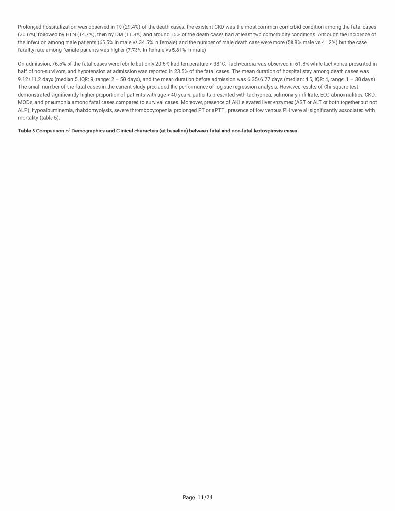
Prolonged hospitalization was observed in 10 (29.4%) of the death cases. Pre-existent CKD was the most common comorbid condition among the fatal cases(20.6%), followed by HTN (14.7%), then by DM (11.8%) and around 15% of the death cases had at least two comorbidity conditions. Although the incidence ofthe infection among male patients (65.5% in male vs 34.5% in female) and the number of male death case were more (58.8% male vs 41.2%) but the casefatality rate among female patients was higher (7.73% in female vs 5.81% in male)
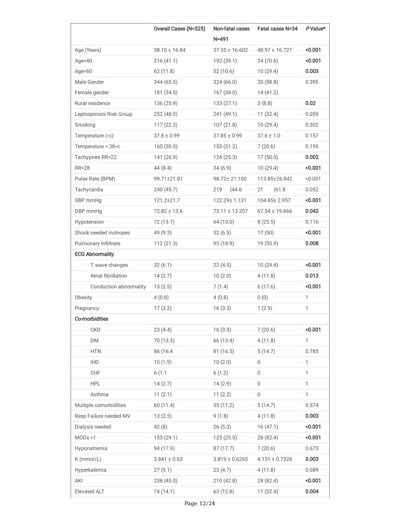
On admission, 76.5% of the fatal cases were febrile but only 20.6% had temperature > 38◦ C. Tachycardia was observed in 61.8% while tachypnea presented inhalf of non-survivors, and hypotension at admission was reported in 23.5% of the fatal cases. The mean duration of hospital stay among death cases was9.12±11.2 days (median:5, IQR: 9, range: 2 – 50 days), and the mean duration before admission was 6.35±6.77 days (median: 4.5, IQR: 4, range: 1 – 30 days).The small number of the fatal cases in the current study precluded the performance of logistic regression analysis. However, results of Chi-square testdemonstrated signi�cantly higher proportion of patients with age > 40 years, patients presented with tachypnea, pulmonary in�ltrate, ECG abnormalities, CKD,MODs, and pneumonia among fatal cases compared to survival cases. Moreover, presence of AKI, elevated liver enzymes (AST or ALT or both together but notALP), hypoalbuminemia, rhabdomyolysis, severe thrombocytopenia, prolonged PT or aPTT , presence of low venous PH were all signi�cantly associated withmortality (table 5).
Table 5 Comparison of Demographics and Clinical characters (at baseline) between fatal and non-fatal leptospirosis cases
Page 12/24
Overall Cases (N=525) Non-fatal cases
N=491
Fatal cases N=34 P Value*
Age (Years) 38.10 ± 16.84 37.35 ± 16.602 48.97 ± 16.727 <0.001
Age>40
Age>60
216 (41.1)
62 (11.8)
192 (39.1)
52 (10.6)
24 (70.6)
10 (29.4)
<0.001
0.003
Male Gender
Female gender
344 (65.5)
181 (34.5)
324 (66.0)
167 (34.0)
20 (58.8)
14 (41.2)
0.395
Rural residence 136 (25.9) 133 (27.1) 3 (8.8) 0.02
Leptospirosis Risk Group 252 (48.0) 241 (49.1) 11 (32.4) 0.059
Smoking 117 (22.3) 107 (21.8) 10 (29.4) 0.302
Temperature (◦c) 37.8 ± 0.99 37.85 ± 0.99 37.6 ± 1.0 0.157
Temperature > 38◦c 160 (30.5) 153 (31.2) 7 (20.6) 0.195
Tachypnea RR>22 141 (26.9) 124 (25.3) 17 (50.0) 0.002
RR>28 44 (8.4) 34 (6.9) 10 (29.4) <0.001
Pulse Rate (BPM) 99.71±21.81 98.72± 21.100 113.85±26.842 <0.001
Tachycardia 240 (45.7) 219 (44.6 21 (61.8 0.052
SBP mmHg 121.2±21.7 122.29± 1.131 104.85± 2.957 <0.001
DBP mmHg 72.82 ± 13.6 73.11 ± 13.207 67.54 ± 19.666 0.042
Hypotension 72 (13.7) 64 (13.0) 8 (23.5) 0.116
Shock needed inotropes 49 (9.3) 32 (6.5) 17 (50) <0.001
Pulmonary In�ltrate 112 (21.3) 93 (18.9) 19 (55.9) 0.008
ECG Abnormality
T wave changes 32 (6.1) 22 (4.5) 10 (29.4) <0.001
Atrial �brillation 14 (2.7) 10 (2.0) 4 (11.8) 0.013
Conduction abnormality 13 (2.5) 7 (1.4) 6 (17.6) <0.001
Obesity 4 (0.8) 4 (0.8) 0 (0) 1
Pregnancy 17 (3.2) 16 (3.3) 1 (2.9) 1
Co-morbidities
CKD 23 (4.4) 16 (3.3) 7 (20.6) <0.001
DM 70 (13.3) 66 (13.4) 4 (11.8) 1
HTN 86 (16.4 81 (16.5) 5 (14.7) 0.785
IHD 10 (1.9) 10 (2.0) 0 1
CHF 6 (1.1 6 (1.2) 0 1
HPL 14 (2.7) 14 (2.9) 0 1
Asthma 11 (2.1) 11 (2.2) 0 1
Multiple comorbidities 60 (11.4) 55 (11.2) 5 (14.7) 0.574
Resp Failure needed MV 13 (2.5) 9 (1.8) 4 (11.8) 0.003
Dialysis needed 42 (8) 26 (5.3) 16 (47.1) <0.001
MODs >1 153 (29.1) 125 (25.5) 28 (82.4) <0.001
Hyponatremia 94 (17.9) 87 (17.7) 7 (20.6) 0.673
K (mmol/L) 3.841 ± 0.63 3.819 ± 0.6265 4.151 ± 0.7328 0.003
Hyperkalemia 27 (5.1) 23 (4.7) 4 (11.8) 0.089
AKI 238 (45.3) 210 (42.8) 28 (82.4) <0.001
Elevated ALT 74 (14.1) 63 (12.8) 11 (32.4) 0.004

Page 13/24
Elevated AST 77 (14.7) 63 (12.8) 14 (41.2) <0.001
AST/ALT Ratio 1.4 ± 1.72 1.37 ± 1.75 1.82 ± 1.23 0.005
Elevated ALP 48 (9.1) 44 (9.0) 4 (11.8) 0.531
Jaundice 77 (14.7) 68 (13.8) 9 (26.5) 0.053
Hypoalbuminemia 203 (38.7) 180 (36.7) 23 (67.6) <0.001
Rhabdomyolysis 40 (7.6) 29 (5.9) 11 (32.4) <0.001
Leukocytosis 224 (42.7) 206 (42.0) 18 (52.9) 0.210
Thrombocytopenia 233 (44.4) 218 (44.4) 15 (44.1) 0.975
Severe Thrombocytopenia 59 (11.2) 51 (10.4) 8 (23.5) 0.042
Anemia 111 (21.1) 101 (20.6) 10 (29.4) 0.166
Prolonged PT 179 (34.1) 157 (32.0) 22 (64.7) <0.001
Prolonged aPTT 44 (8.4) 37 (7.5) 7 (20.6) 0.017
Low venous PH 35 (6.7) 31 (6.3) 4 (11.8) 0.042
Concomitant dengue 37 (7) 34 (6.9) 3 (8.8) 0.676
Pneumonia 85 (16.2) 68 (13.8) 17 (50.0) <0.001
Sepsis 44 (8.4) 36 (7.3) 8 (23.5) 0.004
Late hospitalization 258 (49.1) 241 (49.1) 17 (50.0) 0.918
LOS (days) 6.77±5.68 6.61 ± 5.07 9.12±11.12 0.516
Delayed AB use: 464 (88.4) 431 (87.8) 33 (97.1) 0.161
ICU admission 83 (15.8) 62 (12.6) 21 (61.8) <0.001
RR: respiratory rate, SBP: systolic blood pressure, DBP: diastolic blood pressure, CKD: chronic kidney disease, DM: diabetes mellitus, HTN: hypertension, IHD:ischemic heart disease, CHF: congestive heart failure, HPL: hyperlipidemia, MV: mechanical ventilation, LOS: length of stay, MODs: multiple organdysfunctions.*P values were calculated between fatal and non-fatal cases
The main presenting symptoms in the fatal cases are shown in table 6. Only four baseline presentations were signi�cantly associated with fatal cases andincluded lethargy, dyspnea, pulmonary rales and pulmonary hemorrhage.
Table 6 Comparison of Clinical manifestation (at baseline) between fatal and non-fatal leptospirosis patients

Page 14/24
Overall Cases
N=525
Non-fatal cases N=491 Fatal cases N=34 P value*
Commonly encountered >30%
Fever 492 (93.7) 466 (94.9 26 (76.5 0.001
Myalgia 345 (65.7) 333 (67.8 12 (35.3 <0.001
N & V 321 (61.1) 302 (61.5 19 (55.9 0.515
Arthralgia 284 (54.1) 275 (56.0 9 (26.5 0.001
Headache 253(48.2) 245 (49.9 8 (23.5 0.003
chills 241 (45.9) 235 (47.9 6 (17.6 0.001
Lethargy 213 (40.6) 191 (38.9 22 (64.7 0.003
Abdominal Pain 202 (38.5) 192 (39.1 10 (29.4 0.258
Diarrhea 197 (37.5) 191 (38.9 6 (17.6 0.013
cough 181 (34.5) 166 (33.8 15 (44.1 0.221
Dehydration 141 (26.9) 128 (26.1 13 (38.2 0.122
Less commonly encountered 10-30%
Hemorrhagic Diathesis 95 (18.1) 86 (17.5 9 (26.5 0.19
Hepatomegaly 89 (17.0) 80 (16.3 9 (26.5 0.126
Dyspnea 87 (16.6) 72 (14.7 15 (44.1 <0.001
Anorexia 63 (12.0) 61 (12.4 2 (5.9 0.41
Rarely encountered <10%
Conjunctival Suffusion 42 (8.0) 40 (8.1 2(5.9) 1
Dizziness 36 (6.9) 35 (7.1 1 (2.9 0.5
Retro-Orbital Pain 33 (6.3) 33 (6.7 0 (0) 0.382
Malaise 32 (6.1) 31 (6.3 1 (2.9 0.712
Pulmonary Rales 23 (4.4) 19 (3.9 4 (11.8 0.03
Pulmonary Hemorrhage 18 (3.4) 14 (2.9 4 (11.8 0.023
Sore throat 16 (3.0) 14 (2.9 2 (5.9 0.169
Hematuria 16 (3.0) 15 (3.1 1 (2.9 1
Disturbed Consciousness 15 (2.9) 12 (2.4 3 (8.8 0.066
Petechia Purpura ecchymosis 15 (2.9) 15 (3.1 0 0.614
Chest Pain 12 (2.3) 11 (2.2 1 (2.9 0.428
Hemoptysis 10 (1.9) 9 (1.8 1 (2.9 0.493
convulsion 7 (1.3) 5 (1.0 2 (5.9 0.07
Splenomegaly 6 (1.1) 6 (1.2 0 (0) 1
Gingivorrhagia 6 (1.1) 5 (1.0 1 (2.9 0.334
Malena 6 (1.1) 5 (1.0 1(2.9) 0.334
Encephalitis 4 (0.8) 3 (0.6 1 (2.9) 0.236
N & V: nausea and vomiting *P values were calculated between fatal and non-fatal leptospirosis patients
Determination of complications/causes of death among leptospirosis fatal cases
All leptospirosis death cases were brought alive to hospital and were succumbed to infection within 2-50 days (median: 5 days) of admission. The periodbefore admission ranged from 1-30 days (median: 4.5 days). The diagnosis of leptospirosis infection was con�rmed by the serological test. Only one casewas reported as having previous medical history related to previous leptospirosis infection. More than two third (70.6%) of the fatal cases (n=24/34) diedwithin the �rst seven days of admission and the remaining 10 death cases were reported between day 10 and day 50 of admission. Of the later, 50% (n =

Page 15/24
5/10) died between day 10 and day 15, and another 50% (n = 5/10) died after day 15). We observed multifactorial causes of death including leptospirosisinfection complicated with renal complications (79%), MODs (76.5%), shock (73.5%), pulmonary complications (58.8%), hemorrhagic diathesis ( 52.9%),Cardiac complications (32.4%), altered mental status (26.5%), and GIT bleeding (5.8%). For one patient the data of cause of death was missing.
DiscussionThe incidence of hospital admission due to leptospirosis was signi�cantly higher than that of non-leptospirosis infection admission [17]. Identifying patientswho are at higher risk for prolonged hospitalization due to the disease will help reducing its burden in form of the coast of longer hospital stay. Recognizingthese factors will help not only local practitioners, but also practitioners in other countries where the disease is not endemic but is transmitted through thetravelers and emigrants where the awareness of health care providers regarding the disease burden including the �nancial burden is critically needed [32, 33].This is more important nowadays than ever before as the incidence of the disease is expected to increase as a result of the global warming and increasing theinternational travelling and tourism [5, 34].
The average hospital stays varied in different studies and ranged from less than 5 days [35] to more than 10 days [25]. Our �nding of mean LOS of 6.77 daysis within the international range for the hospital stay in leptospirosis patients. Moreover, the mean LOS for patients with prolonged hospitalization in our study(13.3±7.7 days) was slightly longer comparing to another study (12±9 days) [36], but the mean LOS of patients without prolonged hospitalization in our study(4.46±1.53 days) was near to the LOS in another study (4.34±1.96 days) [37]. Moreover, our results showed more than quarter of the sample investigated(25.9%) had prolonged hospitalization in form of spending more than seven days and was not uncommon to �nd severe leptospirosis patients spentsigni�cantly longer LOS than mild form in a similar manner to the �nding shown by another study [37]. However, 15.4% of patients with prolongedhospitalization had mild form and around half of patients (48.3%) without prolonged hospitalization had severe form in the current study.
Our study showed older patients (age > 40 years) signi�cantly spent more than 7 days comparing to younger patients. However, male patients insigni�cantlyhad more prolonged hospitalization than females. Similarly, one study showed leptospirosis patients of male gender with age > 20 years were insigni�cantlymore and had longer hospitalization, [17]. Moreover, another study showed that males had signi�cantly longer hospital stay compared to females andindividuals aged ≥ 38 had signi�cantly longer hospital stay [38]. Similarly, LOPES, COSTA [39] found that older survivors signi�cantly spent longer hospitalstay than younger survivors. Furthermore, patients from rural areas signi�cantly spent longer hospitalization period than their counterparts from urban area inthe current study. This may be attributed to the longer period they spent before they were admitted to hospital. This delay in seeking care may be due tologistical barriers or de�ciency in the awareness of those people regarding the disease. At the end, this delay is expected to increase the chance for furthercomplications of the disease which necessitate further and longer period of hospital care for those patients. Mean value of the duration before the admissionis signi�cantly longer in patients who had prolonged hospitalization according to data from the current study, and late hospitalization after four days wasinsigni�cantly associated with the prolonged hospitalization. These �ndings underscore the need for awareness campaign particularly in the rural areas wherethe disease is mainly endemic to improve the care seeking behavior for those people and to help them access the hospital care as early as possible [40].
Surprisingly, neither smoking nor any of the studied comorbidities was signi�cantly associated with the prolonged hospitalization, however all of thecomorbidities but CHF (which was insigni�cantly more frequent in patients without prolonged hospitalization) were insigni�cantly more frequentlyencountered in patients with prolonged hospitalization. Moreover, even presence of more than one comorbidity was more frequent in the patients withprolonged hospitalization but did not reach the signi�cant level (p=0.088). This may be attributed to the mild severity of these comorbidities in those patientsor to the well control of these comorbidities in the state where the study was done as the presence of the tertiary level hospitals in this state indicates the easyaccess to high quality health care services there.
Interestingly, patients belonged to leptospirosis risk group, pregnant patients and patients with previous leptospirosis infection insigni�cantly spent < 7 daysmore frequent than their counterparts. This further supports our previous �nding (not shown data) of the association of these features with the mild form ofleptospirosis which is more frequently associated with shorter LOS.
The current study indicated that presence of bleeding symptoms is signi�cantly associated with prolonged hospitalization. Moreover, logistic regressionmodel shows prolonged PT as an independent risk factor for the prolong hospitalization (OR: 1.9). These �ndings underscore the importance of the closemonitoring of the bleeding symptoms during the course of the disease which may indicate the need for longer hospital stay. On the other hand, conjunctivalsuffusion although was considered as pathognomonic symptom for leptospirosis, did not show signi�cant association with the prolonged hospitalizationand this is not surprising as the majority of cases with conjunctival suffusion spent less than seven days in the current study.
The current study indicated that hepatic involvement in form of jaundice, elevation of AST or ALT or both, and hypoalbuminemia was signi�cantly associatedwith extending hospital stay. Moreover, jaundice was the strongest predictor for the prolonged hospitalization (OR: 2.67) among all the factors examined in themultivariate analysis. This �nding could be attributed to the well-known severity of the icteric form which was the cause for the complications and poorprognosis in many studies [10, 41]. Also, elevated ALT was considered as an independent risk factor for prolonged hospitalization in the current study ((OR: 2)and this is not uncommon as ALT although more liver speci�c but its elevation indicates both hepatic and renal dysfunctions and both were seen asindependent risk factors for prolonged hospitalization in the current study. On the other hand, the severe anicteric form when presents as SPHS has rapid andhigh rate of mortality even without presence of jaundice [42]. Moreover, rhabdomyolysis showed signi�cant association with prolonged hospitalization. This isnot uncommon as rhabdomyolysis is a main contributory factor for AKI development [43] which is considered as main cause for the severe form of thedisease and its consequent longer hospital stay.
kidney was the most affected organ in the current study as its injury in form of AKI was the most common among those with prolonged hospitalization; whommore than two thirds of them suffered and signi�cantly associated with this potentially fatal complication. Moreover, AKI was the second strongest predictor(OR: 2.28) for prolonged hospitalization after jaundice similar to another study showed increase in hospital stay was signi�cantly associated with AKI [44].

Page 16/24
The current study also reveals hyponatremia and hyperkalemia were signi�cantly associated with the prolonged hospitalization and this is not unexpected asthese two �ndings indicate further deterioration in the renal function.
Interestingly, AKI and prolonged PT which were identi�ed as independent risk factors for prolonged hospitalization in the current study were previously foundas independent risk factors for prolonged hospitalization in dengue patients as well [45, 46], which may indicate the common causes for the burden ofprolonged hospitalization of the tropical diseases.
The current study showed that although late admission was not an independent risk factor for the prolonged hospitalization, but those patients withprolonged hospitalization spent signi�cantly longer period before being admitted to hospital. Moreover, the initiation of the antibiotic after the 3rd day ofsymptoms onset was strongly associated with prolonged hospitalization. These two �ndings support our previous �ndings which revealed the signi�cantassociation of these two delays with the development of the severe form of the disease (not shown data) which is signi�cantly associated with prolongedhospitalization in the current study. Also, Yang, Yen [47] found delaying of starting the antibiotic therapy was correlated with prolongation of the LOS. Similarly,many studies identi�ed the early admission and starting the proper treatment as the core components for avoiding the poor prognosis and furthercomplications [48-50] which are expected to prolong the hospitalization. It was not unexpected to �nd those who were admitted to ICU had longer hospitalstay (when they kept alive). This is attributed to the more and longer hospital care needed for them and the convalescent period they need after beingdischarged from the ICU.
The burden of the SPHS is mainly presented as higher mortality than as long hospital stay. This was not illustrated in our study and no signi�cant associationof most of the pulmonary complications with the prolonged hospitalization were observed as the predominant form in the current study was the hepato-renalform but not the SPHS.
Bajpai and Bichile [51] found that 22% of the death cases among the acute febrile illnesses in a tertiary care centre after monsoon were because ofleptospirosis. Moreover, a systemic review regarding the burden of leptospirosis and its mortality estimated around 59 thousands deaths annually [52], and thelarge proportion was estimated to occur in young adult male in the tropical regions like south and southeastern Asia and that approaches or exceeds thenumber of deaths caused by other hemorrhagic fever diseases. Furthermore, mortality rates in hospitalized patients with leptospirosis range from 4 to 52%[11] and case fatality of the current study is within that range (6.5%). Early identi�cation of the factors associated with mortality will help clinicians in advanceto keep high level of suspicion to triage those patients at higher risk and immediately commencing the proper course of management needed. Moreover,considering the peculiarities of host, strain, and inoculum in different geographical areas necessitates evaluation of the risk factors of mortality in differentepidemiological settings [53, 54]. No relation between the serogroup and the mortality was established, but serogroup Icterohaemorrhagiae caused much morefrequent icterus and renal insu�ciency than in other serogroups [55, 56]. APACHE II scores for nonsurvivors were signi�cantly higher than survivors [57],however, severity scores were not successful enough to predict mortality in severe leptospirosis [58, 59] and the modi�ed APACHE scoring system, whichincluded multiple clinical and laboratory parameters was not considered as independent risk factor for mortality [60] but can help physician to start theirpharmaceutical care and medical care plans earlier.
The current study is in line with the previous studies which showed old patients were signi�cantly at higher risk for increased mortality due to leptospirosiscompared to younger patients. Moreover, in one study, mortality was higher in adults than pediatrics and old age was independent risk factor for death withinadults [39]. In another study, old age patients were more in need for dialysis (LOPES et al., 2004, Weeratunga et al., 2015) which was associated with themortality in those patients. Moreover, Lopes, Costa [61] showed the mean age of their cohort was signi�cantly higher in patients with hyperkalemia which wassigni�cantly associated with mortality. Moreover, Cetin, Harmankaya [62] found fatal cases were signi�cantly older with signi�cantly higher bilirubin levelcompared to cured patients. On the other hand, male gender although consisted the majority of our cohort (as a common feature in leptospirosis litrature) andof the fatal cases, but was not associated with the fatal cases and, surprisingly, the female gender which consisted the minority in the whole cohort and fatalcases was insigni�cantly more profound in the fatal cases compared to survivors. This was similar to another study showed more fatal cases amongfemales [63]. Moreover, Everard, Edwards [64] found that death from leptospirosis was nearly twice as common among the women as among the men inBarbados. Similarly, Daher, Júnior [65] in their retrospective analysis of two leptospirosis series from 1985-1996, and from 1997-2010 observed the femalegender was signi�cantly associated with nonsurvival cases in those two different periods. However, LOPES, COSTA [39] found a higher, but non-statisticallysigni�cant mortality risk in males, and Chawla, Trivedi [66] observed higher mortality amongst males compared to females in their series which had thehighest mortality (52%) and the main cause of death was the pulmonary hemorrhage and the ARDS. Similarly, Antony, Celine [67] found that case fatality ratewas insigni�cantly higher in males and was signi�cantly increased with age in both sexes. These incompatible �ndings from the different studies includingthe current study regarding the case fatality rate in females compared to males further underscores the need for more investigation of the effect ofleptospirosis on the mortality in females [68].
With the exception of pre-existent CKD, no signi�cant association of any of the comorbidities including DM and HTN with the mortality was shown in thecurrent study. Moreover, and unexpectedly, DM and HTN were more frequent among survivor patients. These �ndings are similar to other �ndings by Dupont,Dupont-Perdrizet [57] who found none of the studied comorbidities which included DM, HTN were signi�cantly associated with mortality. Similarly, Sharp,García [69] found no signi�cant association between any of the comorbidities with fatal cases including diabetes, but the HTN was insigni�cantly morecommon in the fatal cases. Findings of the current study differed partially from that study as our patients with pre-existent CKD were signi�cantly associatedwith mortality (p < 0.001). Also, Sharp, García [69] showed that fatal cases did not differ signi�cantly by reported co-morbidities or chronic medical conditions.Our �nding further illustrates the unique critical impact for the previous renal insu�ciency in increasing the mortality in leptospirosis patients and furtherallocate this group of patients as the highest risk group among all other comorbidities. This supports the ADQI 16 Workshop concepts regarding the criticaleffect of pre-existent CKD in developing AKI which itself associated with mortality in leptospirosis patients [70, 71].

Page 17/24
Interestingly, our study showed obesity was not likely to increase fatality in leptospirosis patients. Although small number of obese patients was reported inthe current study but none of them died. And this �nding is supported by similar �nding of another study which found obesity was insigni�cantly associatedwith the survivors in leptospirosis patients [69]
Similar to Abidi, Dendane [72], pneumonia is likely to increase the risk of mortality in leptospirosis patients according to data from the current report. Anotherstudy showed pneumonia was insigni�cantly more profound in the fatal cases. [57], but nosocomial ventilator-associated pneumonia was signi�cantlyassociated with mortality [73]. Moreover, as expected, presence of sepsis was found to increase the risk of the mortality in the current report similar to anotherstudy [74], this is not uncommon as sepsis is known to cause MODs which is known to increase mortality [70, 75] and this �nding is in line with Chawla,Trivedi [66] who found all the 60 patients entered the ICU had evidence of severe sepsis and more than three quarters of them developed MODs and more thanhalf of the total patients included in that study died.
Current study showed signi�cant association of tachypnea at admission with mortality. Also, RR > 28 was signi�cantly associated with mortality similar toother study [60], but Abidi, Dendane [72] showed RR> 30 as independent risk factors for ICU mortality in leptospirosis patients. Moreover, Hypotension inleptospirosis can result from hypovolemia and decreased systemic vascular resistance induced by vasodilating cytokines and mediators and the current studyshowed signi�cantly lower mean values of SBP and DBP at admission in the fatal cases, and around one quarter of the fatal cases were insigni�cantlyassociated with their hypotension at admission. This �nding is similar to another study where the hypotension at admission was insigni�cantly associatedwith mortality [37], However, other studies showed hypotension at admission was signi�cantly associated with mortality [76, 77].
The current study unexpectedly showed late admission was not likely to increase the risk of mortality, however, this was also identi�ed by other studies [56,57, 61, 78]. Moreover, interestingly, survivors were found to be admitted insigni�cantly later than those who were expired during their hospitalization [56, 69],however, more unexpected results were shown by Spichler, Vilaça [54] who found survivors were signi�cantly admitted later comparing to the fatal cases. Thisapparently bizarre result in that study was attributed to the predominance of the severe pulmonary symptoms in the sample studied and to the fast and highcase fatality in the patients with that severe form. Moreover, the current study also did not show negative consequence of the delay in starting the antibiotic onincreasing mortality due to leptospirosis, and this is similar to the �ndings from other studies in literature which did not �nd signi�cant association of the lateuse of the antibiotic and increasing the mortality [39, 57, 63]. However, this is in contrast to another report showed late antibiotic use was independent riskfactor for mortality [79]. These unexpected �ndings in the current study of the absence of the signi�cant association of delaying the admission or delaying theantibiotic initiation with increasing the mortality should be cautiously interpreted as we had high percent of the whole cohort who were late in admission(around half of all patients) and the high majority were late in starting the antibiotics leading to dissolving the statistical difference between the survivors andfatal cases. It is worthwhile to mention that bene�ts of early use of antibiotic in leptospirosis patients are well documented [65, 80], moreover, �ndings of thepositive effect of using the antibiotic even in the late stage are also observed in different studies as well [61, 81, 82].
The current study also showed the longer LOS is not likely to increase the fatality cases, this is similar to another study [69], but Wagenaar, Goris [83] showedlonger hospitalization period for survivors compared to fatal cases. Moreover, patients with pulmonary involvement were found to insigni�cantly spend longerLOS than those who did not suffered these symptoms and there was no mortality reported in the later group (Martínez García et al., 2000), however, otherstudy showed signi�cant longer stay of the survivor patients [37] with 18.5% case fatality rate, and in another study [76] with 13% case fatality. The shorterLOS of the fatal cases in these studies may be attributed to the severity of the cases or to the late in the admission after the onset of the symptoms [54] whichmake the patients at higher risk for the worse outcomes (death). Our results which showed no association of the late admission with mortality may indicatethe less severity of the disease in our patients and this is supported by the less prevalence of the severe pulmonary involvement in our sample similarly toanother study showed low mortality rate [84] and the less frequent severe pulmonary involvement as well. Moreover, studies showed the shorter LOS in fatalcases also showed higher fatality rate than ours indicating the higher severity of the cases in these studies which may justify the faster and higher mortalityrate in those studies.
The current report showed relatively lower and slower mortality rate but with similar trend in the mortality rate after admission as with many other studies aswe have the majority of the death cases were reported in the �rst seven days of admission and this is in line with another study [73]. This mainly attributed tothe predominance of the less severe hepato-renal but not the pulmonary severe form in our cohort which is known for its higher and faster mortality as themajority of fatal cases were reported within the 24 hours of admission when that form was predominant in the cohorts studied [49, 85]. Moreover, Costa,Lopes [86] showed approximately 44% of the deaths occurred within the �rst three days of hospitalization and another study showed that most of the fatalcases occurred within the �rst �ve days of admission [37]. This mortality rate trend further raises the concerns and increase the need for the fast action as allstudies which investigated the mortality rate in leptospirosis patients including the current study clearly showed the majority of the fatalities occur within the�rst few days of admission. Patrick, Rafaelle [50] attributed the absence of mortality in their cohort to the early diagnosis using the qPCR which providedunequivocal diagnosis and early treatment. This was also concluded from the observations of other studies [48, 87] which showed higher mortality in thosepatients who were diagnosed depending on the clinical symptoms without con�rmation by MAT. This unexpected �nding further encourages the searching forthe predictors of the mortality in those patients to start their treatment in advance (even if the MAT showed negative results).
The current report shows lower rate of mortality (6.5%) compared to many other studies . Although we hope to avoid any death but this percent considered lowcomparing to other studies which showed higher mortality rate [62, 69, 79]. This relatively low rate of mortality in the current report may be attributed to thetreatment measures used in these tertiary care referral centers and the familiarity of the physicians in these referral centers in endemic area (Kelantan) toearly distinguish and diagnose this potentially fatal disease from other similar and less severe diseases, but this level of care and experience of practitionersmay not be the same in other areas with less level of care which may lead to further delaying the diagnosis and the proper treatment which further deterioratethe prognosis and increases the mortality rate.
Page 18/24
In contrast to many studies including the current study, Dupont, Dupont-Perdrizet [57] found hemorrhagic symptoms were not associated with thenonsurvivors. Moreover, they found the mean value of PT was signi�cantly longer in the survivors. Presence of hemorrhagic symptoms in the current study issigni�cantly more frequent in the nonsurvivors and this �nding is similar to the �nding of Durmaz Cetin, Harmankaya [88]. Moreover, Thrombocytopenia is afrequently seen complication in leptospirosis but not always associated with mortality [89], and in the current study the presence of thrombocytopenia in anequally distributed pattern between the survivors and the expired patients and the signi�cant higher urea level and the signi�cant association of the AKI withthe expired patients suggest the uremia as the primary cause of the bleeding in addition to thrombocytopenia. Another suggested cause is the hepaticcholestasis (as our study identi�ed a signi�cant association of the transaminitis and insigni�cant association (p=0.053) of jaundice with the expired patients)which lead to prolonged PT as a result of de�ciency in vitamin K dependent coagulation factors [36]. On the other hand, anemia although was more frequentwith the fatal cases but was not signi�cantly associated with increased mortality in the current study. This �nding is in agreement with other studies found thehigher incidence of anemia in the fatal cases [53, 62]. It is worthwhile to mention that as anemia is known to have higher prevalence in females [90] and theyhad higher mortality rate in the current study, it may have contributing effect on increasing mortality in females but more studies of the effect of anemia onmortality in female leptospirosis patients are needed to support this suggestion.
Goswami, Goswami [79] found all the respiratory involvement symptoms were insigni�cantly more frequent in the fatal cases. Similarly, in the current studymost of these symptoms were more commonly observed in the fatal cases. Hemoptysis was insigni�cantly associated with mortality in the current study aswas concluded in another study [60], and was previously reported as not associated with death although the respiratory insu�ciency was considered aspredictor for mortality in that study [53]. However, hemoptysis was found as signi�cantly associated with mortality in another study [91]. Also, Esen, Sunbul[56] found respiratory symptoms were insigni�cantly more frequent in the expired patients, however, interestingly, they found the pathological auscultation onchest examination was insigni�cantly more frequent in the survivors. The current report showed the severe presentations of pulmonary involvement in formof pulmonary hemorrhage and respiratory failure although were rarely observed in current cohort similar to one study [91], but were signi�cantly associatedwith the fatal cases similar to other studies [54, 72, 92]. Moreover, dyspnea and pulmonary rales were signi�cantly associated with mortality in the currentstudy. These �ndings are similar to other �ndings showed dyspnea was strongly associated with mortality in one study [57] and pulmonary rales wereconsidered the same in another study [60]
Although the ECG abnormalities are commonly reported in leptospirosis patients but the clinical signs of cardiac failure are not commonly reported andalthough it is believed to be underestimated but will have poor prognosis whenever available [75]. In the current report, different ECG abnormalities wereobserved and included T-wave changes, atrial �brillation and conduction abnormalities and all were signi�cantly associated with the fatal cases. Moreover,cardiovascular collapse in form of shock needed inotropes which indicates the cardiac failure was also signi�cantly associated with mortality. This is similarto other study by Pertuiset, Fen [93] who showed the cardiac arrhythmias and cardiovascular collapse as main causes of death after the pneumopathy andperfuse hemorrhage. Another study showed cardiovascular collapse was insigni�cantly associated with the nonservivors [57].
Jaundice in the current study was insigni�cantly associated with the fatal cases, and this is very common as it was not known to be fatal in leptospirosispatients in many studies [53, 57, 61]. Moreover, Spichler, Vilaça [54] found jaundice was not signi�cantly associated with fatal cases but high level of bilirubin> 6 mg/dL was signi�cantly associated with fatal cases in the univariate analysis but did not remain as an independent risk factor for lethal outcome in themultivariate analysis. Contrary to all previous studies including the current report, Goswami, Goswami [79] found that jaundice was signi�cantly associatedwith the fatal cases. Moreover, the current study identi�ed the elevation of any of the transaminases or both of them (transaminitis) or AST/ALT ratio mayincrease the risk of mortality. These �ndings are in line with previous studies [56, 79, 93]. Furthermore, hypoalbuminemia was associated with mortality inmore than two third of the fatal cases and similar �nding was observed by another study [69].
The current report as many previous studies recognizes LAKI as signi�cantly associated with mortality regardless of its severity, moreover, it identi�esstatistically signi�cant higher potassium level at admission in the fatal cases compared to survivors and hyperkalemia was more frequently observed in thefatal cases during their admission. These �ndings are in agreement with other �ndings by Lopes, Costa [61] who found that initial serum potassium waspositively and strongly correlated with the risk of in-hospital death. However, Esen, Sunbul [56] identi�ed hyperkalemia at admission as an independent riskfactor for mortality in leptospirosis patients but oliguria was insigni�cantly more common in the fatal cases in the same study. Contrary, Sharp, García [69] didnot observe elevated potassium level associated with increasing mortality neither at admission nor during hospitalization. Moreover, lower than normal bloodPH was associated with increasing mortality according to data from the current study and this �nding is in line with another study showed metabolic acidosiswith PH <7.2 was signi�cantly associated with fatal cases in the univariate analysis but not in the multivariate analysis [75]. However, Tantitanawat andTanjatham [37] found metabolic acidosis with HCO3 < 20mmol/L signi�cantly associated with severity and mortality.
Unnikrishnan, Pisharody [63] unexpectedly found that neither renal failure nor hepatic involvement were signi�cant predictors for mortality, however theauthors attributed these bizarre results to the selection bias in the sample studied. In a similar unexpected manner, Goswami, Goswami [79] found nosigni�cant difference neither in Scr level nor in urea level between the survivors and the fatal cases. But contrary, Spichler, Vilaça [54] found that even in placeswith a high rate of pulmonary involvement and a high rate of pulmonary hemorrhage as the major cause of death, renal failure remains an importantdeterminant of death.
Inclusion of CNS in leptospirosis was associated with worse prognosis in many studies [70] and altered level of consciousness was found as the strongestpredictor for mortality [53, 56]. However, because of the di�culty in de�ning that term as evident by many terms applied to altered states of consciousness byvarious observers like clouding of consciousness, confessional state, delirium, lethargy, obtundation, stupor, dementia, hypersomnia, vegetative state and coma, presence of any of the symptoms which indicate the inclusion of CNS may suggest the direct contribution of the CNS to the death [94]. Moreover,Pappachan, Mathew [91] identi�ed meningism and disorientation at admission as mortality indicators. The current report showed lethargy was signi�cantlyassociated with mortality in around 65% of the fatal cases. Also, disturbed consciousness although is rarely reported in the current report but showsmarginally signi�cant association with the fatal cases (p=0.066). However, Covic, Goldsmith [95] found the altered level of consciousness was signi�cantly

Page 19/24
associated with all the deceased cases. Moreover, altered level of consciousness was insigni�cantly associated with mortality in the multivariate analysis inanother study [57]. These �ndings further underscore the importance of inclusion of the neurological assessment in leptospirosis patients to accelerate theICU admission for those with lower GCS value [22, 72]. Other CNS symptoms were rarely noted in the current report and included convulsion, encephalitis, butwere insigni�cantly more frequent in the fatal cases in a similar manner to other studies [69, 79]
Around one third of the patients in the current study developed MODs and was not unexpected to �nd presence of MODs was signi�cantly associated withmortality. This is similar to the �nding of Weeratunga, Fernando [75] who showed the presence of MODs was independently associated with the mortality. In asimilar manner, another study found that mortality is increased with increased the number of the dysfunctional systems and critical care management at threeorgan involvement stage would improve the outcome of the patients [96].
Study Limitations
Being retrospective study, we were limited to the data mentioned in the routine medical records, including the physician assessment and diagnosis for thedifferent cases. Moreover, the relatively small number of fatal cases did not allow establishing a reliable logistic prediction model for mortality in leptospirosispatients, but the factors which positively linked with leptospirosis mortality where investigated using univariate analysis. Also, the discharge criteria may varyamong the different practitioners attending patients, which may lead to different LOS depending on physicians’ opinions and this might affect the risk factorsfor prolonged hospitalization identi�ed by the current study. However, patients are always discharged when their clinical and laboratory �ndings are stabilized.Nonetheless, the strength of this study lies on including appropriately powered sample size of leptospirosis patients. Moreover, it is the only large-scale studyin Malaysia investigated the factors associated with prolonged hospitalization and mortality in leptospirosis patients.
ConclusionOur current case series identi�ed signi�cant proportion of patients with prolonged hospitalization and revealed that leptospirosis infection when combinedwith AKI, jaundice, elevated ALT and prolonged PT might denote seriously sick patients who can potentially have more morbidity in form of increased hospitalstay. Mortality rate of 6.5% in the current study is considered within the mortality range of the disease, and as with other studies in literature, the mortality ofthe disease was associated with several factors including age > 40 years, tachypnea at admission particularly when RR exceeds 28, presence of shockrequired vasoactive drugs, respiratory failure required mechanical ventilation, pneumonia, sepsis, pulmonary rales, presence of ECG abnormality, lethargy, pre-existent CKD, development of AKI during the course of the disease, need for dialysis, MODs, elevation of any of the liver transaminases or both together,rhabdomyolysis, severe thrombocytopenia and prolonged PT or aPTT.
Identi�cation of those patients with the abovementioned factors at the earliest and their management with special care will be advantageous in reducingmorbidity and hence their bed occupancy in the hospital and mortality.
Recommendation
The atypical severe form including the fatal pulmonary hemorrhage in the anicteric form of the disease should be further investigated using large scalestudies to identify the risk factors associated with this more form of the disease as the current study showed predominance for the other type of the severeform (hepato-renal). Moreover, more appropriately powered multicenter trials are needed to investigate the factors of prolonged hospital stay in leptospirosispatients to support the �ndings of the current study, and to establish the prediction models for mortality in severe form as well. This will clearly help drawingthe suitable plans in managing those patients and to reduce the burden of the disease in form of mortality and prolonged hospitalization of leptospirosispatients.
AbbreviationsaPTT: activated partial thromboplastin time. AST: aspartate aminotransferase. AKI: Acute kidney injury. ALP: Alkaline phosphatase. ALT: Alanineaminotransferase. CHF: chronic heart disease. CK: Creatine kinase. CNS: Central nervous system. CRP: C-reactive protein. DM: Diabetes mellitus. HPL:Hyperlipidemia. HPRZII: Hospital Perempuan Raja Zainab II. HTN: Hypertension. HUSM: Hospital University Sains Malaysia. IHD: Ischemic heart diseases. INR:International normalized ratio. NTD: Neglected tropical diseases. PT: Prothrombin time. RBCs: Red blood cells. SIRS: systemic in�ammatory responsesyndrome. SPHS: Severe pulmonary hemorrhagic syndrome. ULN: Upper limit of normal. WBCs: White blood cells
DeclarationsEthics approval and consent to participate
This study was approved by Ministry of Health Malaysia Research Ethic Committee (MREC), and by Human Research Ethic Committee USM (HREC) as well.The patients’ informed consents were waived by these ethics committees as the study was observational retrospective analysis. All methods were performedaccording to the guidelines and regulations of the tertiary level teaching hospitals where the study had been done.
Consent for publication
Not applicable
Availability of data and materials
All data generated or analyzed during this study are included in this published article

Page 20/24
Competing interests
The authors declare that they have no competing interests
Funding
This research did not receive any type of fund.
Authors’ contributions
Conceptualization and designing the study: Yassen K. Al Hariri, Syed A.S. Sulaiman, Amer H. Khan. Performing the study and data curation: Yassen K. Al Hariri,Azreen S. Adnan. Analysis of data: Sundos Q. Al-Ebrahem, Yassen K. Al Hariri. Supervision of the methodology: Syed A.S. Sulaiman, Amer H. Khan, Azreen S.Adnan. Writing the 1st draft: Yassen K. Al Hariri, Amer H. Khan. Writing and reviewing the �nal manuscript: Yassen K. Al Hariri, Syed A.S. Sulaiman, Amer H.Khan, Azreen S. Adnan, Sundos Q. Al-Ebrahem
Acknowledgement:
The authors would like to thank the Director General of Health Malaysia for the permission to publish this paper. Moreover, we would like to thank the staff ofmedical records at HUSM and HRBZII for their assistance in retrieving patients’ medical records.
References1. WHO/SEARO. Informal Expert consultation on Surveillance, Diagnosis and Risk Reduction of Leptospirosis. Chennai, 17-18 September 2009.
2. Bharti AR, Nally JE, Ricaldi JN, Matthias MA, Diaz MM, Lovett MA, et al. Leptospirosis: a zoonotic disease of global importance. The Lancet infectiousdiseases. 2003;3(12):757-71.
3. Cruz L, Vargas R, Lopes A. Leptospirosis: a worldwide resurgent zoonosis and important cause of acute renal failure and death in developing nations.Ethnicity & disease. 2008;19(1 Suppl 1):S1-37-41.
4. WHO. Report of the First Meeting of the Leptospirosis Burden Epidemiology Reference Group, Geneva. Geneva: World Health Organization; 2010. ReportNo.: 9241599898.
5. Baylor College of Medicine. Emerging Infectious Diseases 2019 [3rd March 2019]. Available from: https://www.bcm.edu/departments/molecular-virology-and-microbiology/emerging-infections-and-biodefense/emerging-infectious-diseases.
�. Al-Wakeel JS. Accuracy and precision of the CKD-EPI and MDRD predictive equations compared with glomerular �ltration rate measured by inulinclearance in a Saudi population. Annals of Saudi medicine. 2016;36(2):128-34.
7. Torgerson PR, Hagan JE, Costa F, Calcagno J, Kane M, Martinez-Silveira MS, et al. Global Burden of Leptospirosis: Estimated in Terms of DisabilityAdjusted Life Years. PLoS Negl Trop Dis. 2015;9(10):e0004122.
�. Christova I, Tasseva E. Human leptospirosis in Bulgaria, 2010-2014. Probl Infect Parasit Dis. 2016;44(2):23-9.
9. CDC. LEPTOSPIROSIS Fact Sheet for Clinicians 2018 [27th March 2019]. Available from: https://www.cdc.gov/leptospirosis/pdf/fs-leptospirosis-clinicians-eng-508.pdf.
10. Levett PN. Leptospirosis. Clinical Microbiology Reviews. 2001;14(2):296.
11. UpToDate. Leptospirosis: Epidemiology, microbiology, clinical manifestations, and diagnosis 2019 [5th May 2019]. Available from:https://www.uptodate.com/contents/leptospirosis-epidemiology-microbiology-clinical-manifestations-and-diagnosis?search=leptospirosis&source=search_result&selectedTitle=1~79&usage_type=default&display_rank=1.
12. Levett PN, Haake DA. Leptospira species (leptospirosis). Mandell, Douglas and Bennett’s principles and practice of infectious diseases, 7th edn Churchill-Livingstone, Philadelphia. 2010:3059-4005.
13. WHO/SEARO. Leptospirosis : Fact Sheet 2009 [updated 2009. Available from: https://apps.who.int/iris/handle/10665/205437.
14. Hotez PJ, Alvarado M, Basáñez M-G, Bolliger I, Bourne R, Boussinesq M, et al. The Global Burden of Disease Study 2010: Interpretation and Implicationsfor the Neglected Tropical Diseases. PLOS Neglected Tropical Diseases. 2014;8(7):e2865.
15. Gasem MH, Hadi U, Alisjahbana B, Tjitra E, Hapsari M, Lestari ES, et al. Leptospirosis in Indonesia: diagnostic challenges associated with atypical clinicalmanifestations and limited laboratory capacity. BMC infectious diseases. 2020;20(1):1-11.
1�. WHO/WPRO. Emerging disease surveillance and response: Leptospirosis 2019 [Available from:http://origin.wpro.who.int/emerging_diseases/Leptospirosis/en/.
17. Traxler RM, Callinan LS, Holman RC, Steiner C, Guerra MA. Leptospirosis-associated Hospitalizations, United States, 1998-2009. Emerging InfectiousDiseases. 2014;20(8):1273-9.
1�. Lim JK, Murugaiyah VA, Ramli AS, Rahman HA, Mohamed NSF, Shamsudin NN, et al. A case study: leptospirosis in Malaysia. 2011.
19. Tan WL, Soelar SA, Mohd Suan MA, Hussin N, Cheah WK, Verasahib K, et al. LEPTOSPIROSIS INCIDENCE AND MORTALITY IN MALAYSIA. The SoutheastAsian journal of tropical medicine and public health. 2016;47(3):434-40.
20. New Straits Times. Leptospirosis third deadliest after dengue and malaria 2020 [Available from:https://www.nst.com.my/news/nation/2020/03/573627/leptospirosis-third-deadliest-after-dengue-and-malaria#:~:text=KUALA%20LUMPUR%3A%20Leptospirosis%20is%20the,malaria%2C%20said%20a%20leading%20parasitologist.&text=%E2%80%9CThe%
Page 21/24
21. Abdullah NM, Mohammad WMZW, Shafei MN, Sukeri S, Idris Z, Ari�n WN, et al. Leptospirosis and its prevention: knowledge, attitude and practice ofurban community in Selangor, Malaysia. BMC Public Health. 2019;19(1):628.
22. Fann RJ, Vidya RR, Chong HE, Indralingam V, Christopher Chan WS. Clinical presentations and predictors of mortality for leptospirosis - A study fromsuburban area in Malaysia. The Medical journal of Malaysia. 2020;75(1):52-6.
23. Garba B, Bahaman AR, Bejo SK, Zakaria Z, Mutalib AR, Bande F. Major epidemiological factors associated with leptospirosis in Malaysia. Acta Tropica.2018;178:242-7.
24. Benacer D, Thong KL, Verasahib KB, Galloway RL, Hartskeerl RA, Lewis JW, et al. Human Leptospirosis in Malaysia Reviewing the Challenges After 8Decades (1925-2012). Asia-Paci�c Journal of Public Health. 2016;28(4):290-302.
25. Tique V, Mattar S, Miranda J, Oviedo M, Noda A, Montes E, et al. Clinical and Epidemiological Status of Leptospirosis in a Tropical Caribbean Area ofColombia. BioMed Research International. 2018:1-8.
2�. Wahab ZA, editor Epidemiology and current situation of Leptospirosis in Malaysia. Local Authority Conference on Environmental Health; 2015.
27. Hussin N, Jaafar J, Naing NN, Mat HA, Muhamad AH, Mamat MN. A review of dengue fever incidence in Kota Bharu, Kelantan, Malaysia during the years1998-2003. Southeast Asian journal of tropical medicine and public health. 2005;36(5):1179.
2�. Ministry of Health Malaysia. Guidelines for The Diagnosis, Management, Prevention and Control of Leptospirosis, 1st Edition. Malaysia:; 2011.
29. Rajapakse S, Fernando N, Niloofa M, De Silva H, Karunanayake L, Premawansa S, et al. Leptospirosis: challenges in diagnosis, and predictors of severity.Journal of the Ceylon College of Physicians. 2016.
30. Bursac Z, Gauss CH, Williams DK, Hosmer DW. Purposeful selection of variables in logistic regression. Source code for biology and medicine.2008;3(1):17.
31. Mallhi TH, Khan AH, Adnan AS, Sarriff A, Khan YH, Jummaat F. Clinico-laboratory spectrum of dengue viral infection and risk factors associated withdengue hemorrhagic fever: a retrospective study. BMC infectious diseases. 2015;15(1):399.
32. Bandara M, Ananda M, Wickramage K, Berger E, Agampodi S. Globalization of leptospirosis through travel and migration. Globalization and health.2014;10 (1)(1):61.
33. Lau C, Smythe L, Weinstein P. Leptospirosis: An emerging disease in travellers. Travel Medicine and Infectious Disease. 2010;8:33e9.
34. Hotez PJ. Global urbanization and the neglected tropical diseases. PLOS Neglected Tropical Diseases. 2017;11(2):e0005308.
35. Holla R, Darshan B, Pandey L, Unnikrishnan B, Kumar N, Thapar R, et al. Leptospirosis in Coastal South India: A Facility Based Study. BioMed ResearchInternational. 2018;2018.
3�. De Francesco Daher E, Oliveira Neto FH, Ramirez SMP. Evaluation of hemostasis disorders and anticardiolipin antibody in patients with severeleptospirosis. Revista Do Instituto De Medicina Tropical De Sao Paulo. 2002;44(2):85-90.
37. Tantitanawat S, Tanjatham S. Prognostic factors associated with severe leptospirosis. J Med Assoc Thai. 2003;86(10):925-31.
3�. Goris MGA, Kikken V, Straetemans M, Alba S, Goeijenbier M, van Gorp ECM, et al. Towards the Burden of Human Leptospirosis: Duration of Acute Illnessand Occurrence of Post-Leptospirosis Symptoms of Patients in The Netherlands. PloS one. 2013;8(10).
39. LOPES AA, COSTA E, COSTA YA, SACRAMENTO E, de OLIVEIRA JUNIOR ARR, LOPES MB, et al. COMPARATIVE STUDY OF THE IN-HOSPITAL CASE-FATALITY RATE OF LEPTOSPIROSIS BETWEEN PEDIATRIC AND ADULT PATIENTS OF DIFFERENT AGE GROUPS. Rev Inst Med trop S Paulo.2004;46(1):19-24.
40. Pakoa JG, Soupé-Gilbert M-E, Girault D, Takau D, Gaviga J, Gourinat A-C, et al. High incidence of leptospirosis in an observational study of hospitaloutpatients in Vanuatu highlights the need for improved awareness and diagnostic capacities. PLOS Neglected Tropical Diseases. 2018;12(6):e0006564.
41. Gancheva GI. Liver involvement in leptospirosis. population. 2016;2006:8.
42. Gulati S, Gulati A. Pulmonary manifestations of leptospirosis. Lung India: O�cial Organ of Indian Chest Society. 2012;29(4):347.
43. Daher EF, de Abreu KL, da Silva Junior GB. Leptospirosis-associated acute kidney injury. J Bras Nefrol. 2010;32(4):400-7.
44. Bates D, Su L, Yu D, Chertow G, Seger D, Gomes D, et al. Mortality and costs of acute renal failure associated with amphotericin B therapy. ClinicalInfectious Diseases. 2001;32(5):686-93.
45. Khalil MAM, Tan J, Khalil MAU, Awan S, Rangasami M. Predictors of hospital stay and mortality in dengue virus infection-experience from Aga KhanUniversity Hospital Pakistan. BMC research notes. 2014;7(1):473.
4�. Mallhi TH, Khan AH, Sarriff A, Adnan AS, Khan YH. Determinants of mortality and prolonged hospital stay among dengue patients attending tertiary carehospital: a cross-sectional retrospective analysis. BMJ open. 2017;7(7):e016805.
47. Yang H-Y, Yen T-H, Lin C-Y, Chen Y-C, Pan M-J, Lee C-H, et al. Early identi�cation of leptospirosis as an ignored cause of multiple organ dysfunctionsyndrome. Shock (Augusta, Ga). 2012;38(1):24-9.
4�. Ittyachen AM, Krishnapillai TV, Nair MC, Rajan AR. Retrospective study of severe cases of leptospirosis admitted in the intensive care unit. Journal ofPostgraduate Medicine. 2007;53(4):232-5.
49. Tubiana S, Mikulski M, Becam J, Lacassin F, Lefèvre P, Gourinat A-C, et al. Risk factors and predictors of severe leptospirosis in New Caledonia. PLoSneglected tropical diseases. 2013;7(1):e1991.
50. Patrick H, Rafaelle T, Claude O, Pascale B, Guillaume H, Nicolas V, et al. Factors Associated with Severe Leptospirosis, Martinique, 2010–2013. EmergingInfectious Disease journal. 2015;21(12):2221.
51. Bajpai S, Bichile LS. Mortality analysis of patients of acute febrile illness during monsoon in a tertiary care hospital of mumbai. Infectious Diseases inClinical Practice. 2008;16(5):294-7.
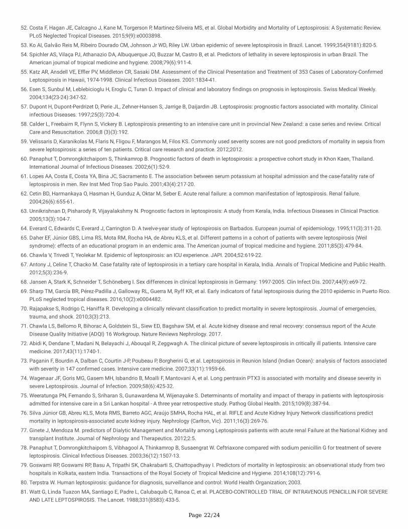
Page 22/24
52. Costa F, Hagan JE, Calcagno J, Kane M, Torgerson P, Martinez-Silveira MS, et al. Global Morbidity and Mortality of Leptospirosis: A Systematic Review.PLoS Neglected Tropical Diseases. 2015;9(9):e0003898.
53. Ko AI, Galvão Reis M, Ribeiro Dourado CM, Johnson Jr WD, Riley LW. Urban epidemic of severe leptospirosis in Brazil. Lancet. 1999;354(9181):820-5.
54. Spichler AS, Vilaça PJ, Athanazio DA, Albuquerque JO, Buzzar M, Castro B, et al. Predictors of lethality in severe leptospirosis in urban Brazil. TheAmerican journal of tropical medicine and hygiene. 2008;79(6):911-4.
55. Katz AR, Ansdell VE, E�er PV, Middleton CR, Sasaki DM. Assessment of the Clinical Presentation and Treatment of 353 Cases of Laboratory-Con�rmedLeptospirosis in Hawaii, 1974-1998. Clinical Infectious Diseases. 2001:1834-41.
5�. Esen S, Sunbul M, Leblebicioglu H, Eroglu C, Turan D. Impact of clinical and laboratory �ndings on prognosis in leptospirosis. Swiss Medical Weekly.2004;134(23-24):347-52.
57. Dupont H, Dupont-Perdrizet D, Perie JL, Zehner-Hansen S, Jarrige B, Daijardin JB. Leptospirosis: prognostic factors associated with mortality. Clinicalinfectious Diseases. 1997;25(3):720-4.
5�. Calder L, Freebairn R, Flynn S, Vickery B. Leptospirosis presenting to an intensive care unit in provincial New Zealand: a case series and review. CriticalCare and Resuscitation. 2006;8 (3)(3):192.
59. Velissaris D, Karanikolas M, Flaris N, Fligou F, Marangos M, Filos KS. Commonly used severity scores are not good predictors of mortality in sepsis fromsevere leptospirosis: a series of ten patients. Critical care research and practice. 2012;2012.
�0. Panaphut T, Domrongkitchaiporn S, Thinkamrop B. Prognostic factors of death in leptospirosis: a prospective cohort study in Khon Kaen, Thailand.International Journal of Infectious Diseases. 2002;6(1):52-9.
�1. Lopes AA, Costa E, Costa YA, Bina JC, Sacramento E. The association between serum potassium at hospital admission and the case-fatality rate ofleptospirosis in men. Rev Inst Med Trop Sao Paulo. 2001;43(4):217-20.
�2. Cetin BD, Harmankaya O, Hasman H, Gunduz A, Oktar M, Seber E. Acute renal failure: a common manifestation of leptospirosis. Renal failure.2004;26(6):655-61.
�3. Unnikrishnan D, Pisharody R, Vijayalakshmy N. Prognostic factors in leptospirosis: A study from Kerala, India. Infectious Diseases in Clinical Practice.2005;13(3):104-7.
�4. Everard C, Edwards C, Everard J, Carrington D. A twelve-year study of leptospirosis on Barbados. European journal of epidemiology. 1995;11(3):311-20.
�5. Daher EF, Júnior GBS, Lima RS, Mota RM, Rocha HA, de Abreu KLS, et al. Different patterns in a cohort of patients with severe leptospirosis (Weilsyndrome): effects of an educational program in an endemic area. The American journal of tropical medicine and hygiene. 2011;85(3):479-84.
��. Chawla V, Trivedi T, Yeolekar M. Epidemic of leptospirosis: an ICU experience. JAPI. 2004;52:619-22.
�7. Antony J, Celine T, Chacko M. Case fatality rate of leptospirosis in a tertiary care hospital in Kerala, India. Annals of Tropical Medicine and Public Health.2012;5(3):236-9.
��. Jansen A, Stark K, Schneider T, Schöneberg I. Sex differences in clinical leptospirosis in Germany: 1997-2005. Clin Infect Dis. 2007;44(9):e69-72.
�9. Sharp TM, García BR, Pérez-Padilla J, Galloway RL, Guerra M, Ryff KR, et al. Early indicators of fatal leptospirosis during the 2010 epidemic in Puerto Rico.PLoS neglected tropical diseases. 2016;10(2):e0004482.
70. Rajapakse S, Rodrigo C, Haniffa R. Developing a clinically relevant classi�cation to predict mortality in severe leptospirosis. Journal of emergencies,trauma, and shock. 2010;3(3):213.
71. Chawla LS, Bellomo R, Bihorac A, Goldstein SL, Siew ED, Bagshaw SM, et al. Acute kidney disease and renal recovery: consensus report of the AcuteDisease Quality Initiative (ADQI) 16 Workgroup. Nature Reviews Nephrology. 2017.
72. Abidi K, Dendane T, Madani N, Belayachi J, Abouqal R, Zeggwagh A. The clinical picture of severe leptospirosis in critically ill patients. Intensive caremedicine. 2017;43(11):1740-1.
73. Paganin F, Bourdin A, Dalban C, Courtin J-P, Poubeau P, Borgherini G, et al. Leptospirosis in Reunion Island (Indian Ocean): analysis of factors associatedwith severity in 147 con�rmed cases. Intensive care medicine. 2007;33(11):1959-66.
74. Wagenaar JF, Goris MG, Gasem MH, Isbandrio B, Moalli F, Mantovani A, et al. Long pentraxin PTX3 is associated with mortality and disease severity insevere Leptospirosis. Journal of Infection. 2009;58(6):425-32.
75. Weeratunga PN, Fernando S, Sriharan S, Gunawardena M, Wijenayake S. Determinants of mortality and impact of therapy in patients with leptospirosisadmitted for intensive care in a Sri Lankan hospital - A three year retrospective study. Pathog Global Health. 2015;109(8):387-94.
7�. Silva Júnior GB, Abreu KLS, Mota RMS, Barreto AGC, Araújo SMHA, Rocha HAL, et al. RIFLE and Acute Kidney Injury Network classi�cations predictmortality in leptospirosis-associated acute kidney injury. Nephrology (Carlton, Vic). 2011;16(3):269-76.
77. Ginete J, Mendoza M. predictors of Dialytic Management and Mortality among Leptospirosis patients with acute renal Failure at the National Kidney andtransplant Institute. Journal of Nephrology and Therapeutics. 2012;2:5.
7�. Panaphut T, Domrongkitchaiporn S, Vibhagool A, Thinkamrop B, Susaengrat W. Ceftriaxone compared with sodium penicillin G for treatment of severeleptospirosis. Clinical Infectious Diseases. 2003;36(12):1507-13.
79. Goswami RP, Goswami RP, Basu A, Tripathi SK, Chakrabarti S, Chattopadhyay I. Predictors of mortality in leptospirosis: an observational study from twohospitals in Kolkata, eastern India. Transactions of the Royal Society of Tropical Medicine and Hygiene. 2014;108(12):791-6.
�0. Terpstra W. Human leptospirosis: guidance for diagnosis, surveillance and control: World Health Organization; 2003.
�1. Watt G, Linda Tuazon MA, Santiago E, Padre L, Calubaquib C, Ranoa C, et al. PLACEBO-CONTROLLED TRIAL OF INTRAVENOUS PENICILLIN FOR SEVEREAND LATE LEPTOSPIROSIS. The Lancet. 1988;331(8583):433-5.
Page 23/24
�2. Pappas G, Cascio A. Optimal treatment of leptospirosis: queries and projections. International journal of antimicrobial agents. 2006;28(6):491-6.
�3. Wagenaar J, Goris M, Partiningrum D, Isbandrio B, Hartskeerl R, Brandjes D, et al. Coagulation disorders in patients with severe leptospirosis areassociated with severe bleeding and mortality. Tropical Medicine & International Health. 2010;15(2):152-9.
�4. Sivaprakasam V, Zochowski WJ, Palmer MF. Clinical spectrum of severe leptospirosis in the UK. JMM case reports. 2014;1(4).
�5. Singh SS, Vijayachari P, Sinha A, Sugunan AP, Rasheed MA, Sehgal SC. Clinico-epidemiological study of hospitalized cases of severe leptospirosis. IndianJournal of Medical Research. 1999;109(MAR.):94-9.
��. Costa E, Lopes AA, Sacramento E, Costa YA, Matos ED, Lopes MB, et al. Penicillin at the late stage of leptospirosis: a randomized controlled trial. Revistado Instituto de Medicina Tropical de São Paulo. 2003;45(3):141-5.
�7. Zubach O, Telegina T, Semenyshyn O, Vasiunets L, Zinchuk A. Leptospirosis in Ukraine (Lviv Oblast): Clinical and Epidemiological Features. Vector BorneZoonotic Dis. 2019;19(5):341-6.
��. Durmaz Cetin B, Harmankaya O, Hasman H, Gunduz A, Oktar M, Seber E. Acute renal failure: a common manifestation of leptospirosis. Renal failure.2004;26(6):655-61.
�9. Daher EF, Silva GB, Silveira CO, Falcão FS, Alves MP, Mota JAAA, et al. Factors associated with thrombocytopenia in severe leptospirosis (Weil's disease).Clinics (Sao Paulo, Brazil). 2014;69(2):106-10.
90. Mayo Clinic. Anemia 2019 [23-July]. Available from: https://www.mayoclinic.org/diseases-conditions/anemia/symptoms-causes/syc-20351360.
91. Pappachan M, Mathew S, Aravindan K, Khader A, Bharghavan P, Kareem M, et al. Risk factors for mortality in patients with leptospirosis during anepidemic in northern Kerala. Natl Med J India. 2004;17(5):240-2.
92. EM P, MI A, RA D, MT M. Outcome of Adults with Leptospirosis and Renal Failure Treated with Pulse Immunosuppression. Journal of Kidney. 2018;4(1).
93. Pertuiset E, Fen MC, Duval G, Genin R. Clinical aspects and prognostic factors of icterohemorrhagic leptospirosis in adults. Apropos of 249 cases in LaReunion. La Revue de medecine interne. 1988;9(5):487-93.
94. Tindall SC. Level of Consciousness. In: Walker HK HW, Hurst JW,, editor. Clinical Methods: The History, Physical, and Laboratory Examinations. 3rd ed.Boston: Butterworths; 1990.
95. Covic A, Goldsmith DJ, Gusbeth‐Tatomir P, Seica A, Covic M. A retrospective 5‐year study in Moldova of acute renal failure due to leptospirosis: 58 casesand a review of the literature. Nephrology Dialysis Transplantation. 2003;18(6):1128-34.
9�. Habaragamuwa B, Piyasiri G. Severe Leptospirosis-A case series and review. Sri Lankan Journal of Anaesthesiology. 2011;19(1).
Figures
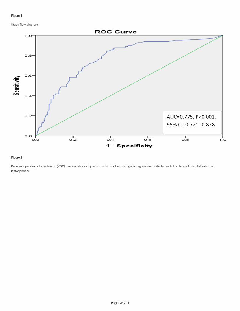
Page 24/24
Figure 1
Study �ow diagram
Figure 2
Receiver operating characteristic (ROC) curve analysis of predictors for risk factors logistic regression model to predict prolonged hospitalization ofleptospirosis