magnetically directed targeting aggregation of
TRANSCRIPT

Hindawi Publishing CorporationJournal of NanomaterialsVolume 2011, Article ID 851520, 5 pagesdoi:10.1155/2011/851520
Research Article
Magnetically Directed Targeting Aggregation ofRadiolabelled Ferrite Nanoparticles
Yuh-Feng Wang,1, 2, 3 Chao-Ming Fu,4 Mei-Hua Chuang,5 Thau-Ming Cham,3
and Mei-Ing Chung3
1 Department of Nuclear Medicine, Buddhist Dalin Tzu Chi General Hospital, Chiayi 62247, Taiwan2 School of Medicine, Tzu Chi University, Hualien 97004, Taiwan3 School of Pharmacy, Kaohsiung Medical University, Kaohsiung 80708, Taiwan4 Department of Physics, National Taiwan University, Taipei 10617, Taiwan5 Department of Pharmacy, Buddhist Dalin Tzu Chi General Hospital, Chiayi 62247, Taiwan
Correspondence should be addressed to Mei-Ing Chung, [email protected]
Received 12 August 2010; Accepted 10 April 2011
Academic Editor: Rakesh Joshi
Copyright © 2011 Yuh-Feng Wang et al. This is an open access article distributed under the Creative Commons AttributionLicense, which permits unrestricted use, distribution, and reproduction in any medium, provided the original work is properlycited.
Ferrite magnetic nanoparticles (Fe3O4 or iron (II,III) oxide; 15–25 nm of diameter) were developed. These magnetic nanoparticlesare a potential vehicle for magnetically induced target aggregation in living animals. In this preliminary study, the radiochemicalpurity for the radiolabeled magnetic nanoparticles was examined, and the possibility of the magnetically induced targetingof the radio-nanoparticles was evaluated. Our results showed that radiolabeled ferrite nanoparticles can be used as magnetictargeting agents with high labeling efficiency and stability. These particles can be distributed within living animals via intravenousinjection, and the biodistribution of the particles can be potentially controlled by external magnetism. These evaluations will bethe groundwork for the future development of delivery techniques for radiopharmaceuticals through external magnetic control.
1. Introduction
Efficient drug delivery to a target organ or tissue achievesthe desired therapeutic outcome with fewer side effects.Therefore, the design of a system for discretelytargeted drugdelivery would be a therapeutic advance. This idea has beenapplied extensively for the control of infections and for themanagement of malignant lesions. Monoclonal antibodiesare examples of the initial development of target therapy [1–4]. They can also serve as drug carriers. Some directedtargetmonoclonal antibody therapies are available commerciallyand have been used in clinical applications [3, 4]. In additionto the biological characteristics of antigen-antibody affinity,the effective localization of applied reagents using histologi-cal characteristics is another approach that has been used [5–8]. Recently, Y-90 microspheres, which were developed forthe treatment of metastatic intrahepatic malignancies, wereimplanted directly in the capillary network in front of thetumor mass [9, 10].
We have developed ferrite magnetic nanoparticles(Fe3O4, or iron (II,III) oxide). These nanoparticles canbe radiolabeled with Tc-99m pertechnetate to Tc-99mnanoferrite [11, 12]. We hypothesize that this radiolabeledmagnetic nanoparticle can be redistributed within an animalby changing the external magnetism. If that can be achieved,a magnetically derived, target aggregation of nanoparticle-carried therapy would be attainable.
2. Materials and Methods
2.1. Preparation of Ferrite Nanoparticles. Ferrite nanoparti-cles were synthesized in an aqueous solution of iron chlorideand ammonium hydroxide using the protocol we previouslydescribed [11, 13]. In brief, the reaction solution of FeCl2(0.05 M), FeCl3 (0.05 M), and a pH-adjusting solution ofNH4OH (50 mL) were simultaneously dropped into an openvessel (200 mL in volume) at room temperature. During thesynthesis, the amount of NH4OH solution was controlled so

2 Journal of Nanomaterials
that the pH of the aqueous solution was near the neutralcondition (7 < pH < 9). The ferrite nanoparticles wereprecipitated in the aqueous solution. X-ray diffraction wasperformed and confirmed that the average diameter ofthe nanoparticles was approximately 15–25 nm, deduced byLangevin’s formulation.
2.2. Radiolabeling of the Tc-99m Pertechnetate to the Nanopar-ticles. After collection of the ferrite nanoparticles from thepreparation solution, radiolabeling was initiated. Sodiumpertechnetate (Tc-99m pertechnetate) was eluted froman Mo-99/Tc-99m generator (Ultra-Techne Kow; DaiichiRadioisotope Laboratories, LTD., Tokyo, Japan), followinginstructions provided by the manufacturer. Stannous solu-tion (Amerscan Stannous Agent, Amersham plc, Bucking-hamshire, UK) was freshly prepared by carefully adding10 mL of normal saline to the kit vial. The final concentrationof the stannous fluoride in the stannous solution was0.4 mg/mL.
Labeling was done by mixing the ferrite nanoparticleswith the stannous solution and Tc-99m pertechnetate. Weplaced 0.5 mL of the ferrite nanoparticle solution in a steriletube (BD Vacutainer; Becton, Dickinson and Company, NJ,USA), and then the stannous solution (0.5 mL) was addedby a volumetric pipette. Tc-99m pertechnetate were eluted(1.11 GBq) and counted by a dose calibrator (CRC-15R; Cap-intec, INC., NJ, USA) and placed in sterile tubes. The ferritenanoparticle and stannous mixtures were then transferredaseptically to the tubes containing Tc-99m pertechnetateusing a syringe and carefully delivering the solution downthe side wall of the tubes. Mixing was performed manuallyby shaking the tubes gently to avoid bubble formation.
2.3. Determining the Optimal Protocol. Ferrite nanoparti-cles labeled with Tc-99m pertechnetate (hereafter “Tc-99mnanoferrite”) was examined in 5 preparation protocols. Thecompositions were as follows (a) nanoferrite 0.5 mL, stan-nous solution 0.5 mL, and Tc-99m pertechnetate 370 MBq;(b) nanoferrite 0.5 mL, stannous solution 0.5 mL, and Tc-99m pertechnetate 1.11 GBq; (c) nanoferrite 0.5 mL, stan-nous solution 0 mL, and Tc-99m pertechnetate 370 MBq; (d)nanoferrite 0.5 mL, stannous solution 1.0 mL, and Tc-99mpertechnetate 370 MBq; (e) nanoferrite 2.0 mL, stannoussolution 0.5 mL, and Tc-99m pertechnetate 370 MBq.
The radiochemical purity (RCP) and stability weredetermined by means of instant thin layer chromatography(ITLC) analysis. One drop of the specimen was placedon a silica gel ITLC strip. The strip was developed byacetone, and the radioactivity distribution over the stripwas determined with a scintillation counter. Radiochemicalpurity was calculated as the fraction of radioactivity thatremained at the origin and was displayed as %RCP. Afterthe mixture was prepared, its stability was acquired bysequentially repeating the RCP procedure at 10 min, 30 min,1 hr, 2 hr, 4 hr, 6 hr, and 24 hr.
2.4. In Vitro Confirmation of the Magnetism Property afterRadiolabeling. Tc-99m nanoferrite preparation was placed
on a plastic plate. A gamma camera (DST-XLi, General Elec-tric Medical Systems, Buc, France) was used to acquire thescintigraphy, and the images were processed and inspectedby the working station (POWERstation SPX, IBM RS6000,Vision 5.2.0, General Electric Medical Systems). The platewas placed on a stable table, and a homogeneous distributionof radioactivity was observed on the monitor. Dynamicacquisition was initiated, and a commercially availablemagnet was then placed at the sidewall of the plate.
2.5. In Vivo Magnetically Derived Aggregation of Tc-99mNanoferrite. The rats (Wistar strain, male, weighing from200 to 250 g) were anesthetized with an intraperitoneal injec-tion of pentobarbital (0.006 mg/100 g). A gamma camera(DST-XLi, General Electric Medical Systems, Buc, France),fitted with a low-energy, high-resolution collimator wasused. Dynamic image acquisitions were set to 600 frames of0.4 seconds, followed by a static image at 5 min, 10 min, and15 min after the initiation of the study.
In addition to the normal biodistribution studies,an examination of magnetically-derived biodistributionchanges was performed. After the study animal was anes-thetized, a magnet was taped in contact with the skin on thelower right abdomen. No sutures were used, and no surgicalprocedure was performed. Image acquisitions followed thesame protocol that was used when there was no externalmagnetic field applied.
2.6. Ethics. All experiments were performed in accordancewith the Animal Protection Act of the Council of Agriculture,and they were approved by the Institutional Animal Care andUse Committee of Dalin Tzu Chi General Hospital.
3. Results
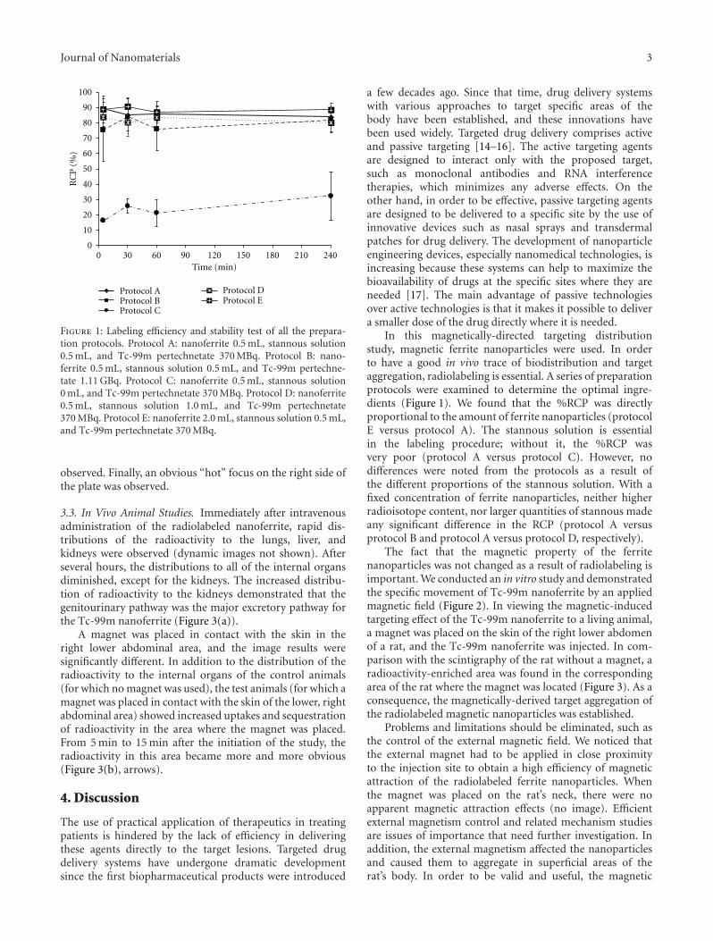
3.1. Optimal Preparation Protocol. Five different radio-labeled composites were examined. Figure 1 demonstratesthe %RCP and the stability of the labeled composites.All showed good RCP and stability after labeling, exceptfor protocol C, which had no stannous solution. ProtocolC showed much poorer labeling efficiency compared tothe other protocols, so it was concluded that preparationprotocol C should not be used. Protocol E, which hadthe highest nanoferrite content, showed the highest %RCPthroughout the study. We also noted that there were nosignificant differences in the %RCP based on the contentof the stannous solution or Tc-99m pertechnetate while thenanoferrite concentration was fixed (protocols A, B, andD). Protocol E appeared to have the highest %RCP for thepreparation of Tc-99m nanoferrite and was acquired for thefollowing studies.
3.2. In Vitro Confirmation of the Magnetic Property afterRadiolabeling. Figure 2 demonstrates the dynamic changeof the radioactivity of the Tc-99m nanoferrite. Initially, thedistribution was homogeneous, but, after the magnet wasplaced adjacent to the sidewall of the plate, a tendency ofgradual movement of radioactivity toward the magnet was
Journal of Nanomaterials 3
0
10
20
30
40
50
60
70
80
90
100
0 30 60 90 120 150 180 210 240Time (min)
RC
P(%
)
Protocol AProtocol BProtocol C
Protocol DProtocol E
Figure 1: Labeling efficiency and stability test of all the prepara-tion protocols. Protocol A: nanoferrite 0.5 mL, stannous solution0.5 mL, and Tc-99m pertechnetate 370 MBq. Protocol B: nano-ferrite 0.5 mL, stannous solution 0.5 mL, and Tc-99m pertechne-tate 1.11 GBq. Protocol C: nanoferrite 0.5 mL, stannous solution0 mL, and Tc-99m pertechnetate 370 MBq. Protocol D: nanoferrite0.5 mL, stannous solution 1.0 mL, and Tc-99m pertechnetate370 MBq. Protocol E: nanoferrite 2.0 mL, stannous solution 0.5 mL,and Tc-99m pertechnetate 370 MBq.
observed. Finally, an obvious “hot” focus on the right side ofthe plate was observed.
3.3. In Vivo Animal Studies. Immediately after intravenousadministration of the radiolabeled nanoferrite, rapid dis-tributions of the radioactivity to the lungs, liver, andkidneys were observed (dynamic images not shown). Afterseveral hours, the distributions to all of the internal organsdiminished, except for the kidneys. The increased distribu-tion of radioactivity to the kidneys demonstrated that thegenitourinary pathway was the major excretory pathway forthe Tc-99m nanoferrite (Figure 3(a)).
A magnet was placed in contact with the skin in theright lower abdominal area, and the image results weresignificantly different. In addition to the distribution of theradioactivity to the internal organs of the control animals(for which no magnet was used), the test animals (for which amagnet was placed in contact with the skin of the lower, rightabdominal area) showed increased uptakes and sequestrationof radioactivity in the area where the magnet was placed.From 5 min to 15 min after the initiation of the study, theradioactivity in this area became more and more obvious(Figure 3(b), arrows).
4. Discussion
The use of practical application of therapeutics in treatingpatients is hindered by the lack of efficiency in deliveringthese agents directly to the target lesions. Targeted drugdelivery systems have undergone dramatic developmentsince the first biopharmaceutical products were introduced
a few decades ago. Since that time, drug delivery systemswith various approaches to target specific areas of thebody have been established, and these innovations havebeen used widely. Targeted drug delivery comprises activeand passive targeting [14–16]. The active targeting agentsare designed to interact only with the proposed target,such as monoclonal antibodies and RNA interferencetherapies, which minimizes any adverse effects. On theother hand, in order to be effective, passive targeting agentsare designed to be delivered to a specific site by the use ofinnovative devices such as nasal sprays and transdermalpatches for drug delivery. The development of nanoparticleengineering devices, especially nanomedical technologies, isincreasing because these systems can help to maximize thebioavailability of drugs at the specific sites where they areneeded [17]. The main advantage of passive technologiesover active technologies is that it makes it possible to delivera smaller dose of the drug directly where it is needed.
In this magnetically-directed targeting distributionstudy, magnetic ferrite nanoparticles were used. In orderto have a good in vivo trace of biodistribution and targetaggregation, radiolabeling is essential. A series of preparationprotocols were examined to determine the optimal ingre-dients (Figure 1). We found that the %RCP was directlyproportional to the amount of ferrite nanoparticles (protocolE versus protocol A). The stannous solution is essentialin the labeling procedure; without it, the %RCP wasvery poor (protocol A versus protocol C). However, nodifferences were noted from the protocols as a result ofthe different proportions of the stannous solution. With afixed concentration of ferrite nanoparticles, neither higherradioisotope content, nor larger quantities of stannous madeany significant difference in the RCP (protocol A versusprotocol B and protocol A versus protocol D, respectively).
The fact that the magnetic property of the ferritenanoparticles was not changed as a result of radiolabeling isimportant. We conducted an in vitro study and demonstratedthe specific movement of Tc-99m nanoferrite by an appliedmagnetic field (Figure 2). In viewing the magnetic-inducedtargeting effect of the Tc-99m nanoferrite to a living animal,a magnet was placed on the skin of the right lower abdomenof a rat, and the Tc-99m nanoferrite was injected. In com-parison with the scintigraphy of the rat without a magnet, aradioactivity-enriched area was found in the correspondingarea of the rat where the magnet was located (Figure 3). As aconsequence, the magnetically-derived target aggregation ofthe radiolabeled magnetic nanoparticles was established.
Problems and limitations should be eliminated, such asthe control of the external magnetic field. We noticed thatthe external magnet had to be applied in close proximityto the injection site to obtain a high efficiency of magneticattraction of the radiolabeled ferrite nanoparticles. Whenthe magnet was placed on the rat’s neck, there were noapparent magnetic attraction effects (no image). Efficientexternal magnetism control and related mechanism studiesare issues of importance that need further investigation. Inaddition, the external magnetism affected the nanoparticlesand caused them to aggregate in superficial areas of therat’s body. In order to be valid and useful, the magnetic

4 Journal of Nanomaterials
1 4 7 10
13 16 19 22
25 28 31 34
37 40 43 46
(a) (b)
Figure 2: In vitro observation of the magnetically forced distribution of Tc-99m nanoferrite using a gamma camera. These data provideevidence that the physical property of magnetism was not distorted by the process of radiolabeling. (a) Dynamic acquisition showed thetendency of the radioactivity to gradually move toward the magnet. (b) An enlarged picture of the dynamic acquisitions: upper, the initialstatus; lower, the final image. An obvious “hot” focus on the right side of the plate was exhibited.
5 min 15 min
(a)
5 min 15 min
(b)
Figure 3: Biodistribution and magnetically-derived biodistribution changes to the Tc-99m nanoferrite particles. Images were acquired at5 and 15 min after injection from the tail vein of the rats, and the acquisition time was 60 seconds. (a) Biodistribution of the Tc-99mnanoferrite; (b) A magnet was placed over the right lower abdominal area of the rat before administration of Tc-99m nanoferrite; an areawith increased radioactivity was demonstrated (arrows).
effect should be centralized and deeply applied to theinternal organ to direct the approach of the pharmaceuticals.Finally, after the therapeutic purpose has been achieved, theaggregated nanoparticles should be dissociated as rapidly aspossible to avoid the formation of thrombosis.
Magnetically radioactive nanoparticles have the advan-tage of being able to highly concentrate radioactivity ina specific area while minimizing the distribution of theradioactivity to surrounding tissues. Our study showed the
possibility of very beneficial usage of radiolabeled ferritenanoparticles in the future. For example, (1) this modelcould be a carrier for precise therapeutic reagents, such as aspecific tumor agent, an antibiotic, or a regeneration activa-tor, and (2) instead of Tc-99m pertechnetate, a radioisotopewith therapeutic effects, such as Re-188, I-131, or Y-90, couldbe applied. Our results suggest the future development oftargeted delivery of radiopharmaceuticals through in vitrocontrol.

Journal of Nanomaterials 5
5. Conclusions
We have reported a novel preparation of Tc-99m nanoferriteparticles. This labeling procedure is achievable as a nuclearmedicine laboratory practice. After radiolabeling, these Tc-99m nanoferrite particles maintained their magnetic prop-erty. This finding was confirmed by in vitro observation, andit was also proven by the results of the in vivo animal study.The radioactivity can be enriched at a specific site by applyingan external magnet. Our results imply that these radiolabelednanoparticles can serve as a vector for magnetically-derivedtargeting of therapeutic pharmaceuticals.
References
[1] R. A. Clynes, T. L. Towers, L. G. Presta, and J. V. Ravetch,“Inhibitory Fc receptors modulate in vivo cytoxicity againsttumor targets,” Nature Medicine, vol. 6, no. 4, pp. 443–446,2000.
[2] C. A. Reade and A. K. Ganti, “EGFR targeted therapy in non-small cell lung cancer: potential role of cetuximab,” Biologics,vol. 3, pp. 215–224, 2009.
[3] M. D. Pescovitz, C. J. Greenbaum, H. Krause-Steinrauf etal., “Rituximab, B-lymphocyte depletion, and preservation ofbeta-cell function,” New England Journal of Medicine, vol. 361,no. 22, pp. 2143–2152, 2009.
[4] B. Li, L. Zhao, H. Guo et al., “Characterization of a rituximabvariant with potent antitumor activity against rituximab-resistant B-cell lymphoma,” Blood, vol. 114, no. 24, pp. 5007–5015, 2009.
[5] M. F. Giblin, G. L. Sieckman, T. D. Shelton, T. J. Hoffman, L.R. Forte, and W. A. Volkert, “In vitro and in vivo evaluationof 177Lu- and 90Y-labeled E. coli heat-stable enterotoxin forspecific targeting of uroguanylin receptors on human coloncancers,” Nuclear Medicine and Biology, vol. 33, no. 4, pp. 481–488, 2006.
[6] Z. Shen, W. Wei, Y. Zhao et al., “Thermosensitive polymer-conjugated albumin nanospheres as thermal targeting anti-cancer drug carrier,” European Journal of PharmaceuticalSciences, vol. 35, no. 4, pp. 271–282, 2008.
[7] J. E. Cyr, D. A. Pearson, D. M. Wilson et al., “Somatostatinreceptor-binding peptides suitable for tumor radiotherapywith Re-188 or Re-186. Chemistry and initial biologicalstudies,” Journal of Medicinal Chemistry, vol. 50, no. 6, pp.1354–1364, 2007.
[8] D. Kopelman and M. Papa, “Magnetic resonance-guidedfocused ultrasound surgery for the noninvasive curativeablation of tumors and palliative treatments: a review,” Annalsof Surgical Oncology, vol. 14, no. 5, pp. 1540–1550, 2007.
[9] B. Sangro, J. I. Bilbao, M. Inarrairaegui, M. Rodriguez, P. Gar-rastachu, and A. Martinez-Cuesta, “Treatment of hepatocel-lular carcinoma by radioembolization using y microspheres,”Digestive Diseases, vol. 27, no. 2, pp. 164–169, 2009.
[10] K. T. Sato, R. J. Lewandowski, M. F. Mulcahy et al., “Unre-sectable chemorefractory liver metastases: radioembolizationwith Y microspheres—safety, efficacy, and survival,” Radiol-ogy, vol. 247, no. 2, pp. 507–515, 2008.
[11] C. M. Fu, Y. F. Wang, Y. C. Chao, S. H. Hung, and M. D.Yang, “Directly labeling ferrite nanoparticles with Tc-99mradioisotope for diagnostic applications,” IEEE Transactions onMagnetics, vol. 40, no. 4, pp. 3003–3005, 2004.
[12] C. M. Fu, Y. F. Wang, Y. F. Guo, T. Y. Lin, and J. S. Chiu, “In vivobio-distribution of intravenously injected Tc-99 m labeledferrite nanoparticles bounded with biocompatible medicals,”IEEE Transactions on Magnetics, vol. 41, no. 10, pp. 4120–4122,2005.
[13] AI. Y. Wang, C. L. Kuo, J. L. Lin, C. M. Fu, and Y. F.Wang, “Study of magnetic ferrite nanoparticles labeled withTc- pertechnetate,” Journal of Radioanalytical and NuclearChemistry, vol. 284, no. 2, pp. 405–413, 2010.
[14] A. Philipp, M. Meyer, and E. Wagner, “Extracellular targetingof synthetic therapeutic nucleic acid formulations,” CurrentGene Therapy, vol. 8, no. 5, pp. 324–334, 2008.
[15] V. Hofmeister, D. Schrama, and J. C. Becker, “Anti-cancertherapies targeting the tumor stroma,” Cancer Immunology,Immunotherapy, vol. 57, no. 1, pp. 1–17, 2008.
[16] K. Manjunath, J. S. Ready, and V. Venkateswarlu, “Solid lipidnanoparticles as drug delivery systems,” Methods and Findingsin Experimental and Clinical Pharmacology, vol. 27, no. 2, pp.127–144, 2005.
[17] K. Cho, X. Wang, S. Nie, Z. Chen, and D. M. Shin, “Therapeu-tic nanoparticles for drug delivery in cancer,” Clinical CancerResearch, vol. 14, no. 5, pp. 1310–1316, 2008.