magnetic resonance imaging spectrum of spinal dysraphisms abstract no. iria- 1037
TRANSCRIPT

MAGNETIC RESONANCE IMAGING SPECTRUM OF
SPINAL DYSRAPHISMS
Abstract No. IRIA- 1037

2
INTRODUCTION
MRI plays an important role in characterising spinal dysraphisms because of excellent soft tissue contrast and the ability to detect fat in the lesion.T2 weighted images provide excellent contrast between the sub-arachnoid spaces and neural tissue while evaluating spinal dysraphisms.
AIM • To study the MRI characteristics of spinal dysraphisms and to categorize
the lesions using non-contrast enhanced Magnetic resonance imaging.

3
MATERIALS AND METHODSSPINAL DYSRAPHISM ON MRI
CLOSED
PRESENT ABSENT
OPEN
12
CLINICAL SUSPICION OF SPINAL DYSRAPHISM
14
111
SUBCUTANEOUS MASS
2 LIPOMYELOCELES1 LIPOMENINGOMYELOCELE
3 8
2 had only simple skin dimple
MENINGOMYELOCELE
DISORDER OF SECONDARY NEURULATION
DISORDER OF NOTOCHORDAL INTEGRATION
1 INTRADURAL LIPOMA1 FILAR LIPOMA
1 CAUDAL AGENESIS1 DIASTEMATOMYELIA2 SEGMENTATION ANOMALY1 DORSAL DERMAL SINUS1 NEUROENTERIC CYST
2 6
RESULTS
MRI

4
EMBRYOLOGICAL CLASSIFICATION OF SPINAL DYSRAPHISMS
PRIMARY NEURULATION

5
TERMINOLOGY

6
MENINGO-MYELOCELE
#
T2 Sagittal image of spine (A) of a 1 day old baby and Axial T2 weighted image at lumbar spine (C) shows neural placode (#) extending above skin surface due to expansion of underlying subarachnoid space (=), which is characteristic of myelomeningocele.T2 Sagittal image of brain (B) showing thinned out corpus callosum, tectal beaking (%), elongated 4th ventricle and tonsillar herniation ($) suggestive of Chiari 2 malformationAlso note the cervical syrinx(*)
1C
1B
1A
#
=
*
$
%
7
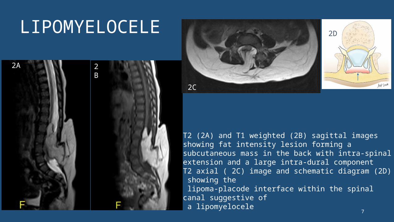
LIPOMYELOCELE
2A 2B
2C
2D
T2 (2A) and T1 weighted (2B) sagittal images showing fat intensity lesion forming a subcutaneous mass in the back with intra-spinal extension and a large intra-dural component T2 axial ( 2C) image and schematic diagram (2D) showing the lipoma-placode interface within the spinal canal suggestive of a lipomyelocele

8
LIPOMYELOCELE
T2 sagittal image (2E) and STIR (2F) image showing herniation of neural tissue which is in contact with fatty tissue forming a subcutaneous massT2 axial image (2G) shows the lipoma-placode interface within the spinal canal. Also note the posterior neural arch defectT2 coronal image (2H) showing associated left hydronephrosis
2F2E
2G
2H
9
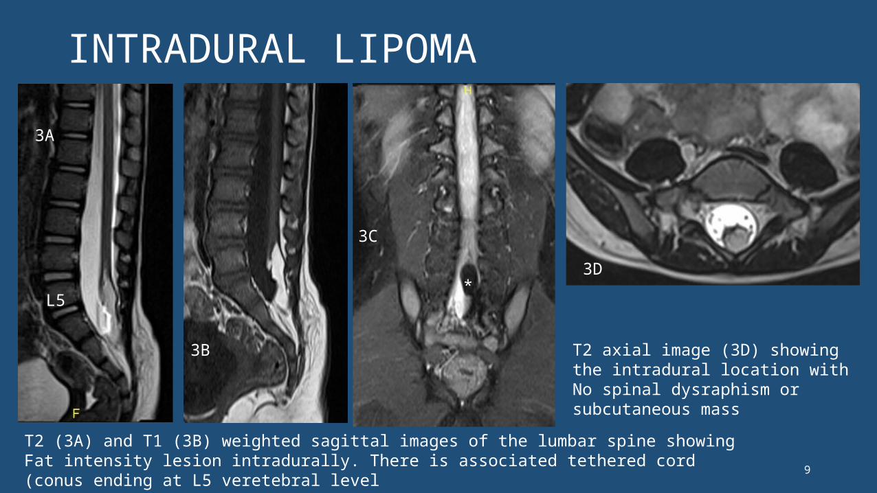
INTRADURAL LIPOMA
T2 (3A) and T1 (3B) weighted sagittal images of the lumbar spine showing Fat intensity lesion intradurally. There is associated tethered cord (conus ending at L5 veretebral levelCoronal STIR image (3C) the lesion showing fat suppression (*)
*
3A
3B
3C
3D
T2 axial image (3D) showing the intradural location with No spinal dysraphism or subcutaneous mass
L5

10
DORSAL DERMAL SINUS
L4
Sagittal T2 weighted (4A) and T1 weighted (4B) image showing a dorsal dermal sinus extending from the skin upto the posterior meninges. Associated tethered cord (conus at lower border of L4). No fat intensity within spinal canal 4C- Schematic diagram of dorsal dermal sinus with intradural dermoid.4D- T2 axial image showing the sinus tract (^)
4A
4B
4C
4D^

DISORDERS OF MIDLINE NOTO CHORDAL INTEGRATION
• Dorsal enteric fistula and Neurenteric cyst- Incomplete regression of neuro-enteric canal with abnormal communication between bowel and spinal canal
• Diastematomyelia - when the persistent neuro-enteric canal splits the spinal cord into 2 hemicords
neurenteric canal transiently connects the yolk sac to the amnion via the primitive knot which regresses during development of the embryo

12
DIASTEMATOMYELIA TYPE 1 WITH TETHERED CORD
L4
T2 axial image (5A) showing 2 hemicords with own dural sheath separated by a fibro-osseous septum. Type 1 diastematomyelia. Notethe left hemicord syrinx. T2 sagittal image (5B)shows tethered cord
5A 5B
Separation of the spinal cord into two hemicords is referred to as diastematomyelia. hemi cords are usually symmetric AND Fuse back distally
PANG Type 1 - the two hemicords - individual dural and arachnoid covering separated by an osseous or cartilaginous septumPANG type 2- there is a single dural tube containing two hemi cords, sometimes with an intervening fibrous septum
13
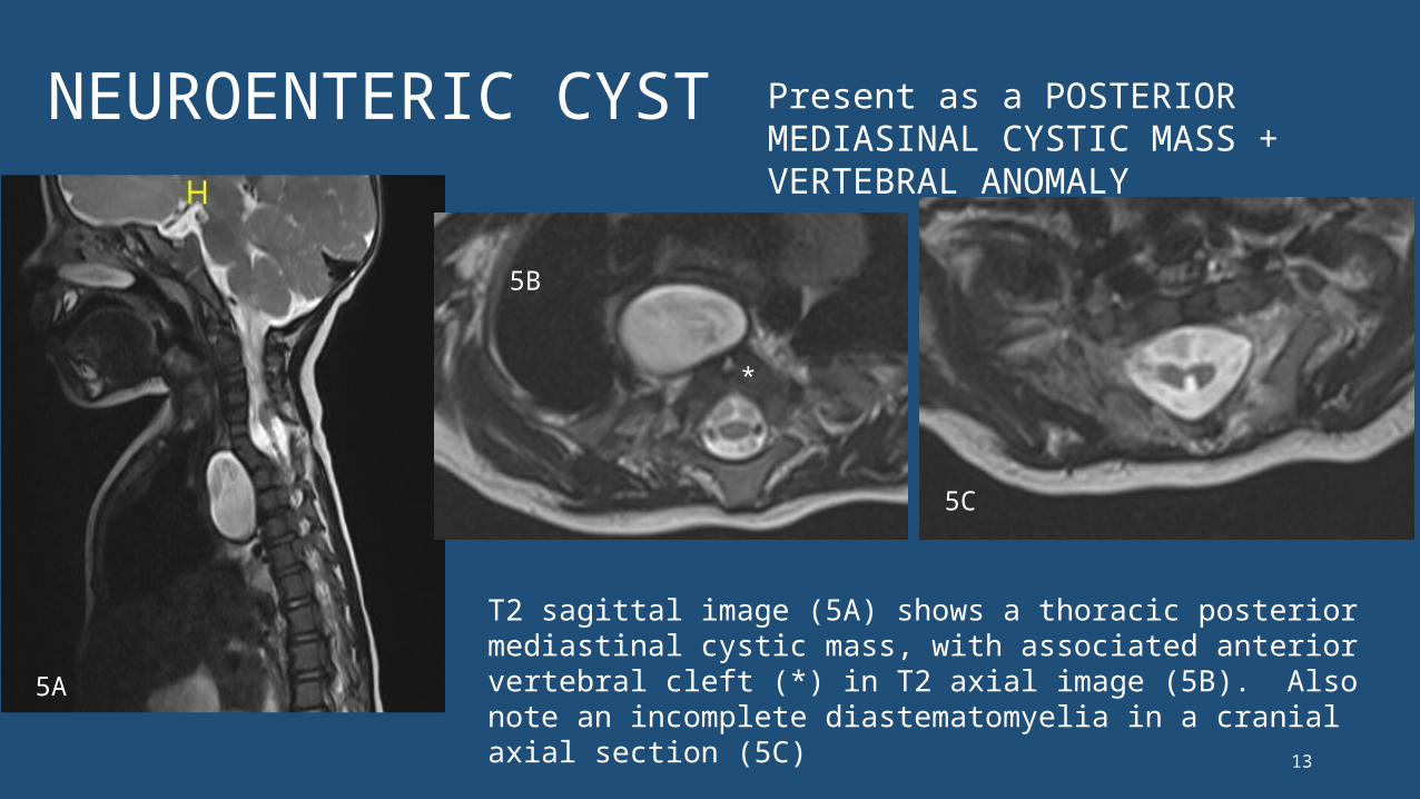
NEUROENTERIC CYST
T2 sagittal image (5A) shows a thoracic posterior mediastinal cystic mass, with associated anterior vertebral cleft (*) in T2 axial image (5B). Also note an incomplete diastematomyelia in a cranial axial section (5C)
*
Present as a POSTERIOR MEDIASINAL CYSTIC MASS + VERTEBRAL ANOMALY
5A
5B
5C

14
Partial SACRAL AGENESIS TYPE 2 (TETHERED CORD)
L4
Figure (6A) showing absent distal sacral vertebral elements and coccyx. (6B) shows the conus terminating at upper border of L4 suggestive of tethered cord
Caudal agenesis can be of two types. In type 1, there is a high position and abrupt termination of the conus medullaris. In type 2, there is a low position and tethering of the conus medullaris( a/w thickened filum / lipoma)
6A 6B

15
COMPLEX DYSRAPHISM
Vertebral segmentation anomaliesLong segment syrinx
Type 2 diastematomyelia at thoracic level with right hemicord syrinx
Thickened filum terminale causing tethered cord. Associated Caudal agenesis
7A
7D
7C
7B
L5

16
CRANIORACHISCHISIS – ENCEPHALOMENINGOCELE WITH TETHERED DERMOID
T2 coronal image (A) showing herniation of the right hemi-brainstem via a defect in the basi-sphenoid along with the right 7 and 8 cranial nerves.T2 axial (8B)and T1 axial (C) image shows associated meningocele. T1 axial image (a more caudal section) shows the fat intensity lesion adhered to the encephalo-meningocele.
17
CRANIORACHISCHISIS – ENCEPHALOMENINGOCELE WITH TETHERED DERMOID
• Cranial dysraphism /Encephaloceles (1 / 40,000 LB) vs spinal dysraphism 1-3 / 1000 LB
• Basal encephaloceles are rare (10% of encephaloceles) and they occur mostly in the fronto-ethmoidal area. (>90%)
• So far in literature, no such case of brainstem herniation with meningocele via a lateral basal, basi-sphenoid bony defect has been described.
• We hence report this as the first case in Literature.

18
CONCLUSION
• MRI is an excellent imaging modality for characterizing spinal dysraphisms. • MRI is indicated even in obvious open neural tube defects, contrary to
the conventional teaching, to look for associated Chiari malformation, syringomyelia etc. • MRI plays a role in the post-operative follow up of many lesions especially
to assess ascent of cord post de-tethering. • It is important to diagnose closed spinal dysraphisms without
subcutaneous masses where no clinical mass is seen, as neurological deficits can be arrested if diagnosed and treated early.

19
CLINICO-RADIOLOGICAL CLASSIFICATION OF SPINAL DYSRAPHISMS

20
REFERENCES
• Congenital Spine and Spinal Cord Malformations—Pictorial Review Stephanie L. Rufener et.al. (2008) – AJR integrative imaging• MRI findings in occult spinal dysraphisms– S. Morrthy et.al (IJRI- 2003)• MR imaging in the tethered cord syndrome - Narasimhachan
Raghavan et.al. (AJNR- 1989)• MRI in Infants and Children with Spinal Dysraphism -P D Barnes et.al
(AJNR -1986)