macromolecularly crowded in vitro microenvironments
TRANSCRIPT

Macromolecularly crowded in vitromicroenvironments accelerate the
production of extracellular matrix-rich supramolecular assemblies
The Harvard community has made thisarticle openly available. Please share howthis access benefits you. Your story matters
Citation Kumar, P., A. Satyam, X. Fan, E. Collin, Y. Rochev, B. J. Rodriguez,A. Gorelov, et al. 2015. “Macromolecularly crowded in vitromicroenvironments accelerate the production of extracellularmatrix-rich supramolecular assemblies.” Scientific Reports 5 (1):8729. doi:10.1038/srep08729. http://dx.doi.org/10.1038/srep08729.
Published Version doi:10.1038/srep08729
Citable link http://nrs.harvard.edu/urn-3:HUL.InstRepos:14351206
Terms of Use This article was downloaded from Harvard University’s DASHrepository, and is made available under the terms and conditionsapplicable to Other Posted Material, as set forth at http://nrs.harvard.edu/urn-3:HUL.InstRepos:dash.current.terms-of-use#LAA

Macromolecularly crowded in vitromicroenvironments accelerate theproduction of extracellular matrix-richsupramolecular assembliesPramod Kumar1, Abhigyan Satyam1, Xingliang Fan1, Estelle Collin1, Yury Rochev1, Brian J. Rodriguez2,Alexander Gorelov3, Simon Dillon4, Lokesh Joshi5, Michael Raghunath6, Abhay Pandit1
& Dimitrios I. Zeugolis1
1Network of Excellence for Functional Biomaterials (NFB), National University of Ireland Galway (NUI Galway), BioscienceResearch Building, Galway, Ireland, 2Conway Institute of Biomolecular & Biomedical Research, University College Dublin, Dublin,Ireland, 3School of Chemistry & Chemical Biology, University College Dublin, Dublin, Ireland, 4BIDMC Genomics, Proteomics,Bioinformatics and Systems Biology Center, Beth Israel Deaconess Medical Center, Harvard Medical School, Boston, USA,5Alimentary Glycoscience Research Cluster, NUI Galway, Galway, Ireland, 6Department of Bioengineering, Faculty of Engineering,National University of Singapore Tissue Engineering Programme, Department of Biochemistry, Yong Loo Lin School of Medicine,National University of Singapore, Singapore.
Therapeutic strategies based on the principles of tissue engineering by self-assembly put forward the notionthat functional regeneration can be achieved by utilising the inherent capacity of cells to create highlysophisticated supramolecular assemblies. However, in dilute ex vivo microenvironments, prolonged culturetime is required to develop an extracellular matrix-rich implantable device. Herein, we assessed theinfluence of macromolecular crowding, a biophysical phenomenon that regulates intra- and extra-cellularactivities in multicellular organisms, in human corneal fibroblast culture. In the presence ofmacromolecules, abundant extracellular matrix deposition was evidenced as fast as 48 h in culture, even atlow serum concentration. Temperature responsive copolymers allowed the detachment of dense andcohesive supramolecularly assembled living substitutes within 6 days in culture. Morphological,histological, gene and protein analysis assays demonstrated maintenance of tissue-specific function.Macromolecular crowding opens new avenues for a more rational design in engineering of clinically relevanttissue modules in vitro.
Tissue engineering by self-assembly1,2, cell-sheet tissue engineering3, scaffold-free tissue engineering4 ormodular tissue engineering5,6 utilise nature’s sophistication to create bottom up supramolecular assemblies.The rationale of either of these approaches lays on the fact that cells are the traditional extracellular matrix
(ECM) builders and create tissue modules with precision and stoichiometric competence still unmatched by man-made devices. Such technologies have led to the development of cohesive cell-assembled prototypes, held togetherby cell-cell and cell-ECM junctions, for skin7–9, blood vessel10,11 and cornea12 that have demonstrated reparative/regenerative efficacy, even in clinical setting.
Over 30 years have passed since the development of the first tissue equivalent in vitro7–9; it is now evidenced thatthis scientific breakthrough was not followed by a similar magnitude technological advancement. Indeed, we havenow recognised that the major culprit in clinical translation and commercialisation of cell-based therapies is thecreation of functional in vitro microenvironments that, by imitating features of the tissue from which the cellswere extracted from, will facilitate cell phenotype, function and therapeutic potential maintenance ex vivo13. Thus,bioinspired research efforts recruit pioneered technologies in biophysical cues (e.g. surface topography, substrateelasticity), biochemical beacons (e.g. oxygen tension, ascorbic acid supplementation) and biological signals (e.g.growth factor supplementation, co-culture systems) to maintain permanently differentiated cells phenotype and/or to accurately direct stem cells lineage commitment for the creation of functional tissue facsimiles in vitro14–17.
Despite the significant advancements that have been achieved to-date, the development of an implantabledevice remains notoriously difficult and prohibitively expensive, as markedly long ex vivo culture times are
OPEN
SUBJECT AREAS:
CELL GROWTH
TRANSLATIONAL RESEARCH
BIOPHYSICAL METHODS
TISSUE ENGINEERING
Received7 August 2014
Accepted27 January 2015
Published4 March 2015
Correspondence andrequests for materials
should be addressed toD.I.Z. (dimitrios.
SCIENTIFIC REPORTS | 5 : 8729 | DOI: 10.1038/srep08729 1

required (e.g. 196 days for blood vessel)10 that are associated with lossof native phenotype and cellular senescence18,19. Herein, we hypothe-sise that macromolecular crowding (MMC), a biophysical phenom-enon known to accelerate biological processes by several orders ofmagnitude20–22, will enhance protein turnover and amplify the pro-duction of ECM-rich supramolecularly assembled tissue equivalents.Specifically, in vivo, cells inhabit in the dense intertwined network ofthe ECM, where the proteinase-specific conversion of procollagen tocollagen type I is rapid23. In the dilute culture media, the conversionof water soluble procollagen to water insoluble collagen type I is veryslow; thus prolonged culture times are required to create an implan-table device (Fig. 1). Thus, the addition of inert macromolecules inthe culture media, by imitating the dense extracellular space, willenable the accelerated production of ECM-rich living substitutes(Fig. 1). To validate our hypothesis, human corneal fibroblasts(HCFs) were used for very first time as exemplary and the influenceof a FicollTM cocktail (FC) as a means of MMC on collagen type I, themost abundant ECM protein of corneal stromal, biosynthesis wasassessed.
ResultsIdentification of optimal newborn calf serum (NBCS) concen-tration for maximum ECM deposition. Sodium dodecyl sulphatepolyacrylamide gel electrophoresis (SDS-PAGE; Fig. S1) andcomplementary densitometric analysis (Fig. S2) demonstrated thatat all NBCS concentrations collagen type I remained in the media inthe absence of FC, whilst in the presence of FC, collagen type I wasdeposited in the cell layer. It was further observed that collagen type Icontent remained constant or reduced as a function of increased
NBCS concentration at given time point in culture. This reductionin collagen type I content was attributed to the enhanced matrixmetalloproteinase-2 (MMP-2) activity, as revealed by gelatinzymography (Fig. S3). The highest (p , 0.0001) collagen type Ideposition was achieved after culturing HCFs for 6 days in thepresence of FC. No significant difference (p . 0.05) in collagentype I deposition was observed among the different NBCSconcentrations (0 to 10%) at any time point tested (2, 4 and 6days). Phase contrast microscopy (Fig. S4) revealed that HCFsmaintained their spindle-shaped morphology for all experimentalconditions (e.g. presence/absence of FC; 0%–10% NBCS; 2–6 daysin culture). Further, the addition of FC did not affect cell metabolicactivity (Fig. S5A) and cell viability (Fig. S5B and Fig. S5C)independently of the experimental condition.
Identification of optimal serum origin for maximum ECM depo-sition. To avoid xenogeneic contaminants, the influence of humanserum (HS), as an alternative to NBCS, was assessed at 0.5%, inpresence and absence of FC for 2, 4 and 6 days. SDS-PAGE (Fig.S6) and complementary densitometric analysis (Fig. 2A) showed thatin the absence of FC collagen type I remained in the media and itscontent remained constant as a function of time in culture. Thisindifference in collagen type I content was attributed to theincreased MMP-2 content, as revealed by gelatin zymography(Fig. 2B). In the presence of FC, collagen type I was deposited atthe cell layer and its content was increased as a function of time inculture. The highest (p , 0.0001) collagen type I deposition wasachieved after culturing HCFs for 6 days in the presence of FC.Significant increase in collagen type I (p , 0.001) deposition wasobserved in the presence of HS, as compared to NBCS at 4 and 6 days.
Figure 1 | Schematic representation of how MMC enhances ECM deposition in vitro and its application in the development of ECM-rich cell sheet.Under standard cell culture conditions, the conversion of the water-soluble procollagen to insoluble collagen is very slow as the proteinases are
deactivated before they cleave the specific N- and C- propeptides and/or the procollagen is dissolved before its conversion to collagen. After substantially
long culture time, the cells will self-crowd the media and collagen is deposited in the cell layer. The addition of inert polydispersed macromolecules
(presented as spheres with different diameters) in the culture media results in most effective volume occupancy; in increased relative density of
procollagen and proteinases; in cleavage of the propeptides by their respectful proteinases; and finally in accelerated procollagen conversion to collagen
and deposition of the former.
www.nature.com/scientificreports
SCIENTIFIC REPORTS | 5 : 8729 | DOI: 10.1038/srep08729 2

The increase in collagen deposition in the presence of HS wasattributed to the lower MMP-2 content of HS (Fig. S7).
Immunocytochemistry (ICC) analysis (Fig. 2C) further corrobo-rated the high collagen type I deposition in the presence of FC and itsrelative increase as a function of time in culture, independently of theserum origin. No difference in fibronectin deposition was observed.Phase contrast microscopy (Fig. S8) revealed that HCFs maintainedtheir spindle-shaped morphology for all experimental conditions(e.g. presence/absence of FC; 0.5% NBCS/HS; 2–6 days in culture).Further, the presence of FC did not affect cell metabolic activity (Fig.S9A), cell viability (Fig. S9B and Fig. S9C) and DNA content (Fig.S9D), at any given time point, independently of the serum origin.
Production and characterisation of ECM-rich HCFs supramolecularassemblies. Due to the abundant ECM deposition in the presence ofFC, commercially available temperature-responsive N-isopropyla-crylamide (NIPAAM) coating was not suitable for the detachment ofintact ECM-rich HCFs supramolecular assemblies after 6 days inculture, although cell attachment and growth was not prohibited(Fig. S10). A 65% N-isopropylacrylamide/35% N-tert-butylacrylamide(NTBA) copolymer [poly(NIPAAM-co-NTBA)] allowed attachment(Fig. 3A) and detachment (Fig. 3B) of intact ECM-rich HCFssupramolecular assemblies after 6 days in culture in the presence ofFC and 0.5% HS. Auxiliary time-lapse microscopy revealed that due tothe abundance in deposited ECM under MMC conditions, thedetachment rate of the ECM-rich HCFs supramolecular assemblieswas slower than their non-MMC counterparts (Fig. 3C and S11).
Histological analysis using van Gieson’s (Fig. 4A) and Masson’strichrome (Fig. 4B) staining and ICC analysis (Fig. 4C) further cor-roborated the enhanced collagen type I deposition under MMC con-ditions (0.5% HS, FC, 6 days in culture). Transmittance analysis(Fig. 4D) revealed no significant difference (p . 0.05) between thegroups (non-MMC, 0.5% HS, 6 days in culture; MMC, 0.5% HS, 6days in culture; PBS; control surface). Further atomic force micro-scopy analysis (Fig. 4E and S12) revealed the presence of abundantquartered staggered collagen type I fibrils at the intercellular regionsof the MMC HCFs (0.5% HS, 6 days in culture).
MMC (0.5% HS, FC) did not affect (p . 0.05) gene expressionanalysis (Fig. 5) for collagens (I, III, IV, V, VI); fibronectin; pheno-typic markers (CD34, CHST6); and unintended differentiation mar-
ker (a SMA) for all time points assessed (2, 4 and 6 days).Proteomics analysis revealed that under MMC conditions (0.5%HS, FC, 6 days), the total protein content in the cell layer wassignificantly increased (Table S2). Subsequent validation usingICC (Fig. 6) and complementary fluorescence intensity (Fig.S13) further confirmed the high deposition of collagenous pro-teins (I, III, IV, V, VI) and fibronectin under MMC conditions(0.5% HS, FC, 6 days in culture), whilst no difference wasobserved for unintended differentiation markers (a SMA) andphenotypic markers (CD34, keratocan; Fig. S14) for all timepoints (2, 4 and 6 days in culture).
Low-density versus high-density culture. At day 6, MMC at low-density (25,000 cells/cm2) culture induced higher ECM depositionthan non-crowded high-density (50,000 cells/cm2) culture (Fig. 7).The highest ECM deposition was detected in the high-density culturein the presence of MMC (Fig. 7).
DiscussionDuring development, cellular secretome creates a tissue-specificmicroenvironment that governs the various functions of the tissue.Cellular secretome also provides necessary signals for tissue repairand regeneration during or following injury. Therapeutic strategiesbased on the principles of tissue engineering by self-assembly aim tocapture this specific reparative potential of cellular secretome.However, for such approaches to succeed, it is essential to create afunctional ex vivo microenvironment that by emulating the tissuefrom which the cells were extracted from or are to be implanted inwill maintain cell phenotype, function and therapeutic potential.Unfortunately, the still primitive culture conditions not only areassociated with phenotypic drift, but also require a prolonged periodof time for the development of an implantable device. Herein, wehypothesised that MMC, by imitating the dense extracellular space,will accelerate the deposition of tissue-specific ECM, whilst preserv-ing cell phenotype. Given that tissue engineering by self-assemblystrategies for corneal stromal require 4 to 8 weeks for the productionof supramolecularly assembled monolayers24–26 and the readiness ofcorneal fibroblasts to trans-differentiate towards unintended myo-fibroblast lineage27–32, we used this cell type as model.
Figure 2 | MMC accelerates ECM deposition in HCFs culture in the presence of NBCS and HS. (A) Densitometric analysis of SDS-PAGE confirmed the
high collagen type I deposition in the cell layer as early as 2 days in culture. Collagen type I deposition was consistently increased up to 6 days in
culture (longer time point assessed). (B) Gelatin zymography detected the 68–72 kDa proMMP-2 and the 62 kDa MMP-2 active form. Both forms of
enzyme were higher in NBCS, which explains the enhanced ECM deposition in the presence of HS. (C) ICC analysis further collaborated the enhanced
collagen type I deposition in the presence of FC, whilst fibronectin, the template for collagen deposition, was not affected.
www.nature.com/scientificreports
SCIENTIFIC REPORTS | 5 : 8729 | DOI: 10.1038/srep08729 3

In the absence of MMC, collagen type I remained in the media,whilst in the presence of FC ample ECM deposition was evidenced inthe cell layer as early as 48 h in culture, which persisted up to 6 daysin culture (longer time point tested). This is in accordance to pre-vious observations, where FC accelerated ECM deposition in humanlung fibroblast culture33 and human bone marrow stem cell culture34.The efficacy of FC to induce accelerated ECM deposition lays on thefact that effectively excludes volume35,36, facilitating that way cleavageof C- and distant N-propeptides by their respective proteinases andtherefore accelerated conversion of procollagen to collagen type I.
Tissue engineering by self-assembly supramolecular aggregatesare customarily produced using copious amounts of animal sera(up to 30%). Our data indicate that excessive sera supplementationis counter-effective, due to the high amount of MMP-2 present thatdegrades deposited ECM. Given though that sera contain survivalsignals, mitogens and growth factors, all necessary for cell expansion,a balance needs to be achieved that will allow both cell survival andECM deposition. Herein, we demonstrate that low serum concentra-
tion appears to achieve optimal balance between matrix depositionand cell growth, which is in accordance to previous results, wherehigh serum supplementation resulted in phenotypic drift of cornealstromal cells37–39, tenocytes40 and retinal progenitor cells41, whilst lowserum or serum-free media resulted in phenotype maintenance ofdental-derived stem cells42, embryonic stem cells43 and mesenchymalstem cells44,45. Further, given the potential of interspecies transmis-sion of disease and severe immune reactions associated with animalsera, our data further corroborate previous studies supporting the useof HS for clinically relevant cell therapies46–50.
The ample deposition of ECM in the presence of FC prohibitedharvesting of intact supramolecularly assembled ECM-rich HCFssubstitutes from the brush-like commercially available NIPAAMdishes. The efficiency of poly(NIPAAM-co-NTBA) copolymer toproduce intact ECM-rich cell sheets is attributed to the decreasednumber of N – H residues, due to the additional steric hindranceinduced by the addition of the NTBA group, which decreased hydro-gen bonding and consequently decreased protein adsorption51, mak-ing cell detachment easier. Although the detachment rate of theMMC samples was slower than the non-MMC counterparts, a fullycharacterised poly(NIPAAM-co-NTBA) copolymer52–54 allowedharvesting of dense and cohesive tissue-like modules as a continuoussheet within 45 min by a simple switch of temperature from 37uC to10uC. Subsequent morphological analysis revealed that these denseand cohesive supramolecularly assembled living substitutes pro-duced within 6 days in culture in the presence of FC had intactcell-cell and cell-ECM junctions and achieved similar level of biomi-micry as scaffold and scaffold-free substitutes that were producedafter 355, 424, 956 or even 1157 weeks in culture. Further, althoughtraditional self-assembled living substitutes require prolonged cul-ture time to form and mature, in the presence of macromolecularcrowding, such processes are enhanced, as evidenced by the presenceof b-bands on the gels.
Further gene and protein analysis assays demonstrated that FC didnot affect expression levels of collagenous proteins, glycoproteins,phenotypic markers and unintended differentiation markers. It isworth pointing out that both MMC and non-MMC counterpartswere negative for CD34 and keratocan at protein level and verylow expression at gene level. It has been reported that the expressionof these markers rapidly declines in low-density cultures58 after expo-sure to serum37–39,59 and as a function of time in culture56,60,61.
Here, we demonstrate that modulation of the in vitro micro-environment of HCFs with FC results in ECM-rich supramolecularassemblies within days rather than weeks or months in culture, with-out compromising fundamental cellular functions. This technologynot only requires a lower cell number than multilayer cell sheets orhigh-density cultures, which is often not available, but also bypassesaltogether such approaches that due to poor nutrient, oxygen andwaste transport result in cell necrosis in the central cell-layers62. Itfurther evades complex and expensive culture media that only tem-porarily will maintain phenotype, without actually increasing ECMdeposition63–67.
MethodsMaterials. Tissue culture consumables were purchased from Sarstedt (Ireland) andNunc (Denmark). All other materials, including cell culture media, reagents andmacromolecular crowders (FicollTM 70, FicollTM 400) were purchased from SigmaAldrich (Ireland), unless otherwise stated. Live/DeadH viability/cytotoxicity kit andalamarBlueH cell metabolic activity kit were purchased from BioSource International,Invitrogen (Ireland).
Corneal fibroblast culture. Primary HCFs (Innoprot, Spain) were cultured as persupplier protocol. Briefly 5,000 cells/cm2 were seeded on poly (L-lysine) coated tissueculture flasks maintained at 37uC with 5% CO2/95% air in a humidified incubator.Cells used in all experiments were between 3 and 5 passage.
Macromolecular crowding treatment. HCFs were seeded in 24-well plates at 25,000cells/cm2 density. When we compared low-density and high-density cultures,25,000 cells/cm2 and 50,000 cells/cm2 were used, respectively. The next day, the media
Figure 3 | A poly(NIPAAM-co-NTBA) temperature-responsivecopolymer allowed production and detachment of dense and cohesivecorneal stromal layers. (A) The poly(NIPAAM-co-NTBA) temperature-
responsive copolymer allowed attachment and spreading of HCFs under
MMC and non-MMC conditions. (B) The poly(NIPAAM-co-NTBA)
temperature-responsive copolymer allowed intact detachment of the de
novo produced ECM-rich HCFs sheets after 6 days in culture. (C) The
presence of FC, which resulted in enhanced ECM deposition, delayed the
intact detachment of the ECM-rich HCFs sheets; nonetheless, complete
detachment achieved within 45 min after temperature reduction.
www.nature.com/scientificreports
SCIENTIFIC REPORTS | 5 : 8729 | DOI: 10.1038/srep08729 4

was replaced with fresh containing FC (FicollTM 70 1 FicollTM 400: 37.5 mg/ml 1 25 mg/ml) in the presence of 0.0 to 10.0% NBCS or HS. 100 mM L ascorbic acid phosphatesupplement was added in the media during the crowding experiment to enhance ECMsynthesis. The influence of MMC was assessed at 2, 4 and 6 days in culture.
Collagen deposition analysis. Media and cell layers were digested with porcinegastric mucosa pepsin (Sigma Aldrich, Ireland) for 2 h at 37uC with continuousshaking and subsequent neutralisation with 1 N NaOH. The samples for SDS-PAGEwere prepared using appropriate dilution with distilled water and 5 3 sample buffer.Finally, 15 ml per sample solution per well was loaded on the gel (5% running gel/3%stacking gel) after 5 min heating at 95uC. Electrophoresis was performed in a Mini-PROTEAN Tetra Electrophoresis System (Bio-Rad, Ireland) by applying potentialdifference of 50 mV for the initial 30 min and then 120 mV for the remaining time(approximately 1 h). The gels were washed gently with ultra pure water and stainedusing silver stain kit (SilverQuestTM, Invitrogen, Ireland) according to themanufacturer’s protocol. Images of the gels were taken after brief washing with water.In order to quantify the cell-produced collagen type I deposition, the relative densities(GeneTools software, Syngene, Ireland) of collagen a1(I) and a2(I) chains wereevaluated and compared to the a1(I) and a2(I) chain bands densities of standardcollagen type I (Symatese Biomateriaux, France).
Matrix metalloproteinase analysis. The presence of MMPs was evaluated usinggelatin zymography. Briefly, at the end of each cell culture time point, media werecollected and mixed with non-reducing SDS sample buffer (125 mM Tris-HCl, pH6.8; 20% glycerol; 2% SDS; 0.002% bromophenol blue) and fractionated by SDS-PAGE using 10% gels containing 0.1% gelatin. After electrophoresis, the gels were
washed with two incubations in 2.5% Triton X-100 for 30 min. The gels wereincubated for 18 h at 37uC in a reaction buffer containing 50 mM Tris, pH 7.4; 5 mMCaCl2; 1 mM ZnCl2 to promote recovery of protease activity were and then stainedwith 0.5% Coomassie G250 brilliant blue for 30 min. Images of the gels were takenafter de-staining with 30% ethanol/10% acetic acid. These gelatin zymography gelsbands were compared for relative expression of MMP-2. Uncultured media withvarious percentages of NBCS and HS were also used as controls in respectiveexperiments.
Cell morphology analysis. Cell morphology was assessed using phase contrastmicroscopy (Olympus IX81 inverted microscope, Japan).
Cell metabolic activity and viability assessment. The influence of MMC on cellmetabolic activity and viability was assessed using alamarBlueH and Live/DeadHassays, as per manufacturer’s guidelines. Briefly, cell culture media containing 10%alamarBlueH reagent was added to various samples after removing the culturemedium and brief washing with PBS and then incubated for 4 h to allow for thereduction of resazurin dye by active cells in cellular metabolism assay. Followingincubation, 100 ml of medium samples were transferred into a black 96-well plate.Fluorescence of the media was determined using a micro-plate reader (VarioskanFlash, Thermo Scientific, UK) at excitation and emission 570 nm/600 nmwavelength. The level of metabolic activity was calculated using % reduction of dye,according to the supplier’s protocol and compared with the respective controlsamples. The cell viability was determined using Live/DeadH viability kit (Invitrogen,Ireland). The cells were incubated with calcium AM and ethidium homodimersolution (2 mM calcein-AM and 4 mM EthD-1) in PBS according to manufacturer’s
Figure 4 | MMC allowed production of HCFs sheets with physiological, tissue-like characteristics. (A and B) Histological analysis using van Gieson’s
and Masson’s trichrome staining demonstrate enhanced ECM deposition and tissue-like organization of the HCFs substitutes under MMC
conditions. (C) ICC analysis for collagen type I further corroborates the enhanced collagen type I deposition under MMC conditions. (D) No significant
difference in transparency between MMC HCFs sheets and PBS, control surface and non-MMC sheets was observed at 0.5% HS and after 6 days in culture.
(E) AFM analysis at the intracellular regions (shown are amplitude images) further confirmed the high deposition of collagenous proteins exhibiting
characteristic D-periodicity in the presence of FC.
www.nature.com/scientificreports
SCIENTIFIC REPORTS | 5 : 8729 | DOI: 10.1038/srep08729 5
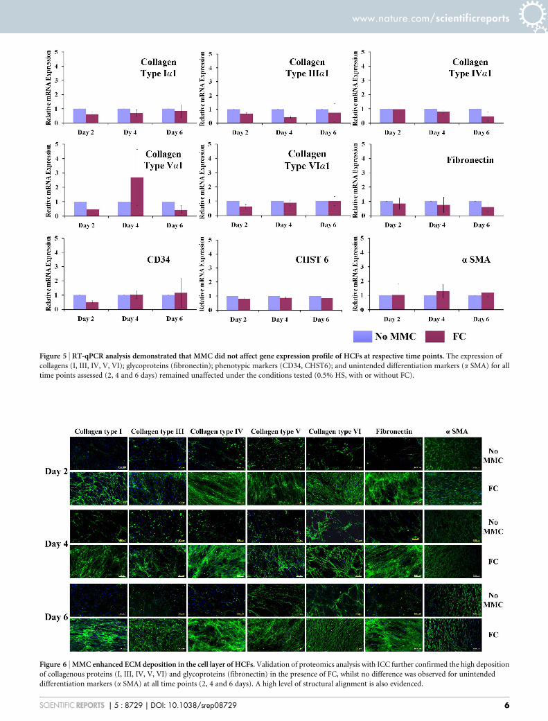
Figure 5 | RT-qPCR analysis demonstrated that MMC did not affect gene expression profile of HCFs at respective time points. The expression of
collagens (I, III, IV, V, VI); glycoproteins (fibronectin); phenotypic markers (CD34, CHST6); and unintended differentiation markers (a SMA) for all
time points assessed (2, 4 and 6 days) remained unaffected under the conditions tested (0.5% HS, with or without FC).
Figure 6 | MMC enhanced ECM deposition in the cell layer of HCFs. Validation of proteomics analysis with ICC further confirmed the high deposition
of collagenous proteins (I, III, IV, V, VI) and glycoproteins (fibronectin) in the presence of FC, whilst no difference was observed for unintended
differentiation markers (a SMA) at all time points (2, 4 and 6 days). A high level of structural alignment is also evidenced.
www.nature.com/scientificreports
SCIENTIFIC REPORTS | 5 : 8729 | DOI: 10.1038/srep08729 6

staining protocol for at least 30 min. The cell layers were washed in fresh PBS toremove excess dye. Following that, fluorescence images were taken using an OlympusIX81 inverted fluorescence microscope.
DNA quantification. DNA quantification was carried out using Quant-iTTM
PicoGreenH dSDNA assay kit (Invitrogen, Ireland) according to the manufacturer’sprotocol. Briefly, DNA was extracted using three freeze-thaw cycles after adding200 ml of nucleic acid free water per well (24 well plate). The cell suspension wassubsequently transferred to cold eppendorf tubes and was centrifuged for 5 minutesat 12000 rpm. 25 ml were then transferred into 96-well plate containing 75 ml of 1 3
TE buffer. A standard curve was generated using 0, 7.8, 15.6, 31.2, 62.5, 125, 250 and500 mg/mL DNA concentrations. 100 ml of a 15200 dilution of Quant-iTTM
PicoGreenH reagent was added to each sample and the plate was read using a micro-plate reader (Varioskan Flash, Thermo Scientific, Ireland) with an excitationwavelength of 480 nm and an emission wavelength of 525 nm.
Immunocytochemistry analysis. Cells were seeded in 4-well-chamber Lab-TekTM IIslide and MMC was carried out as described above. At the end of each culture timepoint, cells were fixed with 3% formaldehyde in PBS (pH 7.4). After fixation, the
specimens were washed three times in PBS. The fixed cell layers were incubated with3% BSA for 30 min to stop the non-specific binding of proteins. Cell layers wereincubated with primary antibodies diluted in PBS for 90 min at room temperature.Rabbit collagen type I, III, IV, V and VI (Abcam, UK; 15200) and mouse anti-fibronectin antibody (Sigma Aldrich, Ireland; F7387 15200) were used to detect theexpression of various collagens and fibronectin respectively. Mouse anti-actin, asmooth muscle antibody (15400 dilution), mouse CD34 antibody (1550 dilution) andmouse keratocan antibody (1550 dilution) were used to detect a SMA, CD34 andkeratocan expression in HCFs culture. Methanol fixing (cold methanol at 220uC for5 min) was also used after fixation to detect CD34. After primary incubation, thesamples were incubated with secondary antibody for 30 min at room temperature.The secondary antibodies used were Alexa FluorH 488, chicken anti rabbit or donkeyanti mouse respectively for rabbit and mouse antibodies at the 15400 dilutions(Invitrogen, Ireland). Antibody incubation was followed by three washes in PBS. Fornuclear staining, DAPI (49,6-diamidino-2-phenylindole) was used at 154000 dilution(Invitrogen, Ireland). Finally, the cover slips were mounted on glass slides withVectaShield (Vector Laboratories, UK) for direct observation. The images were takenusing an Olympus IX81 inverted fluorescence microscope using 10 3 objective. Theintensity of the fluorescence was evaluated using the Scope-Pro Plus software.
Figure 7 | MMC is more effective in producing ECM-rich living substitutes than high-density non-crowded cultures. At day 6, MMC at low-density
(LD; 25,000 cells/cm2) culture induced higher ECM deposition than non-crowded high-density (HD; 50,000 cells/cm2) culture and the highest
ECM deposition was detected in the high-density culture in the presence of MMC, as evidenced by SDS-PAGE (A) and complementary densitometric (B)
analysis, immunocytochemistry analysis (C, D, E) and staining assays (F, G).
www.nature.com/scientificreports
SCIENTIFIC REPORTS | 5 : 8729 | DOI: 10.1038/srep08729 7

Cell sheet production. Poly(NIPAAM-co-NTBA) was dissolved in anhydrousethanol at 40 mg/ml and subjected to continuous shaking overnight. This acrylamidesolution was mixed with poly(L-lysine) (100 mg/ml) in 151 V/V ratio and againallowed to shake overnight to mix properly. 100 ml of the mixed solution wasdeposited onto each of the petri dishes, which were left in ethanol soaked desiccatorovernight. The dishes were further dried in 600 mBar vacuum oven at 40uC for atleast 4 h. Cell culture was carried out as described above after mild UV sterilisation ofthe petri dishes for 2 h. Following culture, a temperature-controlled plate was used toinduce detachment and subsequent cell-sheet harvesting.
Histological analysis using van Gieson staining. After 6 days in culture, sampleswere fixed with 3% paraformaldehyde for at least 30 min. The samples were thenincubated with Weigert working solution for 10 min after 3 brief PBS washings.Finally, the samples were incubated in van Gieson’s solution for 2–3 min, after briefPBS washing. The samples were dehydrated using 95% alcohol, absolute alcohol andxylene. Images were taken using the Olympus microscope (BX 51) at 1003.
Histological analysis using Masson’s Trichrome staining. Samples were fixed inBouin’s solution for 1 h at 56uC after fixation. The samples were then incubated inWeigert’s iron haematoxylin staining for 10 min, followed by rinsing with runningtap water. The samples were then washed with distilled water, after 10 min in warmrunning tap water. The samples were incubated in Biebrich scarlet-acid fuchsinsolution for 10 to 15 min and washed again with distilled water. Phosphomolybdic-phosphotungstic acid solution was used for further 15 min and the samples were thentransferred directly to aniline blue solution and stained for 10 min, rinsed briefly indistilled water and then in 1% acetic acid solution for 5 min. The samples weredehydrated quickly through 95% ethyl alcohol, absolute ethyl alcohol (these stepwipes off Biebrich scarlet-acid fuchsin staining) and cleared in xylene after briefwashing in distilled water. Images were then taken using the Olympus invertedmicroscope (BX 51) at 3 100.
Atomic force microscopy analysis. HCFs were seeded in 4-well-chamber Lab-TekTM
II slides and cell culture took place as described above. After six days in culture, celllayers were washed with HBSS and fixed at room temperature for 15 min. The celllayers were then washed three times with PBS and serially dehydrated with 30%, 50%,70%, 90% and 100% ethanol. Atomic force microscopy (MFP-3D, Asylum Research,USA) analysis was then performed using rectangular Si cantilevers (SSS-NCH,Nanosensors, Switzerland), each having a nominal resonance frequency of 330 kHzand a spring constant of 42 N/m. Images were recorded using amplitude modulationmode in an ambient environment after drying the samples with nitrogen.
Light transmission. Light transmission was assessed using a previously describedmethod26. Briefly, the media was aspirated and 100 ml PBS was added in each samplewell after PBS washing. PBS alone was used as control blank. For zero absorbance and100% absorbance, the wells without PBS or cells and black dye were taken as control;this black dye showed approximately 100% absorbance (0% transmittance), while theabsorbance value in air was taken as 100% transmittance. The optical density of thesamples was measured using a spectrophotometer (Varioskan Flash, ThermoScientific, Ireland) at 380–780 nm wavelength with a resolution of 5 nm. The %transmission was calculated as: % Transmittance 5 100 (10ˆ-A), where A isabsorbance measured.
Gene expression (RT-qPCR) analysis. Total RNA was extracted using a modifiedTrizol isolation method at the given time points. Briefly, TriReagentH (Invitrogen,Ireland) was added to the cell layer after aspiration of medium from the culture andbrief washing with PBS. The cellular layer was mechanically disrupted using gentlepipetting of tissue culture plate. Phase separation was conducted with chloroform andthe total RNA contained in the aqueous phase was purified using RNeasyH mini kitcolumn (QIAGEN, Hilden, Germany), according to the supplier’s protocol. Threeextractions were carried out for each sample and pooled at the end of the RNeasyprotocol. The purity and quantity of total RNA were evaluated using an ultravioletspectrometer (NanoDrop ND-1000 Spectrophotometer, Thermo Scientific, Ireland).Reverse transcription (RT) was performed using MJ Research PTC-200 DNA Enginesystem according to the manufacturer’s protocol (Promega RT System, UK). Theprepared cDNA was monitored using SYBRGreen master mix (QIAGEN, Hilden,Germany) by real-time PCR using StepOnePlusTM Real-Time PCR System (AppliedBioscience, Switzerland). Gene transcription was normalised to the transcription ofhousekeeping human 18S gene. The 22DDCt method was used to calculate relative geneexpression for each target gene at respective time point. Primers used for variouscollagens, fibronectin, CD34, CHST6 and a SMA are given in Table S1.
Proteomics analysis. Total protein extraction from the cell layers was carried outusing QproteomeTM mammalian protein preparation kit (Qiagen, UK). Briefly, celllayers were washed twice with PBS and scraped gently, using a cell scraper, in thepresence of ice cold PBS and transferred to pre-chilled 1.5 ml tubes. This solution wascentrifuged at 450 g for five min at 4uC. The cell layer was lysed gently with cell lysisbuffer containing BenzonaseH nuclease and protease inhibitor after discarding thesupernatant. This suspension was centrifuged again at 14,000 g at 4uC for 10 min,after agitation in a rotary shaker for 5 min. After centrifugation, the supernatant wastransferred into pre-chilled 0.5 ml tubes (Protein LoBind Tubes, UK) and freeze-dried. High-throughput proteomic profiling was performed using the 4-plex iTRAQlabelling kit, equipped with the 4700 MALDI TOF/TOF system (Applied Biosystems,
USA). Briefly, the protein pellet from each sample was re-suspended in a proteindissolution buffer (10 mM triethylammonium bicarbonate, pH 8.5) and each proteinsample was assigned to one of four isobaric tags as per the manufacturer’sinstructions. The tagged samples were pooled together and subjected to massspectrometry analysis. Samples were fractionated using strong cation exchange andseparated by reverse phase chromatography using the UltimatePlus NanoLC system(Dionex, USA). After reverse phase chromatography, the eluted protein fractionswere spotted onto a MALDI plate (ABI 4800 OptiTOF, Applied Biosystems, USA).Using a Probot printing robot (Dionex, USA), the spotted plate was mixed with a-Cyano-4-hydroxycinnamic acid ionization matrix (Sigma Aldrich, Ireland) at a ratioof 152. Mass spectrometry analysis of the spotted plate was carried out an ABI4800Plus MALDI-TOF/TOF tandem MS system (Applied Biosystems, USA). Thetime-of-flight of the protein was proportional to the molecular weight. The final read-out graph was a multi-peak spectrum with different peak intensities, whichcorrespond to the relative protein amount. The peak location corresponds to theprecise protein or peptide molecular weight. Data analysis was performed usingProtein Pilot 2.0 software (Applied Biosystems, USA) on SWISS-PROT, TrEMBL(www.ebi.ac.uk/swissprot) and NCBI (www.ncbi.nlm.nih.gov/) non-redundantprotein databases. The unused score was kept at more than 1.3 to get the confidenceinterval more than 90; relative peptide value at 95% was compared among samples.Selected ECM proteins detected in proteomics results were validated using ICC andfluorescent intensity, using Olympus IX81 inverted microscope.
Statistical analysis. All results presented are mean 6 SD. Statistical analysis(MINITABTM version 16, Minitab, Inc., USA) was performed using two-sample t-testfor pair wise comparisons or one away analysis of variance (ANOVA) for multiplecomparisons after confirming: (a) the normal distribution from which each of thesamples (Anderson-Darling normality test); and (b) the variances of the populationof the samples were equal to one another (Bartlett’s and Levene’s tests forhomogenicity of variance). Non-parametric statistics were used if above assumptionswere violated and consequently Kruskal-Wallis test for multiple comparisons orMann-Whitney test for 2-samples used. Each experiment was performed in biologicaltriplicates in minimum three samples. Differences between selective experimentalgroups were considered statistically significant at p value , 0.05.
1. Guillame-Gentil, O. et al. Engineering the extracellular environment: Strategiesfor building 2D and 3D cellular structures. Adv Mater 22, 5443–5462 (2010).
2. Peck, M., Dusserre, N., McAllister, T. N. & L’Heureux, N. Tissue engineering byself-assembly. Mater Today 14, 218–224 (2011).
3. Matsuda, N., Shimizu, T., Yamato, M. & Okano, T. Tissue engineering based oncell sheet technology. Adv Mater 19, 3089–3099 (2007).
4. Kelm, J. M. & Fussenegger, M. Scaffold-free cell delivery for use in regenerativemedicine. Adv Drug Deliv Rev. 62, 753–764 (2010).
5. Dankers, P., Harmsen, M., Brouwer, L., Luyn, M. V. & Meijer, E. A modular andsupramolecular approach to bioactive scaffolds for tissue engineering. Nat Mater4, 568–574 (2005).
6. Nichol, J. W. & Khademhosseini, A. Modular tissue engineering: Engineeringbiological tissues from the bottom up. Soft Matter 5, 1312–1319 (2009).
7. Green, H., Kehinde, O. & Thomas, J. Growth of cultured human epidermal cellsinto multiple epithelia suitable for grafting. Proc Natl Acad Sci U S A 76,5665–5668 (1979).
8. Gallico, G., O’Connor, N., Compton, C., Kehinde, O. & Green, H. Permanentcoverage of large burn wounds with autologous cultured human epithelium.N Engl J Med. 311, 448–451 (1984).
9. Phillips, T., Kehinde, O., Green, H. & Gilchrest, B. Treatment of skin ulcers withcultured epidermal allografts. J Am Acad Dermatol. 21, 191–199 (1989).
10. L’Heureux, N. et al. Human tissue-engineered blood vessels for adult arterialrevascularization. Nat Med. 12, 361–365 (2006).
11. L’Heureux, N., McAllister, T. N. & de la Fuente, L. M. Tissue-engineered bloodvessel for adult arterial revascularization. N Engl J Med. 357, 1451–1453 (2007).
12. Nishida, K. et al. Corneal reconstruction with tissue-engineered cell sheetscomposed of autologous oral mucosal epithelium. N Engl J Med. 351, 1187–1196(2004).
13. Cigognini, D. et al. Engineering in vitro microenvironments for cell basedtherapies and drug discovery. Drug Discov Today 18, 1099–1108 (2013).
14. Bates, C. J., Bailey, A. J., Prynne, C. J. & Levene, C. I. The effect of ascorbic acid onthe synthesis of collagen precursor secreted by 3T6 mouse fibroblasts in culture.Biochim Biophys Acta. 278, 372–390 (1972).
15. Engler, A. J., Sen, S., Sweeney, H. L. & Discher, D. E. Matrix elasticity directs stemcell lineage specification. Cell 126, 677–689 (2006).
16. Ivanovic, Z. Hypoxia or in situ normoxia: The stem cell paradigm. J Cell Physiol.219, 271–275 (2009).
17. Edalat, F., Bae, H., Manoucheri, S., Cha, J. & Khademhosseini, A. Engineeringapproaches toward deconstructing and controlling the stem cell environment.Ann Biomed Eng. 40, 1301–1315 (2012).
18. Campisi, J. & Fagagna, F. Cellular senescence: When bad things happen to goodcells. Nat Rev Mol Cell Biol 8, 729–740 (2007).
19. Beltrami, A., Cesselli, D. & Beltrami, C. Stem cell senescence and regenerativeparadigms. Clin Pharmacol Ther. 91, 21–29 (2012).
www.nature.com/scientificreports
SCIENTIFIC REPORTS | 5 : 8729 | DOI: 10.1038/srep08729 8

20. Zimmerman, S. B. & Harrison, B. Macromolecular crowding increases binding ofDNA polymerase to DNA: An adaptive effect. Proc Natl Acad Sci U S A 84,1871–1875 (1987).
21. Minton, A. P., Colclasure, G. C. & Parker, J. C. Model for the role ofmacromolecular crowding in regulation of cellular volume. Proc Natl Acad SciU S A 89, 10504–10506 (1992).
22. Zhou, Z. et al. Crowded cell-like environment accelerates the nucleation step ofamyloidogenic protein misfolding. J Biol Chem. 284, 30148–30158 (2009).
23. Canty, E. G. & Kadler, K. E. Procollagen trafficking, processing and fibrillogenesis.J Cell Sci. 118, 1341–1353 (2005).
24. Guo, X. et al. Morphologic characterization of organized extracellular matrixdeposition by ascorbic acid–stimulated human corneal fibroblasts. InvestOphthalmol Vis Sci. 48, 4050–4060 (2007).
25. Proulx, S. et al. Reconstruction of a human cornea by the self-assembly approachof tissue engineering using the three native cell types. Mol Vis. 16, 2192–2201(2010).
26. Grobe, G. M. & Reichl, S. Characterization of vitamin C-induced cell sheetsformed from primary and immortalized human corneal stromal cells for tissueengineering applications. Cells Tiss Org. 197, 283–297 (2013).
27. Jester, J. V., Petroll, W. M., Barry, P. A. & Cavanagh, H. D. Expression of alpha-smooth muscle (alpha-SM) actin during corneal stromal wound healing. InvestOphthalmol Vis Sci. 36, 809–819 (1995).
28. Fini, M. E. Keratocyte and fibroblast phenotypes in the repairing cornea. ProgRetin Eye Res. 18, 529–551 (1999).
29. Espana, E., Kawakita, T., Liu, C. & Tseng, S. CD-34 expression by cultured humankeratocytes is downregulated during myofibroblast differentiation induced byTGF-beta1. Invest Ophthalmol Vis Sci. 45, 2985–2991 (2004).
30. Pei, Y., Sherry, D. M. & McDermott, A. M. Thy-1 distinguishes human cornealfibroblasts and myofibroblasts from keratocytes. Exp Eye Res. 79, 705–712 (2004).
31. Yoshida, S., Shimmura, S., Shimazaki, J., Shinozaki, N. & Tsubota, K. Serum-freespheroid culture of mouse corneal keratocytes. Invest Ophthalmol Vis Sci. 46,1653–1658 (2005).
32. West-Mays, J. A. & Dwivedi, D. J. The keratocyte: Corneal stromal cell withvariable repair phenotypes. Int J Biochem Cell Biol. 38, 1625–1631 (2006).
33. Lareu, R. R. et al. Collagen matrix deposition is dramatically enhanced in vitrowhen crowded with charged macromolecules: The biological relevance of theexcluded volume effect. FEBS Lett. 581, 2709–2714 (2007).
34. Zeiger, A. S., Loe, F. C., Li, R., Raghunath, M. & Van Vliet, K. J. Macromolecularcrowding directs extracellular matrix organization and mesenchymal stem cellbehavior. Plos One 7, e37904 (2012).
35. Satyam, A. et al. Macromolecular crowding meets tissue engineering by self-assembly: A paradigm shift in regenerative medicine. Adv Mater 26, 3024–3034(2014).
36. Zeugolis, D. & Satyam, A. inventors. National Universty of Ireland, Galway,assignee. Engineered living tissue substitute. European patent EP 2 532 736. 2012December 12.
37. Funderburgh, J. L., Mann, M. M. & Funderburgh, M. L. Keratocyte phenotypemediates proteoglycan structure: A role for fibroblasts in corneal fibrosis. J BiolChem. 278, 45629–45637 (2003).
38. Guerriero, E. et al. Loss of alpha3(IV) collagen expression associated with cornealkeratocyte activation. Invest Ophthalmol Vis Sci. 48, 627–635 (2007).
39. Kawakita, T. et al. Keratocan expression of murine keratocytes is maintained onamniotic membrane by down-regulating transforming growth factor-betasignaling. J Biol Chem. 280, 27085–27092 (2005).
40. Imada, K. et al. Serum-dependent osteoblastic changes in cultured tenocytesisolated from rat Achilles tendon. J Tokyo Med Uni. 71, 143–150 (2013).
41. Hu, Y. et al. An in vitro comparison study: The effects of fetal bovine serumconcentration on retinal progenitor cell multipotentiality. Neurosc Lett. 534,90–95 (2013).
42. Tarle, S. A., Shi, S. & Kaigler, D. Development of a serum-free system to expanddental-derived stem cells: PDLSCs and SHEDs. J Cellul Physiol. 226, 66–73 (2011).
43. Hannoun, Z. et al. The comparison between conditioned media and serum-freemedia in human embryonic stem cell culture and differentiation. Cellular Reprogr.12, 133–140 (2010).
44. Meuleman, N. et al. Human marrow mesenchymal stem cell culture: Serum-freemedium allows better expansion than classical a-MEM medium. Eur J Haemat.76, 309–316 (2006).
45. Kuznetsov, S. A., Mankani, M. H. & Robey, P. G. Effect of serum on human bonemarrow stromal cells: Ex vivo expansion and in vivo bone formation. Transplant70, 1780–1787 (2000).
46. Ang, L., Tan, D., Seah, C. & Beuerman, R. The use of human serum in supportingthe in vitro and in vivo proliferation of human conjunctival epithelial cells. Br JOphthalmol. 89, 748–752 (2005).
47. Mannello, F. & Tonti, G. Concise review: No breakthroughs for humanmesenchymal and embryonic stem cell culture: Conditioned medium, feederlayer, or feeder-free; medium with fetal calf serum, human serum, or enrichedplasma; serum-free, serum replacement nonconditioned medium, or ad hocformula? All that glitters is not gold! Stem Cells 25, 1603–1609 (2007).
48. Tateishi, K. et al. Comparison of human serum with fetal bovine serum forexpansion and differentiation of human synovial MSC: Potential feasibility forclinical applications. Cell Transplant 17, 549–557 (2008).
49. Aldahmash, A. et al. Human serum is as efficient as fetal bovine serum insupporting proliferation and differentiation of human multipotent stromal(mesenchymal) stem cells in vitro and in vivo. Stem Cell Rev and Rep. 7, 860–868(2011).
50. Dreher, L. et al. Cultivation in human serum reduces adipose tissue-derivedmesenchymal stromal cell adhesion to laminin and endothelium and reducescapillary entrapment. Stem Cells Dev. 22, 791–803 (2013).
51. Lynch, I. et al. Correlation of the adhesive properties of cells to N-isopropylacrylamide/N-tert-butylacrylamide copolymer surfaces with changes insurface structure using contact angle measurements, molecular simulations, andRaman spectroscopy. Chem Mater 17, 3889–3898 (2005).
52. Allen, L. T. et al. Interaction of soft condensed materials with living cells:Phenotype/transcriptome correlations for the hydrophobic effect. Proc Natl AcadSci U S A 100, 6331–6336 (2003).
53. Gilcreest, V. et al. Thermoresponsive poly(N-isopropylacrylamide) copolymers:Contact angles and surface energies of polymer films. Langmuir 20, 10138–10145(2004).
54. Moran, M. T., Carroll, W. M., Selezneva, I., Gorelov, A. & Rochev, Y. Cell growthand detachment from protein-coated PNIPAAm-based copolymers. J BiomedMater Res A. 81, 870–876 (2007).
55. Du, Y. et al. Secretion and organization of a cornea-like tissue in vitro by stem cellsfrom human corneal stroma. Invest Ophthalmol Vis Sci. 48, 5038–5045 (2007).
56. Wu, W. J. et al. Bioengineering organized, multilamellar human corneal stromaltissue by growth factor supplementation on highly aligned synthetic substrates.Tissue Eng. 19, 2063–2075 (2013).
57. Ren, R. et al. Human primary corneal fibroblasts synthesize and depositproteoglycans in long-term 3-D cultures. Developm Dyn. 237, 2705–2715 (2008).
58. Musselmann, K., Alexandrou, B., Kane, B. & Hassell, J. R. Maintenance of thekeratocyte phenotype during cell proliferation stimulated by insulin. J Biol Chem.280, 32634–32639 (2005).
59. Kawakita, T. et al. Preservation and expansion of the primate keratocytephenotype by downregulating TGF-beta signaling in a low-calcium, serum-freemedium. Invest Ophthalmol Vis Sci. 47, 1918–1927 (2006).
60. Builles, N. et al. Development of an optimised culture medium for keratocytes inmonolayer. Biomed Mater Eng. 16, S95–S104 (2006).
61. Perrella, G. et al. Expression of haematopoietic stem cell markers, CD133 andCD34 on human corneal keratocytes. Br J Ophthalmol. 91, 94–99 (2007).
62. Shimizu, T. et al. Polysurgery of cell sheet grafts overcomes diffusion limits toproduce thick, vascularized myocardial tissues. FASEB J. 20, 708–710 (2006).
63. Pancholi, S., Tullo, A., Khaliq, A., Foreman, D. & Boulton, M. The effects ofgrowth factors and conditioned media on the proliferation of human cornealepithelial cells and keratocytes. Graefes Arch Clin Exp Ophthalmol. 236, 1–8(1998).
64. Jester, J. V. & Ho-Chang, J. Modulation of cultured corneal keratocyte phenotypeby growth factors/cytokines control in vitro contractility and extracellular matrixcontraction. Exp Eye Res. 77, 581–592 (2003).
65. Etheredge, L. et al. Enhanced cell accumulation and collagen processing bykeratocytes cultured under agarose and in media containing IGF-I, TGF-b orPDGF. Matrix Biol. 29, 519–524 (2010).
66. Kim, A., Lakshman, N., Karamichos, D. & Petroll, W. M. Growth factor regulationof corneal keratocyte differentiation and migration in compressed collagenmatrices. Invest Ophthalmol Vis Sci. 51, 864–875 (2010).
67. Lakshman, N. & Petroll, W. Growth factor regulation of corneal keratocytemechanical phenotypes in 3-D collagen matrices. Invest Ophthalmol Vis Sci. 53,1077–1086 (2012).
AcknowledgmentsWe thank Dr Oliver Carroll for technical support and Mr Maciej Doczyk (http://doczykdesign.com) for the preparation of Fig. 1. This work is supported by ScienceFoundation Ireland, Research Frontiers Programme, Project Number:SFI-09-RFP-ENM2483 to D.Z.; Science Foundation Ireland, E.T.S. Walton Visitor AwardsProgramme, Project Number: 08/W.1/B2568 to M.R., A.P. and D.Z.; Health ResearchBoard, Project Number: HRA_POR/2011/84 to D.Z.; and College of Engineering &Informatics, National University of Ireland, Galway, Postgraduate College FellowshipScheme to P.K. and D.Z.
Author contributionsP.K. and D.Z. designed research; P.K., A.S., X.F. and E.C. performed experimental work;P.K., A.S., X.F., A.G. and Y.R. developed temperature responsive polymers and cell sheets;P.K., A.S., S.D. and D.Z. designed, performed, analysed and validated the proteomics study;P.K., A.S. and B.J.R. designed, performed and analysed AFM study; P.K., A.S. and D.Z.analysed data; A.P., M.R., L.J. and D.Z. discussed results; P.K. and D.Z. wrote the paper.
Additional informationSupplementary information accompanies this paper at http://www.nature.com/scientificreports
Competing financial interests: The authors declare no competing financial interests.
How to cite this article: Kumar, P. et al. Macromolecularly crowded in vitro
www.nature.com/scientificreports
SCIENTIFIC REPORTS | 5 : 8729 | DOI: 10.1038/srep08729 9

microenvironments accelerate the production of extracellular matrix-rich supramolecularassemblies. Sci. Rep. 5, 8729; DOI:10.1038/srep08729 (2015).
This work is licensed under a Creative Commons Attribution 4.0 InternationalLicense. The images or other third party material in this article are included in the
article’s Creative Commons license, unless indicated otherwise in the credit line; ifthe material is not included under the Creative Commons license, users will needto obtain permission from the license holder in order to reproduce the material. Toview a copy of this license, visit http://creativecommons.org/licenses/by/4.0/
www.nature.com/scientificreports
SCIENTIFIC REPORTS | 5 : 8729 | DOI: 10.1038/srep08729 10