m. - journal of bacteriologyjb.asm.org/content/25/3/309.full.pdfthe gram stain and differential...
TRANSCRIPT

THE GRAM STAIN AND DIFFERENTIAL STAININGM. L. KAPLAN AND LEAH KAPLAN
Bureau of Laboratories, Department of Health, New York City
Received for publication, June 22, 1932
The Gram test is the most commonly employed method fordifferential staining of bacteria. Despite this fact, and the con-siderable time which has elapsed since its discovery, its mechanismis clouded in doubt. The r6le of iodine in the test and the differ-ence between positives and negatives are still controversial mat-ters. It seems to be generally known that the triphenylmethanedyes form water-insoluble compounds with iodine; but Stearnand Stearn (1930) attribute the fixation of the dye in Gram-posi-tives to an oxidation of the cell material, rather than to the forma-tion of this compound. Burke and Barnes (1929), on the otherhand, ascribe the retention of the dye to the increased size of thedye-iodine molecule, and postulate that the essential differencebetween positives and negatives resides in the lower permeabilityof the cells of the former type to the compound. This conceptionis negatived by the observation of Stearn and Stearn that the dye-iodine compound dissociates into its components in alcohol solu-tion. Among other views of the Gram mechanism are those ofBrudny (1908) who considers that Gram-negatives are imper-meable to iodine; and of Benians (1912), who suggests a lack ofpermeability of positives to alcohol.
Benians' concept of a difference between positives and negativesin alcohol permeability would explain the so-called "pseudo-Gram" reaction obtained by the use of Victoria Blue and otheralcohol-soluble dyes, such as Spirit Blue and Night Blue, if weassume that the dissolved dye molecule unites with severalmolecules of the solvent. This would involve the passage of aconsiderable number of solvent molecules into and out of thestained cell. However, since alcohol does extract methyl and
309
on July 7, 2018 by guesthttp://jb.asm
.org/D
ownloaded from
M. L. KAPLAN AND LEAH KAPLAN
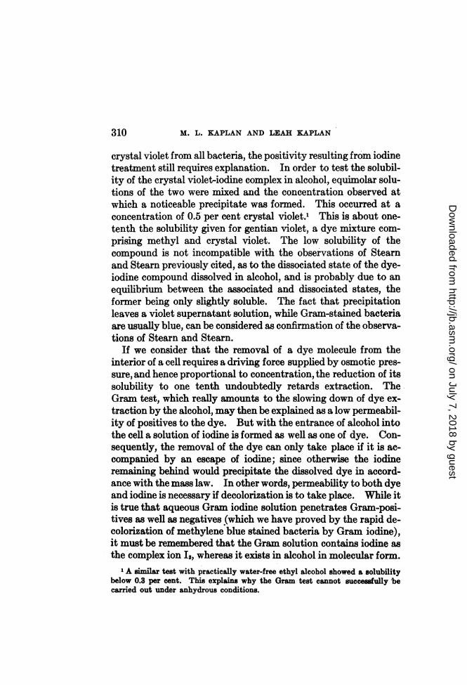
crystal violet from all bacteria, the positivity resulting from iodinetreatment still requires explanation. In order to test the solubil-ity of the crystal violet-iodine complex in alcohol, equimolar solu-tions of the two were mixed and the concentration observed atwhich a noticeable precipitate was formed. This occurred at aconcentration of 0.5 per cent crystal violet.' This is about one-tenth the solubility given for gentian violet, a dye mixture com-prising methyl and crystal violet. The low solubility of thecompound is not incompatible with the observations of Stearnand Stearn previously cited, as to the dissociated state of the dye-iodine compound dissolved in alcohol, and is probably due to anequilibrium between the associated and dissociated states, theformer being only slightly soluble. The fact that precipitationleaves a violet supernatant solution, while Gram-stained bacteriaare usually blue, can be considered as confirmation of the observa-tions of Stearn and Stearn.
If we consider that the removal of a dye molecule from theinterior of a cell requires a driving force supplied by osmotic pres-sure, and hence proportional to concentration, the reduction of itssolubility to one tenth undoubtedly retards extraction. TheGram test, which really amounts to the slowing down of dye ex-traction by the alcohol, may then be explained as a low permeabil-ity of positives to the dye. But with the entrance of alcohol intothe cell a solution of iodine is formed as well as one of dye. Con-sequently, the removal of the dye can only take place if it is ac-companied by an escape of iodine; since otherwise the iodineremaining behind would precipitate the dissolved dye in accord-ance with the mass law. In other words, permeability to both dyeand iodine is necessary if decolorization is to take place. While itis true that aqueous Gram iodine solution penetrates Gram-posi-tives as well as negatives (which we have proved by the rapid de-colorization of methylene blue stained bacteria by Gram iodine),it must be remembered that the Gram solution contains iodine asthe complex ion Is, whereas it eists in alcohol in molecular form.
1 A similar test with practically water-free ethyl alcohol showed a solubilitybelow 0.3 per cent. This explains why the Gram test cannot succesfully becarried out under anhydrous conditions.
310
on July 7, 2018 by guesthttp://jb.asm
.org/D
ownloaded from

THE GRAM STAIN AND DIFFERENTIAL STAINING 311
Moreover, the permeability of the cell in. water is probably differ-ent than in alcohol.
If the above reasoning is valid, retention of the dye by positivesmay be due not only to a low permeability to the dye, but to a lowpermeability to iodine as well. Asan indication of the importanceof the latter factor may be cited our observation that the decolori-zation of Gram-negatives is prevented by the addition of 0.2 to0.3 per cent of iodine to the alcohol. Still more striking is thefact that methyl alcohol, which decolorizes most Gram-positivesalmost instantly, does not, when as little as 0.01 per cent iodine is
TABLE 1
PER CZNT IODINEORGANISM _
0.01 0.02 0.04 0.06 0.08
B. subtili s.+ + + + +M. tetragenus .. + + + +
Sarcina aurantiaca.................. -} + - + + +
Staph. aureus .. +:} - + +
Strep. hemolyticus.ou _ -_
D. pneumoniae............. -.j.+i +
added, decolorize such positives as B. subtilis, M. tetragenus, etc.,while only slightly larger additions of iodine are necessary to pre-vent decolorization of other positives.
Table 1 shows the results of tests on six organisms obtainedfrom Bellevue Medical School.2 A set of slides was prepared,on each of which were placed, side by side, the six organisms.After staining with a 0.5 per cent solution of crystal violet in 2 percent sodium acetate, they were treated with Gram iodine solu-tion, washed and dried, and then placed in small jars containingvarious concentrations of iodine in methyl alcohol (C. P. Eimer
2 These organisms were supplied to us through the kindness of Dr. Wm. H.Park to whom we owe thanks.
JOURNAL OF BACTERIOLOGY, VOL. XXV, NO. 3
on July 7, 2018 by guesthttp://jb.asm
.org/D
ownloaded from
1M. L. KAPLAN AND LEAH KAPLAN
and Amend) to which 5 per cent of water was added. Smallquantities of freshly prepared and dried calcium carbonate wereplaced in each jar to insure neutrality of the methanol. Theslides were kept in motion in the solutions for two minutes andthen promptly washed and counterstained with Safranin fortwenty to thirty seconds.We see from the table that Gram-positive bacteria may be dif-
ferentiated according to their degree of positivity as measured byiodine concentration in methyl alcohol necessary to prevent ex-traction of iodine and dye from the cell.
Similar tests with other organisms gave approximately constantvalues for each species, so long as the water content of the alcoholremained constant. The more aqueous the methanol, the moreiodine was required to prevent extraction.Organisms were frequently encountered which showed varying
degrees of positivity in different parts of the cell body. Figure 1is the photograph of a twelve-hour culture of a soil spore-bearerwhich had been Gram stained and then treated as above withmethyl alcohol containing 0.04 per cent iodine. A strain of B.diphtheriae isolated from a clinical case and tested in the same waygave positive terminal metachromatic granules at 0.01 to 0.02per cent iodine, while the rest of the cell body required muchhigher iodine concentrations. It is interesting to note that astrain of B. Hoffmani isolated from a nasal discharge required 0.05to 0.06 per cent iodine to prevent dye-iodine escape.Whether this difference is due to a difference in permeability
to dye or to iodine is hard to say off hand, since a decrease in eitherwould probably have the same effect. In order to investigatethis point, we exposed crystal violet stained slides, omittingGram iodine treatment, to ethyl alcohol solutions of iodine, since,under these conditions, iodine and dye move in opposite directionsand cell permeabilities to them have opposite effects. For exam-ple, table 1 shows M. tetragenus to be more highly positive thanSarcina aurantiaca. If the greater positivity were ascribable tolower permeability to iodine, one would expect M. tetragenus torequire the higher concentration of iodine to insure dye retention.The test, however, gave the opposite result. On the other hand,
312
on July 7, 2018 by guesthttp://jb.asm
.org/D
ownloaded from

THE GRAM STAIN AND DIFFERENTIAL STAINING 3
B. subtilis, which shows the highest degree of positivity, does notretain the dye in all parts of the cell body even when 0.2 per centiodine has been added to the alcohol, an amount which is sufficientto suppress the escape of dye even from B. coli. The arrangementof stained and unstained spots obtained in this way from certain
/..
..... .. evtw~~~~~~~~~~~~~~~~~~~~~~~~~~~~~~~~~~~~~~~~~~~~~~~~~~~. ......... .. :.. ...::s:
....~~~~~~~~~~~;T... ..............,U.,;:.:::*~~~~~~~~~~~~~~~~~~~~~~~~ .*.;S.
M b
..:.:.;.....
FIG. 1. TWELVE-HOUR CULTURE OF SOIL SPORE BEARER, GRAM-TREATED ANDEXPOSED TO THE ACTION OF METHYL ALCOHOL CONTAINING 0.04 PER
CENT IODINE
members of the subtilis group is striking. Figures 2 and 3 arephotographs of slides of B. subtilis and a soil spore bearer, respec-tively. The slides had been stained with crystal violet and thentreated for two minutes with 0.08 per cent cent iodine in 85 percent alcohol. The unequal behavior of different parts of the cellwas also observed in other organisms. Thus, B. diphtheriaeshows retention of dye in the terminal bodies, while B. Hoffmani,in the early transplants after isolation from the human body,showed decolorization on one side and dye retention on the otherside of the cell.
313
on July 7, 2018 by guesthttp://jb.asm
.org/D
ownloaded from

..
4,F..* Pa. v
W.'t,I,,,
iA
FIG. 2. B. SUBTILIS STAINED WITH CRYSTAL VIOLET AND TREATED WITH 0.08 PER
CENT IODINE IN 85 PER CENT ETHYL ALCOHOL
Note stained and unstained spots. Compare with figure 3.
N '.::
¶ill
a.
'a
(aa
¾.
't
FIG. 3. SOIL SPORE BEARER STAINED WITH CRYSTAL VIOLET AND TREATED WITH0.08 PER CENT IODINE IN 85 PER CENT ETHYL ALCOHOL
314
Mwr1IV16'I.4
on July 7, 2018 by guesthttp://jb.asm
.org/D
ownloaded from
THE GRAM STAIN AND DIFFERENTIAL STAINING
These facts point to different degrees of permeability to iodineof the Gram-positives. The comparative resistance of M. tetra-genus to decolorization by ethyl alcohol containing iodine indi-cates that its resistance to dye escape is higher than that ofSarcina without a corresponding increased resistance to iodinepassage. This shows that the resistance is due not only to thesize of the moving molecule but also to factors specific to the cell.Instances of a certain degree of parallelism between the two re-sistances can be cited, though limited to interior parts of the celland aqueous iodine solutions. Treatment of bacteria with dilutealkali (0.1 normal NaOH) developed a difference in differentparts of the cell which becomes pronounced by either of the follow-ing staining methods: (1) Staining with methylene blue and thencovering with Gram iodine for one minute; (2) staining withcrystal violet and counterstaining with safranin for two minutes.
Figures 4 and 5 illustrate the first method applied to B. subtilisand a soil spore-bearer respectively; and figures 6 and 7 show thesame organisms treated by the second method. The similaritiesof the two photographs of each organism are striking, since thedark bodies in the photographs. obtained by the first method aredue to the failure of iodine to penetrate to those parts, while inthose obtained by the second method they are due to the failureof crystal violet to escape from those parts. We see here ex-amples of parallelism between the two resistances. Another ex-ample is the strain of B. diphtheriae mentioned above which, whenstained by these two methods without previous alkali treatment,gave good pictures of the terminal metachromatic granules(see figure 8, from a photograph after staining this organism withcrystal violet and counterstaining with safranin for two minutes).These granules also appear when Gram stained slides are treatedwith iodine in methyl alcohol.Such parallelisms, however, do not exclude the possibility of
divergence. The behavior of Gram-negatives may be regarded asan extreme case. When suspended in water they often exhibitgreater resistance to dye escape than many positives. B. colistained with crystal violet and counterstained with safranin(dye displacement method called method 2 above) lost its crystal
315
on July 7, 2018 by guesthttp://jb.asm
.org/D
ownloaded from

M. L. KAPLAN AND LEAH KAPLAN
...t ...
$s ' U .
S -F
* #,
j#
FIG. 4. B. SUBTILIS TREATED WITH 0.1 N NaOH, STAINED WITH METHYLENE BLUE,AND DECOLORIZED WITH GRAM IODINE SOLUTION
irt.twuEp#d
B. ffi.- .4*.U*6sr
o 44
8*v,
FIG. 5. SOIL ORGANISM TREATED WITH 0.1 N NaOH, STAINED WITH METHYLENE
BLUE, AND DECOLORIZED WITH GRAM IODINE SOLUTION
316
on July 7, 2018 by guesthttp://jb.asm
.org/D
ownloaded from

THE GRAM STAIN AND DIFFERENTIAL STAINING 317
F-- ~-. f$
it
FIG. 6. B. SUBTILIS TREATED WITH 0.1 N NaOH, STAINED WITH CRYSTAL VIOLETAND COUNTERSTAINED WITH SAFRANIN FOR Two MINUTES
4L NsM
A~~~~~~J
it < ...'4':~~~~~~~~~~~~~~~~~~~~.....: .:~~~~A T
q ::' .'R.,' . b. .',.....M.'..' :'
*. .. s ' . . ' . 'S . E . >iiiUt @
FIG. 7. SOIL SPORE BEARER TREATED WITH 0.1 N NaOH, STAINED WITH CRYSTALVIOLET, AND COUNTERSTAINED WITH SAFRANIN FOR Two MINUTES
I z," I
i.:k..RZ I
on July 7, 2018 by guesthttp://jb.asm
.org/D
ownloaded from

M. L. KAPLAN AND LEAH KAPLAN
violet less readily than Sarcina and many soil spore bearers.Since the quantity of the counterstain is great compared to theoriginally absorbed dye, preferential affinities are not likely toplay any role. We can conclude, therefore, that B. coli offersconsiderable resistance to dye passage. Since B. coli and otherGram-negatives stained with crystal violet and treated with iodineaccording to the Gram method show remarkable acid-fastness, the
w
Of ~ :
FI D C Ve
4t 1
WITH SAFRANIN FOR Two MINUTES
possibility of a failure of iodine to enter, as suggested by Brudny,is excluded. From this we conclude that Gram negativity is dueto low resistance to iodine escape (that is, high permeability toiodine). Attempts to measure this resistance by iodine back-pressure, so effective in the case of the Gram-positives, did notgive clearly marked results. B. coli and others tested with lowiodine concentrations in ethyl alcohol showed retention of the dyeonly in a thin line in the interior of the cell. As the iodine concen-
318
on July 7, 2018 by guesthttp://jb.asm
.org/D
ownloaded from
THE GRAM STAIN AND DIFFERENTIAL STAINING
tration is increased, the line grows in thickness until it reaches theouter visible border of the cell at about 0.2 per cent iodine. ManyGram-positives when subjected to the action of methyl alcoholcontaining iodine in insufficient amount to prevent the escape ofthe dye completely, show, in contrast to the Gram-negatives, somedecolored cells and some cells in which the dye is retained.The lack of a sharp transition in the case of the negatives, and thecontrasting behavior of the positives, gives the impression that thelatter have a limiting membrane of higher resistance or, to useChurchman's phraseology (1929), a cortex, which the Gram-nega-tives lack.Another indication of a structural difference between Gram-
positives and negatives is the ease with which the latter are dis-solved by alkali in contrast to the resistance offered by the former(Smith, 1922). We have observed that even 0.1 N sodium hy-droxide converts emulsions of Gram-negatives into slimy masses.Some members of the group, such as B. typhosus, Proteus andothers have formed slime when treated with alkali as dilute as0.025 N. We have encountered an exception to this rule in thecase of an unidentified borderline organism which formed slimewith 0.2 N alkali and yet, in view of its behavior when Gram-treated and immersed in ethyl alcohol containing low concentra-tions of iodine, should be classed with the positives. Despite thisexception, the only one encountered, we consider this slime forma-tion to be a fair test for Gram negativity.The test is performed by emulsifying scrapings from an agar
culture on a slide in a drop of water and mixing a small loopfulof the emulsion with an equal quantity of 0.2 N NaOH on anotherslide. B. proteus, B. typhosus, and para A and B are convertedalmost instantly into a heavy, sticky mass; while B. coli some-times requires a few seconds for complete conversion. The alkalitreatment usually does not interfere with ordinary staining, andbrings about the differentiation of the interior parts which be-comes visible as beading when treated by methods 1 or 2 de-scribed above. In the cases of B. coli treated with quantities ofalkali small in proportion to the number of organisms present,
319
on July 7, 2018 by guesthttp://jb.asm
.org/D
ownloaded from
M. L. KAPLAN AND LEAH KAPLAN
methylene blue and erythrosin are effective in bringing out thebeading, the body appearing faintly pink while the beads take on adeep blue.
In general, the combination of a basic dye such as methyleneblue and an acid dye such as eosin or erythrosin has been found topossess excellent differentiation possibilities, though dividingbacteria along entirely new lines. Thus, B. subtilis and otherspore bearers lose the initial blue basic dye and become pink, withoccasional round blue bodies in the interior of the cells. Gram-negatives, on the other hand, retain the original blue dye, althoughB. proteus and B. typhosus give a mixture of blue and pink cells.All the streptococci we could obtain stainedpink; while the staphy-lococci varied, some staining entirely blue while others showedmixtures of pink and blue cells. Previous treatment of smearswith sufficient quantities of alkali resulted in Gram-negativesstaining pink exclusively, while blue-staining Gram-positivesgave mixtures of blue and pink cells. Acid treatment prior tostaining also affects different organisms differently. In short,there seem to be considerable possibilities of differentiating bac-teria by means of dye pairs.
It should be noted that the capacity of cells to retain methyleneblue after treatment with erythrosin is completely lost if smearsare treated with methyl alcohol, which fact points to the removalof some important bacterial constituent from the cell, and elimi-nates the possibility of fixing with alcohol.
Since most Gram-negative bacteria retain the original bluedye, the method described above should be advantageous insearching for these organisms in smears containing pus. Wetried it on a pus-containing nasal discharge of a child sick with thegrippe and were able to see numerous small blue, stained bacilli,probably of the influenza type, against a pink background ofmucus and pus cells.
Alluring as the prospects of research in this direction appear,the lack of a sufficient number of labelled bacteria and a propercollection of dyes has prevented our continuing along these lines.We are at present investigating the effect of solutions of iodine inother solvents.
320
on July 7, 2018 by guesthttp://jb.asm
.org/D
ownloaded from
THE GRAM STAIN AND DIFFERENTIAL STAINING
SUMMARY
Gram-positives act in a manner indicating a low permeabilityto iodine in alcoholic solution. This, combined with the lowalcohol solubility of the dye-iodine compound, is probably thecause of dye retention by positives in the Gram test. A methodis described for measuring the degree of permeability to alcoholiciodine. This makes possible the subdivision of Gram-positivesinto groups according to their degree of permeability. In con-trat to the Gram-negatives, positives are shown to have proper-ties indicating a high resistance to iodine passage along the outerlimits of the cell, in harmony with the view of Benians (1919-20).This assumption is further supported by differences in behavioron alkali treatment, which readily converts negatives into slime.Although positives do not form slime under these conditions,both types give rise to beading when treated with alkali. Meth-ods are described for bringing out this beading. Alkali treatmentresulting in slime formation does not interfere with ordinarystaining reactions. The method described, of studying resistanceto dye movement by displacing one basic dye with another, whileoften differentiating bacteria of the same group, does not show aparallelism between resistance to dye movement and Gram posi-tivity. Remarkable results in differentiation of bacteria alongnew lines were obtained by the dye replacement method when anacid dye was used to replace a basic dye. These results with dyepairs indicate further possibilities.
REFERENCES
BENIANS 1912 Jour. Path. and Bacteriol. 17, 199.BENIANS 1919-20 Jour. Path. and Bacteriol., 23, 411.BRUDNY 1908 Central. f. Bakteriol., 21, 62.BURKE AND BARNES 1929 Jour. Bacteriol., 18, 69.CHURCHMAN 1929 Jour. Bacteriol., 18, 413.SMITH, H. 1922 Amer. Jour. Hyg., 2, 607.STEARN AND STEARN 1930 Jour. Bacteriol., 20, 287.
321
on July 7, 2018 by guesthttp://jb.asm
.org/D
ownloaded from