lysinuric protein intolerance (lpi) gene maps to the long arm of chromosome 14
TRANSCRIPT

8/8/2019 Lysinuric Protein Intolerance (LPI) Gene Maps to the Long Arm of Chromosome 14
http://slidepdf.com/reader/full/lysinuric-protein-intolerance-lpi-gene-maps-to-the-long-arm-of-chromosome 1/8
Am. J. Hum. Genet. 60:1479–1486, 1997
Lysinuric Protein Intolerance (LPI) Gene Maps to the Long Arm of Chromosome 14
Tuija Lauteala,1 Pertti Sistonen,3 Marja-Liisa Savontaus,1 Juha Mykkanen,1 Jaakko Simell,1
Mari Lukkarinen,2 Olli Simell,2 and Pertti Aula1
Departments of 1Medical Genetics and 2Pediatrics, University of Turku, Turku, Finland; and 3Finnish Red Cross Blood Transfusion Service,Helsinki
Summary renal failure may occur at any age (Parto et al. 1993,1994).
Lysinuric protein intolerance (LPI) is an autosomal re- LPI patients excrete large amounts of cationic aminocessive disease characterized by defective transport of acids into the urine, whereas the corresponding plasmacationic amino acids and by hyperammonemia. Linkage concentrations are often subnormal. The molecularanalysis in 20 Finnish LPI families assigned the LPI gene mechanisms leading to the clinical symptoms and signslocus to the proximal long arm of chromosome 14. Re- of LPI have remained uncharacterized, and the gene lo-combinations placed the locus between framework cus and the gene itself are unknown.
markers D14S72 and MYH7, a 10-cM interval in which The first patients with LPI were described in Finlandthe markers D14S742, D14S50, D14S283, and TCRA in 1965 (Perheentupa and Visakorpi 1965). The diseaseshowed no recombinations with the phenotype. The shows worldwide distribution, but excess cases arephenotype was in highly significant linkage disequilib- known, in Finland (33 families), southern Italy (Incertirium with markers D14S50, D14S283, and TCRA. The et al. 1993), and Japan (Kato et al. 1984).strongest allelic association obtained with marker Since three cationic amino acid–transporter proteinsTCRA, resulting in a P excess value of .98, suggests that encoded by two genes have been described (Yoshimotothe LPI gene locus lies in close proximity to this marker, et al. 1991; Closs et al. 1993a, 1993b), we first appliedprobably within a distance of õ100 kb. the candidate-gene approach to map the LPI gene. Link-
age studies, with flanking microsatellite markers, ex-cluded both hCAT-1 and hCAT-2 (Lauteala et al., in
Introduction press) as the mutated gene in LPI. The random-mappingapproach, with the use of highly informative microsatel-
Lysinuric protein intolerance (LPI; MIM 222700) is an lite markers in samples from 18 LPI patients and 48 of autosomal recessive disease in which transport of the their healthy family members, localized the gene causingcationic amino acids lysine, ornithine, and arginine is LPI to the proximal part of the long arm of chromosomedefective at the basolateral (antiluminal) membranes of 14. Linkage-disequilibrium data suggest that oneepithelial cells in the intestine (Desjeux et al. 1980; Ra- founder mutation is responsible for the majority of thejantie et al. 1980) and in the renal tubules (Rajantie et LPI cases in the Finnish population.al. 1981; Simell 1995). Through unexplained mecha-nisms, the transport defect also leads to hyperammo- Families, Material, and Methodsnemia after high-protein meals. The clinical signs of LPI
Families comprise failure to thrive, vomiting, growth retardation,and muscular hypotonia. Episodes of stupor may occur For the initial screening, two generations of 10 Finnish
LPI families and three generations of 1 family were cho-after high-protein meals, but strong protective aversionto dietary protein develops usually at an early age. The sen. After linkage had been confirmed, two generations
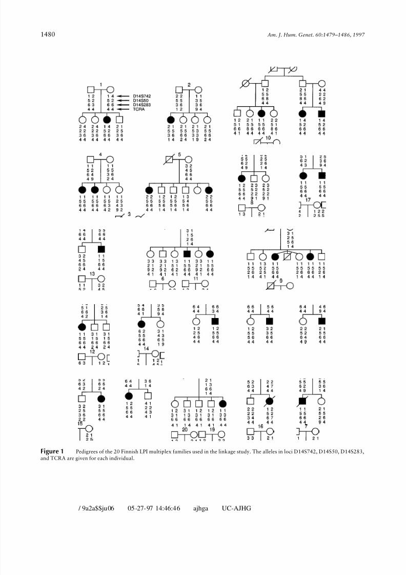
of 9 additional families were included. Altogether, thepatients show hepatosplenomegaly and osteoporosis.Potentially fatal interstitial lung disease and progressive linkage study consisted of samples from 27 patients with
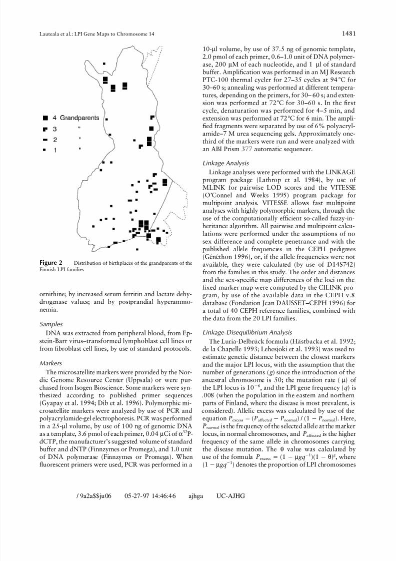
LPI and from 77 healthy family members (fig. 1). Tenadditional families with a single LPI child were includedin the linkage-disequilibrium calculations. The birth-Received December 10, 1996; accepted for publication March 10,
1997. places of the grandparents of the patients were distrib-Address for correspondence and reprints: Dr. Tuija Lauteala, De- uted unevenly throughout Finland (fig. 2). Diagnosis of
partment of Medical Genetics, University of Turku, Kiinamyllynkatu LPI in all index patients was confirmed by clinical evalu-10, SF-20520, Turku, Finland.
ation; by documentation of increased urinary excretion 1997 by The American Society of Human Genetics. All rights reserved.0002-9297/97/6006-0027$02.00 and low plasma concentrations of lysine, arginine, and
1479
/ 9a2a$$ju06 05-27-97 14:46:46 ajhga UC-AJHG
8/8/2019 Lysinuric Protein Intolerance (LPI) Gene Maps to the Long Arm of Chromosome 14
http://slidepdf.com/reader/full/lysinuric-protein-intolerance-lpi-gene-maps-to-the-long-arm-of-chromosome 2/8
1480 Am. J. Hum. Genet. 60:1479–1486, 1997
Figure 1 Pedigrees of the 20 Finnish LPI multiplex families used in the linkage study. The alleles in loci D14S742, D14S50, D14S283,and TCRA are given for each individual.
/ 9a2a$$ju06 05-27-97 14:46:46 ajhga UC-AJHG
8/8/2019 Lysinuric Protein Intolerance (LPI) Gene Maps to the Long Arm of Chromosome 14
http://slidepdf.com/reader/full/lysinuric-protein-intolerance-lpi-gene-maps-to-the-long-arm-of-chromosome 3/8
1481Lauteala et al.: LPI Gene Maps to Chromosome 14
10-ml volume, by use of 37.5 ng of genomic template,2.0 pmol of each primer, 0.6–1.0 unit of DNA polymer-ase, 200 mM of each nucleotide, and 1 ml of standardbuffer. Amplification was performed in an MJ ResearchPTC-100 thermal cycler for 27–35 cycles at 94C for30–60 s; annealing was performed at different tempera-
tures, depending on the primers, for 30– 60 s; and exten-sion was performed at 72C for 30–60 s. In the firstcycle, denaturation was performed for 4–5 min, andextension was performed at 72C for 6 min. The ampli-fied fragments were separated by use of 6% polyacryl-amide–7 M urea sequencing gels. Approximately one-third of the markers were run and were analyzed withan ABI Prism 377 automatic sequencer.
Linkage Analysis
Linkage analyses were performed with the LINKAGEprogram package (Lathrop et al. 1984), by use of MLINK for pairwise LOD scores and the VITESSE
(O’Connel and Weeks 1995) program package formultipoint analysis. VITESSE allows fast multipointanalyses with highly polymorphic markers, through theuse of the computationally efficient so-called fuzzy-in-heritance algorithm. All pairwise and multipoint calcu-lations were performed under the assumptions of nosex difference and complete penetrance and with thepublished allele frequencies in the CEPH pedigrees(Ge ´ne ´thon 1996), or, if the allele frequencies were not
Figure 2 Distribution of birthplaces of the grandparents of the available, they were calculated (by use of D14S742)Finnish LPI families from the families in this study. The order and distances
and the sex-specific map differences of the loci on thefixed-marker map were computed by the CILINK pro-
ornithine; by increased serum ferritin and lactate dehy- gram, by use of the available data in the CEPH v.8drogenase values; and by postprandial hyperammo- database (Fondation Jean DAUSSET–CEPH 1996) fornemia. a total of 40 CEPH reference families, combined with
the data from the 20 LPI families.Samples
Linkage-Disequilibrium Analysis DNA was extracted from peripheral blood, from Ep-stein-Barr virus–transformed lymphoblast cell lines or The Luria-Delbru ¨ ck formula (Ha ¨ stbacka et al. 1992;from fibroblast cell lines, by use of standard protocols. de la Chapelle 1993; Lehesjoki et al. 1993) was used to
estimate genetic distance between the closest markersMarkers and the major LPI locus, with the assumption that the
number of generations ( g ) since the introduction of theThe microsatellite markers were provided by the Nor-dic Genome Resource Center (Uppsala) or were pur- ancestral chromosome is 50; the mutation rate (m) of
the LPI locus is 1006, and the LPI gene frequency (q) ischased from Isogen Bioscience. Some markers were syn-
thesized according to published primer sequences .008 (when the population in the eastern and northernparts of Finland, where the disease is most prevalent, is(Gyapay et al. 1994; Dib et al. 1996). Polymorphic mi-
crosatellite markers were analyzed by use of PCR and considered). Allelic excess was calculated by use of theequation Pexcess Å (Paffected0 Pnormal) / (1 0 Pnormal). Here,polyacrylamide-gel electrophoresis. PCR was performed
in a 25-ml volume, by use of 100 ng of genomic DNA Pnormal is the frequency of the selected allele at the markerlocus, in normal chromosomes, and Paffected is the higheras a template, 3.6 pmol of each primer, 0.04mCi of a32P-
dCTP, the manufacturer’s suggested volume of standard frequency of the same allele in chromosomes carryingthe disease mutation. The u value was calculated bybuffer and dNTP (Finnzymes or Promega), and 1.0 unit
of DNA polymerase (Finnzymes or Promega). When use of the formula Pexcess Å (1 0 m gq01)(1 0 u) g , where(10 m gq01) denotes the proportion of LPI chromosomesfluorescent primers were used, PCR was performed in a
/ 9a2a$$ju06 05-27-97 14:46:46 ajhga UC-AJHG

8/8/2019 Lysinuric Protein Intolerance (LPI) Gene Maps to the Long Arm of Chromosome 14
http://slidepdf.com/reader/full/lysinuric-protein-intolerance-lpi-gene-maps-to-the-long-arm-of-chromosome 4/8
1482 Am. J. Hum. Genet. 60:1479–1486, 1997
Table 1
Pairwise LOD Scores between the LPI Locus and Nine Marker Loci
LOD SCORE AT u Å
MARKER 90% CONFIDENCE
LOCUS .00 .001 .01 .05 .10 .20 .30 Zmax umax LIMITS
D14S261 0 03.25 .57 2.55 2.74 2.03 1.07 2.76 .09 . . .D14S1023 0 0.17 1.87 3.01 3.00 2.16 1.13 3.08 .07 .01 õ u õ .21D14S72 0 1.50 3.36 4.08 3.79 2.61 1.36 4.08 .05 .007 õ u õ .16D14S742 5.82 5.81 5.65 4.94 4.09 2.53 1.24 5.82 .00 .00 õ u õ .06D14S50 4.02 4.01 3.91 3.45 2.88 1.82 .91 4.02 .00 .00 õ u õ .09D14S283 6.91 6.89 6.75 6.07 5.17 3.36 1.72 6.91 .00 .00 õ u õ .06TCRA 5.90 5.89 5.82 5.29 4.48 2.80 1.34 5.90 .00 .00 õ u õ .07MYH7 0 02.00 .04 1.38 1.69 1.43 .82 1.70 .11 . . .D14S64 0 2.98 3.85 3.96 3.52 2.36 1.23 4.04 .03 .001 õ u õ .14
carrying the same ancestral mutation (Lehesjoki et al. (with a {1-LOD-unit support interval of 2.7–8.6 cM)and TCRA, respectively (fig. 4).1993).
Linkage DisequilibriumResultsLinkage-disequilibrium mapping is based on the ob-
Linkage Analysis servation that affected chromosomes descending from acommon ancestral mutation show a distinctive haplo-A total of 317 microsatellite markers randomly spreadtype in the immediate vicinity of the gene, reflecting thein all autosomes were analyzed in the primary familieshaplotype of the ancestral chromosome. We thereforeuntil highly promising LOD scores (ú3) eventually weretested for linkage disequilibrium in LPI families, withobtained with the microsatellite marker GATA74E02,the completely linked markers D14S742, D14S50,defining the locus D14S742 on the long arm of chromo-D14S283, and TCRA. The alleles of these markers, assome 14, near the centromere. No obligatory recombi-well as allele distribution in LPI and non-LPI chromo-nation events were found between the LPI locus and this
marker. Pairwise linkage analysis revealed a maximumLOD score (Zmax) of 4.40 (u Å .00), for the initial mate-
rial from 11 LPI families. Consequently, new markersfrom this region were analyzed, and linkage was con-firmed with six other microsatellite markers (for LODscores in pairwise linkage analyses between the LPI locusand the chromosome 14 markers, see table 1). No oblig-atory recombinants were observed between the LPI locusand the loci D14S742, D14S50, D14S283, and TCRA.Pairwise linkage analysis revealed Zmax values of 5.82,4.02, 6.91, and 5.90, respectively, at u Å .00 for eachof the four markers.
Recombinations with the proximal marker D14S72and the distal marker MYH7 preliminarily defined thegenomic region harboring the LPI locus within a 10-
cM chromosomal region on a sex-averaged genetic map.The 9-point fixed-marker map based on the combinedCEPH-LPI data revealed no significant sex-specific dif-ferences (data not shown). The best-supported order and
Figure 3 The order and estimated sex-averaged distances (inthe estimated sex-averaged distances (in cM) betweencM) between the markers showing linkage to the LPI locus. The best-the analyzed markers are shown in figure 3. By the com-supported sex-averaged 9-point order is shown. The odds that thisbination of information obtained by use of markersorder is correct are at least 2.6 1 104 better than those of the other
D14S72, D14S742, D14S50, D14S283, TCRA, and 9-point orders (excluding pairs with no recombinants) produced byMYH7, in multipoint analyses, the highest LOD scores pairwise inversion of all loci. No sex differences, as estimated in the
study by Hellsten et al. (1993), could be shown within this map region.of 9.32 and 9.31 were measured at markers D14S283
/ 9a2a$$ju06 05-27-97 14:46:46 ajhga UC-AJHG

8/8/2019 Lysinuric Protein Intolerance (LPI) Gene Maps to the Long Arm of Chromosome 14
http://slidepdf.com/reader/full/lysinuric-protein-intolerance-lpi-gene-maps-to-the-long-arm-of-chromosome 5/8
1483Lauteala et al.: LPI Gene Maps to Chromosome 14
somes, are shown in table 2. Allele 4 of marker TCRAwas present in 60 (98.4%) of the 61 LPI chromosomes,which is in contrast with its presence in 22 (35.5%) of the 62 non-LPI chromosomes. Similarly, allele 6 of marker D14S283 was present in 57 (93.6%) of the 61LPI chromosomes and in 14 (22.6%) of the 62 non-LPI
chromosomes. The marker alleles on the chromosomescarrying the LPI mutation also were distributed nonran-domly at loci D14S50 and D14S742. The Pexcess valuesfor the loci D14S742, D14S50, D14S283, and TCRAwere .47, .76, .92, and .98, respectively (table 2). Theestimated genetic region showing allelic association withthe LPI locus spans 1.7 cM (fig. 3). To further estimatethe distance between the disease and the marker loci,we applied a modified Luria-Delbru ¨ ck method. When50 generations of expansion were assumed, a mutationfrequency of 1006 for the LPI locus and an LPI genefrequency of .008 resulted in genetic-distance estimatesof 0.04 cM, 0.17 cM, and 0.54 cM, for the loci TCRA,
D14S283, and D14S50, respectively (fig. 5). In the LPI Figure 5 Luria-Delbru ¨ ck-based analysis of the location of thechromosomes, ten different haplotypes were detectedLPI gene, with regard to marker loci showing no recombinations inwith four markers (D14S742, D14S50, D14S283, andlinkage analysis. Genetic-distance estimates are shown as a function
TCRA), but only five haplotypes were detected when of the number of generations. The 95% confidence interval for TCRAthe alleles of the closest markers, D14S283 and TCRA, (lower limit at 0) is based on the sampling error for chromosomes.
were used. The haplotype x–y–6–4 was present in 57(93.4%) of the 61 LPI chromosomes but in only 5(8.8%) of the 57 non-LPI chromosomes. The haplotype 1–5–6–4 was carried by 31 (50.8%) of the 61 LPI
chromosomes and by 2 (3.5%) of the 57 non-LPI chro-mosomes (table 3). Haplotype x–y–6–4 homozygositywas found in 34 (91.9%) of the 37 LPI patients.
Discussion
The gene for LPI was assigned to the proximal part of chromosome 14 (14q11-13), by use of linkage analysisbased on random screening of the autosomal genome.The closest flanking markers, D14S72 on the centro-meric side and MYH7 on the telomeric side, defined aninterval of Ç10 cM. Strong linkage disequilibrium, withmarkers TCRA, D14S283, and D14S50, was presentin a genomic region of 1.7 cM, and, furthermore, theapplication of the Luria-Delbru ¨ ck-based formula sug-gested that the distance between the closest marker(TCRA) and the LPI locus is only 0.04 cM. The resultsare completely consistent with autosomal recessive in-
heritance and full penetrance, in agreement with pre-viously published conclusions based on segregation pat-terns and on careful clinical evaluation. We found noevidence of genetic heterogeneity, suggesting that, atFigure 4 Seven-point linkage analysis between the LPI locus and
six marker loci. The sex-averaged distances between markers on the least in Finland, the disease is caused by mutations at ahorizontal axis are based on combined data from the LPI and CEPH single locus or at closely related loci. It remains to befamilies. Locus D14S72 was selected as a starting point for the map. determined if LPI families from other countries showThe family data include 20 Finnish families with more than one child.
evidence of mapping to the same region.The Zmax and the corresponding map location were 9.3 and 6.1 cM,Our results are in line with previous linkage studiesrespectively, with a {1-LOD-unit support interval of 2.7–8.6 cM.
The centromere is to the left. of recessive disorders in the isolated population of Fin-
/ 9a2a$$ju06 05-27-97 14:46:46 ajhga UC-AJHG

8/8/2019 Lysinuric Protein Intolerance (LPI) Gene Maps to the Long Arm of Chromosome 14
http://slidepdf.com/reader/full/lysinuric-protein-intolerance-lpi-gene-maps-to-the-long-arm-of-chromosome 6/8
1484 Am. J. Hum. Genet. 60:1479–1486, 1997
Table 2
Distribution of Alleles of TCRA, D14S283, D14S742, and D14S50, in LPI and Non-LPI Chromosomes
TCRA D14S283
No. (%) of No. (%) of
ALLELE LPI Non-LPI Pexcess LPI Non-LPI Pexcess
1 1 (1.6) 15 (24.2) 0 1 (1.6)2 0 10 (16.1) 0 9 (14.5)3 0 3 (4.8) 1 (1.6) 14 (22.6)4 60* (98.4) 22 (35.5) .98 1 (1.6) 9 (14.5)5 . . . . . . 0 10 (16.1)6 . . . . . . 57* (93.6) 14 (22.6) .927 . . . . . . 1 (1.6) 1 (1.6)8 . . . . . . 0 2 (3.2)9 0 12 (19.4) 0 2 (3.2)
. . . . . . 1 (1.6) 010Total 61 62 61 62
D14S50 D14S742
1 1 (1.6) 9 (14.3) 36* (58.1) 17 (27.0) .472 4 (6.6) 26 (41.3) 17 (32.3) 24 (38.1)3 4 (6.6) 6 (9.5) 3 (4.8) 17 (27.0)4 2 (3.2) 6 (9.5) 2 (3.2) 5 (7.9)5 50* (82.0) 16 (25.4) .76 . . . . . .
. . . . . . 1 (1.6) 06Total 61 63 59 63
* P õ .001.
land and again exemplify the power of linkage-disequi- application of the Luria-Delbru ¨ ck formula, originallydeveloped to analyze mutations in rapidly growing bac-librium mapping in the study of such populations. Ap-
proximately 30 recessive diseases (called ‘‘the Finnish terial populations, has been considered justified for thestudy of isolated human populations as well. Dia-disease heritage’’; Norio et al. 1973) are exceptionally
common in the Finnish population. The genes for at strophic dysplasia was a good example for the use of linkage-disequilibrium mapping in the Finnish popula-least 20 of these diseases have been mapped to a specific
locus (Hellsten 1995). In six diseases (progressive myo- tion: Luria-Delbru ¨ ck-based analysis predicted that thediastrophic dysplasia gene lies Ç64 kb from the locusclonus epilepsy [Pennacchio et al. 1996], diastrophic
dysplasia [Ha ¨ stbacka et al. 1994], choroideremia [San- of the closest marker, CSF1R. When the diastrophicdysplasia gene subsequently was cloned, its location waskila et al. 1992], infantile neuronal ceroid lipofuscinosis
[Vesa et al. 1995], hypergonadotropic ovarian dysgene- 70 kb from CSF1R (Ha ¨ stbacka et al. 1992, 1994).The age of the ancestor mutation may be extrapolatedsis [Aittoma ¨ ki et al. 1995], and congenital chloride diar-
rhea [Ho ¨ glund et al. 1996]), the genes have been cloned from the distribution of the grandparents’ birthplaces inFinland or from the size of the genetic region showingby use of the positional cloning strategy. For the last four
diseases listed above, the founder mutation has been linkage disequilibrium. In the diastrophic dysplasia anal-
ysis, the number of generations was assumed to be 100.described further. For four other diseases for which pro-tein data was available (aspartylglucosaminuria [Ikonen This estimate was based on the geographical distribution
of the birthplaces of the grandparents, in the so-calledet al. 1991], hereditary fructose intolerance [Cross etal. 1988], gyrate atrophy of the choroid and the retina earlier settlement areas in the southern and central parts
of the country. In the LPI families, the distribution of [Mitchell et al. 1989], and nonketotic hyperglycinemia[Kure et al. 1992]), the gene was cloned by use of the the grandparents’ birthplaces in the southeastern and
northern parts of the country—that is, both the olderfunctional cloning principle, followed by detection of the prevalent mutations. and recent settlement areas — supports the estimate of
Ç50 generations as the age of the mutation. The geneticSince its founding 100–200 generations ago, the pop-ulation of Finland has grown in isolation. Therefore, region of 1.7 cM that is in strong linkage disequilibrium
/ 9a2a$$ju06 05-27-97 14:46:46 ajhga UC-AJHG

8/8/2019 Lysinuric Protein Intolerance (LPI) Gene Maps to the Long Arm of Chromosome 14
http://slidepdf.com/reader/full/lysinuric-protein-intolerance-lpi-gene-maps-to-the-long-arm-of-chromosome 7/8
1485Lauteala et al.: LPI Gene Maps to Chromosome 14
follicle-stimulating hormone receptor gene causes hereditaryTable 3hypergonadotropic ovarian failure. Cell 82:959–968
Distribution of Combined Haplotypes for the D14S742–D14S50– Closs EI, Albritton LM, Kim JW, Cunningham JM (1993 a)D14S283–TCRA Loci, in LPI and Non-LPI Chromosomes Identification of a low affinity, high capacity transporter of
cationic amino acids in mouse liver. J Biol Chem 268:7538–NO. (%) OF
7544Closs EI, Lyons R, Kelly C, Cunningham JM (1993b) Charac-LPI Non-LPI
terization of the third member of the MCAT family of cat-HAPLOTYPE [n Å 61] [n Å 57]ionic amino acid transporters. J Biol Chem 268:20796–
1– 5–6 –4 31 (50.8) 2 (3.5) 20800x– 5–6 –4 48 (78.7) 3 (5.3) Collins A, Teague J, Keats BJ, Morton NE (1996) Linkagex– y–6 –4 57 (93.4) 5 (8.8) map integration. Genomics 36:157–162Others 4 (6.6) 52 (91.2) Cross N, Tolan D, Cox T (1988) Catalytic deficiency of human
aldolase B in hereditary fructose intolerance caused by acommon missense mutation. Cell 53:881–885
Davis MM, Bjorkman PJ (1988) T-cell antigen receptor genesalso supports evidence of a relatively high number of and T-cell recognition. Nature 334:395–402generations since expansion of the LPI mutation. The
de la Chapelle A (1993) Disease gene mapping in isolatednumber of generations strongly influences the distancehuman populations: the example of Finland. J Med Genet
estimation (fig. 5). If the number of generations since30:857–865
the founding of the LPI mutation is set to be 20 instead Desjeux J-F, Rajantie J, Simell O, Dumontier A-M, Perheen-
of 50, the distance between the LPI and TCRA loci tupa J (1980) Lysine fluxes across the jejunal epithelium inwould increase from 0.04 cM to 0.12 cM. lysinuric protein intolerance. J Clin Invest 65:1382–1387The critical region harboring the LPI locus has three Dib C, Faure S, Fizames C, Samson D, Drouot N, Vignal A,
Millasseau P, et al (1996) A comprehensive genetic map of known gene assignments, TCRA, CTLA1, and TRP1the human genome based on 5,264 microsatellites. Nature(Morton et al. 1992; Collins et al. 1996). None of these380:152–154genes has any known amino acid–transport function.
Fondation Jean DAUSSET– CEPH (1996) http://wwwTRCA is a large 97.6-kb region containing the human.cephb.frT cell–receptor a and d polypeptide-chain coding re-
Ge ´ne ´thon (1996) http://www.genethon.frgions. T cell antigen receptor polypeptides (a, b, g , andGyapay G, Morissette J, Vignal A, Dib C, Fizames C, Milas-
d) form two different heterodimers, ab and gd. The func-seau P, Bernardi G, et al (1994) The 1993–1994 Ge ´ne ´thon
tion of T cell receptor ab is well established in antigen human genetic linkage map. Nat Genet 7:246–339recognition, but less is known about the gd receptor Ha ¨stbacka J, de la Chapelle A, Kaitila I, Sistonen P, Weaver A,(Davis and Bjorkman 1988; Koop et al. 1994). Some Lander E (1992) Linkage disequilibrium mapping in isolated
LPI patients have associated immunological abnormali- founder populations: diastrophic dysplasia in Finland. NatGenet 2:204–211ties, but a clear T cell defect is not a feature of the disease
Ha ¨stbacka J, de la Chapelle A, Mahtani MM, Clines G, Reeve-(Nagata et al. 1987). TRP1 is a tRNA proline 1 (MitchellDaly MP, Daly M, Hamilton BA, et al (1994) The dia-et al. 1991). The third gene in this region, CTLA1, codesstrophic dysplasia gene encodes a novel sulfate transporter:for cytotoxic T-lymphocyte–associated serine esterase 1positional cloning by fine-structure linkage disequilibrium(Heusel et al. 1991). Whether any of these genes is themapping. Cell 78:1073–1087disease-causing gene in LPI remains to be seen. If not,
Hellsten E (1995) Positional cloning of the infantile ceroidthe assignment of the critical LPI region to a chromo-
lipofuscinosis gene. PhD thesis, National Public Health In-somal area of õ100 kb provides a good starting point stitute, Helsinkifor physical cloning and gene identification. Hellsten E, Vesa J, Speer MC, Ma ¨ kela ¨ T, Ja ¨rvela ¨ I, Alitalo K,
Ott J, et al (1993) Refined assignment of the infantile neu-ronal ceroid lipofuscinosis (INCL, CLN1) locus at 1p32:Acknowledgmentsincorporation of linkage disequilibrium in multipoint analy-
We thank the lysinuric protein– intolerance families for their sis. Genomics 16:720– 725excellent cooperation. We also thank Mrs. A. Reinikainen and Heusel JW, Hanson RD, Silverman GA, Ley TJ (1991) Struc-Mrs. Y. Bullen for their expert technical assistance. This study ture and expression of a cluster of human hematopoieticwas supported by the Ulla Hjelt Fund, the Foundation for serine protease genes found on chromosome 14q11.2. J BiolPediatric Research, in Finland, the Sigrid Juselius Foundation, Chem 266:6152 –6158and the Signe and Arne Gyllenberg Foundation. Ho ¨ glund P, Haila S, Socha J, Tomaszewski L, Saarialho-Kere
U, Karjalainen-Lindsberg M-L, Airola K, et al (1996) Muta-tions of the Down-regulated in adenoma (DRA) gene causeReferencescongenital chloride diarrhea. Nat Genet 14:316–319
Ikonen E, Baumann M, Gro ¨ n K, Syva ¨ nen A-C, Enomaa N,Aittoma ¨ ki K, Lucena J, Pakarinen P, Sistonen P, Tapanainen J, Gromoll J, Kaskikari R, et al (1995) Mutation in the Halila R, Aula P, et al (1991) Aspartylglucosaminuria:
/ 9a2a$$ju06 05-27-97 14:46:46 ajhga UC-AJHG

8/8/2019 Lysinuric Protein Intolerance (LPI) Gene Maps to the Long Arm of Chromosome 14
http://slidepdf.com/reader/full/lysinuric-protein-intolerance-lpi-gene-maps-to-the-long-arm-of-chromosome 8/8
1486 Am. J. Hum. Genet. 60:1479–1486, 1997
cDNA encoding human aspartylglucosaminidase and the guchi S, Aoki K (1987) Immunological abnormalities in apatient with lysinuric protein intolerance. Eur J Pediatr 146:missense mutation causing the disease. EMBO J 10:51– 58427–428Incerti B, Andria G, Parenti G, Sebastio G, Ghezzi M, Strisciug-
Norio R, Nevanlinna HR, Perheentupa J (1973) Hereditarylio P, Sperlı D, et al (1993) Lysinuric protein intolerance:diseases in Finland: rare flora in rare soil. Ann Clin Res 5:studies on 17 Italian patients. Am J Hum Genet Suppl 53:109–141908
O’Connel JR, Weeks DE (1995) The VITESSE algorithm forKato T, Mizutani N, Ban M (1984) Hyperammonemia in ly-
rapid exact multilocus linkage analysis via genotype set-sinuric protein intolerance. Pediatrics 73:489–492recording and fuzzy inheritance. Nat Genet 11:402–408Koop BF, Rowen L, Wang K, Kuo CL, Seto D, Lenstra JA,
Parto K, Kallajoki M, Aho H, Simell O (1994) PulmonaryHoward S, et al (1994) The human T-cell receptor TCRAC/ alveolar proteinosis and glomerulonephritis in lysinuric pro-TCRDC (C-alpha/C-delta) region: organization, sequence,tein intolerance: case reports and autopsy findings of fourand evolution of 97.6 kb of DNA. Genomics 19:478–493pediatric patients. Hum Pathol 25:400– 407Kure S, Takayanagi M, Narisawa K, Tada K, Leisti J (1992)
Parto K, Svedstro ¨ m E, Majurin M-L, Ha ¨ rko ¨ nen R, Simell OIdentification of a common mutation in Finnish patients(1993) Pulmonary manifestations in lysinuric protein intol-with nonketotic hyperglycinemia. J Clin Invest 90:160–164erance. Chest 104:1176– 1182Lathrop GM, Lalouel JM, Julier C, Ott J (1984) Strategies for
Pennacchio LA, Lehesjoki A-E, Stone NE, Willour VL, Virta-multilocus linkage analysis in humans. Proc Natl Acad Scineva K, Miao J, D’Amato E, et al (1996) Mutations in theUSA 81:3443–3446gene encoding cystatin B in progressive myoclonus epilepsyLauteala T, Horelli-Kuitunen N., Closs EI, Savontaus M-L,(EPM1). Science 271:1731– 1734Lukkarinen M, Simell O, Cunningham J, et al. Human cat-
Perheentupa J, Visakorpi JK (1965) Protein intolerance withionic amino acid transporter gene hCAT-2 is assigned to
deficient transport of basic amino acids: another inborn er-8p22 but is not the causative gene in lysinuric protein intol-ror of metabolism. Lancet 2:813– 816
erance. Hum Genet (in press)Rajantie J, Simell O, Perheentupa J (1980) Basolateral-mem-
Lehesjoki A-E, Koskiniemi M, Norio R, Tirrito S, Sistonen P,brane transport defect for lysinuric protein intolerance. Lan-
Lander E, de la Chapelle A (1993) Localization of the EPM1cet 7:1219–1221
gene for progressive myoclonus epilepsy on chromosome(1981) Basolateral transport defect in renal tubuli. J
21: linkage disequilibrium allows high resolution mapping.Clin Invest 67:1078–1082
Hum Mol Genet 2:1229–1234Sankila E-M, Tolvanen R, van den Hurk J, Cremers F, de la
Mitchell A, Bale AE, Wang-ge M, Yi HF, White R, Pirtle Chapelle (1992) Aberrant splicing of the CHM gene is aRM, McBride OW (1991) Localization of a DNA segment significant cause of choroideremia. Nat Genet 1:109–113encompassing four tRNA genes to human chromosome Simell O (1995) Lysinuric protein intolerance and other cat-14q11 and its use as an anchor locus for linkage analysis. ionic aminoacidurias. In: Scriver CR, Beaudert AL, Sly WS,Genomics 11:1063– 1070 Valle D (eds) The metabolic and molecular bases of inherited
Mitchell G, Brody L, Sipila ¨ I, Looney J, Wong C, Engelhardt disease, 7th ed. McGraw-Hill, New York, pp 3603– 3627 J, Patel A, et al (1989) At least two mutant alleles of orni-
Vesa J, Hellsten E, Verkruyse LA, Camp LA, Rapola J, San-thine d-aminotransferase cause gyrate atrophy of the cho- tavuori P, Hofmann SL, et al (1995) Mutations in the pal-roid and retina in Finns. Proc Natl Acad Sci USA 86:197– mitoyl protein thioesterase gene causing infantile neuronal201 ceroid lipofuscinosis. Nature 376:584–587
Morton NE, Collins A, Lawrence S, Shields DC (1992) Algo- Yoshimoto T, Yoshimoto E, Meruelo D (1991) Molecularrithms for a location database. Ann Hum Genet 56:223– cloning and characterization of a novel human gene homolo-232 gous to the murine ecotropic retroviral receptor. Virology
185:10–17Nagata M, Suzuki M, Kawamura G, Kono N, Koda N, Yama-
/ 9a2a$$ju06 05-27-97 14:46:46 ajhga UC-AJHG