lynn m. anderanin, cpc,cpc-i,cosc -...
TRANSCRIPT

1
Case Studies of the
Upper and Lower Extremities
Lynn M. Anderanin, CPC,CPC-I,COSC
Setting the Rules
• Coding is subjective
• Guidelines and policies differ
• Every provider dictates using their own style
• Complete operative reports are available for review
2

2
3 Steps to Coding
• Where - is the procedure being performed
• How - is the procedure being performed
• What - is being done
3
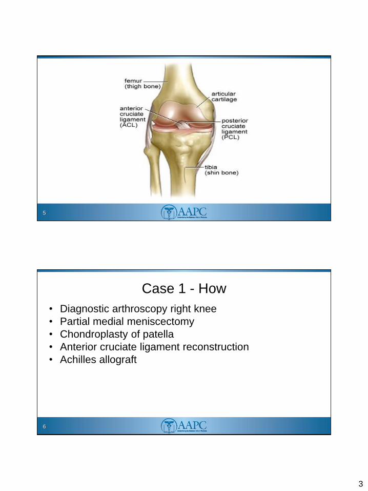
Case Study 1 - Where
• Right anterior cruciate ligament rupture
• Medial meniscus repair
• Chondromalacia of the patella
4
3
5
Case 1 - How
• Diagnostic arthroscopy right knee
• Partial medial meniscectomy
• Chondroplasty of patella
• Anterior cruciate ligament reconstruction
• Achilles allograft
6
4
Case Study 1 - What
• Standard diagnostic arthroscopy at the anterolateral portal site
• Anterior medial working portal made
• A partial medial meniscectomy carried out
• The lateral compartment normal
• Adequate notchplasty performed
• Chondroplasty at the patellofemoral joint
• Simultaneous thawing of a 10 mm preshaped achilles tendon
allograft
7
Case Study 1 - What
• Placement of the tibial tunnel guide through the medial portal
• 2-3cm vertical stab incision made at the medial face of the
proximal tibia
• Over-reaming with a 9.5mm solid reamer
• Placement of a femoral socket on the lateral wall of the right
femur
8

5
Case Study 1 - What
• Over-reaming to a depth of 25 mm to match the bone plug
• 8x25 mm screw selected
• Vertical stab incision made percutaneously
• Distal fixation performed
• Whipstitch four ends spread with the tensioner
9
Case Study 1 - What
• Four arms of the suture to the wall of the tibial
tunnel
• Placement of a large sheath.
• Excess graft cut flush to the tibia
10
6
Case Study 1 - Coding
CPT®
• 29888
• 29881
ICD-9-CM
• 844.2
• 836.0
• 717.7
11
Case Study 2 - Where
• Left endstage ankle arthritis
• Left subtalar arthritis
• Left talonavicular arthritis
12

7
Bones of the Foot
13
Case Study 2 - How
• Subtalar arthrodesis
• Total ankle arthroplasty
• Debridement and capsular release talonavicular
joint
14

8
Case Study 2 - What
• A minimally invasive lateral incision created along the
subtalar joint
• Subtalar joint exposed through a lateral sinus tarsi incision
• Fishtailing of the arthrodesis surfaces
• The joint repaired
• Two 6.7 cannulated screws fixated
15
Case Study 2 - What
• Standard anterior incision created
• Joint surfaces prepared through a stepwise
procedure
• Final implants trialed
• There was no need for gastrocnemius or achilles
lengthening
• Implants placed
16

9
Case Study 2 - What
• The talonavicular joint identified through the distal
end of the anterior incision
• Cheilectomy and release of the dorsal capsule
• Osteophyte resected at the level of the dorsal
talonavicular joint
17
Case Study 2 - Coding
CPT®
• 27702
• 28725
• 28120
ICD-9-CM
• 715.97
18
10
Case Study 3 - Where
• History of right ankle arthrodesis with nonunion
19
Case Study 3 - How
• Right ankle revision arthrodesis
• Fibular osteotomy
• Right calcaneal bone graft
• Fibular autograft
• Removal of hardware, right ankle
20

11
Case Study 3 - What
• Percutaneous medial incision utilized in line with the previous
surgical incision
• Remove medial screws
• The fibula removed and utilized for bone graft
• Percutaneous calcaneal bone graft performed mixing
allograft bone graft in a mill
• Bony edges prepared using a saw blade
21
Case Study 3 - What
• Bur used to prepare the nonunion site
• Medial incision made to prepare medial gutter
• Cannulated screws placed across the arthrodesis
site
• The external compressor applied
• Laterally applied plate
22

12
Case Study 3 - Coding
CPT®
• 27870
• 20680
• 20900
• 27707
ICD-9-CM• 996.78
23
Case Study 4 - Where
• Degenerative arthritis secondary to avascular
necrosis, left femoral head of the hip
• Degenerative arthritis of the right knee
24

13
Case Study 4 - How
• Left total hip arthroplasty
• Right total hip arthroplasty
25
Case Study 4 - What
Left Hip
• Incision made on the lateral aspect of the hip centering on the posterior aspect of the trochanteric region
• Bursal tissue excised
• Short external rotators released off the posterior aspect of the femoral head and neck region
• Femoral head removed
• Capsule and labrum excised
26

14
Case Study 4 - What
• Reaming performed on the acetabulum
• Bone chips obtained to graft cystic areas of the anterior
acetabulum region
• Acetabular cup was implanted with 2 screws
• Intramedullary canal was reamed
• Femoral stem was placed
• Trial head and neck combination placed in position
• Permanent components placed
27
Case Study 4 - What
Right Knee
• Incision made in the anterior aspect of the knee
• Osteophytes removed with a ronguer
• Saw used to remove undersurface of the patella
• Protective metal plate then positioned
28

15
Case Study 4 - What
• Opening in the distal femur with drill hole
• An intramedullary rod placed
• Saw used to make a femoral cut
• Large osteophyte from the anterior edge of the tibia removed
• Awl used to make an opening in the tibial plafond
29
Case Study 4 - What
• Drill hole made at the anterior aspect of the tibia
• An intramedullary rod placed
• Bone cut below the medial tibial plateau
• Femoral and tibial cuts made
• Femoral trial and trial liner fitted
30

16
Case Study 4 - What
• Lug holes drilled in the patella
• Trial patellar button fitted
• Drilling and broaching of the tibia
• Tibial tray and patella cemented
• Permanent components put into place
31
Case Study 4 - Coding
CPT®
• 27447
• 27130
ICD-9-CM
• 715.96
• 715.25
• 733.42
32

17
Case Study 5 - Where
• Sustained a left knee distal quadriceps rupture
when she mis-stepped going down the stairs.
• X-ray evidence showed avulsion fragment of bone.
33
Case Study 5 - How
• Distal quadriceps repair of the left knee
34

18
Case Study 5 - What
• Midline incision made
• Small bone fragments excised
• Soft tissue debrided from the distal pole of the patella
• Bone chips and periosteum created corticoccancellous
bleeding bed to prepare reattachment
• Torn tendon tissue excised
35
Case Study 5 - What
• 3 holes drilled from mid proximal pole of the patella to the
inferior pole
• Medial and lateral parallel drill holes performed
• Shuttle sutures placed
• 2 modified Krakow stitches created
• The four arms shuttled through the drill holes
• Knots tied over a bony bridge at the distal pole of the patella
36

19
Case Study 5 - Coding
CPT®
• 27385
ICD-9-CM
• 844.8
• E880.9
37
Case Study 6 - Where
• Fracture dislocation of the left elbow of person who fell on an
outstretched arm.
• Closed manipulation of the ulna humeral joint performed in the
ED.
• X-rays showed significant abnormalities of the radiocapitellar joint
and comminuted fracture at the inferior hemisphere and mild
anterior subluxation.
• Posterolateral rotator instability and lateral ulnar collateral
ligament injury.
38

20
Case Study 6 - How
• Left elbow exam under anesthesia
• Open excision of communited Mason Type radial head and
coronoid process type I fragment.
• Radial head arthroplasty
39
Case Study 6 - What
• Incision made at the mid capitellar line and equator
of the radial capitellar joint
• Sharp elevation performed at anterior joint capsule
• Radial head missing from its anatomical location
• Capsulectomy performed
• Removal of multiple cartilaginous fragments inferior
to the radial head
40

21
Case Study 6 - What
• The radiocapitellar joint reduced with a retractor
• Distal capsulotomy extended
• Neck of the radial head excised
• Implant sized
• Type I coronoid process fracture removed
• Awl used as a canal finder followed by broaching
41
Case Study 6 - What
• Trial placed and tried
• Final implant placed
42

22
Case Study 6 - Coding
CPT®
• 24666
ICD-9-CM
• 813.05
• 832.01
43
Case Study 7 - Where
• Grade IIIA open distal tibia and fibular fracture of
the right leg
• Large wound with tibia exposed
44

23
Case Study 7 - How
• Irrigation and debridement right leg
• Closure of wound
45
Case Study 7 - What
• Soft tissue, subcutaneous tissue, and lower tissues including
loose fascia, and bone debrided
• Pulse lavage of 6 liters used to wash out the wound
• Complex closure of 8 cm wound closed
• More proximal wound of 5 cm closed with simple sutures
46
24
Case Study 7- Coding
CPT®
• 11012 (11044)
• 13121-58
• 13122-58
• 12002-59-58
ICD-9-CM• 891.0
• 823.92
47
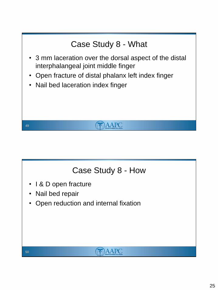
The Hand
48
25
Case Study 8 - What
• 3 mm laceration over the dorsal aspect of the distal
interphalangeal joint middle finger
• Open fracture of distal phalanx left index finger
• Nail bed laceration index finger
49
Case Study 8 - How
• I & D open fracture
• Nail bed repair
• Open reduction and internal fixation
50

26
Case Study 8 - What
• Skin and subcutaneous tissue debrided
• Sutures placed to radial and ulnar aspects of a bone fragment to fix the bone into place
• Skin then sutured
• Remaining nail removed from the nail bed
• Nail bed repaired with sutures
• Nail replaced onto the nail bed with sutures
• Band Aid applied to middle finger
51
Case Study 8 - Coding
CPT®
• 26765
• 11010-51
• 11760-51
ICD-9-CM
• 816.12
• 883.1
52

27
Case Study 9 - Where
• Left distal radius fracture
• Malunion with dorsal tilt
53
Case Study 9 - How
• Osteotomy with tricortical iliac crest allograft
54
28
Case Study 9 - What
• Volar approach made of the distal radius
• Osteotomy with a micro oscillating saw
• Wide osteotome utilized to cut to correct the deformity
• Tricortical allograft cut into shape
• 4 locking screws, a plate, and interfragmentary screw placed
through the distal radius an allograft
55
Case Study 9 - Coding
CPT®
• 25405
ICD-9-CM
• 733.81
56

29
Case Study 10 - Where
• Stenosing tenosynovitis of the right index finger
• Stenosing tenosynovitis of the right middle finger
• Stenosing tenosynovitis of the right ring finger
57
Case Study 10 - How
• Open release of stenosing tenosynovitis
58

30
Case Study 10 - What
• Transverse incision made in line with distal palmar crease of the index finger
• A1 pulley released
• Oblique incision made in line with distal palmar crease of the middle finger
• A1 pulley released
• Transverse incision made in line with the distal palmarcrease of the ring finger
• A1 pulley released
59
Case Study 10 - Coding
CPT®
• 26055-F6
• 26055-F7
• 26055-F8
ICD-9-CM
• 727.03
60
31
Case Study 11 - Where
• Right 5th and 4th metacarpal fractures
– Displaced and comminution of the shaft 5th MC
– Nondisplaced diaphyseal junction fracture 4th MC
61
Case Study 11 - How
• Open reduction internal fixation of the right 5th metacarpal
• Closed treatment of the right 4th metacarpal
62

32
Case Study 11 - What
• Longitudinal incision over the dorsal and ulnar aspect of the 5th MC
• Periosteum incised
• Bone subperiosteally dissected except for a fragment
• Open reduction obtained and secured
• 6 hole plate applied to the dorsal ulnar aspect of the fracture and secured using AO technique
• 4th MC remained nondisplaced
63
Case Study 11 - Coding
CPT®
26615
26600-59
ICD-9-CM
• 815.03
• 815.00
• E888.9
64
33
Case Study 12 - Where
• Distal biceps tendon rupture right elbow
65
Case Study 12 - What
• Incision made over the antecubital fossa
• Palpated a stump of the biceps tendon distally
• Incision made over the radial tuberosity
• A trough made for the insertion of the biceps tendon
• Biceps tendon end freshened
66

34
Case Study 12 - What
• Two drill holes made for sutures
• A stitch made with fiberwire
• Sutures delivered through the tunnel and drill holes
• Tucked tendon into the trough
• Tied the tendon to the radius
67
Case Study 12- Coding
CPT®
• 24342
ICD-9-CM
• 840.8
68
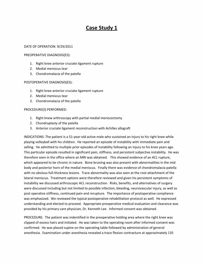
Case Study 1
DATE OF OPERATION: 9/29/2011
PREOPERATIVE DIAGNOSIS(ES):
1. Right knee anterior cruciate ligament rupture 2. Medial meniscus tear 3. Chondromalacia of the patella
POSTOPERATIVE DIAGNOSIS(ES):
1. Right knee anterior cruciate ligament rupture 2. Medial meniscus tear 3. Chondromalacia of the patella
PROCEDURE(S) PERFORMED:
1. Right knew arthroscopy with partial medial meniscectomy 2. Chondroplasty of the patella 3. Anterior cruciate ligament reconstruction with Achilles allograft
INDICATIONS: The patient is a 51‐year‐old active male who sustained an injury to his right knee while playing volleyball with his children. He reported an episode of instability with immediate pain and selling. He admitted to multiple prior episodes of instability following an injury to his knee years ago. This particular episode resulted in significant pain, stiffness, and persistent subjective instability. He was therefore seen in the office where an MRI was obtained. This showed evidence of an ACL rupture, which appeared to be chronic in nature. Bone bruising was also present with abnormalities in the mid body and posterior horn of the medial meniscus. Finally there was evidence of chondromalacia patella with no obvious full‐thickness lesions. Trace abnormality was also seen at the root attachment of the lateral meniscus. Treatment options were therefore reviewed and given his persistent symptoms of instability we discussed arthroscopic ACL reconstruction. Risks, benefits, and alternatives of surgery were discussed including but not limited to possible infection, bleeding, neurovascular injury, as well as post operative stiffness, continued pain and rerupture. The importance of postoperative compliance was emphasized. We reviewed the typical postoperative rehabilitation protocol as well. He expressed understanding and elected to proceed. Appropriate preoperative medical evaluation and clearance was provided by his primary care physician, Dr. Kenneth Lee. Informed consent was obtained.
PROCEDURE: The patient was indentified in the preoperative holding area where the right knee was clipped of excess hairs and initialed. He was taken to the operating room after informed consent was confirmed. He was placed supine on the operating table followed by administration of general anesthesia. Examination under anesthesia revealed a trace flexion contracture at approximately 135

degrees of passive flexion. There was evidence of 2+ Lachman and a pivot glide with no significant collateral ligament instability. He was therefore stabilized in an arthroscopic leg holder and placed in a well‐padded proximal tight tourniquet perset to 275mmHg. The contralateral leg was placed in a sequential compression device at the lower calf and stabilized in the hemilithotomy position with the lateral proximal knee well padded. The entire right lower extremity was then prepped and draped in the usual sterile fashion. After confirming consent, side, and prophylactic antibiotics, standard diagnostic arthroscopy was initiated through a horizontal stab incision at the anterolateral portal site. Blunt atraumatic entry into the patellofemoral joint revealed mild synovitis and posttraumatic effusion. There was also irregularity at the apex and lateral facet of the patella consistent with the MRI findings of chondromalacia. The trochlea was intact. No obvious loose bodies were seen. The medial gutter demonstrated a moderately robust synovial plica. A spinal needle was used to localize an anterior medial working portal. This was used to probe a clear abnormality at the medial meniscus. There was a complex tear of the mid body and posterior horn of the medial meniscus consistent with the MRI findings. A partial medial meniscectomy was therefore carried out using a combination of up‐biting and straight basket resectors and the 4.0 synovial resector. Ir required resection of the inferior leaflet of a horizontal cleavage tear of the posterior horn. 30‐40% of the mid body of the meniscus required resection due to a parrot‐beaked flap tear. The anterior horn was intact. The partial medial meniscectomy was balanced by placing the scope in the medial portal and approaching the mid body laterally. Chondral surface of the tibiofemoral joint demonstrated grade1 softening and areas of grade2 fibrillation but no significant high grade lesions.
Inspection of the notch demonstrated scar tissue consistent with a chronic anterior cruciate ligament rupture. The lateral wall was empty in the notch and there was a stump of ACL displaced posteriorly. The PCL was intact. The lateral compartment was then inspected with normal‐appearing tibiofemoral articular cartilage with only mild softening. The inner margin of the lateral meniscus was intact with no obvious macro tearing. There was some mild fibrillation at the root attachment which was very difficult to access, but there was no palpable instability at this location. The popliteus tendon was visualized and intact. The lateral gutter was free of loose bodies.
At this point in the procedure chondroplasty at the patellofemoral joint was performed. A 4.0 synoval resector was used on both oscillate and forward modes to debride unstable cartilage at the apex, lateral and medial facets of the patella. This involved approximately 2.5cm square with grade 2 and grade 3 involvement. No subchondral bone was exposed. After adequate irrigation the femoral notch was addressed. The soft tissues were removed with a 4.0 synovial resector and it was clear that an ACL reconstruction would be necessary. Simultaneous thawing of a 10mm preshaped Achilles tendon allograft was performed at the back table by my assistant. It was thawed in antibiotic impregnated saline, followed by appropriate shaping. It was measured at 9.5mm in diameter and precut to approximately 15cm in length. Allograft was then whipstitch where the tail of the tendon was split into four arms. The four tendon arms also measured approximately 9.5mm in ideal size. This was then appropriately tensioned on the back table and draped with a sterile sponge. Simultaneous preparation of the notch was performed by me using a 5.5 ball‐tipped bur to create a hemi‐cathedral to allow for the over the top position to be visualized. Adequate notchplasty was performed to accept at least a 10mm

graft. I then placed the retrograde tibial tunnel guide through the medial portal and 2‐3cm vertical stab incision was made at the medial face of the proximal tibia. Blunt dissection was carried out to the periosteum of the tibia followed by retrograde insertion of the tibial guidepin at 55 degrees of inclination at the anatomic insertion of the ACL on the tibial plateau. This was adjusted with a second pin slightly posteriorly to ensure no lateral wall or ceiling impingement. This was overreamed with a 9.5mm solid reamer followed by placement of an over‐the‐top guide. This allowed for placement of a femoral socket at approximately the 10 to 10:30 position on the lateral wall of the right femur. This was overreamed with a reamer to a depth of 25mm to match the bone plug. A pin was passed to deliver traction sutures at the bone socket of the Achilles allograft, which was placed into the knee retrograde. It seated nicely into the socket. I selected an 8 x 25mm titanium interference screw which was placed over a guidepin at percutaneously to allow for parallel insertion of the screw in relation to the femoral socket. This was performed with excellent bite and fixation. The knee was cycled 10 times and it was felt to be isometric. Distal fixation was then carried out. The whipstitch four ends were spread with the tensioner and the knee was cycled 10 times. An appropriate large dilator was used to spread the four arms of the suture to the wall of the tibial tunnel followed by placement of a large sheath. With the knee in extension and approximately 15 pounds of distal traction as well as posterior drawer pressure on the proximal tibia a large taper screw was placed with excellent fixation and bite. Intraoperative Lachman and palpation of the graft was extremely satisfying. Final photographs were obtained and the excess graft was cut flush to the medial face of the tibia. The wounds were copiously irrigated and closure was performed with 3‐0 vicryl for this medial tibial incision followed by simple 3‐0 nylons. Approximately 2oml of 1% xylocaine with epinephrine and 10ml of 0.25% marcaine with epinephrine and 3mg of duramorph were then infused into the knee for postoperative pain control. A sterile nonadhesive dressing was applied using betadine ointment, fluffs, ABDs,and sterile webril. He was placed in a knee brace locked in full extension. All sponge, needle, and instrument counts were correct, and the patient tolerated the procedure well with no evidence of intraoperative complication. He was taken to the postanesthesia recovery room in stable condition.
Elapsed tourniquet time was approximately 90 minutes.
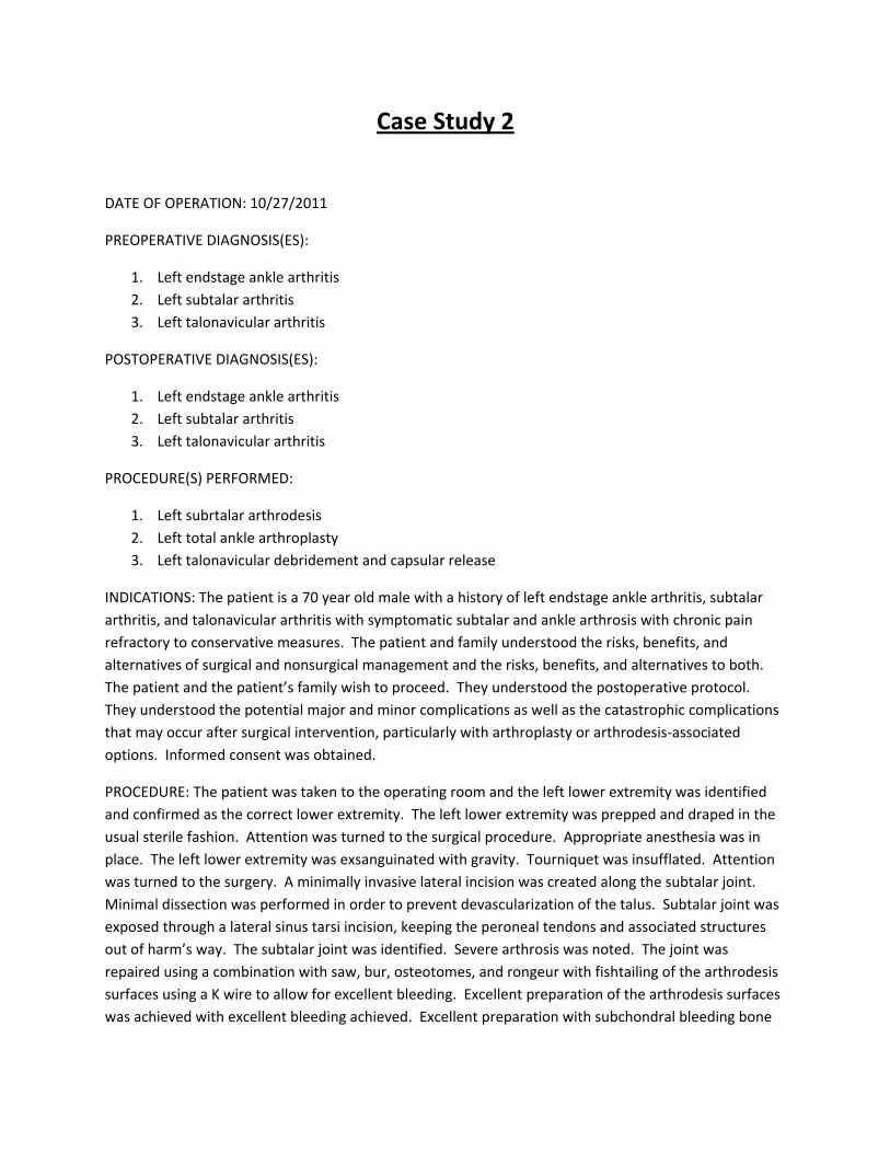
Case Study 2
DATE OF OPERATION: 10/27/2011
PREOPERATIVE DIAGNOSIS(ES):
1. Left endstage ankle arthritis 2. Left subtalar arthritis 3. Left talonavicular arthritis
POSTOPERATIVE DIAGNOSIS(ES):
1. Left endstage ankle arthritis 2. Left subtalar arthritis 3. Left talonavicular arthritis
PROCEDURE(S) PERFORMED:
1. Left subrtalar arthrodesis 2. Left total ankle arthroplasty 3. Left talonavicular debridement and capsular release
INDICATIONS: The patient is a 70 year old male with a history of left endstage ankle arthritis, subtalar arthritis, and talonavicular arthritis with symptomatic subtalar and ankle arthrosis with chronic pain refractory to conservative measures. The patient and family understood the risks, benefits, and alternatives of surgical and nonsurgical management and the risks, benefits, and alternatives to both. The patient and the patient’s family wish to proceed. They understood the postoperative protocol. They understood the potential major and minor complications as well as the catastrophic complications that may occur after surgical intervention, particularly with arthroplasty or arthrodesis‐associated options. Informed consent was obtained.
PROCEDURE: The patient was taken to the operating room and the left lower extremity was identified and confirmed as the correct lower extremity. The left lower extremity was prepped and draped in the usual sterile fashion. Attention was turned to the surgical procedure. Appropriate anesthesia was in place. The left lower extremity was exsanguinated with gravity. Tourniquet was insufflated. Attention was turned to the surgery. A minimally invasive lateral incision was created along the subtalar joint. Minimal dissection was performed in order to prevent devascularization of the talus. Subtalar joint was exposed through a lateral sinus tarsi incision, keeping the peroneal tendons and associated structures out of harm’s way. The subtalar joint was identified. Severe arthrosis was noted. The joint was repaired using a combination with saw, bur, osteotomes, and rongeur with fishtailing of the arthrodesis surfaces using a K wire to allow for excellent bleeding. Excellent preparation of the arthrodesis surfaces was achieved with excellent bleeding achieved. Excellent preparation with subchondral bleeding bone

was achieved. This was then prepared and transfixed using two 6.7 cannulated screws with rigid purchase and fixation with excellent compression and alignment achieved. The preoperative severe valgus was corrected through the subtalar arthrodesis to approximately two or three degrees of hindfoot valgus with appropriate correction anatomically. Excellent correction of the hindfoot was achieved. The screws were placed in appropriate position to prevent impingement on the planned ankle replacement.
Attention was now turned to the ankle arthroplasty. Standard anterior incision was created. No touch technique was utilized in order to prevent an injury to the skin. Dissection was carried out. The superficial peroneal nerve was identified and kept out of harm’s way. The extensor retinaculum was incised in line with the skin incision longitudinally. The deep peroneal nerve was identified. This was appropriately mobilized out of harm’s way. The joint was visualized. The articular surfaces were identified. Severe endstage arthrosis was noted. The joint surfaces were appropriately prepared. The medial and lateral gutters were identified. Standard technique was utilized for preparing the joint surfaces through a stepwise procedure, using the ankle replacement system. Excellent preparation of the surfaces was achieved. Final implants were trialed with a size 2 talus, 8mm polyethylene with excellent range of motion to approximately 20 degrees of dorsiflexion intraoperatively and 40 degrees of plantar flexion with excellent motion achieved. There was no need for gastrocnemius or Achilles lengthening, given the excellent motion intraoperatively. The prosthesis was noted to be in appropriate position and excellent alignment in all planes as confirmed fluoroscopically as well as by direct anatomic visualization. The talonavicular joint was then identified through the distal end of the anterior incision. A cheilectomy and release of the dorsal capsule was performed and the osteophyte was resected at the level of the dorsal talonavicular joint. Copious irrigation of all wounds was carried out. Layered closure was performed. A well padded soft tissue dressing with nonweightbearing cam walker boot was applied. The patient tolerated the procedure well without complication and was transferred to the recovery room in stable condition with appropriate followup instructions given.

Case Study 3
DATE OF OPERATION: 8/02/2011
PREOPERATIVE DIAGNOSIS(ES):
1. Right ankle arthrodesis nonunion
POSTOPERATIVE DIAGNOSIS(ES):
1. Right ankle arthrodesis nonunion
PROCEDURE:
1. Right ankle revision arthrodesis 2. Fibular osteotomy 3. Right Calcaneal bone graft as well as fibular autograft, separate incision. 4. Removal of hardware, right ankle
INDICATIONS: this patient is a male with a history of right ankle arthrodesis with nonunion, with continued pain and failure of improvement with conservative measures. The patient wished to proceed with the above‐names procedures. He understood the risks, benefits, and alternatives, including but not limited to infection, bleeding, nerve injury, persistent pain, failure of improvement, need for additional surgery, as well as other associated both major and minor complications, as well as catastrophic complications that may occur. The patient and patient’s family wished to proceed. The patient understood postoperative protocol. The patient understood the potential catastrophic complications. Informed consent was obtained.
PROCEDURE: The patient was taken to the operating room and the right lower extremity was identified and confirmed as the correct lower extremity. Care was taken to pad the bilateral lower extremities and torso. Attention was turned to the surgical procedure. Appropriate anesthesia was in place. The tourniquet was insufflated to 250mmHg calf tourniquet, after the limb was exsanguinated. A percutaneous medial incision was utilized in line with the previous surgical incision to remove the medial screws. The lateral screws were removed using the standard lateral incision. A fibular osteotomy was performed. The fibula was removed and utilized for bone graft. Percutaneous calcaneal bone graft was performed using bone graft reamer harvester was utilized for the bone graft. The bone graft was prepared within a bone mill. In addition allograft was impacted into the arthrodesis site. The bone edges were prepared using a saw blade. The arthrodesis surfaces were appropriately prepared using a saw blade until excellent bleeding bony edges were achieved. The surfaces were then prepped using a bur until excellent preparation of the nonunion site was achieved. There was noted to be significant fibrous nonunion. Separate medial incision was created to prepare the medial gutter. Excellent preparation of bleeding bony surfaces was achieved. Excellent bleeding was achieved on each

level. After this, 2 4.5 cannulated screws were placed across the arthrodesis site from the lateral shoulder of the talus extending proximally with bicortical purchase fixation and the tibia with excellent rigid purchase. At this point the ankle was corrected to valgus to neutral sagittal plane positioning, and an appropriate rotational alignment with approximately 5 degrees of valgus and excellent positioning in all planes. The rigid fixation was achieved with percutaneous screw fixation followed by use of a compression system.
Next, the external compressor was applied using compression technique followed by laterally applied plate along the lateral shoulder of the talus and along the tibia. Care was taken to place the screws extra‐articular within the talus. Rigid purchase fixation was achieved. Rigid compression was achieved at the arthrodesis site and excellent alignment in all planes. Copious irrigation of the wound was then carried out. Layered closure was performed. Complete hemostasis was obtained prior to closure. A well‐padded soft tissue dressing followed by a short leg cast was applied with appropriate alignment. The patient tolerated the procedure well. The patient was transferred to the recovery room in stable condition.

Case Study 4
DATE OF OPERATION: 9/15/2011
PREOPERATIVE DIAGNOSIS(ES):
1. Degenerative arthritis secondary to avascular necrosis of the femoral head, left hip.
2. Degenerative arthritis of the right knee.
POSTOPERATIVE DIAGNOSIS(ES):
1. Degenerative arthritis secondary to avascular necrosis of the femoral head, left hip.
2. Degenerative arthritis of the right knee.
PROCEDURES PERFORMED:
1. Left total hip arthroplasty
2. Right total hip arthroplasty
PROCEDURE: Following administration of right femoral nerve block and general anesthesia, the patient
was then placed In the right lateral decubitus position with a deflatable bean bag either in this position
and an axillary roll beneath the right lateral thorax. The patient was kept in the decubitis position with
padded bumps anteriorly and posteriorly as well. The left lower extremity and left hip were then
prepped and draped in the usual sterile fashion. A curvilinear incision was made on the lateral aspect of
the hip centering on the posterior aspect of the trochanteric region with a #10 blade. The incision was
approximately 20cm in length. The incision was carried down to the dermis and subcutaneous tissue
down to the iliotibial band and gluteus maximus fascia. These were incised in line with the skin incision.
A retractor was then used to retract these edges anteriorly and posteriorly.
Attention was then paid to the bursal region, which was noted to have markedly thickened bursal tissue.
This was excised. The short external rotators were then released off the posterior aspect of the femoral
head and neck region. The distance between the mark placed over the iliac crest and th rrochanteric
ridke measured 125mm. It was determined that preoperatively the length of approximately a
centimeter would be appropriate. The piriformis tendon was identified and released after being tagged
with #5 ethibond sutures. The capsule was then incised in a T fashion. There was a moderate amount
of inflammatory fluid expressed from the knee, but there was also calcified synovial tissue present. This
was excised and sent to pathology. The femoral head was noted to be markedly deformed. The
osteotomy guide for the hip system was then used to make a cut in the femoral neck. The dead was
removed, which was as mentioned deformed. Attention was then paid to the acetabulum and
thickened capsule and labrum were ecxised from around the perimeter. Reaming was then begun with
a size 46 reamer, then advanced up to a 50 reamer. There was good punctuate bleeding bone
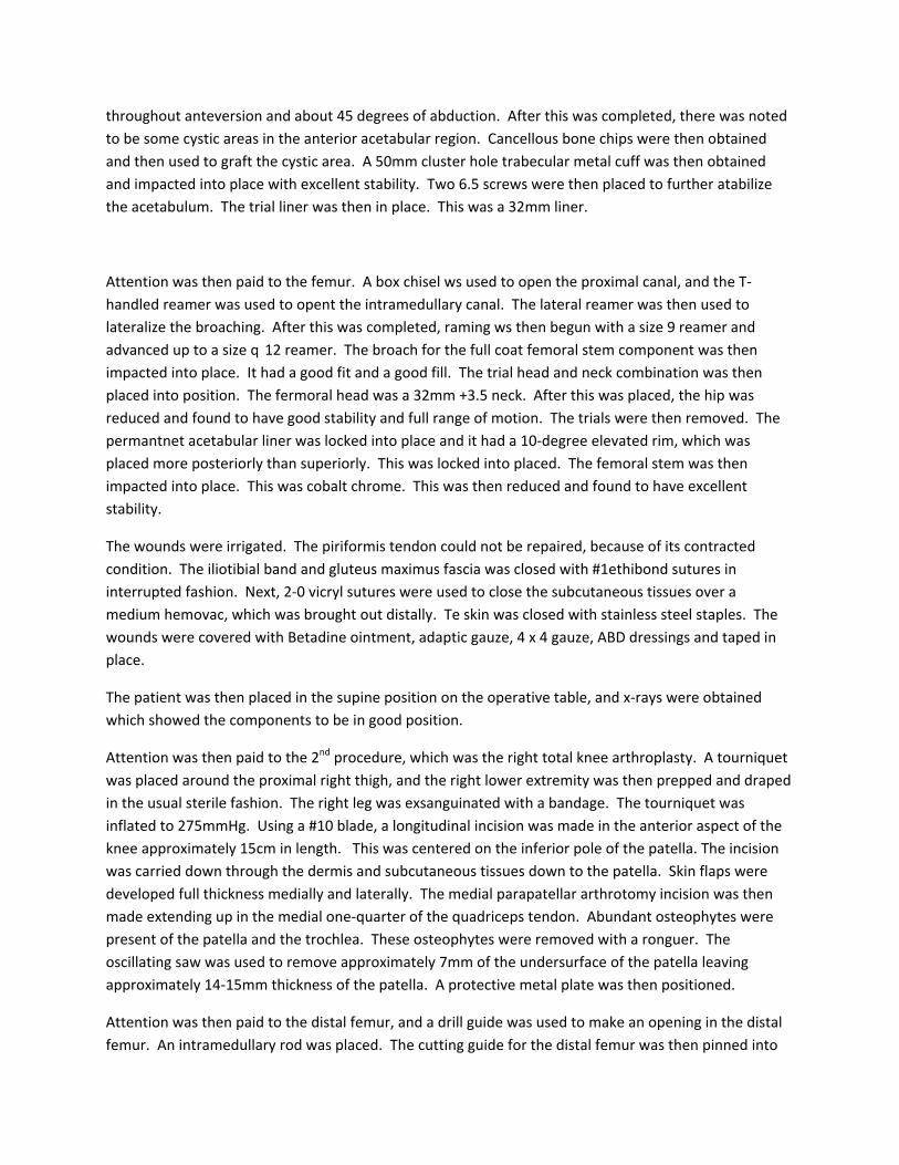
throughout anteversion and about 45 degrees of abduction. After this was completed, there was noted
to be some cystic areas in the anterior acetabular region. Cancellous bone chips were then obtained
and then used to graft the cystic area. A 50mm cluster hole trabecular metal cuff was then obtained
and impacted into place with excellent stability. Two 6.5 screws were then placed to further atabilize
the acetabulum. The trial liner was then in place. This was a 32mm liner.
Attention was then paid to the femur. A box chisel ws used to open the proximal canal, and the T‐
handled reamer was used to opent the intramedullary canal. The lateral reamer was then used to
lateralize the broaching. After this was completed, raming ws then begun with a size 9 reamer and
advanced up to a size q 12 reamer. The broach for the full coat femoral stem component was then
impacted into place. It had a good fit and a good fill. The trial head and neck combination was then
placed into position. The fermoral head was a 32mm +3.5 neck. After this was placed, the hip was
reduced and found to have good stability and full range of motion. The trials were then removed. The
permantnet acetabular liner was locked into place and it had a 10‐degree elevated rim, which was
placed more posteriorly than superiorly. This was locked into placed. The femoral stem was then
impacted into place. This was cobalt chrome. This was then reduced and found to have excellent
stability.
The wounds were irrigated. The piriformis tendon could not be repaired, because of its contracted
condition. The iliotibial band and gluteus maximus fascia was closed with #1ethibond sutures in
interrupted fashion. Next, 2‐0 vicryl sutures were used to close the subcutaneous tissues over a
medium hemovac, which was brought out distally. Te skin was closed with stainless steel staples. The
wounds were covered with Betadine ointment, adaptic gauze, 4 x 4 gauze, ABD dressings and taped in
place.
The patient was then placed in the supine position on the operative table, and x‐rays were obtained
which showed the components to be in good position.
Attention was then paid to the 2nd procedure, which was the right total knee arthroplasty. A tourniquet
was placed around the proximal right thigh, and the right lower extremity was then prepped and draped
in the usual sterile fashion. The right leg was exsanguinated with a bandage. The tourniquet was
inflated to 275mmHg. Using a #10 blade, a longitudinal incision was made in the anterior aspect of the
knee approximately 15cm in length. This was centered on the inferior pole of the patella. The incision
was carried down through the dermis and subcutaneous tissues down to the patella. Skin flaps were
developed full thickness medially and laterally. The medial parapatellar arthrotomy incision was then
made extending up in the medial one‐quarter of the quadriceps tendon. Abundant osteophytes were
present of the patella and the trochlea. These osteophytes were removed with a ronguer. The
oscillating saw was used to remove approximately 7mm of the undersurface of the patella leaving
approximately 14‐15mm thickness of the patella. A protective metal plate was then positioned.
Attention was then paid to the distal femur, and a drill guide was used to make an opening in the distal
femur. An intramedullary rod was placed. The cutting guide for the distal femur was then pinned into

place, and the oscillating saw was used to make the femoral cut. One additional note was that the right
knee had range of motion which lacked about 5‐10 degrees of extension and had flexion to about 75‐80
degrees preoperatively.
After the distal femoral cut was made, attention was then paid to the tibia, and the oscillating saw was
used to remove a large osteophyte from the anterior edge of the tibia. An awl was used to make an
opening in the tibial plafond 1.5cm from the anterior aspect of the tibia. The three‐eighths step drill was
used to make a drill hole at this location.
An intramedullary rod was placed and the cutting guide was then set for approximately 2‐3mm of bone
resection below the medial tibial plateau. The oscillating saw was used to make this cut after the citting
guide was pinned into place. It was elected to use a posterior stabilized component due to the severe
osteophytes of the knee and the attenuation of the cruciate ligaments. The sizing guide for the femur
was then set in place. A size F component had the best fit. The cutting guide was then used to make the
femoral cuts except for the trochlear recession cut. One additional note, the oscillating saw was used
also to make a tibial cut and these cuts were completed using osteotomes and with the removal of the
bone fragment using a bone ronguer. The femoral trial was placed, and this was also used to make the
box cut for the posterior stabilized component and the trochlear recession cut. After this was
completed, the trials were placed in position. A size 5 tibial tray had good coverage. The size F standard
femoral component had good coverage as well. The size 10mm trial liner was placed.
Attention was then addressed to the patella, and the 32mm lug hole drill guide was used to drill the lug
holes. The trial patellar button also had good tracking and good coverage. These all trials were
removed.
Then, attention was paid to drilling and broaching the tibia for the stemmed component. After this was
completed, pusatile lavage was then used and cement was mixed, and the tibial tray was cemented into
place. The knee was then brought into extension. Pressure was applied during the curing phase while
the patella was cemented. Similarly, excess cement was removed from around the patella and the tibial
components. After bone cement hardened, the porous‐coated posterior stabilized size F component
was impacted into place. A size 10 linear was then placed and this is highly cross linked. The knee was
brought into full extension. It had excellent fit and stability. The patella tracked well. The knee was
then irrigated free of any loose debris. The knee was also irrigated with a diluted mixture of Betadine
solution and saline. The arthrotomy incision was closed with no. 1 ethibond sutures in a interrupted
fashion. The hemovac was placed in the subcutaneous space, and the subcutaneous space was closed
with 2‐0 vicryl sutures. The skin was closed with stainless steel staples.
The blood loss during the case for the hip was 1000ml and for the knee 0ml.
The patient did receive 1 unit of blood, packed RBCs. The patient also received crystalloid. The wounds
were covered with Betadine ointment, adaptic gauze, 4x4 gaueze, ABD dressings, steile webril and an
ace bandage. The tourniquet was deflated after less than 2 hours and 2 minutes of tourniquet time.

The x‐rays were obtained postoperatively, showed the component in good position. There were no
complications. Sponge and needle counts were correct at the end of the procedure. The patient was
transferred to postanesthesia recovery room in stable condition.

Case Study 5
DATE OF OPERATION: 9/16/2011
PREOPERATIVE DIAGNOSIS(ES): Left distal quadriceps rupture
POSTOPERATIVE DIAGNOSIS(ES):Left distal quadriceps rupture
PROCEDURES PERFORMED: Left knee distal quadriceps repair
INDICATIONS: The patient is a 75‐year‐old who sustained a left knee distal quadriceps rupture earlier
today when she misstepped going down the stairs. Immediate pain, swelling, and the inability to
ambulate prompted a visit directly to the emergency room. Workup included physical exam and plain x‐
rays, which demonstrated a palpable defect at the distal quadriceps insertion at the superior pole of the
patella. There was also x‐ray evidence of an avulsion fragment of bone from the superior pole
suggestive of quadriceps tendon rupture. We therefore had a long discussion regarding her treatment
options. A poor prognosis associated with conservative management was reviewed and, therefore, I
recommended repair. Risk, benefits, and alternatives to repair were reviewed in detail, including but
not limited to possible infection, bleeding, neurovascular injury, as well as postop stiffness and
continued pain. The possibility of rerupture was also reviewed. Risks associated with anesthesia were
also reviewed. Appropriate preoperative medical evaluation and clearance was provided by the
internist on call in preparation for surgery today. Informed consent was obtained.
Urinalysis obtained in the emergency room was suspicious for subclinical infection and, therefore, IV
PROCEDURE: The patient was identified in the preoperative holding area where the left lower extremity
was initialed. She was taken to the operating room and placed on the operating table in the supine
position. She was successfully placed under general LMA anesthesia, followed by application of a well‐
padded proximal thigh tourniquet preset to 275mmHg. The left lower extremity was then prepped and
draped in the usual sterile fashion to the proximal thigh with all other bony prominences well padded.
After confirming consent, side, and prophylactic antibiotics, the left lower extremity was marked with a
pen beginning at the inferior pole of the patella extending proximally 8‐10cm. The leg was
exsanguinated and the tourniquet was elevated. A midline incision was then made through skin and
subcutaneous tissues exposing a hematoma and a clear defect at the distal quadriceps insertion at the
superior pole of the patella. This was irrigated and the tear was inspected. There was a complete
avulsion of the distal quadriceps from the superior pole of the patella with small fragments of bone
encased within the ruptured tendon proximal stump. This was sharply excised with a 15 blade. The
subcutaneous layer was elevated medially and proximally to assess the extent of the quadriceps
rupture. It did extend down the medial and lateral retinacular gutters with avulsion of the vastus
medialis muscle. Once the tear was clearly identified, the distal pole of the patella was initially
addressed. A ronguer, sharp 15 blade, and a 6mm ball tipped bur was used to debride soft tissue.

Unstable chips of bone and periosteum to create corticocancellous bleeding bed in preparation for
reattachment of the torn tendon. Attention was then drawn to the tendon to where fibrillated torn
tendon tissue was excised to a clean healthy‐appearing tendinous bleeding bed. I then addressed the
patella initially at the mid proximal pole of the patella extending to the inferior pole. A medial and
lateral parallel drill hole was also achieved, followed by grasping sutures. Once this was prepared, a #2
fiberwire was used to place a modified stitch beginning medially at the margin of the quadriceps tendon
and the vastus medialis. This was extended proximally and then marched back distally at the midaspect
of the quadriceps tendon. In a similar fashion, a second #2 fiberwire was begun at the lateral margin
with a modified stitch marching proximally and then returning distally at the midsection of the tendon.
All 4 arms were then carefully shuttled through the previously placed drill holes. The 2 central arms
were delivered in the central hole with the medial arm extending medially and the lateral arm extending
laterally. The knee was hyperextended and the tendon was reduced to the prepared bony bed.
Excellent reduction and anatomic alignment was felt to be achieved. Knots were then tied over a bony
bridge at the distal pole of the patella. The knee was then taken through a passive range of motion and
the repair appeared stable and rigid through at least 45 degrees of knee flexion. This was felt to be due
to the fairly immediate intervention from the time of injury. Excellent mobility was felt to be present. I,
therefore, continued with the repair oversewing the medial and lateral aspects of the retinaucular tissue
with figure‐of‐eight #1 vicryl. The wound was copiously irrigated and suctioned dry. Layered closure
was then performed with 0‐vicryl and 2‐0 vicryl reapproximating the subcutaneous adipose layer and
the dermis. The skin was reapproximated with skin staples. A sterile nonadhesive dressing was applied
using betadine ointment, adaptic, ABDs, and sterile webril. The entire lower extremity was padded well
with additional webril, and compressive ace wrap was placed from the metatarsals to the proximal
thigh. She was securely immobilized in full extension in a knee immobilizer, and awoken without
complication. All sponge, needle, and instrument counts were correct. Elapsed tourniquest time was 54
minutes. She was taken to the postanesthesia recovery room in stable condition.
Case Study 6
DATE OF OPERATION: 09/13/2011
PREOPERATIVE DIAGNOSIS(ES): Left elbow fracture‐dislocation of the radial head and coronoid process
POSTOPERATIVE DIAGNOSIS(ES): Left elbow fracture‐dislocation of the radial head Mason type III and
coronoid process type I without posterolateral rotatory instability
PROCEDURES PERFORMED:
1. Left elbow exam under anesthesia with open excision comminuted Mason Type III radial head
and coronoid process type I fragment.
2. Radial head arthroplasty
INDICATIONS: The patient is a 59‐year‐old right hand dominant female who sustained a fracture‐
dislocation of her left elbow on 9/1/2011. She fell on an outstretched arm and was initially managed in
the emergency room with closed manipulation. This resulted in reduction of the ulna humeral joint.
She was found to be stable per the ER staff and was admitted for pain control. Followup x‐rays
demonstrated a reduced ulnohumeral joint, but there were significant abnormalities at the
radiocaptellar joint with the evidence of a comminuted fracture at the inferior hemisphere and mild
anterior subluxation. She was seen in the office and a long discussion took place. Given significant
blocks at terminal flexion and pronation, my recommendation was for open treatment of this terrible
triad‐type injury. We discussed treatment options including open reduction and internal fixation of the
radial head fragment if at all repairable. If significant, posterolateral rotatory instability and lateral ulnar
collateral ligament injury was also indentified. This would be repaired as well. I felt that the coronoid
fragment was likely too small for repair, estimated at 1‐2mm in size. Finally if the elbow was felt to be
unstable in the operating room, an ulnar collateral ligament repair would also be carried out. The risks,
benefits, and alternatives to surgery were discussed including but not limited to possible infection,
bleeding, neurovascular injury, as well as persistent instability, loss of motion and possible complications
related to anesthesia. She expressed understanding and elected to proceed. Informed consent was
obtained.
PROCEDURE: The patient was identified in the preoperative holding area where the left upper extremity
was initiated. She was taken to the operating room, placed supine on the operating table followed by
general anesthesia. The left upper extremity was then extended on a hand table with a fluoroscopic
imaging and exam under general anesthesia demonstrated stable ulnohumeral joint from 20‐100
degrees of passive flexion. There was block to further flexion presumably due to an anterior medially
subluxed radial head. There was a tiny chip fracture at the tip of the coronoid, which moved with a ulna
through its range of motion. Gentle varus and valgus stess demonstrated valgus instability which
incongruency of the radiocapitellar joint. No significant varus instability was noted. I also did not
appreciate any posterolateral rotatory instability on gentle supination and pivot shift. The patient was

therefore prepped and draped in the usual sterile fashion with plans for a sterile proximal arm
tourniquet.
After confirming consent, side, and prophylactic antibiotics, an 8cm curvilinear incision was marked
beginning just proximal to the lateral epicondyle extending toward Lister tubercle. The arm was
exsanguinated and the tourniquest was elevated. The skin was incised as marked exposing the deep
fascia of the extensor digitorum comminus. This was incised at the mid capitellar line and equator of
the radial capitellar joint in line with the fibers. Sharp elevation was performed anteriorly to expose the
anterior joint capsule. I was able to palpate the capitellum, but the radial head was noticeably missing
at its anatomic location.
A capsulotomy was therefore performed to expose the superior lateral aspect of the capitellum. Care
was taken to incise the capsule superior to the lateral ulnar collateral ligament, which was apparently
intact despite this injury. Fracture hematoma was encountered. This was evacuated and soft clot was
removed. There was clear subluxation of the radial head superiorly and medially. This clearly blocked
flexion beyond 100 degrees as well a pronation. There were multiple cartilaginous fragments inferior to
the radial head which were removed. These were no larger than 3‐4mm in diameter. I was able to
reduce the radial capitellar joint with the assistance of a small angled retractor. The distal capsulotomy
was extended no further than 2.5cm form the radiocaptellar joint line to protect the posterior
interosseous nerve. Due to the blockage and pronation, I was only able to pronate the forearm to
approximately 20‐30 degrees; however, as the radiocapitellar joint was reduced. Better supination and
pronation was achieved. The distal extension was performed in full pronation thereafter. The annular
ligament was carefully noted for later repair. At this point in the procedure, the radial head was
inspected. It was a clear Mason type III comminuted radial head fracture with no significant remnant of
the posterolateral rim of the radial head. It was impacted and the multiple fragments were
unsalvageable.
At this point in the procedure, I elected to proceed with radial head arthroplasty and excision of this
irrepairable type III injury. A 10mm microsagittal saw was therefore used to perform a surgical neck
excision of the comminuted radial head approximately 8‐10mm from the proximal articular surface of
the radial head. The superior hemisphere of the radial head was intact and provided a guide to length.
After head excision, it was inspected and sized on the back table and felt to be ideally fit for a 20mm
diameter implant. I also copiously irrigated the joint medially, and there was evidence of a congruent
ulnohumeral joint. Image instensification confirmed tiny type I coronoid process fracture, which was
easily indentified and found loose in the synovium. This was easily removed and found to be 2x1mm in
size. This was discarded. The elbow was taken through a passive range of motion, and there was no
significant instability in the ulnohumeral joint from 20‐120 degrees of flexion. Given satisfactory
stability and evidence of what appeared to be an intact lateral ulnar collateral ligament, I elected to
proceed with arthroplasty as planned. The 5mm awl was used as a canal finder followed by sequential
broaching. A 7mm diameter implant was felt to be ideal to engage the cortex of the radial shaft.
Trial was therefore placed after appropriate stepwise broaching with a zero stem length. The
radiocapitellar joint was carefully reduced and taken through a range of motion. It was felt to be

somewhat lacing in length and therefore a +2mm stem was inserted as a second trial. This provided
excellent reestablishment of the joint with no significant opening of the ulnohumeral joint at the
coronoid. There was evidence of full range of motion with a stable radiocapitellar joint on the
posterolateral rotatory testing. I was therefore satisfied with this length, which was selected for the
final implant. The neck cut was felt to be essentially perpendicular to the shaft and therefore cortical
reaming was not necessary. At this point in the procedure, the final implant was prepared on the back
table including a 20mm in diameter head impacted on a 7x2mm stem. This was impacted onto the
taper with the appropriate alignment of the laser marks to be positioned at the mid equator with the
forearm in neutral supination pronation. The final module implant was then impacted into the canal
with excellent fit and motion I was extremely satisfied with the stability, congruity, and position of the
radial head implant. At this point in the procedure, the wound was copiously irrigated. Final
radiographs confirmed ideal placement of the implant. The elbow was stressed in valgus in full
extension and 30 degrees flexion and felt to be stable at the ulnohumeral joint with mild 1 to 2+ valgus
instability, presumably due to an ulnar collateral ligament injury. I felt that this would heal on its own
with early rehabilitation and range of motion with protection from valgus stress.
At this point in the procedure, layered closure was performed. The annular ligament was repaired with
2‐0 ethibond followed by 0 vicryl for reapproximation of the capsulotomy. The extensor communis
tendon was also reapproximated with 0 vicryl. Skin was reapproximated with 2‐0 vicryl followed by 4‐0
monocryl and steri‐strips. A sterile nonadhesive dressing was applied using betadine ointment, adaptic,
fluffs, ABD and steril webril. She was placed in a fiberglass posterior mold in 90 degrees of elbow flexion
and neutral supination pronation.
All sponge, needle, and instrument counts were correct, and she tolerated the procedure well with no
evidence of intraoperative complication.
Total tourniquet time was 99 minutes.

Case Study 7
DATE OF OPERATION: 10/19/2011
PREOPERATIVE DIAGNOSIS(ES):
1. Grade 3 open distal tibia and fibular fracture
2. Right leg wound, 18cm
POSTOPERATIVE DIAGNOSIS(ES):
1. Grade 3 open distal tibia and fibular fracture
2. Right leg wound, 18cm
PROCEDURES PERFORMED:
1. Irrigation and debridement of wound tibia right leg.
2. Complex closure of wound, 18cm
INDICATIONS: The patient sustained a severe injury to her right lower extremity with a right hip
dislocation and a grade 3A open distal tibia and fibular fracture. She is status post intramedullary
fixation right leg. She also had undergone I and D of the right leg. At this point she is brought back here
for irrigation and debridement right leg and closure of wound. Informed consent was obtained. She
received appropriate preoperative intravenous antibiotic.
PROCEDURE: The patient was taken to the operating room, placed on the operating table in supine
position. General anesthesia was successfully administered. Right lower extremity was prepped and
draped in the usual sterile fashion. Noted was a large wound with tibia bone exposed. We debrided
some of the soft tissues including some of the loose fascia. We also continued down to the bone. We
debrided the bone and surrounding soft tissue. We made sure that the wound was thoroughly debrided
and cleaned. We used pulse lavage a total of 6 liters to wash out the wound after debridement. Then
we proceeded forward with this complex wound closure. We were able to bring the soft tissues and
take a soft tissue flap and approximated it and bring it out distally, and closed the wound with 3‐0 nylon
interrupted fashion. The total wound size was 8cm. The more proximal wound measured 5cm. We
were able to close with simple interrupted nylon. The procedure went well. There were no
complications. Sterile soft dressings applied and a posterior splint was applied with the ankle in neutral
position. After soft dressing was applied, ace wrap was applied. There was no complication. The
patient did well. She was taken to the recovery room in satisfactory condition

Case Study 8
DATE OF OPERATION: 11/27/2011
PREOPERATIVE DIAGNOSIS(ES):
1. Complex wound left index finger
2. Open fracture distal phalanx left index finger
3. Nail bed laceration left index finger
POSTOPERATIVE DIAGNOSIS(ES):
1. Complex wound left index finger
2. Open fracture distal phalanx left index finger
3. Nail bed laceration left index finger
PROCEDURES PERFORMED:
1. Irrigation and debridement open fracture distal phalanx left index finger
2. Open reduction and internal fixation of left index finger
3. Nailbed repair
INDICATIONS: This is a 20 month old who sustained an injury today to this left index finger. He is noted
to have lacerated through the nail, nailbed. There is a flap of tissue hanging at the tip of the finger. The
patient therefore is being taken to the operating room for treatment of this condition.
PROCEDURE: The patient was seen in the preoperative holding area, marking confirmed the left index
finger the operative site. The patient taken to the operating room. Time‐out was called, confirming left
index finger as the operative site. The patient’s left upper extremity was cleaned, prepped and draped
In the usual sterile manner. The arm was exsanguinated and the tourniquet inflated to 250mmHg.
There was a 3mm laceration over the dorsal aspect of the distal interphalangeal joint middle finger; this
was irrigated and a band‐aid was placed on this at the end of the procedure. The index finger itself was
noted to have laceration which went through about mid nailbed from proximal distal, the paronychial
skin also involved all the way circumferentially. There was a little bit of tissue coming into this area,
which I think was probably a vessel and nerve more on the volar ulnar aspect. This was carefully
preserved. Loupe magnification was used. This was untwisted as it had spiraled and twisted around.
Wound irrigated. Contamination was none. The skin and subcutaneous tissue were debrided. It was
noted that there was bone in the distal portion of the skin. There was nothing in it to revascularize at
this distal level, but it was felt that it would be worthwhile to sew the fragment into place in this young
individual and see if we get some composite graft healing. The most critical aspect or helpful portion I
think would be to get the nailbed to heal back into place and bone to heal into place and I think this is
certainly possible and likely to happen.

After wound was irrigated completely, a couple of 6‐0 vicryl sutures were placed to radial and ulnar
aspects of the bone with needle going easily through the soft bone at the distal phalanx and these were
pulled taught radially and ulnarly and the bone was fixed into place. Sutures were cut. The skin was
then reapproximated with 6‐0 gut using simple interrupted suture technique form the paronychial skin
on the ulnar side and going around the volar side over to the ulnar side. Lastly, the remaining nail was
removed from the nail bed. It was noted that the nail was avulsed form its origin. The nail was kept on
the side, cleaned up. The nail bed repaired with loupe magnification, 6‐0 gut using simple interrupted
suture technique at the 4 corners and then the nail itself was replaced onto the nail bed to hold shape
and to keep the eponychial fold open it is anticipated that this nail will fall out as the new one grows in.
The finger was irrigated. Band‐aid was placed on the middle finger as described previously. Xeroform
gauze placed around the tip of the index finger but not to circumferentially to allow for some swelling
and vascularity. This was then dressed dorsally, palmarly, and ulnarly with folded over cut 4x4’s and
then wrapped gently with stretch kling one inch, followed by light layer of coban which was not tight
and then aluminum foam splint in the shape of a C was fitted to the index finger, hand, and wrist with
coban.
Tourniquet was deflated prior to the end of the case. The estimated blood loss minimal. There was no
drain. No specimen. No complications. The patient awakened at the end of the procedure, taken to
recovery room, awake, and stable.
Case Study 9
DATE OF OPERATION:12/1/2011
PREOPERATIVE DIAGNOSIS(ES): Malunion of left distal radius fracture
POSTOPERATIVE DIAGNOSIS(ES): Malunion of left distal radius fracture
PROCEDURES PERFORMED:
1. Left distal radius osteotomy
2. Tricortical iliac crest bone allograft
INDICATIONS: This is a 78 year old with a left distal radius fracture. She was noted to have with dorsal
tilt. She has some really secondary DC deformity. The patient is therefore being taken to the operating
room for treatment of this condition. I discussed the diagnosis, planned operative procedure, risks,
benefits, and alternatives as well as potential complications with her. Questions were answered. She
voiced understanding and agreement and we are proceeding with surgery today.
PROCEDURE: The patient was seen in the preoperative holding area, marking and confirming the left
wrist as the operative site. The patient was taken to the operating room where a time out was called,
confirming the left wrist to be the operative site. The patient was placed under general anesthesia, the
left upper extremity cleaned, prepped, and draped in the usual sterile manner. She received one gram
of Ancef prior to the onset of the procedure. A volar approach was made of the distal radius along the
distal portion of the flexor carpi radialis, going to the floor of this, and down to identify the flexor
pollicus longus and pronator quadrates. The thumb flexor was dissected and retracted ulnarly. The
radial aspect of the pronator quadrates was incised and the distal radius was subperiosteally exposed.
Fluoroscopy was utilized to view where the osteotomy should be made. It was felt that it was fairly
distal but all metaphyseal and I wanted to keep enough bone distally to get the screws for the plate in.
An osteotomy was made with a micro oscillating saw, cooling the blade and the osteotomy site and
performed this under fluoroscopic guidance.
After this was obtained, a wide osteotome was utilized to complete the cut and then open up the
osteotomy and correct the deformity. An allograft tricortical wedge allograft was utilized, cut into shape
in trapezoidal fashion and placed at the oteotomy site, provisionally fixed with Kirschner wires and then
a synthes locking plate was applied. The distal four screws were placed first into the distal fragment,
again under fluoroscopic guidance, all locking screws, and then the place was reduced to and fixed down
to the distal shaft of the radius and finally an interfragmentary screw 3.5mm diameter was placed
through the volar native distal radius and into the dorsal allograft to cinch this down to the shaft and
proximal fragment. The final x‐rays demonstrated good alignment, length and fixation with good radial
tilt and volar tilt accomplished.

The wound was irrigated copiously. The pronator quadratus was at least partially closed over the plate
and most of the radial aspect could not be closed over. The pronator quadratus was reapproximated
with 4‐0 vicryl. The fascia over the flexor carpi radialis was reapproximated with 5‐0 vicryl and skin
closed with 6‐0 nylon using simple running suture technique in these cases. The wound was instilled
with 0.5% marcaine for postoperative pain relief, dressed with xeroform gauze. Fluffs were placed
dorsally and palmarly and a volar padded splint was applied and secured with an ace wrap. The
tourniquet was deflated at the end of the case. The estimate blood loss was minimal. There were no
drain, no specimen, and no complication. The patient was awakened at the end of the procedure and
taken to recovery room awake and stable.

Case Study 10
DATE OF OPERATION: 10/19/2011
PREOPERATIVE DIAGNOSIS(ES):Stenosing tenosynovitis right index finger, middle finger, ring finger.
POSTOPERATIVE DIAGNOSIS(ES): Stenosing tenosynovitis right index finger, middle finger, ring finger.
PROCEDURES PERFORMED:
1. Right index finger trigger finger release
2. Right middle finger trigger release
3. Right ring finger trigger release
INDICATIONS: The patient is a 68 year old lady with a history of pain and difficulty bending her index,
middle, and ring fingers. She had thickness and swelling of the tendons of the A1 pulley of the right
index, and middle finger consistent with stenosing tenosynovitis of the index, middle, and ring finger
right hand. Informed consent was obtained.
PROCEDURE: The patient was taken to the operating and placed on the operating room table in a supine
position. One percent lidocaine mixed with soda bicarbonate at a ratio of 10:1 was injected locally in
the area of the A1 pulley of the right index finger, middle finger, ring finger. All three injections were
given under sterile conditions, and she tolerated that well. A pneumatic tourniquet over webril was
placed over the right proximal arm. Right upper extremity was prepped and draped in the usual sterile
fashion. The extremity was exsanguinated. The tourniquet was inflated to 250mmHg. A transverse
incision was made in line with the distal palmar crease in line with the index finger. The incision was
carried down through the dermis and subcutaneous tissue, and continued our dissection down to the A1
pulley. We retracted both neurovascular bundles. We indentified the A1 pulley and released it
longitudinally. As we released it, we noted it was significantly thickened. Then she was able to fully flex
the index finger. There was no further triggering. An oblique incision was then made in line with the
distal palmar crease for the middle finger. The incision was carried out through the dermis and
subcutaneous tissue. Neurovascular bundles were retracted. We identified the A1 pulley, and it was
released. We also noted it was significantly thickened. Similarly, she was able to fully flex the middle
finger. Then lastly the right finger trigger release was performed. A transverse incision was made in line
with the distal palmar crease in line with the ring finger. The incision was carried down through dermis
and subcutaneous tissue and down to the A1 pulley. We retracted the neurovascular bundles and
protected the neurovascular bundles . We identified the A1 pulley and we released them. As we
released the A1 pulley, we noted it was significantly thickened. She had full passive and active range of
motion of the index finger. There was no further trigger. She was able to make a full fist wheras
preoperatively she was not able to do that. We irrigated each of the wounds, and the incision was
repaired with interrupted 4‐0 nylon suture for the index, middle, and ring finger. Sterile soft dressing
was applied. Marcaine 0.25% plain was injected in the index, middle, ring finger for postop pain relief.

A sterile soft dressing was applied. The tourniquet was deflated. She had normal cap refill, normal
vascularity. There were no complications. The patient tolerated the procedure well. She was given
written and oral instructions and asked to follow up in our office.
Case Study 11
DATE OF OPERATION: 12/8/2011
PREOPERATIVE DIAGNOSIS(ES): Right 5th and 4th metacarpal fractures
POSTOPERATIVE DIAGNOSIS(ES): Right 5th and 4th metacarpal fractures
PROCEDURES PERFORMED:
1. Open reduction internal fixation of the right 5th metacarpal fracture with modular hand system
2. Closed treatment of right 4th metacarpal fracture
INDICATIONS: This is a 40 year old with a fall injuring his right hand. He is noted to have fracture with
displacement and comminution of the shaft of the 5th metacarpal. There is also a metaphysic diaphyseal
junction fracture which is nondisplaced for the 4th metacarpal. The patient is not being taken the
operating room for treatment of this unstable 5th metacarpal fracture. I discussed the diagnosis,
planned procedure, risks, benefits, alternatives, as well as potential complications with him. Questions
were answered He voiced understanding and agreement with proceeding with surgery today.
PROCEDURE: The patient was seen in the preoperative holding area, marking confirming the right hand
as the operative site. The patient taken to the operating room. A time out was called confirming the
right hand as the operative site. The patient was placed under general anesthesia, received 1g of Ancef.
Prior to the onset of the procedure the right upper extremity cleaned, prepped and draped in the usual
sterile manner, the arm exsanguinated and the tourniquet inflated to approximately 250mmHg. A
longitudinal incision was made over the dorsal and ulnar aspect of the right 5th metacarpal, carried down
through the subcutaneous tissue with a knife and tenotomy scissors. Loupe magnification used
throughout for identification of structures and hemostasis obtained with bipolar electrocautery. The
periosteum was incised just ulnar to the extensor digitorum and extensor digiti quinti of the small finger
and the bone was subperiosteally dissected except for a dorsal butterfly fragment. The hematoma was
cleared from the fracture site. Open reduction was obtained and secured with clamps. A 1.5 six hole
plate was selected, applied to the dorsal ulnar aspect of the fracture and secured using standard AO
technique. X‐rays demonstrated acceptable alignment and fixation and good position for the right 5th
metacarpal. The 4th metacarpal fracture remained undisplaced. Wound was irrigated. Periosteum
reapproximated with 5‐0 vicryl using simple running suture technique. The wound instilled with 0.5%
marcaine for postoperative pain relief, dressed with xeroform gauze, 4x4s, kerlix, and the ulnar gutter
splint was applied and secured with ace wrap. I should note that clinically at the end of the procedure
there was also good clinical rotational alignment of the metacarpal with no scissoring or spreading of
the 5ht and 4th fingers going into flexion. The tourniquet was deflated at the end of the case. The
estimated blood loss was minimal. There is no drain, no specimen. No complications. The patient was
awakened at the end of the procedure, taken to the recovery room awake and stable.

Case Study 12
DATE OF OPERATION: 10/19/2011
PREOPERATIVE DIAGNOSIS(ES): Distal biceps tendon rupture right elbow
POSTOPERATIVE DIAGNOSIS(ES): Distal biceps tendon rupture right elbow
PROCEDURES PERFORMED: Reattachment of biceps tendon right elbow
INDICATIONS: The patient is a 46 year young gentleman who sustained an injury to the right upper
extremity resulting in a distal biceps tendon rupture. He had weakness of his elbow flexion as well as
weakness of forearm supination as well as positive popeye sign. He received prophylactic intravenous
antibiotic preoperatively.
PROCEDURE: The patient was taken to the operating room, placed on table in supine positon. General
anesthesia was successfully administered. The right upper extremity was prepped and draped in sterile
fashion. A sterile pneumatic tourniquet over webril was placed over the right proximal arm. Extremity
was exsanguinated. The tourniquet was inflated to 250mmHg. An incision was made over the
antecubital fossa. It was carried down through dermis, subcutaneous tissue. We identified the biceps
tendon, the brachialis and the musculocutaneous nerve. We protected the musculocutaneous nerve
throughout the procedure. Of course we also protected the interosseous nerve throughout the
procedure. We were able to palpate the biceps tendon distally and noted there was a stump, but there
was still some few fibers that prevented it from retracting. One of our concerns is that this has been
delayed and as to whether the tendon would retraction and whether he would need a tendon graft, but
luckily it did not retract, but there was a complete biceps tendon rupture. A longitudinal incision was
made over the radial tuberosity. The incision was carried down through dermis and subcutaneous tissue
down to fascia and down to the radial tuberosity. We protected the posterior interosseous nerve
throughout the procedure. We made a trough for the insertion of the biceps tendon. Then we made
two drill holes for eventual insertion of the sutures and eventual insertion of the biceps tendon to the
trough. The biceps tendon was placed in a tongue blade and we freshened the end. Then we inserted
the #2 fiberwire in a stitch. After we made the trough, we irrigated the wound thoroughly to prevent
any proximal radial ulnar synostosis. Then we delivered the sutures through the tunnel into the area of
the radial tuberosity. Again, protected muscular tedious nerve and the posterior interosseous nerve
throughout the procedure. Then we placed the suture through the two drill holes and then sucked
down the tendon into the trough and tied firmly the biceps tendon to the radius. We had an excellent
repair. As we repaired, the forearm was in pronation. After the repair, then we placed the forearm in
supination and maintained the elbow in flexion. Then we repaired the wounds in layers including the
fascia, then subcutaneous tissue and skin was closed subcuticular monocryl suture. Then the elbow wa
also repaired similarly. Subcutaneous tissue closed with vicryl sutures. Skin closed with subcuticular
monocryl sutures. Steri‐strips applied. Marcaine 0.25% with epinephrine injected in surgical sites for

postop pain relief. Then a sterile soft dressing was applied along with an anterior posterior splint and
side strut maintaining the elbow at 90 degrees of flexion and the forearm in supination. After
tourniquet was deflated, he had normal cap refill, normal vasularity. There were no complications. The
patient did was taken to recovery in satisfactory addition. I talked to the patient’s wife postoperatively,
answered questions. They will be given further written and verbal instructions and asked to follow up
in our office.