lymphocyte version 2.0! upgrading the immune...
TRANSCRIPT

Lymphocyte Version 2.0!
Upgrading the Immune System to Fight Metastatic Melanoma
Pharmacotherapy Rounds
John Malamakal Pharm.D., M.S.
PGY2 Hematology/Oncology Pharmacy Resident
South Texas Veterans Health Care System, San Antonio, Texas
Division of Pharmacotherapy, The University of Texas at Austin College of Pharmacy
Pharmacotherapy Education and Research Center
The University of Texas Health Science Center at San Antonio
September 11, 2015
Learning Objectives:
1. Describe the epidemiology and pathophysiology of melanoma
2. Review the fundamentals of immunotherapy
3. Discuss current pharmacotherapy options for the treatment of metastatic melanoma
4. Evaluate the immunotherapeutic agents for metastatic melanoma
5. Recommend the appropriate sequence and dosing of the new immunotherapeutic agents

2 | M a l a m a k a l
BACKGROUND
I. Epidemiology1-3
A. Types of skin cancer
i. Basal cell carcinoma
ii. Squamous cell carcinoma
iii. Kaposi’s sarcoma – Rare cancer of cells lining blood vessels
iv. Melanoma – Neoplasm of pigment (melanin) producing cells
B. Incidence
i. Average age at diagnosis is 62 years
ii. Melanoma is the fifth leading cancer in men and seventh in women
iii. Melanoma accounts for less than two percent of skin cancers, though it is responsible for the
vast majority of deaths from skin cancers
iv. Melanoma is more than 20 times more common in fair skinned individuals
v. Incidence is rising dramatically
a. Fastest growing cancer incidence rates in the United States – Incidence has doubled
1982-2011
b. Lifetime probability of developing melanoma in the United States (from 2004-2006)
1) 1 in 37 men
2) 1 in 56 women
C. Geographical representation
i. Australia and New Zealand have the highest incidence and mortality rates of melanoma in
the world
a. High rates of predominantly fair skinned population
b. High levels of ambient ultraviolet (UV) radiation
D. Survival
i. Melanoma survival rates depend on stage of diagnosis
a. Metastatic melanoma has a five-year survival rate at about 15% to 20%. The 10-year
survival is about 10% to 15%
b. Comparison of other stages of melanoma (Figure 1)
Figure 1: Survival comparison between melanoma cancer stages

3 | M a l a m a k a l
E. Risk factors4
i. UV-radiation exposure
a. UVB Radiation > UVA Radiation
b. History, timing, and pattern of sun exposure (intense, intermittent exposure)
c. Tanning beds
d. Skin and hair color – fair and pale skinned individuals pose a higher risk
ii. Family history of melanoma
iii. History of non-melanoma skin cancer
iv. Immunosuppression
a. HIV patients
b. Organ transplant recipients
II. Pathophysiology5
A. The sequence of events (melanomagenesis) is poorly understood
B. Likely arises from the epidermal melanocytes of the skin through a multistep process involving both
(Figure 2)
i. UV radiation exposure
a. Can directly affect mutagenesis
b. Promote growth factor secretion – leads to cell proliferation
1) FGF – (Fibroblast growth factor)
2) TGF-α – (Transforming growth factor – alpha)
ii. Genetic susceptibility – mutations altering cell proliferation, differentiation, and death
a. Mutations take place in
1) Specific oncogenes responsible in regulating cell survival, growth, and
differentiation
BRAF – ( v-raf murine sarcoma viral oncogene homolog B) common
mutation found in 40-60% of patients – inhibitors on the market
MEK – (mitogen activated protein kinase ) – inhibitors on the market
NRAS – (neuroblastoma RAS viral [v-ras] oncogene homolog)
2) Tumor suppressor genes
CDKN2A – (cyclin-dependent kinase inhibitor 2A)
Figure 2: Mechanisms involved in melanoma carcinogenesis

4 | M a l a m a k a l
C. Melanocytes synthesize melanin to protect various tissues, such as the skin, from UV radiation-
induced damage
D. In adults, most melanocytes are located in two main areas
i. Epidermal – dermal junction of skin
ii. Choroid of the eye
E. Primary melanoma can arise in any area of the body with melanocytes and has low tendency to
metastasize
F. Vertical growth of melanoma and high nevus count (moles) have a very high potential for metastasis
G. Melanocytes require growth factors for proliferation
i. They secrete growth autocrine and paracrine factors that can also facilitate growth
ii. The PI3K-AKT pathway is often over-active
H. Tumor progression by the Clark Model6
i. Starts with benign nevus and can eventually spread vertically to surrounding tissue (Figure 3)
ii. In situ and locally invasive melanomas are typically curable by surgery, advanced disease is
difficult to treat and often lethal
Figure 3: Tumor progression by the Clark Model
6
III. Prevention and Screening 7 A. “ABCDE” – acronym used when screening for tumors
i. A – Asymmetry
ii. B – irregular Borders
iii. C – Color of lesions
iv. D – Diameter (>6 mm)
v. E – Evolving
5 | M a l a m a k a l
IV. Signs and symptoms7
A. Local disease – usually asymptomatic
i. Pruritus
ii. Bleeding
iii. Pain
B. Metastatic disease – symptoms based on location of tumor spread
i. Common metastatic sites – lungs, liver, brain, bone
ii. Rare sites – adrenal glands, spleen, gastrointestinal tract
V. Diagnosis and staging8
A. Diagnosis is based on excisional biopsy
i. Requires one to three mm margins of normal skin and part of subcutaneous fat
ii. Complete history
iii. Positron Emission Tomography (PET)/Computed Tomography (CT) scan if suspicion of
stage III or higher
iv. Mutation analysis – helps guide treatment decision
a. Specifically BRAF V600
b. Pharmacologic agents available for this specific mutation
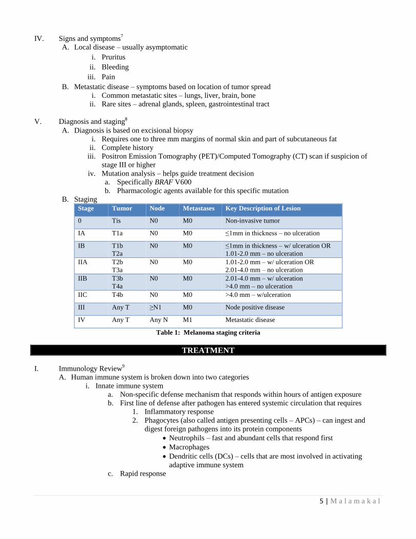
B. Staging
Stage Tumor Node Metastases Key Description of Lesion
0 Tis N0 M0 Non-invasive tumor
IA T1a N0 M0 ≤1mm in thickness – no ulceration
IB T1b
T2a
N0 M0 ≤1mm in thickness – w/ ulceration OR
1.01-2.0 mm – no ulceration
IIA T2b
T3a
N0 M0 1.01-2.0 mm – w/ ulceration OR
2.01-4.0 mm – no ulceration
IIB T3b
T4a
N0 M0 2.01-4.0 mm – w/ ulceration
>4.0 mm – no ulceration
IIC T4b N0 M0 >4.0 mm – w/ulceration
III Any T ≥N1 M0 Node positive disease
IV Any T Any N M1 Metastatic disease
Table 1: Melanoma staging criteria
TREATMENT
I. Immunology Review9
A. Human immune system is broken down into two categories
i. Innate immune system
a. Non-specific defense mechanism that responds within hours of antigen exposure
b. First line of defense after pathogen has entered systemic circulation that requires
1. Inflammatory response
2. Phagocytes (also called antigen presenting cells – APCs) – can ingest and
digest foreign pathogens into its protein components
Neutrophils – fast and abundant cells that respond first
Macrophages
Dendritic cells (DCs) – cells that are most involved in activating
adaptive immune system
c. Rapid response

6 | M a l a m a k a l
ii. Adaptive immune system
a. Divided into humoral and cell-mediated immunity (Figure 4)
Figure 4: Humoral vs. cellular immunity
b. Creates defense against specific invaders
c. Involves antigen-specific immune responses consisting of lymphocytes
1. B-lymphocytes (B-cells)
Produced in the bone marrow
Involved in the humoral response – production of antibodies to ‘tag’
foreign pathogens for phagocytosis or pathogen dysfunction
2. T-lymphocytes (T-cells)
Produced in bone marrow but mature in the thymus
Consist of helper T-cells and cytotoxic T-lymphocytes (CTLs)
Involved in cell-mediated response
d. Much slower response
e. Eliminating cancer cells require activation of CTL
1. Antigen bound to Major Histocompatibility Complex I (MHCI) interacts with
the T-cell Receptor (TCR) of CTL
2. B7 protein binds to CD28 receptor on CTL to provide a costimulatory signal
that maintains CTL activation
3. Activated CTLs travel to site of cancer cell
4. CTLs target specific antigen on cancer cell surface
5. Causes apoptosis of cancer cell
6. Intricate balance of checkpoints that accelerate or dampen immune responses
f. Helper T-cells provide support by secreting cytokines to keep CTLs activated

7 | M a l a m a k a l
II. Melanoma Immunotherapy
A. Historical perspective10
i. Over 100 years of research and development
a. 1890’s – Dr. William Coley found success in injecting bacteria directly into tumors
1. Caused complete remission in a 21 year old man with malignant sarcoma
2. Results were unpredictable
b. 1900-1957 – Immuno-surveillance was a theory formulated suggesting that
lymphocytes act as ‘sentinels’ in recognizing and eliminating continuously arising,
nascent, transformed cells11, 12
1. Surveillance provides important host protection
2. Decreases cancer rates by inhibiting carcinogenesis and maintaining regular
cell homeostasis
c. 1976 – Dacarbazine became the first approved alkylating agent for use in metastatic
melanoma
1. Use was limited due to low response rates of 10-15% (Appendix I)
2. Median survival 6-9 months
3. Five year overall survival – 6%
4. Toxicities of myelosuppression, nausea/vomiting, flu-like symptoms
5. Other chemotherapy agents approved
d. 1986-1992 – Interferon-α (IFN- α) and high dose interleukin-2 (IL-2) introduced as
therapeutic options for melanoma
e. Late 2000’s – Development of novel effective agents
B. Four general approaches to immunotherapy in cancer13
i. Non-specific immune stimulation (applying the gas)
a. Creates generalized activation of immune system in-vivo
b. APCs can be activated using pharmacological cytokines
1. IFN-α
Historically used as adjuvant therapy in those with high risk of relapse
after surgery
Many intolerable toxicities – fevers, chills, malaise, arthralgias, weight
loss, and depression
2. IL-2
15-25% response rates with 2-5% achieving complete response
5-10% of patients can last for decades
Many toxicities – capillary leak syndrome, bone marrow suppression,
hepatotoxic, renal toxic, nausea/vomiting
c. T-cells become activated to kill tumor cells
d. Disadvantage
1. Many adverse effects due to non-specific activation of immune system
2. Subsequent treatments not well-tolerated
ii. Vaccines (eliciting an immune response to cancer antigens)
a. Intended to specifically initiate or amplify a host response against evolving tumors
1. Glycoprotein 100 (gp100) – studied in Phase II trials with IL-2
Minimal response rates: ~16%
2. Melanoma-associated antigen 3 (MAGE-3) – protein expressed on around 70%
of melanoma cells
Study vaccine was associated with significantly prolonged times to
treatment failure
b. Two important limitations
1. Lack of understanding of how to immunize patients to achieve potent cytotoxic
T-cell responses

8 | M a l a m a k a l
2. Presence of inhibitor signaling in the tumor microenvironment may decrease
or disable antitumor immune responses before clinically relevant tumor killing
can take place
c. Overall, vaccines have failed to induce significant objective tumor shrinkage and more
research is needed
iii. Adoptive cell transfer (engineered CTLs)
a. Extract immune cells from patient
b. Activates the extracted cells outside the body
c. Engineered to specifically target cancer tissue
d. Preliminary results are promising
e. Disadvantages
1. More research needed
2. Therapy likely to be very costly
iv. Immune-checkpoint blockade (removing the brakes)
a. Receptor upregulation on tumor cells and APCs that dampen immune response
b. Normal function is to prevent collateral damage to healthy tissue
c. Blocking interaction allows for immune system to continue fighting
C. The cancer-immunity cycle13
i. Model of stepwise events required for an appropriate immune response to killing cancer cells
Figure 5: Cancer immunity cycle
ii. Cycle steps
a. Step 1 – Neoantigens created by oncogenesis are released and captured by DCs for
processing
b. Step 2 – DCs present the captured antigen on MHCI & MHC II to T-cells
1. DCs having antigen bound to MHCI presents to CTL
2. DCs having antigen bound to MHCII present to helper T-cells
c. Step 3 – Priming and activation of effector T-cell responses against cancer specific
antigens that are viewed as foreign

9 | M a l a m a k a l
1. Helper T-cells become activated through interaction of co-stimulatory
interactions
2. Activation causes produce cytokines to active other CTL
d. Step 4 - Activated effector T-cells traffic to tumor site
e. Step 5 – Activated effector T-cells infiltrate the tumor bed
f. Step 6 – Recognition of cancer cells by T-cells through interaction between T-cell
receptors (TCRs) and cognate antigen bound to MHCI
g. Step 7 – Killing of targeted cancer cells
h. Return to Step 1 – Killing of the cancer cells releases additional tumor-associated
antigens that will cause the cycle to repeat again
iii. Problems
a. In melanoma, the immunity cycle does not work optimally
b. Tumor antigens may not be detected
c. DCs and T-cells may treat antigens as self rather than foreign
1. PD-L1 on tumor binding to PD-1 on CTL plays a role
2. Suppresses CTL ability to recognize foreign cancer cells
iv. Goals of cancer immunotherapy
a. Initiate or re-initiate a self-sustaining cycle of cancer immunity
b. Enabling the immunity to amplify and propagate
c. Have appropriate restraints to prevent autoimmune inflammatory responses
d. Modulation of the immune system may allow for better host response to clearing
tumors
D. Immune checkpoints – function to restrain or dampen over-exuberant responses14, 25-27
i. CTLA-4 inhibitors (cytotoxic T-lymphocyte-associated antigen 4)
a. Ipilimumab is the only approved agent
b. Site of action – Priming phase of T-cells (Step 3)
c. Mechanism of action: blocks the interaction CTLA-4 found on CTL with B7 protein
on dendritic cells (See Figure 6)
d. CTLA-4 receptors are upregulated during T-cell activation
e. CTLA-4 bound to B7 normally prevents helper T-cell activation
f. This interaction binds with higher affinity than the stimulatory interaction of CD28
with B7
g. Blockade may be more non-specific
h. Reported toxicities
1. Common: Fatigue, diarrhea, pruritus, colitis
2. Many are immune-mediated adverse events that may require supportive care
ii. Approval of CTLA-4 inhibitors – ipilimumab
a. Efficacy first shown in two large Phase III trials 15- 17

10 | M a l a m a k a l
Population Interventions Primary
Endpoint Results
676 patients with
unresectable metastatic
disease (stage III or IV)
that progressed during
systemic therapy with
dacarbazine,
temozolomide,
fotemustine, carboplatin,
or IL-2
Ipilimumab + gp100
vaccine
OR
Ipilimumab alone
OR
gp100 alone
Overal Survival
(OS)
Ipilimumab significantly increased
overall survival
Ipilimumab+gp100 compared to
gp100 alone: Median OS 10.0; HR
= 0.68, p<0.001
Ipilimumab alone compared to
gp100 alone: Median OS 10.1
months; HR = 0.66, p=0.003
Gp100 alone: OS 6.4 months
502 patients with
previously untreated
metastatic melanoma
Dacarbazine + ipilimumab
OR
Dacarbazine + placebo
OS
Dacarbazine + Ipilimumab
showed longer OS than control arm
(11.2 vs 9.1 months)
Three-year survival was also
improved with the dacarbazine +
ipilimumab group compared to
control arm (20.8% vs. 12.2%; HR
= 0.72, p<0.001)
Table 2: CTLA-4 inhibitor approval trial summary
iii. PD-1 inhibitors (Programmed-Death 1)
a. Two FDA approved agents
1. Pembrolizumab
2. Nivolumab
b. Site of action: priming and effector phases (Steps 3 and 6 respectively)
c. Mechanism of action: inhibits interaction of PD-L1 and PD-L2 found on tumors
cells/antigen presenting cells with PD-1 found on CTLs (See Figure 6)
d. PD-L1 is naturally upregulated on host cells like B-cells and macrophages
e. Tumor cells can express the PD-L1 protein and are thought to ‘escape’ killing by
binding to the PD-1 protein on an activated CTL
f. Blocking this interaction can allow lysis to continue
g. May be more specific therapy than CTLA-4 inhibitors
h. Reported toxicities
1. Fatigue, peripheral edema, chills, constipation, pruritus, diarrhea
2. Immune mediated adverse effects: pneumonitis, colitis, hepatitis, hypophysitis,
nephritis, hypo/hyperthyroidism
iv. Approal of PD-1 inhibitors18,19
Population Interventions Primary
Endpoint Results
KEYNOTE-002
540 patients with ipilimumab-
refractory metastatic
melanoma
Pembrolizumab
• 2 mg/kg Q 3 weeks
• 10 mg/kg Q 3 weeks
OR
Chemotherapy
• Carboplatin plus paclitaxel
• Paclitaxel alone
• Dacarbazine
• Temozolomide
6 month
Progression Free
Surivival (PFS)
And
Overall Response
Rates (ORR)
6 month PFS
Pembrolizumab
• 34% 2 mg/kg
• 38% 10 mg/kg Chemotherapy group
• 16%
---------------------------------
ORR Pembrolizumab
• 21% 2 mg/kg
• 26% 10 mg/kg Chemotherapy group
• 4%

11 | M a l a m a k a l
CHECKMATE-066 trial
418 previously untreated
patients with metastatic
melanoma without BRAF
mutation
Nivolumab
3 mg/kg Q 2 weeks
vs.
Dacarbazine
1000 mg/m2 Q 3 weeks
1 year OS
And
PFS
And
ORR
OS
Nivolumab 72.9%
Dacarbazine 42.1%
HR for death 0.42 (99.8%
CI 0.25-0.73)
PFS
Nivolumab 5.1 months
Dacarbazine 2.2 months
ORR
Nivolumab 40%
Dacarbazine 13.9%
Table 3: PD-1 inhibitor approval trial summary
v. Pembrolizumab is currently approved as second-line option in ipilimumab refractory patients
vi. Nivolumab approved as first line option in metastatic melanoma
Figure 6: Immune checkpoint blockade of PD-1 and CTLA-4
E. Combination checkpoint inhibition
i. Preliminary evidence from phase 1 and phase 2 clinical trials have shown the combination to
be effective 20, 21
Population Interventions Primary
Endpoint Results
Phase I Trial
86 patients with stage III or
IV melanoma with no
previous T-cell modulating
antibody therapy (except
ipilimumab for patients in
sequenced-regimen
cohorts).
Various dosing regimens involving
ipilimumab and nivolumab ORR
Depending upon the dose
cohort, ORR ranged from
21-53%
Nivolumab 1 mg/kg +
ipilimumab 3 mg/kg
yielded the most substantial
ORR
Supported the utilization of
those doses in Phase II trial

12 | M a l a m a k a l
Phase II Trial
142 patients with
Stage III or IV melanoma
Combination Therapy Nivolumab 1 mg/kg Q 3 weeks x 4
doses
Ipilimumab 3 mg/kg Q 3 weeks x 4
doses
VS
Monotherapy Ipilimumab 3 mg/kg Q 3 weeks x 4
doses
ORR
Nivolumab + ipilimumab
ORR
61% (95% CI: 49-72)
Ipilimumab
11% (95% CI: 3-25).
Table 5: Combination immune checkpoint inhibitor trial summary
III. Current Treatment Guideline Recommendations for Metastatic Melanoma
i. National Comprehensive Cancer Network – NCCN 8
Figure 7: NCCN Melanoma treatment recommendations for metastatic melanoma
ii. Current guidance provides no preference for any checkpoint inhibitor agent over another
IV. Clinical Question
A. Which checkpoint inhibitor/combinations should be used initially in first-line treatment of metastatic
melanoma? What evidence if any supports this?
B. Of the PD-1 inhibitors, which agent is preferred?
C. Are the toxicities of combination treatment manageable?
LITERATURE REVIEW
Robert C, Schachter J, Long GV, et al. Pembrolizumab versus Ipilimumab in Advanced Melanoma. N
Engl J Med. 2015; 372(26):2521-32.22
Objective To compare PD-1 inhibition with CTLA-4 blockade involving patients with advanced
melanoma
Study Design Randomized, controlled, phase III study
Population Inclusion
o Patients 18 years of age or older
o Histologically confirmed, unresectable Stage III or IV melanoma
o No previous systemic therapy for advanced disease
o Known BRAF V600 mutation status required
o Eastern Cooperative Oncology Group (ECOG) performance status of 0 or 1
(Appendix 2)
Exclusion
o Previous therapy with CTLA-4, PD-1, or PD-L1 inhibitors
Nivolumab (Category 1)
Ipilimumab (Category 1)
Pembrolizumab
High-dose IL-2
Unresectable or metastatic
melanoma BRAF V600
wild type
First Line Therapy

13 | M a l a m a k a l
o Patients with ocular melanoma
o Active brain metastases
o History of serious autoimmune disease
Primary
Outcome PFS – defined as time from randomization to documented disease progression
OS – defined as time from randomization to death from any cause
Secondary
Outcomes ORR– defined as percentage of patients with complete or partial response
Duration of response – defined as the time from the first documented response to
radiologic progression
Safety as assessed in as-treated population
Efficacy in intent-to-treat population
Methods Patients randomly assigned in a 1:1:1 ratio to receive
o Pembrolizumab 10 mg/kg of body weight either:
Every 2 weeks
Every 3 weeks
o Ipilimumab –3 mg/kg every 3 weeks (4 cycles)
Pembrolizumab administered IV during 30 min infusion and continued until either
o Disease progression
o Onset of unacceptable side effects
o Investigators decision to discontinue treatment
o Maximum of 24 months of therapy
Patients with confirmed complete response (CR) who received 6 months of
pembrolizumab could discontinue therapy – with two more additional doses beyond
CR
Ipilimumab administered during a 90-minute period and continued for the same
reasons as pemrbolizumab
Response measured at week 12 and every 6 weeks thereafter
Survival assessed every 3 months after discontinuation of study drug
Statistics Kaplan-Meier method to calculate estimates of PFS and OS
Hazard ratios (HR) and associated 95% confidence intervals (CI) assessed with Cox-
proportional-hazards model
Response rates studied using Miettinen and Nurminen method
Enrollment Pembrolizumab every 2 week arm - 279 patients
Pemrbolizumab every 3 week arm – 277 patients
Ipilimumab every 3 weeks arm – 278 patients
Baseline Well matched between groups
Among enrolled patients, 68.7% had no previous treatment for advanced melanoma
68.7% had an ECOG performance status of 0
BRAF V600 mutation was observed in 36.2% of patients (with ~50% receiving
previous BRAF inhibitor treatment)
80.5% of patients had PD-L1-positive tissue samples
Results Estimated 6 month PFS rates

14 | M a l a m a k a l
Median estimates of PFS:
o Pembrolizumab every 2 weeks: 5.5 months (95% CI 3.4-6.9 months)
o Pembrolizumab every 3 weeks 4.1 months (95% CI 2.9-6.9 months)
o Ipilimumab: 2.8 (95% CI 2.8-2.9 months)
HR for disease progression for both pembrolizumab groups vs. ipilimumab: 0.58
(95% CI 0.46-0.72) – p<0.001
Benefit for pembrolizumab over ipilimumab seen in both PD-L1-positive and PD-L1
negative subgroups (PD-L1 negative subgroup not statistically significant)
OS
o Pembrolizumab every 2 weeks and 3 weeks longer than ipilimumab
o 1 year OS: ~70% pembrolizumab vs. 58.2% ipilimumab
RR
o ~33% for pembrolizumab groups and 11.9% in ipilimumab group (p<0.0001)
Adverse Events
o Grade 3-5 toxicities attributed to study drugs pembrolizumab every 2 weeks,
every 3 weeks, and ipilimumab: 13.3%, 10.1%, and 19.9% respectively
Hypo/hyperthyroidism was more frequent in the pembrolizumab groups
Colitis and hypophysitis were more frequent in the ipilimumab group
Conclusions
Pembrolizumab prolonged progression free survival and overall survival compared to
ipilimumab
Discussion
Strengths Trial design – Phase 3, randomized, controlled
Multi-centered – 16 countries
Efficacy determined in intent-to-treat population
Safety assessed in as-treated (per-protocol) population
First trial comparing head-to-head immune checkpoint inhibitors
Limitations Pembrolizumab did not show statistical benefit in overall survival for
o Female population
o PD-L1 negative patients
o BRAF mutant gene with or without previous therapy
Long-term duration of benefit not known for pembrolizumab
Median overall-survival benefit data not reached
10 mg/kg dose is 5 times the dose used in the approval of drug. Potentially much more
costly
Take Home
Points Pembrolizumab is superior to ipilimumab in specific patient populations for frontline
treatment of patients with metastatic melanoma
First study to give insight that PD-1 inhibitors can be used first line
Dose of pemrbolizumab every 3 weeks = better patient convience
10 mg/kg dose is not currently FDA approved and confers more patient costs
0
10
20
30
40
50
Pembrolizumab Q 2
Weeks
Pembrolizumab Q 3
Weeks
Ipilimumab Q 3
Weeks
Per
cen
t
Estimated 6 month PFS

15 | M a l a m a k a l
Larkin J, Chiarion-sileni V, Gonzalez R, et al. Combined Nivolumab and Ipilimumab or Monotherapy
in Untreated Melanoma. N Engl J Med. 2015;373(1):23-34.23
Objective To determine the efficacy of combination therapy of ipilimumab plus nivolumab compared to
ipilimumab
Study
Design
Double blind, randomized, placebo-controlled, Phase III trial
Population Inclusion
o Patients 18 years of age or older
o Histologically confirmed, unresectable Stage III or IV melanoma
o No previous systemic therapy for advanced disease
o Known BRAF V600 mutation status required
o Eastern Cooperative Oncology Group (ECOG) performance status of 0 or 1
Exclusion
o ECOG performance status of 2 or higher
o Previous therapy with CTLA-4, PD-1, or PD-L1 inhibitors
o Patients with ocular melanoma
o Active brain metastases
o History of serious autoimmune disease
Primary
Outcome PFS – defined as the time between date of randomization and date of documented
progression or death
OS – defined as time from randomization to death from any cause
Secondary
Outcomes ORR
PD-L1 expression as a predictable biomarker for efficacy outcomes
Safety
Methods Patients randomly assigned in 1:1:1 ratio to receive one of the following
o Nivolumab 3mg/kg every 2 weeks (plus ipilimumab matched placebo)
o Nivolumab 1mg/kg every 3 weeks plus 3 mg of ipilimumab every 3 weeks for 4 doses
– followed by nivolumab 3mg/kg for cycle 3 and beyond
o Ipilimumab 3mg/kg IV every 3 weeks for 4 doses (plus nivolumab-matched placebo)
Randomization was stratified according to
o PD-L1 status
o BRAF mutation status
o Cancer stage
Assessment of tumor response at 12 weeks, then every 6 weeks for 49 weeks
Then assesed every 12 weeks until progression or treatment discontinuation
Statistics PFS calculated by – Two-sided log rank test with stratification
Hazard ratios (HR) and two sided 99.5% CI calculated using Cox proportional-hazard
model
Kaplan-Meier method used to measure PFS
Enrollment 945 patients randomized 1:1:1
Patients balanced on age and gender
Majority of patients were male (~64%) and <65 years old
Most patients (~65%) were PD-L1 negative with no BRAF mutation (~68%)
Results
Most frequent reason for discontinuation in nivolumab and ipilimumab group was disease
progression (49.2% and 65% respectively)
Most frequent reason for discontinuation of nivolumab plus ipilimumab group was toxicity
(38.3%)
61% of patients had measurable tumor shrinkage
Only ~14% of patients did not respond
PFS

16 | M a l a m a k a l
o HR of combination compared to ipilimumab – 0.42 (99.5% CI 0.31-0.57)
o HR of nivolumab compared to ipilimumab – 0.57 (99.5% CI 0.43-0.76)
o HR of combination compared to nivolumab – 0.74 (95% CI 0.60-0.92) –study not
designed for formal comparison Secondary Endpoints
ORR – Combination (57.6%), nivolumab (43.7%), ipilimumab (19.0%)
Complete response (CR) – Combination (11.5%), nivolumab (8.9%), ipilimumab (2.2%)
PFS in pre-specified subgroups showed consistently longer survival with nivolumab or
combination therapy for
o PD-L1 status
Positive
Negative
o BRAF mutation status – positive or wild type – median PFS 11.2-11.7 months
PD-L1 negative status showed differerence in PFS for nivolumab compared to PD-L1
positive status
Adverse events
o Treatment of adverse events of any grade occurred in 95.5% of those in the
combination group
o Most common adverse event in the combination – diarrhea, fatigue, pruritis
o Incidence of treatment-related adverse events of grade 3 or 4 higher in
combination group, 55%, compared to 16.3% in nivolumab and 27.3 % in
ipilimumab
0 5 10 15
Nivolumab plus ipilimumab
Nivolumab
Ipilimumab
Months
PFS
0
5
10
15
Nivolumab +
ipilimumab
Nivolumab Ipilimumab
Mo
nth
s
PD-L1 Positive PFS
0
5
10
15
Nivolumab +
ipilimumab
Nivolumab Ipilimumab
Mo
nth
s
PD-L1 Negative PFS
(95% CI 2.8-3.4)
(95% CI 4.3-9.5)
(95% CI 8.9-16.7)

17 | M a l a m a k a l
Conclusions
In previously untreated advanced melanoma, longer PFS and higher rates of ORR with
nivolumab alone and with combination of nivolumab and ipilimumab compared to
ipilimumab alone
Discussion
Strengths Results of PD-L1 positive/negative results provide insight as to who would benefit from
combination therapy the most
Relatively large sample size
Multi-centered trial (137 centers)
Study design – Phase III, randomized, controlled
Confirmation of superiority of PD-1 inhibitor class over CTLA-4 class
Limitations Overall survival results are still pending
Toxicities of combination therapy may limit first line use
Majority of patients <65 years old – benefit in elderly may be questionable
Majority of patients male gender
o No breakdown in efficacy between genders
Majority of patients were ECOG 0-1
Take Home
Points Combination therapy with nivolumab plus ipilimumab and nivolumab alone in metastatic
melanoma produced far better response rate compared to ipilimumab alone
Results are too early to be standard of care due to no overall survival data
PD-1 inhibitor class is superior to CTLA-4 and should be category 1 recommendation
This combination therapy comes at the price of more toxicities and tremendous cost of
therapy
I. Cost Effective Analysis (CEA) with Checkpoint Inhibitors
A. No analysis has been performed to date
B. Wholesale Acquisition Cost (WAC) Comparison 24-27
i. Nivolumab vs. pembrolizumab
ii. Cost of ipilimumab ~$160,000 for 3 months of therapy
iii. Combination therapy would be very costly for patients
Nivolumab Pembrolizumab
Cost for 3 months of therapy (WAC*^)
$41,437 $41,434
Frequency of Administration
Every 2 weeks Every 3 weeks
Infusion Time
1 hour 30 minutes
Efficacy (no official comparison between agents)
Median OS at 12 months: 63%
Median OS at 24 months: 48%
OS at 12 months: 66%
OS at 24 months: 49%
Median PFS: 4.4 months
Progression free at 12 months: 35%
Table 6: Comparison of nivolumab and pembrolizumab
* WAC – Wholesale Acquisition Cost
^ Acquisition cost is based on the average wholesale price (AWP) for the most common or FDA-approved dosage and a patient weighing 80 kg, and it does
not include the cost of supportive care for immune-related toxicity.
COSTS & CONSIDERATIONS

18 | M a l a m a k a l
CONCLUSIONS
I. Optimal first-line sequence of therapy in unresectable metastatic melanoma
A. PD-1 inhibitor Ipilimumab
B. Combination therapy (PD-1 inhibitor + CTLA-4) offers greater response rates than either agent alone
i. Results are very exciting
ii. No OS data to support first-lint use
iii. Although generally tolerable, patients are subjected to many more toxicities
iv. Very high cost combining two very expensive therapies
v. Wait until more data is available to try this option in most patients
i. May consider if patient’s ECOG is 0-1
ii. May be an option in PD-L1 negative patients
iii. No significant co-morbidities
iv. If patient is able to afford therapy
C. Pembrolizumab vs. Nivolumab?
i. Costs are virtually comparable
ii. Patient convenience favors pembrolizumab due to dosing frequency and infusion time
iii. Nivolumab may have better market advantage right now due to fixed dosing recommendation
in other types of malignancies
iv. Pembrolizumab likely preferred in metastatic melanoma due to slightly longer market
experience, ease of administration, and patient convenience
D. Toxicities
i. PD-1 inhibitors – have shown to be rare and manageable
ii. CTLA-4 inhibitors – much more prevalent in terms of immune mediated reactions
II. Expect NCCN guideline update for metastatic melanoma soon
III. Proposed Algorithm
Figure 8: Proposed NCCN treatment algorithm for metastatic melanoma
Pembrolizumab or Nivolumab (Category 1)
Nivolumab plus ipilimumab*
Ipilimumab (Category 2)
High-dose IL-2 (Category 3)
Unresectable or
metastatic melanoma
BRAF V600 wild type
First Line Therapy
Disease Progression
Supportive Care
Subsequent Therapy
ECOG
3-4
ECOG
0-2
Pembrolizumab or Nivolumab (Category 1)
Nivolumab plus ipilimumab*
Ipilimumab (Category 2)
High-dose IL-2 (Category 3)
Subsequent Therapy

19 | M a l a m a k a l
*Considerations for nivolumab plus ipilimumab
Excellent performance status
No co-morbidities
Possible PD-L1 negative status
Affordability of high cost of dual therapy
IV. The future is bright for immunotherapy and is now the standard of care over chemotherapy in metastatic
melanoma
A. Immune checkpoint inhibitors are being studied in many different cancers
i. Non-small cell lung cancer (nivolumab already approved)
ii. Renal cell carcinoma
iii. Many hematologic malignancies
REFERENCES
1. Siegel R, Ma J, Zou Z, Jemal A. Cancer statistics, 2014. CA Cancer J Clin. 2014;64(1):9-29.
2. American Cancer Society | Information and Resources for Cancer: Breast, Colon, Lung, Prostate, Skin. Web. 28 Aug. 2015.
http://www.cancer.org/cancer/skincancer-melanoma/detailedguide/melanoma-skin-cancer-key-statistics
3. Guy GP, Thomas CC, Thompson T, Watson M, Massetti GM, Richardson LC. Vital signs: melanoma incidence and mortality
trends and projections - United States, 1982-2030. MMWR Morb Mortal Wkly Rep. 2015;64(21):591-6.
4. Miller AJ, Mihm MC. Melanoma. N Engl J Med. 2006;355(1):51-65.
5. Demierre MF, Nathanson L. Chemoprevention of melanoma: an unexplored strategy. J Clin Oncol. 2003 Jan 1. 21(1):158-65.
6. Thompson JF, Scolyer RA, Kefford RF. Cutaneous melanoma. Lancet. 2005;365(9460):687-701.
7. Abbasi NR, Shaw HM, Rigel DS, et al. Early diagnosis of cutaneous melanoma: revisiting the ABCD criteria. JAMA.
2004;292(22):2771-6.
8. National Comprehensive Cancer Network. Melanoma (Version 3.2015). Accessed July 16th, 2015.
9. Sompayrac LM. How the Immune System Works. Wiley-Blackwell; 2008.
10. Eggermont AM, et al. Eur J Cancer. 2004 Aug;40(12):1825-36.
11. Burnet M. Cancer; a biological approach. I. The processes of control. Br Med J. 1957;1(5022):779-86.
12. Odunsi K, Old LJ. Tumor infiltrating lymphocytes: indicators of tumor-related immune responses. Cancer Immun. 2007;7:3.
13. Chen DS, Mellman I. Oncology meets immunology: the cancer-immunity cycle. Immunity. 2013;39(1):1-10.
14. Drake CG, Lipson EJ, Brahmer JR. Breathing new life into immunotherapy: review of melanoma, lung and kidney cancer. Nat
Rev Clin Oncol. 2014;11(1):24-37.
15. Hodi FS, O'Day SJ, McDermott DF, et al. Improved survival with ipilimumab in patients with metastatic melanoma. N Engl J
Med 2010; 363:711.
16. Robert C, Thomas L, Bondarenko I, et al. Ipilimumab plus dacarbazine for previously untreated metastatic melanoma. N Engl
J Med 2011; 364:2517.
17. Maio M, Grob JJ, Aamdal S, et al. Five-year survival rates for treatment-naive patients with advanced melanoma who received
ipilimumab plus dacarbazine in a phase III trial. J Clin Oncol 2015; 33:1191.
18. Ribas A, Puzanov I, Dummer R, et al. Pembrolizumab versus investigator-choice chemotherapy for ipilimumab-refractory
melanoma (KEYNOTE-002): a randomised, controlled, phase 2 trial. Lancet Oncol. 2015;16(8):908-18.
19. Robert C, Long GV, Brady B, et al. Nivolumab in previously untreated melanoma without BRAF mutation. N Engl J Med.
2015;372(4):320-30.
20. Wolchok JD, Kluger H, Callahan MK, et al. Nivolumab plus ipilimumab in advanced melanoma. N Engl J Med 2013;
369:122.
21. Postow MA, Chesney J, Pavlick AC, et al. Nivolumab and ipilimumab versus ipilimumab in untreated melanoma. N Engl J
Med. 2015;372(21):2006-17.
22. Robert C, Schachter J, Long GV, et al. Pembrolizumab versus Ipilimumab in Advanced Melanoma. N Engl J Med. 2015;
372(26):2521-32.
23. Larkin J, Chiarion-sileni V, Gonzalez R, et al. Combined Nivolumab and Ipilimumab or Monotherapy in Untreated Melanoma.
N Engl J Med. 2015;373(1):23-34.
24. Walko, CM, Harvey, RD. Treatment of Advanced Melanoma. CE in the Midday: ASHP Advantage.
http://www.cemidday.com/melanoma. Accessed July 28th, 2015.
25. Yervoy (ipilimumab) prescribing information. Princeton, NJ: Bristol-Myers Squibb Company; 2013 Dec.
26. Opdivo (nivolumab) prescribing information. Princeton, NJ: Bristol-Myers Squibb Company; 2015 Mar.
27. Keytruda (pembrolizumab) prescribing information. Whitehouse Station, NJ: Merck & Co., Inc; 2015 Jan.

20 | M a l a m a k a l
APPENDIX
I. Glossary of terms used in oncology studies8
A. Objective response (OR) or Overall response rate (ORR): Objective response means either a
partial or complete response (In the literature, frequently see "CR+PR" which means the same thing)
B. Complete response (CR): All detectable tumor has disappeared
C. Partial response (PR): This roughly corresponds to at least a 50% decrease in the total tumor
volume but with evidence of some residual disease still remaining
D. Progression free survival (PFS): Measures the length of time that a patient is both alive and without
worsening of their cancer
E. Overall survival (OS): Time from randomization until death from any cause
II. Eastern Cooperative Oncology Group (ECOG) Performance Status
8
GRADE ECOG PERFORMANCE STATUS
0 Fully active, able to carry on all pre-disease performance without restriction
1 Restricted in physically strenuous activity but ambulatory and able to carry out work of a light or
sedentary nature, e.g., light house work, office work
2 Ambulatory and capable of all selfcare but unable to carry out any work activities; up and about more
than 50% of waking hours
3 Capable of only limited selfcare; confined to bed or chair more than 50% of waking hours
4 Completely disabled; cannot carry on any selfcare; totally confined to bed or chair
5 Dead