lymph node mapping and beyond in the surveillance...
TRANSCRIPT
Lymph Node Mapping and Beyond in the Surveillance/Detection of
Thyroid Cancer
Elizabeth Westfall, MD Banner University Medical Center Phoenix
Department of Medical Imaging

No disclosures.

Objectives: 1. Discuss the role of cervical lymph node
mapping in thyroid cancer surveillance. 2. Describe the ultrasound appearance of
normal and abnormal cervical lymph nodes. 3. Identify other imaging modalities which may have a role in thyroid cancer surveillance.

2015 American Thyroid Association Management Guidelines for Adult Patients with Thyroid Nodules
and Differentiated Thyroid Cancer
The American Thyroid Association Guidelines Task Force on Thyroid Nodules and Differentiated Thyroid Cancer
Bryan R. Haugen,1,* Erik K. Alexander,2 Keith C. Bible,3 Gerard M. Doherty,4 Susan J. Mandel,5
Yuri E. Nikiforov,6 Furio Pacini,7 Gregory W. Randolph,8 Anna M. Sawka,9 Martin Schlumberger,10 Kathryn G. Schuff,11 Steven I. Sherman,12 Julie Ann Sosa,13 David L. Steward,14
R. Michael Tuttle,15 and Leonard Wartofsky16
Role of Preoperative Neck Ultrasound (Recommendation 32)
(A) Preoperative neck US for cervical (central and especially lateral neck compartments) lymph nodes is recommended or all patients undergoing thyroidectomy for malignant or suspicious for malignancy cytologic or molecular findings. (Strong recommendation, Moderate-quality evidence)
(B) US-guided FNA of sonographically suspicious lymph nodes ‡8–10mm in the smallest diameter should be performed to confirm malignancy if this would change management. (Strong recommendation, Moderate-quality evidence)
(C) The addition of FNA-Tg washout in the evaluation of suspicious cervical lymph nodes is appropriate in select patients, but interpretation may be difficult in patients with an intact thyroid gland. (Weak recommendation, Low-quality evidence)
Preoperative/Initial Lymph Node Status Most Important In Evaluation for LN Recurrence
• Most recurrences in already involved compartments. • Increased risk with higher number of N1 and higher number of N1 with
extracapsular extension. • Increased risk with macroscopic rather than microscopic lymph node metastases.
Role of cervical ultrasonography during follow-up (Recommendation 65)
(A) Following surgery, cervical US to evaluate the thyroid bed and central and lateral cervical nodal compartments should be performed at 6–12 months and then periodically, depending on the patient’s risk for recurrent disease and Tg status. (Strong recommendation, Moderate-quality evidence)
(B) If a positive result would change management, ultrasonographically suspicious lymph nodes ‡8–10mm (see Recommendation 71) in the smallest diameter should be biopsied for cytology with Tg measurement in the needle washout fluid. (Strong recommendation, Low-quality evidence)
(C) Suspicious lymph nodes less than 8–10mm in smallest diameter may be followed without biopsy with consideration for FNA or intervention if there is growth or if the node threatens vital structures. (Weak recommendation, Low-quality evidence)
(D) Low-risk patients who have had remnant ablation, negative cervical US, and a low serum Tg on thyroid hormone therapy in a sensitive assay (<0.2 ng/mL) or after TSH stimulation (Tg <1 ng/mL) can be followed primarily with clinical examination and Tg measurements on thyroid hormone replacement. (Weak recommendation, Low-quality evidence)

How To Order A Cervical Lymph Node Mapping US
• “Cervical Lymph Node Mapping Ultrasound”
• Be specific about where you want it done or it may be scheduled at the facility closest to the patient.
• This is even better than specifying where you don’t want it done.

Technique
• High frequency transducer (>10 MHz) used to evaluate all lymph node compartments and the thyroid bed.
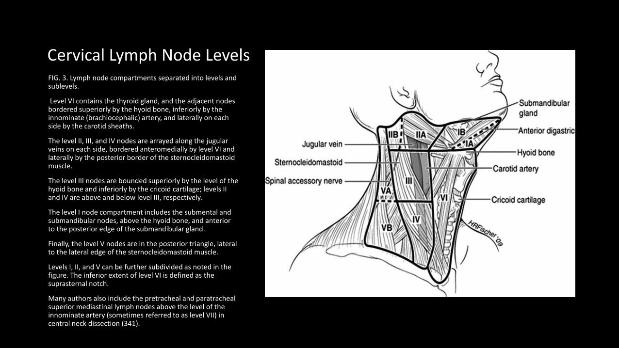
Cervical Lymph Node Levels FIG. 3. Lymph node compartments separated into levels and sublevels.
Level VI contains the thyroid gland, and the adjacent nodes bordered superiorly by the hyoid bone, inferiorly by the innominate (brachiocephalic) artery, and laterally on each side by the carotid sheaths.
The level II, III, and IV nodes are arrayed along the jugular veins on each side, bordered anteromedially by level VI and laterally by the posterior border of the sternocleidomastoid muscle.
The level III nodes are bounded superiorly by the level of the hyoid bone and inferiorly by the cricoid cartilage; levels II and IV are above and below level III, respectively.
The level I node compartment includes the submental and submandibular nodes, above the hyoid bone, and anterior to the posterior edge of the submandibular gland.
Finally, the level V nodes are in the posterior triangle, lateral to the lateral edge of the sternocleidomastoid muscle.
Levels I, II, and V can be further subdivided as noted in the figure. The inferior extent of level VI is defined as the suprasternal notch.
Many authors also include the pretracheal and paratracheal superior mediastinal lymph nodes above the level of the innominate artery (sometimes referred to as level VII) in central neck dissection (341).
Normal lymph node appearance on ultrasound
• Oval shape • Hypoechoic, homogeneous peripheral cortex • Hyperechoic/echogenic central fatty hilum • Central hilar vessel • Short axis size less than 10 mm • No concerning characteristics
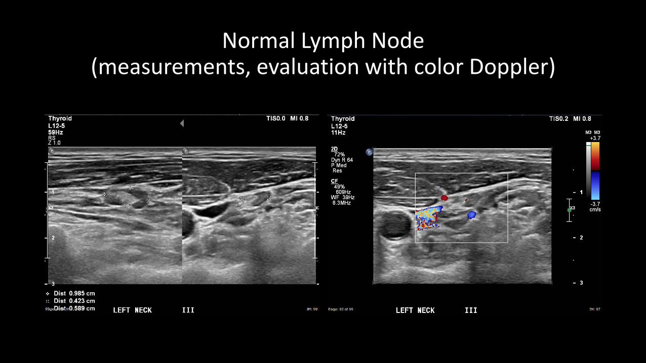
Normal Lymph Node (measurements, evaluation with color Doppler)

Normal Thyroid Bed
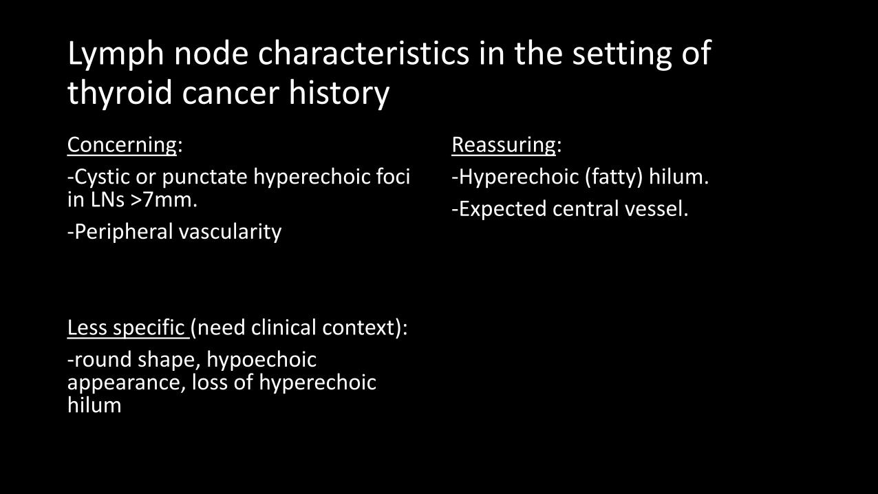
Lymph node characteristics in the setting of thyroid cancer history Concerning: -Cystic or punctate hyperechoic foci in LNs >7mm. -Peripheral vascularity Less specific (need clinical context): -round shape, hypoechoic appearance, loss of hyperechoic hilum
Reassuring: -Hyperechoic (fatty) hilum. -Expected central vessel.
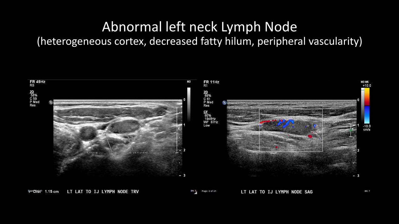
Abnormal left neck Lymph Node (heterogeneous cortex, decreased fatty hilum, peripheral vascularity)
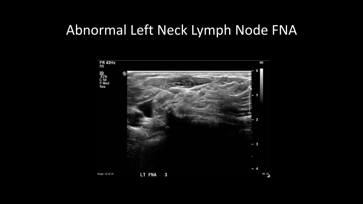
Abnormal Left Neck Lymph Node FNA
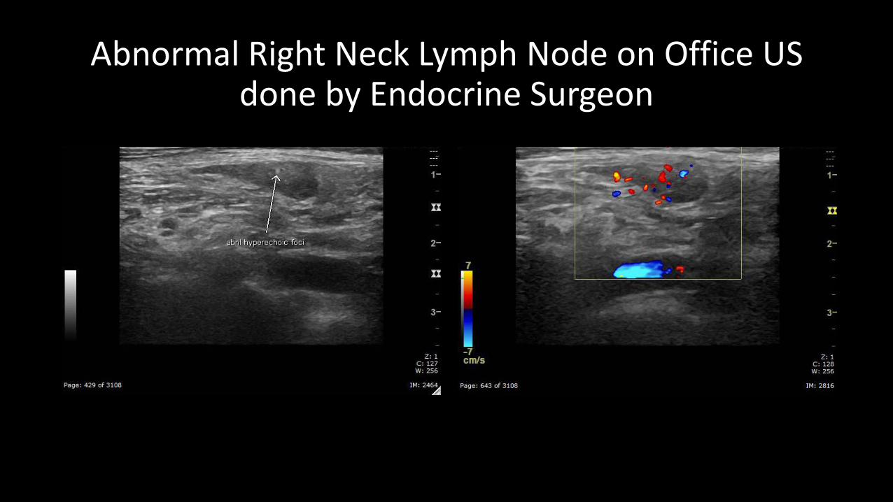
Abnormal Right Neck Lymph Node on Office US done by Endocrine Surgeon

Abnormal Right Neck Lymph Node FNA
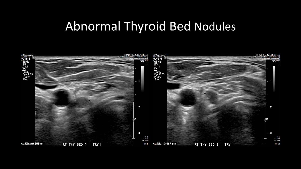
Abnormal Thyroid Bed Nodules

CT done for further evaluation confirms thyroid bed nodules

CT done for further evaluation confirms thyroid bed nodules
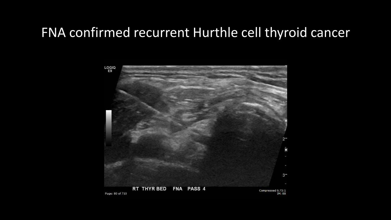
FNA confirmed recurrent Hurthle cell thyroid cancer
Additional imaging modalities may have a role (Recommendations 66-69) • Whole body RAI scans with SPECT/CT • FDG-PET scan • CT, MRI of the neck and upper chest -when US may not visualize all disease -in high risk DTC patients with elevated Tg or rising Tg antibodies with or without negative RAI imaging -imaging other organs in high risk DTC patients with elevated serum TG and negative neck and chest imaging with symptoms referable to those organs or who are being prepared for TSH-stimulated RAI therapy and may be at risk for complications of tumor swelling