lung adenocarcinoma: sustained subtyping with ...rihuc.huc.min-saude.pt/bitstream/10400.4/2065/1/rev...
TRANSCRIPT
Rev Port Pneumol. 2015;21(3):113---125
www.revportpneumol.org
ORIGINAL ARTICLE
Lung adenocarcinoma: Sustained subtyping withimmunohistochemistry and EGFR, HER2 and KRASmutational status
Vitor Sousaa,b,∗, Carolina Rodriguesa, Maria Silvaa, Ana Maria Alarcãoa, Lina Carvalhoa,b
a IAP-FMUC-Institute of Pathology, Faculty of Medicine, University of Coimbra, Coimbra, Portugalb Pathology Institute, Coimbra University Hospital, Coimbra, Portugal
Received 18 March 2014; accepted 1 September 2014Available online 11 March 2015
KEYWORDSLung;EGFR;KRAS;HER2;Immunohistochemistry;Adenocarcinomas
Abstract Pulmonary adenocarcinomas are still in the process of achieving morphological,immunohistochemical and genetic standardization. The ATS/ERS/IASLC proposed classificationfor lung adenocarcinomas supports the value of the identification of histological patterns,specifically in biopsies.
Thirty pulmonary adenocarcinomas were subjected to immunohistochemical study (CK7, CK5,6, 18, CK20, TTF1, CD56, HER2, EGFR and Ki-67), FISH and PCR followed by sequencing andfragment analysis for EGFR, HER2 and KRAS.
Solid pattern showed lower TTF1 and higher Ki-67 expression. TTF1 expression was higherin non-mucinous lepidic and micropapillary patterns when compared to acinar and solid andacinar, solid and mucinous respectively. Higher Ki67 expression was present in lepidic and solidpatterns compared to mucinous. EGFR membranous staining had increasing expression fromnon-mucinous lepidic/BA pattern to solid pattern and micropapillary until acinar pattern. EGFRmutations, mainly in exon 19, were more frequent in females, together with non-smoking status,while KRAS exon 2 mutations were statistically more frequent in males, especially in solidpattern. FISH EGFR copy was correlated gross, with mutations. HER2 copy number was raisedin female tumours without mutations, in all cases. Although EGFR and KRAS mutations aregenerally considered mutually exclusive, in rare cases they can coexist as it happened in one ofthis series, and was represented in acinar pattern with rates of 42.9% and 17.9%, respectively.EGFR mutations were more frequent in lepidic/BA and acinar patterns. Some cases showed
d between the adenocarcinoma patterns reinforce the need torns present, with implications in diagnosis and in pathogenic under-tational status can be determined in biopsies representing bronchial
different EGFR mutations.The differences identifie
carefully identify the pattestanding. EGFR and KRAS mu
∗ Corresponding author.E-mail address: [email protected] (V. Sousa).
http://dx.doi.org/10.1016/j.rppnen.2014.09.0092173-5115/© 2014 Sociedade Portuguesa de Pneumologia. Published by Elsevier España, S.L.U. All rights reserved.
114 V. Sousa et al.
pulmonary carcinomas because when a mutation is present it is generally present in all thehistological patterns.© 2014 Sociedade Portuguesa de Pneumologia. Published by Elsevier España, S.L.U. All rightsreserved.
I
TwrBccrnpr
emcpmgiApscaabt
fri(ea
ratmKpcaicYw
tor
pDpbmmtdtEt
Kblpnrd
M
M
AfittH
indr
E
TtM
I
RpCbronchial-pulmonary adenocarcinomas mainly to validate
ntroduction
here are 1.3 million deaths from lung cancer annuallyorldwide and it is the leading cause of cancer-
elated mortality in USA, Japan and Western-countries.1---4
ronchial-pulmonary carcinomas were classified as smallell lung cancer (SCLC) and non-small cell lung can-er (NSCLC), representing 13% and 85% of lung cancersespectively. Squamous cell carcinoma (SQCC) and ade-ocarcinoma (ADC) subdivision become mandatory due toersonalized therapy and NSCLC designation should not beeported.5,6
Bronchial-pulmonary adenocarcinomas are malignantpithelial tumours with glandular differentiation, and/orucin production, acinar, papillary, micropapillary, bron-
hioloalveolar (BA)/lepidic, or solid with mucin growthatterns, complementing the morphological spectrum ofixed-type adenocarcinoma. The incidence differs with
ender and population, being roughly 28% in men and 42%n women. Recently a new classification was proposed byTS/ERS/IASLC. This new classification recognizes severalatterns such as lepidic instead of BA, acinar, papillary,olid, and micropapillary. It also recognizes mucinous adeno-arcinomas including the former mucinous BA carcinomand colloid carcinoma. It also highlighted the need for anssertive diagnosis specially in biopsy material, supportedy immunohistochemical study, with clinical, prognostic andherapeutic implications.5---8
The transmembrane tyrosine kinase epidermal growthactor receptor (EGFR) belonging to the EGFR family ofeceptor tyrosine kinases (TKs) called the HER or ErbB fam-ly (consisting of four members --- EGFR (HER1/ErbB1), HER2ErbB2), HER3 (ErbB3) and HER4 (ErbB4))9,10 may be over-xpressed and this correlates with poor prognosis, withggressive disease and decreased survival.3,11---14
KRAS mutations are associated with poor prognosis,eported since 1990, occurring in codon 12, occasion-lly at codon 13 and rarely at codon 61.10,15,16 Accordingo the current data, EGFR and KRAS mutations areutually exclusive10,17,18; the explanation is related toRAS-MAPK pathway inserts in the downstream signallingathway of EGF.10 This mutation appears in 30% of Cau-asian patients with lung cancer and 10% of East Asiandenocarcinomas.16,18,19 KRAS mutations are more frequentn smokers and are related to poor prognosis.10,16,17 Lungancers with KRAS mutations are resistant to EGFR-TKIs andatabe confirmed in his study that none of the lung cancersith KRAS mutation achieved clinical response.10,20
Due to what has been said, the EGFR-MAPK signal
ransduction pathway is important to understand the rolef individual somatic changes in tumours, predicting theesponse to EGFR-TKIs. EGFR status is then a favourablesiw
redictive factor in the case of sensitizing mutations.espite the positive response observed in up to 70% ofatients, different data concluded that not every patientenefitted from treatment with TKIs, probably due toutations in the downstream effectors of EGFR signalling,ore frequently KRAS gene. Mutations in this intermediate
ransduction pathway may also select patients; as KRAS actsownstream of EGFR receptor, its somatic changes can leado a non-response to EGFR-TKIs as response rate to anti-GFR therapy is less than 3% in patients with KRAS mutantumours as opposed to 20% in NSCLC with wild-type KRAS.
The objectives of this work are to evaluate EGFR andRAS mutational status, EGFR and HER2 gene copy num-er and immunohistochemical EGFR and HER2 expression inung adenocarcinomas according to the patterns/subtypesresent, in order to understand the value of pattern recog-ition, supported by an immunohistochemical set used inoutine, in adenocarcinoma diagnosis and anti-EGFR therapyecision.
aterials and methods
aterials
series of 30 bronchial-pulmonary adenocarcinomas classi-ed according with WHO 2004 histological classification andhe new ATS/ERS/IASLC classification were selected fromhe archive of the Pathology Service of Coimbra Universityospital.
The patterns present were registered (namely lep-dic/BA, acinar, papillary, micropapillary, solid and muci-ous). Metastases were also registered by patterns. Clinicalata like age, gender, smoking habits and stage were alsoegistered.
thical standards
he principles of Helsinki Declaration were respected andhe study was developed according to the Faculty ofedicine of the University of Coimbra.
Ethical Committee rules for PhD theses were followed.
mmunohistochemistry (IHC)
epresentative sections of the adenocarcinomas and theiratterns were submitted to IHC (CK7, TTF1, CK5/6,D56 and CK20) to validate the pure condition of
olid pattern. Ki67-MIB1 antibody was used to character-ze proliferation index. C-erbB-1/EGFR and c-erbB-2/HER2ere applied to evaluate protein expression of these
muta
ap
tawwi
gma2Ai
8cimis
nsuw
sb
P
GeiFwfi(moifiaa
dptamloptM
Lung adenocarcinoma: subtyping and EGFR, HER2 and KRAS
molecules. Endogenous peroxidase activity was quenchedusing 15 min incubation in 3% diluted hydrogen peroxide(H2O2). For blocking nonspecific binding with primary anti-bodies we used Ultra V Block (Ultra Vision Kit; TP-125-UB;Lab Vision Corporation; Fremont CA; USA). Primary anti-bodies against CK7 (clone OV-TL12/30; DakoCytomation,Glostrup, Denmark) at a dilution of 1/50 for 30 min, Cyto-keratin 5,6,18 (clone LP34; Novocastra Laboratories Ltd,Newcastle, United Kingdom) at a dilution of 1/100 for60 min, TTF1 (clone 8G7G3/1; DakoCytomation, Glostrup,Denmark) at a dilution of 1/100 for 60 min, CD56 (cloneCD564; Novocastra Laboratories Ltd, Newcastle, UnitedKingdom) at a dilution of 1:75 for 60 min, CK20 (cloneKS20.8; DakoCytomation, Glostrup, Denmark) at a dilutionof 1/50 for 30 min, Ki67 (clone MIB-1; DakoCytomation,Glostrup, Denmark) at a dilution of 1/50 for 30 min, c-erbB-2 (Polyclonal; DakoCytomation, Glostrup, Denmark) ata dilution of 1/200 for 30 min, and c-erB-1 (clone 31G7;Invitrogen, Camarillo, California, USA) at a dilution of 1:20for 30 min were applied to the cells and incubated at roomtemperature. They were washed with phosphate-bufferedsaline (PBS) (Ultra Vision; TP-125-PB; Lab Vision Corpora-tion; Fremont CA; USA); and after this, for 15 min, slideswere incubated with biotin-labelled secondary antibody(Ultra Vision Kit; TP-125-BN; Lab Vision Corporation; Fre-mont CA; USA). Primary antibody binding was localizedin tissues using peroxidase-conjugated streptavidin (UltraVision Kit; TP-125-HR; Lab Vision Corporation; Fremont CA;USA) and 3,3-diaminobenzidine tetrahydrochloride (DAB)(RE7190-K; Novocastra Laboratories Ltd, Newcastle, UnitedKingdom) was used as chromogen, according to manufac-turer’s instructions. Pretreatment was done with Pronase,10′ for CK7, CK5,6,18, CK20 and c-erbB-1, with MW ---microwave, PH6, 20′ for Ki67 and c-erbB-2 and with MW, EDTA,40′ for TTF1 and CD56. Haematoxylin was used to counter-stain the slides which were then dehydrated and mounted.In parallel, known positive and negative controls wereused.
The intensity of the staining was graded semi-quantitatively on a four point scale (0;1+,2+,3+). Thepercentage of immunostained cells was also registered. Afinal score was obtained multiplying the intensity by the per-centage of cells with immunohistochemical expression andthe cut off considered was 10% positive cells.
Fluorescent in situ hybridization --- FISH
The Vysis LSI EGFR/CEP7 probe assay (Vysis; Abbott Molecu-lar, USA) was applied to tumour sections of 4 �m thickness,baked overnight at 56 ◦C, deparaffinized in xylol, rehy-drated in 100%, 70% ethanol and bidistilled water. A pressurecooker with 10 mM citric acid-trisodium salt buffer pH 6,for 4 min, was used to submit slides to a pre-treatment.They were washed in 2× SSC salts (sodium chloride andsodium citrate) pH 7 for 5 min at room temperature. At15 min slides were immersed in proteinase K solution at
37 ◦C and then, they were rinsed in 2× SSC pH 7 for 5 minat room temperature. The slides were then dehydratedin 70%, 90% and 100% ethanol, and then air dried. Tenmicroliters of probe mixture were applied on the targets
ad
tional status 115
reas and a 22 mm × 22 mm glass coverslip was placed overrobe.
After being sealed with rubber cement and codenatura-ion at 83 ◦C for 5 min, coverslips were incubated overnightt 37 ◦C in a humidity chamber. Post-hybridization they hadashes in buffer (50% formamide 2× SCC pH 7) at 46 ◦C andere also washed with 2× SCC pH 7. Slides were air-dried
n the dark and counterstained with DAPI.FISH was used to analyze the chromosome 7 and EGFR
ene, and they were scored according to Cappuzzo’s (2005)ethod. Positive FISH cases showed high polysomy or
mplification and the same procedure was followed to HER- probe (HER-2/Neu (17q12)/SE17; Kreatech diagnostics;msterdam). Positive and negative FISH cases were accord-
ng to Varella-Garcia et al.21
The microscopic analyses were done in a Nikon Eclipse0i of brilliant field and epifluorescent microscope (LUCIAytogenetics software). Images were captured and reg-stered with a digital camera (Nikon DXM 1220F), inonochromatic images/layers posterior joint in one single
mage. This process was assisted by Nikon ACT-1 captureoftware.
Overlapping cells were excluded from analysis. Two sig-als were counted as adjacent or fused only if they wereeparated by less than one domain. Two different individ-als examined one hundred spindle cells interphase nucleiith strong and well-delineated signals.
Fluorescent signals were observed and quantified with acore previously defined using DAPI, FITC, Texas Red (uniqueand) and triple band (DAPI, FITC and Texas Red) filters.
CR, sequencing and fragment analysis
enomic DNA was extracted from 5 �m section of paraffin-mbedded tissue after manual microdissection of allndependent patterns separation as supported in Table 1.or that, the QIAmp DNA Mini Kit (Qiagen, IZAZA, Germany),as used. One hundred nanograms (ng) of DNA were ampli-ed in a 50 �l reaction solution containing 5 �l of 10× bufferRoche, Germany), 2.5 mM MgCl2, 0.2 �M of each comple-entary primer, 200 �M deoxynucleoside triphosphate and
ne unit of DNA polymerase (Roche, Germany). A 5 minnitial denaturation at 95 ◦C was used to perform the ampli-cations; this was followed by 40 cycles, 30 s at 95 ◦C, 1 mint 60 ◦C (for exon 19) or 57 ◦C (for exon 21), 1 min at 72 ◦Cnd 10 min of final extension at 72 ◦C.
The EGFR gene mutations located at exons 19 and 21 wereetermined using the intron-based primers according to theublished method.22 Mutational analysis of exon 19 dele-ion L858R point mutation of the EGFR gene was explored,s described.23 The determination of exon 19 deletion wasade by common fragment analysis using PCR with an FAM-
abelled primer set, and the products were electrophoresedn ABI PRISM 3100 (Applied Biosystems®) and all eletro-herograms were reanalyzed by visual inspection in ordero check for mutations. To evaluate the L858R mutationyCycler (Bio-Rad) was also used and its products were then
tudied by direct sequencing.The same procedure was applied to KRAS except for
mplification, which we performed using a 5 min initialenaturation at 95 ◦C, followed by 40 cycles, 30 s at 95 ◦C,
116 V. Sousa et al.
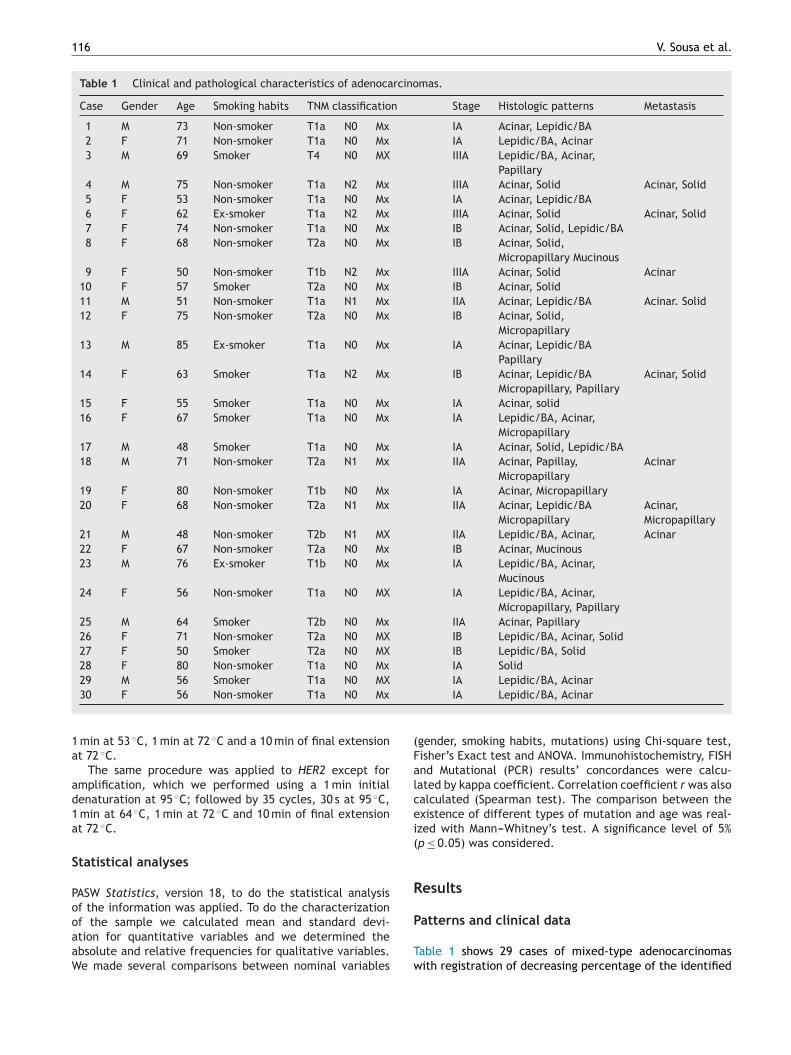
Table 1 Clinical and pathological characteristics of adenocarcinomas.
Case Gender Age Smoking habits TNM classification Stage Histologic patterns Metastasis
1 M 73 Non-smoker T1a N0 Mx IA Acinar, Lepidic/BA2 F 71 Non-smoker T1a N0 Mx IA Lepidic/BA, Acinar3 M 69 Smoker T4 N0 MX IIIA Lepidic/BA, Acinar,
Papillary4 M 75 Non-smoker T1a N2 Mx IIIA Acinar, Solid Acinar, Solid5 F 53 Non-smoker T1a N0 Mx IA Acinar, Lepidic/BA6 F 62 Ex-smoker T1a N2 Mx IIIA Acinar, Solid Acinar, Solid7 F 74 Non-smoker T1a N0 Mx IB Acinar, Solid, Lepidic/BA8 F 68 Non-smoker T2a N0 Mx IB Acinar, Solid,
Micropapillary Mucinous9 F 50 Non-smoker T1b N2 Mx IIIA Acinar, Solid Acinar
10 F 57 Smoker T2a N0 Mx IB Acinar, Solid11 M 51 Non-smoker T1a N1 Mx IIA Acinar, Lepidic/BA Acinar. Solid12 F 75 Non-smoker T2a N0 Mx IB Acinar, Solid,
Micropapillary13 M 85 Ex-smoker T1a N0 Mx IA Acinar, Lepidic/BA
Papillary14 F 63 Smoker T1a N2 Mx IB Acinar, Lepidic/BA
Micropapillary, PapillaryAcinar, Solid
15 F 55 Smoker T1a N0 Mx IA Acinar, solid16 F 67 Smoker T1a N0 Mx IA Lepidic/BA, Acinar,
Micropapillary17 M 48 Smoker T1a N0 Mx IA Acinar, Solid, Lepidic/BA18 M 71 Non-smoker T2a N1 Mx IIA Acinar, Papillay,
MicropapillaryAcinar
19 F 80 Non-smoker T1b N0 Mx IA Acinar, Micropapillary20 F 68 Non-smoker T2a N1 Mx IIA Acinar, Lepidic/BA
MicropapillaryAcinar,Micropapillary
21 M 48 Non-smoker T2b N1 MX IIA Lepidic/BA, Acinar, Acinar22 F 67 Non-smoker T2a N0 Mx IB Acinar, Mucinous23 M 76 Ex-smoker T1b N0 Mx IA Lepidic/BA, Acinar,
Mucinous24 F 56 Non-smoker T1a N0 MX IA Lepidic/BA, Acinar,
Micropapillary, Papillary25 M 64 Smoker T2b N0 Mx IIA Acinar, Papillary26 F 71 Non-smoker T2a N0 MX IB Lepidic/BA, Acinar, Solid27 F 50 Smoker T2a N0 MX IB Lepidic/BA, Solid28 F 80 Non-smoker T1a N0 Mx IA Solid
1a
ad1a
S
PooaaW
(Falcei(
R
29 M 56 Smoker T1a N0 MX30 F 56 Non-smoker T1a N0 Mx
min at 53 ◦C, 1 min at 72 ◦C and a 10 min of final extensiont 72 ◦C.
The same procedure was applied to HER2 except formplification, which we performed using a 1 min initialenaturation at 95 ◦C; followed by 35 cycles, 30 s at 95 ◦C,
min at 64 ◦C, 1 min at 72 ◦C and 10 min of final extensiont 72 ◦C.
tatistical analyses
ASW Statistics, version 18, to do the statistical analysisf the information was applied. To do the characterization
f the sample we calculated mean and standard devi-tion for quantitative variables and we determined thebsolute and relative frequencies for qualitative variables.e made several comparisons between nominal variablesP
Tw
IA Lepidic/BA, AcinarIA Lepidic/BA, Acinar
gender, smoking habits, mutations) using Chi-square test,isher’s Exact test and ANOVA. Immunohistochemistry, FISHnd Mutational (PCR) results’ concordances were calcu-ated by kappa coefficient. Correlation coefficient r was alsoalculated (Spearman test). The comparison between thexistence of different types of mutation and age was real-zed with Mann---Whitney’s test. A significance level of 5%p ≤ 0.05) was considered.
esults
atterns and clinical data
able 1 shows 29 cases of mixed-type adenocarcinomasith registration of decreasing percentage of the identified
Lung adenocarcinoma: subtyping and EGFR, HER2 and KRAS mutational status 117
F2
tt(lh(
e5(
cipecslscd(tMp(
detsi
imt
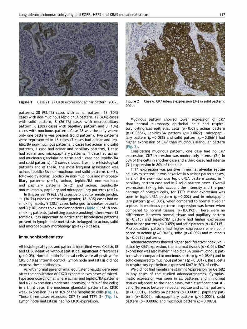
Figure 1 Case 21: 2+ CK20 expression; acinar pattern. 200×.
patterns: 28 (93.4%) cases with acinar pattern, 18 (60%)cases with non-mucinous lepidic/BA pattern, 12 (40%) caseswith solid pattern, 8 (26.7%) cases with micropapillarypattern, 6 (20%) cases with papillary pattern and 3 (10%)cases with mucinous pattern. Case 28 was the only whereonly one pattern was present (solid pattern). Two patternswere represented in 16 cases (7 cases had acinar and lep-idic/BA non-mucinous patterns, 5 cases had acinar and solidpatterns, 1 case had acinar and papillary patterns, 1 casehad acinar and micropapillary patterns, 1 case had acinarand mucinous glandular patterns and 1 case had lepidic/BAand solid patterns); 13 cases showed 3 or more histologicalpatterns and of these, the most frequent association wasacinar, lepidic/BA non-mucinous and solid patterns (n = 3),followed by acinar, lepidic/BA non-mucinous and micropap-illary patterns (n = 2), acinar, lepidic/BA non-mucinousand papillary patterns (n = 2) and acinar, lepidic/BAnon-mucinous, papillary and micropapillary patterns (n = 2).
In this series, 19 (63.3%) cases belonged to female gender,11 (36.7%) cases to masculine gender, 18 (60%) cases had nosmoking habits, 9 (30%) cases belonged to smoker patientsand 3 (10%) cases to ex-smoker patients (Table 1); of 18 non-smoking patients (admitting passive smoking), there were 13females. It is important to notice that histological patternspresent in lymph node metastasis belonged to acinar, solidand micropapillary morphology (pN1/2---8 cases).
Immunohistochemistry
All histological types and patterns identified were CK 5,6,18and CD56 negative without statistical significant differences(p > 0.05). Normal epithelial basal cells were all positive forCK5,6,18 as internal control; lymph node metastasis did notexpress these antibodies.
As with normal parenchyma, equivalent results were seenafter the application of CK20 except: in two cases of mixed-type adenocarcinoma, where acinar and lepidic/BA patternshad a 2+ expression (moderate intensity) in 50% of the cells;
in a third case, the mucinous glandular pattern had CK20weak expression (1+) in 10% of the neoplastic cells (Fig. 1).These three cases expressed CK7 3+ and TTF1 3+ (Fig. 1).Lymph node metastasis had no CK20 expression.c(tp
igure 2 Case 6: CK7 intense expression (3+) in solid pattern.00×.
Mucinous pattern showed lower expression of CK7han normal pulmonary epithelial cells and respira-ory cylindrical epithelial cells (p = 0.09); acinar patternp = 0.0584), lepidic/BA pattern (p = 0.0822), micropapil-ary pattern (p = 0.086) and solid pattern (p = 0.0661) hadigher expression of CK7 than mucinous glandular patternFig. 2).
Considering mucinous pattern, one case had no CK7xpression; CK7 expression was moderately intense (2+) in0% of the cells in another case and a third case, had intense3+) expression in 80% of the cells.
TTF1 expression was positive in normal alveolar septaeells as expected; it was negative in 6 acinar pattern cases,n 2 of the non-mucinous lepidic/BA pattern cases, in 1apillary pattern case and in 2 solid pattern cases. Overallxpression, taking into account the intensity and the per-entage of positive cells, for TTF1 higher expression waseen in lepidic/BA pattern (p = 0.002) and in micropapil-ary pattern (p = 0.005), when compared to normal alveolareptae. In mucinous patterns, expression was lower whenompared to normal tissues (p = 0.0192). There were noifferences between normal tissue and papillary patternp = 0.315) and lepidic/BA pattern had higher expressionhan acinar pattern (p = 0.059) and solid pattern (p = 0.0115).icropapillary pattern had higher expression when com-ared to acinar (p = 0.0413), solid (p = 0.009) and mucinousp = 0.0225) patterns.
Adenocarcinomas showed higher proliferative index, vali-ated by Ki67 expression, than normal tissues (p < 0.05). Ki67xpression was also higher in lepidic/BA (non-mucinous) pat-ern when compared to mucinous pattern (p = 0.0845) and inolid compared to mucinous patterns (p = 0.0817). Basal cellsn respiratory epithelium expressed Ki67 in 50% of cells.
We did not find membrane staining/expression for CerbB2n any cases of the studied adenocarcinomas. Cytoplas-atic expression was seen in all patterns and in normal
issues adjacent to the neoplasias, with significant statisti-al differences between alveolar septae and acinar patterns
p < 0.0001), lepidic/BA pattern (p < 0.0001), papillary pat-ern (p = 0.004), micropapillary pattern (p < 0.0001), solidattern (p = 0.0006) and mucinous pattern (p = 0.0073).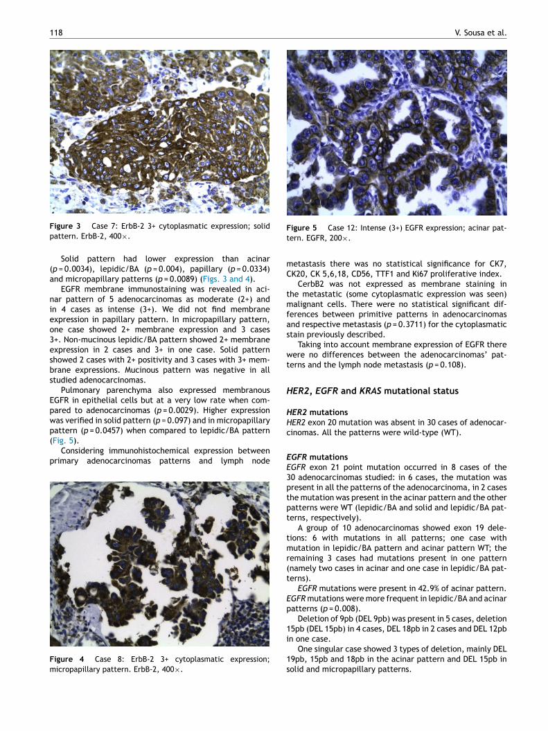
118 V. Sousa et al.
Fp
(a
nieo3esbs
Epwp(
p
Fm
Ft
mC
tmfas
wt
H
HH
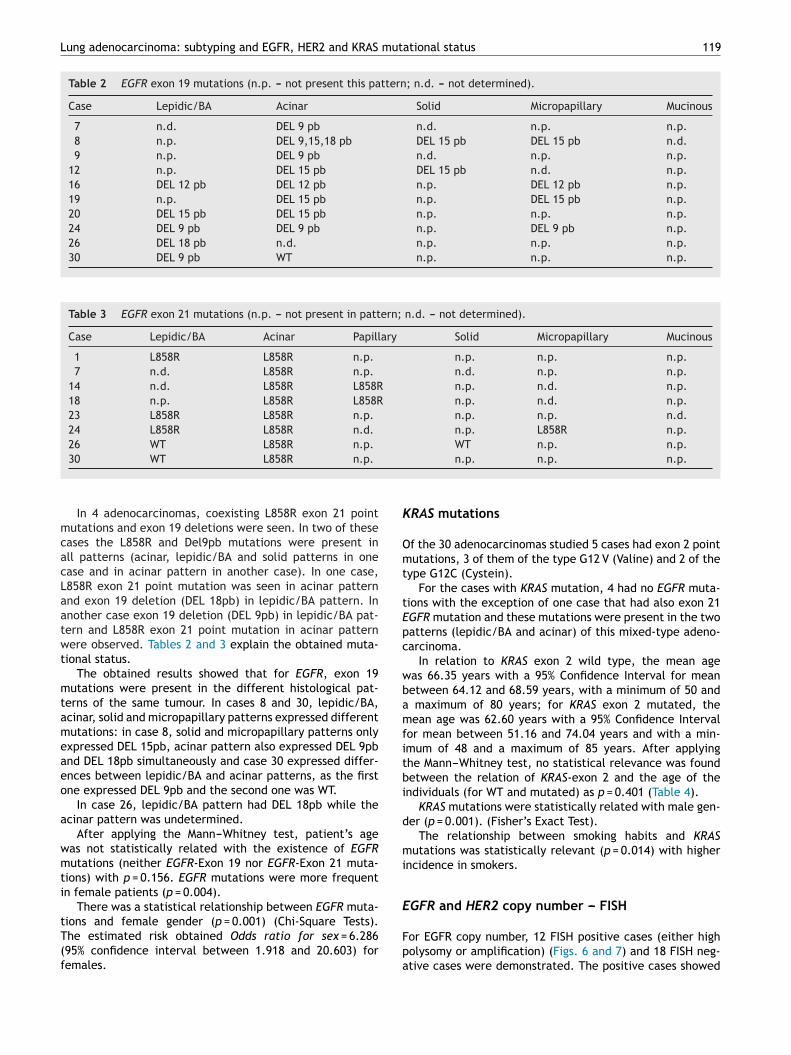
igure 3 Case 7: ErbB-2 3+ cytoplasmatic expression; solidattern. ErbB-2, 400×.
Solid pattern had lower expression than acinarp = 0.0034), lepidic/BA (p = 0.004), papillary (p = 0.0334)nd micropapillary patterns (p = 0.0089) (Figs. 3 and 4).
EGFR membrane immunostaining was revealed in aci-ar pattern of 5 adenocarcinomas as moderate (2+) andn 4 cases as intense (3+). We did not find membranexpression in papillary pattern. In micropapillary pattern,ne case showed 2+ membrane expression and 3 cases+. Non-mucinous lepidic/BA pattern showed 2+ membranexpression in 2 cases and 3+ in one case. Solid patternhowed 2 cases with 2+ positivity and 3 cases with 3+ mem-rane expressions. Mucinous pattern was negative in alltudied adenocarcinomas.
Pulmonary parenchyma also expressed membranousGFR in epithelial cells but at a very low rate when com-ared to adenocarcinomas (p = 0.0029). Higher expressionas verified in solid pattern (p = 0.097) and in micropapillary
attern (p = 0.0457) when compared to lepidic/BA patternFig. 5).Considering immunohistochemical expression betweenrimary adenocarcinomas patterns and lymph node
igure 4 Case 8: ErbB-2 3+ cytoplasmatic expression;icropapillary pattern. ErbB-2, 400×.
c
EE3ptpt
tmr(t
Ep
1i
1s
igure 5 Case 12: Intense (3+) EGFR expression; acinar pat-ern. EGFR, 200×.
etastasis there was no statistical significance for CK7,K20, CK 5,6,18, CD56, TTF1 and Ki67 proliferative index.
CerbB2 was not expressed as membrane staining inhe metastatic (some cytoplasmatic expression was seen)alignant cells. There were no statistical significant dif-
erences between primitive patterns in adenocarcinomasnd respective metastasis (p = 0.3711) for the cytoplasmatictain previously described.
Taking into account membrane expression of EGFR thereere no differences between the adenocarcinomas’ pat-
erns and the lymph node metastasis (p = 0.108).
ER2, EGFR and KRAS mutational status
ER2 mutationsER2 exon 20 mutation was absent in 30 cases of adenocar-inomas. All the patterns were wild-type (WT).
GFR mutationsGFR exon 21 point mutation occurred in 8 cases of the0 adenocarcinomas studied: in 6 cases, the mutation wasresent in all the patterns of the adenocarcinoma, in 2 caseshe mutation was present in the acinar pattern and the otheratterns were WT (lepidic/BA and solid and lepidic/BA pat-erns, respectively).
A group of 10 adenocarcinomas showed exon 19 dele-ions: 6 with mutations in all patterns; one case withutation in lepidic/BA pattern and acinar pattern WT; the
emaining 3 cases had mutations present in one patternnamely two cases in acinar and one case in lepidic/BA pat-erns).
EGFR mutations were present in 42.9% of acinar pattern.GFR mutations were more frequent in lepidic/BA and acinaratterns (p = 0.008).
Deletion of 9pb (DEL 9pb) was present in 5 cases, deletion5pb (DEL 15pb) in 4 cases, DEL 18pb in 2 cases and DEL 12pb
n one case.One singular case showed 3 types of deletion, mainly DEL9pb, 15pb and 18pb in the acinar pattern and DEL 15pb inolid and micropapillary patterns.
Lung adenocarcinoma: subtyping and EGFR, HER2 and KRAS mutational status 119
Table 2 EGFR exon 19 mutations (n.p. --- not present this pattern; n.d. --- not determined).
Case Lepidic/BA Acinar Solid Micropapillary Mucinous
7 n.d. DEL 9 pb n.d. n.p. n.p.8 n.p. DEL 9,15,18 pb DEL 15 pb DEL 15 pb n.d.9 n.p. DEL 9 pb n.d. n.p. n.p.
12 n.p. DEL 15 pb DEL 15 pb n.d. n.p.16 DEL 12 pb DEL 12 pb n.p. DEL 12 pb n.p.19 n.p. DEL 15 pb n.p. DEL 15 pb n.p.20 DEL 15 pb DEL 15 pb n.p. n.p. n.p.24 DEL 9 pb DEL 9 pb n.p. DEL 9 pb n.p.26 DEL 18 pb n.d. n.p. n.p. n.p.30 DEL 9 pb WT n.p. n.p. n.p.
Table 3 EGFR exon 21 mutations (n.p. --- not present in pattern; n.d. --- not determined).
Case Lepidic/BA Acinar Papillary Solid Micropapillary Mucinous
1 L858R L858R n.p. n.p. n.p. n.p.7 n.d. L858R n.p. n.d. n.p. n.p.
14 n.d. L858R L858R n.p. n.d. n.p.18 n.p. L858R L858R n.p. n.d. n.p.23 L858R L858R n.p. n.p. n.p. n.d.24 L858R L858R n.d. n.p. L858R n.p.26 WT L858R n.p. WT n.p. n.p.
K
Omt
tEpc
wbamfitbi
d
mi
E
30 WT L858R n.p.
In 4 adenocarcinomas, coexisting L858R exon 21 pointmutations and exon 19 deletions were seen. In two of thesecases the L858R and Del9pb mutations were present inall patterns (acinar, lepidic/BA and solid patterns in onecase and in acinar pattern in another case). In one case,L858R exon 21 point mutation was seen in acinar patternand exon 19 deletion (DEL 18pb) in lepidic/BA pattern. Inanother case exon 19 deletion (DEL 9pb) in lepidic/BA pat-tern and L858R exon 21 point mutation in acinar patternwere observed. Tables 2 and 3 explain the obtained muta-tional status.
The obtained results showed that for EGFR, exon 19mutations were present in the different histological pat-terns of the same tumour. In cases 8 and 30, lepidic/BA,acinar, solid and micropapillary patterns expressed differentmutations: in case 8, solid and micropapillary patterns onlyexpressed DEL 15pb, acinar pattern also expressed DEL 9pband DEL 18pb simultaneously and case 30 expressed differ-ences between lepidic/BA and acinar patterns, as the firstone expressed DEL 9pb and the second one was WT.
In case 26, lepidic/BA pattern had DEL 18pb while theacinar pattern was undetermined.
After applying the Mann---Whitney test, patient’s agewas not statistically related with the existence of EGFRmutations (neither EGFR-Exon 19 nor EGFR-Exon 21 muta-tions) with p = 0.156. EGFR mutations were more frequentin female patients (p = 0.004).
There was a statistical relationship between EGFR muta-
tions and female gender (p = 0.001) (Chi-Square Tests).The estimated risk obtained Odds ratio for sex = 6.286(95% confidence interval between 1.918 and 20.603) forfemales.Fpa
n.p. n.p. n.p.
RAS mutations
f the 30 adenocarcinomas studied 5 cases had exon 2 pointutations, 3 of them of the type G12 V (Valine) and 2 of the
ype G12C (Cystein).For the cases with KRAS mutation, 4 had no EGFR muta-
ions with the exception of one case that had also exon 21GFR mutation and these mutations were present in the twoatterns (lepidic/BA and acinar) of this mixed-type adeno-arcinoma.
In relation to KRAS exon 2 wild type, the mean ageas 66.35 years with a 95% Confidence Interval for meanetween 64.12 and 68.59 years, with a minimum of 50 and
maximum of 80 years; for KRAS exon 2 mutated, theean age was 62.60 years with a 95% Confidence Interval
or mean between 51.16 and 74.04 years and with a min-mum of 48 and a maximum of 85 years. After applyinghe Mann---Whitney test, no statistical relevance was foundetween the relation of KRAS-exon 2 and the age of thendividuals (for WT and mutated) as p = 0.401 (Table 4).
KRAS mutations were statistically related with male gen-er (p = 0.001). (Fisher’s Exact Test).
The relationship between smoking habits and KRASutations was statistically relevant (p = 0.014) with higher
ncidence in smokers.
GFR and HER2 copy number --- FISH
or EGFR copy number, 12 FISH positive cases (either higholysomy or amplification) (Figs. 6 and 7) and 18 FISH neg-tive cases were demonstrated. The positive cases showed
120 V. Sousa et al.
Table 4 KRAS exon 2 mutations (n.p. --- not present in this pattern; n.d. --- not determined).
Case Lepidic/BA Acinar Papillary Solid Mucinous
13 n.d. G12C G12C n.p. n.p.17 n.d. G12V n.p. G12V n.p.21 G12V G12V n.p. n.p. n.p.23 G12V
(EGFRL858R)
G12V(EGFRL858R)
n.p. n.p. n.d.
29 G12C G12C
Ft7
piOETc
Ftk
Ew
FFs
bf
Eqc(ca
i2atmnn
D
TsbssC
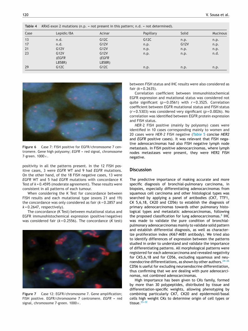
igure 6 Case 7: FISH positive for EGFR/chromosome 7 cen-romere. Gene high polysomy; EGFR --- red signal, chromosome-green. 1000×.
ositivity in all the patterns present. In the 12 FISH pos-tive cases, 3 were EGFR WT and 9 had EGFR mutations.n the other hand, of the 18 FISH negative cases, 13 wereGFR WT and 5 had EGFR mutations with concordance Kest of k = 0.4595 (moderate agreement). These results wereonsistent in all patterns of each tumour.
When considering the K Test for concordance betweenISH results and each mutational type (exons 21 and 19)he concordance was only considered as fair (k = 0.2857 and
= 0.2647, respectively).The concordance (K Test) between mutational status and
GFR immunohistochemical expression (positive/negative)as considered fair (k = 0.2556). The concordance (K test)
igure 7 Case 12: EGFR/chromosome 7. Gene amplification;ISH positive. EGFR/chromosome 7 centromere. EGFR --- redignal, chromosome 7-green. 1000×.
pltwpaitsorfrCtn
bdcct
n.p. n.p. n.p.
etween FISH status and IHC results were also considered asair (k = 0.2635).
Correlation coefficient between immunohistochemicalGFR expression and mutational status was considered notuite significant (p = 0.0561) with r = 0.3525. Correlationoefficient between EGFR mutational status and FISH statusr = 0.5303) was considered very significant (p = 0.0026). Noorrelation was identified between EGFR protein expressionnd FISH status.
HER-2 FISH positive (mainly by polysomy) cases weredentified in 10 cases corresponding mainly to women and0 cases were HER-2 FISH negative (Table 5 concise HER2nd EGFR positive cases). It was relevant that FISH nega-ive adenocarcinomas had also FISH negative lymph nodeetastasis. In FISH positive adenocarcinomas, where lymph
odes metastases were present, they were HER2 FISHegative.
iscussion
he predictive importance of making accurate and morepecific diagnosis of bronchial-pulmonary carcinoma, iniopsies, especially differentiating adenocarcinomas fromquamous cell carcinoma and other histological types wasearched by applying a panel of antibodies (CK7, TTF1,K 5,6,18, CK20 and CD56) to establish the diagnosis ofrimary adenocarcinomas towards other pulmonary histo-ogical types and metastatic adenocarcinomas, followinghe proposed classification for lung adenocarcinomas.8 IHCas made to validate the pure condition of bronchial-ulmonary adenocarcinomas mainly to validate solid patternnd establish differential diagnosis, as well as character-ze proliferation index (Ki67-MIB1 antibody). We tried alsoo identify differences of expression between the patternstudied in order to understand and validate the importancef differentiating patterns. All morphological patterns wereegistered for each adenocarcinoma and revealed negativityor CK5,6,18 and for CD56, excluding squamous and neu-oendocrine differentiations, as shown by other authors.24---26
D56 is useful for excluding neuroendocrine differentiation,hus confirming that we are dealing with pure adenocarci-omas, not combined adenocarcinomas.
High importance has been given to CKs family, formedy more than 30 polypeptides, distributed by tissue and
ifferentiation-specific weights, allowing phenotyping byombining particularly CK7, CK20 and epidermoid/basalells high weight CKs to determine origin of cell types orissue.25---32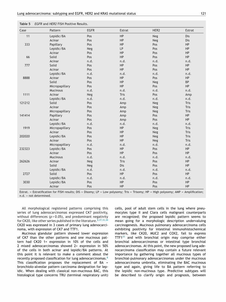
Lung adenocarcinoma: subtyping and EGFR, HER2 and KRAS mutational status 121
Table 5 EGFR and HER2 FISH Positive Results.
Case Pattern EGFR Estrat HER2 Estrat
11 Lepidic/BAAcinar
PosPos
HPHP
NegNeg
DisDis
333 PapillaryLepidic/BAAcinar
PosNegPos
HPLPHP
PosPosPos
HPHPHP
66 SolidAcinar
Posn.d.
HPn.d.
Posn.d.
HPn.d.
777 SolidAcinarLepidic/BA
PosPosn.d.
HPHPn.d.
PosPosn.d.
HPHPn.d.
8888 AcinarSolidMicropapillaryMucinous
PosPosPosn.d.
HPHPHPn.d.
PosNegPosn.d.
HPBPHPn.d.
1111 AcinarLepidic/BA
Negn.d.
Trisn.d.
Posn.d.
Ampn.d.
121212 SolidAcinarMicropapillary
PosPosPos
AmpAmpAmp
NegNegNeg
TrisTrisTris
141414 PapillaryAcinarLepidic/BA
PosPosn.d.
AmpAmpn.d.
PosPosn.d.
HPHPn.d.
1919 MicropapillaryAcinar
PosPos
HPHP
NegNeg
TrisTris
202020 Lepidic/BAAcinarMicropapillary
PosPosn.d.
HPHPn.d.
NegNegn.d.
TrisTrisn.d.
232323 Lepidic/BAAcinarMucinous
PosPosn.d.
HPHPn.d.
PosPosn.d.
HPHPn.d.
262626 AcinarSolidLepidic/BA
NegNegn.d.
TrisDisn.d.
PosPosn.d.
HPHPn.d.
2727 SolidLepidic/BA
Posn.d.
HPn.d.
Posn.d.
HPn.d.
3030 Lepidic/BAAcinar
PosPos
HPHP
PosPos
HPHP
poly
cmamcemTbanib
Estrat. --- Estratification for FISH results; DIS --- Disomy; LP --- Lown.d. --- not determined.
All morphological registered patterns comprising thisseries of lung adenocarcinomas expressed CK7 positivity,without differences (p > 0.05), and predominant negativityfor CK20, like other series published in the literature.8,27,33,34
CK20 was expressed in 3 cases of primary lung adenocarci-noma, with expression of CK7 and TTF1.
Mucinous glandular pattern showed lower expressionof CK7 than the other patterns and one mucinous pat-tern had CK20 1+ expression in 10% of the cells and2 mixed adenocarcinomas showed 2+ expression in 50%of the cells in both acinar and lepidic/BA patterns. Atthis point it is relevant to make a comment about therecently proposed classification for lung adenocarcinomas.8
This classification proposes the replacement of BA ---bronchiolo-alveolar pattern carcinoma designation for lep-idic. When dealing with classical non-mucinous BAC, thishistological type concerns TRU (terminal respiratory unit)
attb
somy; Tris --- Trisomy; HP --- High polysomy; AMP --- Amplification;
ells, pool of adult stem cells in the lung where pneu-ocytes type II and Clara cells malignant counterparts
re recognized; the proposed lepidic pattern seems toean going for a morphologic description undervaluing
arcinogenesis. Mucinous pulmonary adenocarcinoma cellsxhibiting positivity for intestinal immunohistochemicalarkers, like CK20, MUC2 and COX2, fail to expressTF135 and with bronchial origin may comprise eitherronchial adenocarcinomas or intestinal type bronchialdenocarcinomas. At this point, the new proposed lung ade-ocarcinoma classification may contain a future relevantmportance by gathering together all mucinous types ofronchial-pulmonary adenocarcinomas under the mucinous
denocarcinoma umbrella, eliminating the BAC mucinousype and again, giving life to BA pattern referring tohe lepidic non-mucinous type. Predictive subtypes wille described to clarify origin and prognosis, between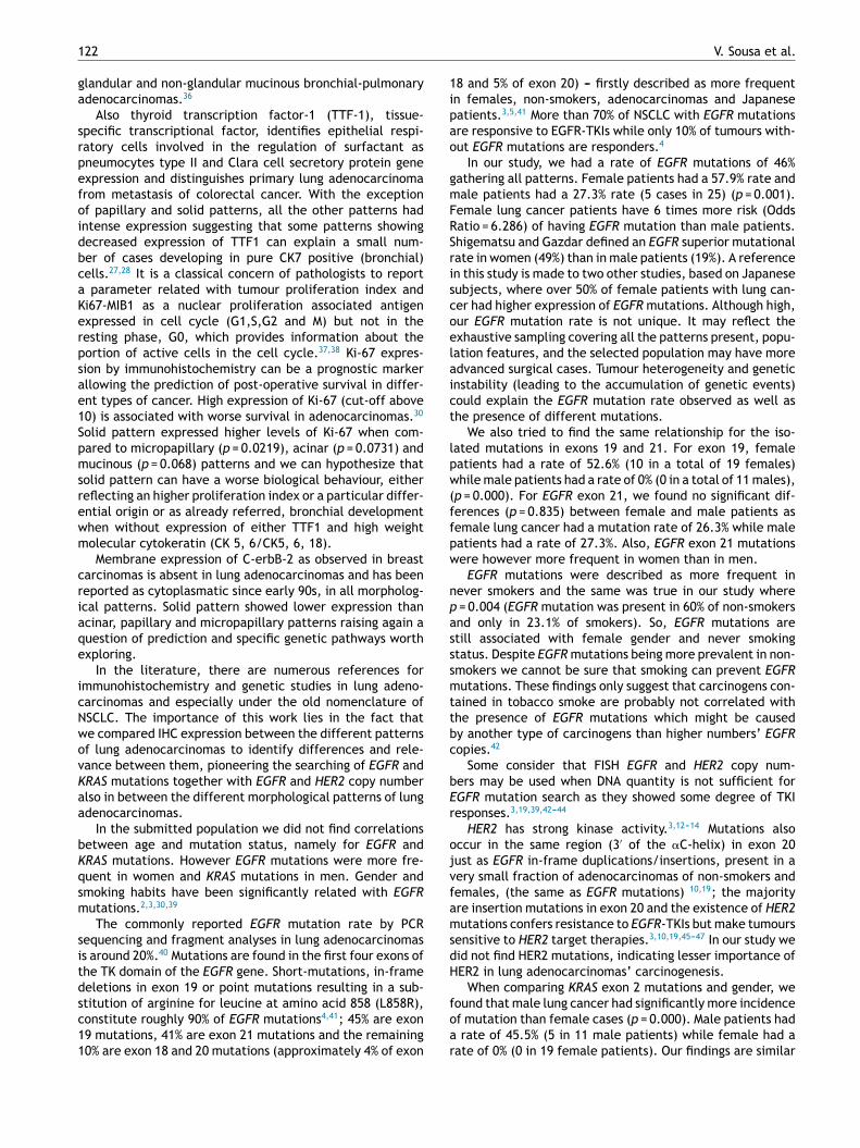
1
ga
srpefoidbcaKerpsae1Spmsrewm
criaqe
icNwovKaa
bKqsm
sitdsc11
1ipao
gmFRSriscoelaict
lpw(ffpw
npasssmttbc
bEr
ojvfamsdH
22
landular and non-glandular mucinous bronchial-pulmonarydenocarcinomas.36
Also thyroid transcription factor-1 (TTF-1), tissue-pecific transcriptional factor, identifies epithelial respi-atory cells involved in the regulation of surfactant asneumocytes type II and Clara cell secretory protein genexpression and distinguishes primary lung adenocarcinomarom metastasis of colorectal cancer. With the exceptionf papillary and solid patterns, all the other patterns hadntense expression suggesting that some patterns showingecreased expression of TTF1 can explain a small num-er of cases developing in pure CK7 positive (bronchial)ells.27,28 It is a classical concern of pathologists to report
parameter related with tumour proliferation index andi67-MIB1 as a nuclear proliferation associated antigenxpressed in cell cycle (G1,S,G2 and M) but not in theesting phase, G0, which provides information about theortion of active cells in the cell cycle.37,38 Ki-67 expres-ion by immunohistochemistry can be a prognostic markerllowing the prediction of post-operative survival in differ-nt types of cancer. High expression of Ki-67 (cut-off above0) is associated with worse survival in adenocarcinomas.30
olid pattern expressed higher levels of Ki-67 when com-ared to micropapillary (p = 0.0219), acinar (p = 0.0731) anducinous (p = 0.068) patterns and we can hypothesize that
olid pattern can have a worse biological behaviour, eithereflecting an higher proliferation index or a particular differ-ntial origin or as already referred, bronchial developmenthen without expression of either TTF1 and high weightolecular cytokeratin (CK 5, 6/CK5, 6, 18).Membrane expression of C-erbB-2 as observed in breast
arcinomas is absent in lung adenocarcinomas and has beeneported as cytoplasmatic since early 90s, in all morpholog-cal patterns. Solid pattern showed lower expression thancinar, papillary and micropapillary patterns raising again auestion of prediction and specific genetic pathways worthxploring.
In the literature, there are numerous references formmunohistochemistry and genetic studies in lung adeno-arcinomas and especially under the old nomenclature ofSCLC. The importance of this work lies in the fact thate compared IHC expression between the different patternsf lung adenocarcinomas to identify differences and rele-ance between them, pioneering the searching of EGFR andRAS mutations together with EGFR and HER2 copy numberlso in between the different morphological patterns of lungdenocarcinomas.
In the submitted population we did not find correlationsetween age and mutation status, namely for EGFR andRAS mutations. However EGFR mutations were more fre-uent in women and KRAS mutations in men. Gender andmoking habits have been significantly related with EGFRutations.2,3,30,39
The commonly reported EGFR mutation rate by PCRequencing and fragment analyses in lung adenocarcinomass around 20%.40 Mutations are found in the first four exons ofhe TK domain of the EGFR gene. Short-mutations, in-frameeletions in exon 19 or point mutations resulting in a sub-
titution of arginine for leucine at amino acid 858 (L858R),onstitute roughly 90% of EGFR mutations4,41; 45% are exon9 mutations, 41% are exon 21 mutations and the remaining0% are exon 18 and 20 mutations (approximately 4% of exonfoar
V. Sousa et al.
8 and 5% of exon 20) --- firstly described as more frequentn females, non-smokers, adenocarcinomas and Japaneseatients.3,5,41 More than 70% of NSCLC with EGFR mutationsre responsive to EGFR-TKIs while only 10% of tumours with-ut EGFR mutations are responders.4
In our study, we had a rate of EGFR mutations of 46%athering all patterns. Female patients had a 57.9% rate andale patients had a 27.3% rate (5 cases in 25) (p = 0.001).
emale lung cancer patients have 6 times more risk (Oddsatio = 6.286) of having EGFR mutation than male patients.higematsu and Gazdar defined an EGFR superior mutationalate in women (49%) than in male patients (19%). A referencen this study is made to two other studies, based on Japaneseubjects, where over 50% of female patients with lung can-er had higher expression of EGFR mutations. Although high,ur EGFR mutation rate is not unique. It may reflect thexhaustive sampling covering all the patterns present, popu-ation features, and the selected population may have moredvanced surgical cases. Tumour heterogeneity and geneticnstability (leading to the accumulation of genetic events)ould explain the EGFR mutation rate observed as well ashe presence of different mutations.
We also tried to find the same relationship for the iso-ated mutations in exons 19 and 21. For exon 19, femaleatients had a rate of 52.6% (10 in a total of 19 females)hile male patients had a rate of 0% (0 in a total of 11 males),
p = 0.000). For EGFR exon 21, we found no significant dif-erences (p = 0.835) between female and male patients asemale lung cancer had a mutation rate of 26.3% while maleatients had a rate of 27.3%. Also, EGFR exon 21 mutationsere however more frequent in women than in men.
EGFR mutations were described as more frequent inever smokers and the same was true in our study where
= 0.004 (EGFR mutation was present in 60% of non-smokersnd only in 23.1% of smokers). So, EGFR mutations aretill associated with female gender and never smokingtatus. Despite EGFR mutations being more prevalent in non-mokers we cannot be sure that smoking can prevent EGFRutations. These findings only suggest that carcinogens con-
ained in tobacco smoke are probably not correlated withhe presence of EGFR mutations which might be causedy another type of carcinogens than higher numbers’ EGFRopies.42
Some consider that FISH EGFR and HER2 copy num-ers may be used when DNA quantity is not sufficient forGFR mutation search as they showed some degree of TKIesponses.3,19,39,42---44
HER2 has strong kinase activity.3,12---14 Mutations alsoccur in the same region (3′ of the �C-helix) in exon 20ust as EGFR in-frame duplications/insertions, present in aery small fraction of adenocarcinomas of non-smokers andemales, (the same as EGFR mutations) 10,19; the majorityre insertion mutations in exon 20 and the existence of HER2utations confers resistance to EGFR-TKIs but make tumours
ensitive to HER2 target therapies.3,10,19,45---47 In our study weid not find HER2 mutations, indicating lesser importance ofER2 in lung adenocarcinomas’ carcinogenesis.
When comparing KRAS exon 2 mutations and gender, we
ound that male lung cancer had significantly more incidencef mutation than female cases (p = 0.000). Male patients hadrate of 45.5% (5 in 11 male patients) while female had aate of 0% (0 in 19 female patients). Our findings are similar
muta
soE
aan(tlt
psoaln
spifh
C
Tptabaiap
C
T
E
Pda
hp
d
A
Vh
Lung adenocarcinoma: subtyping and EGFR, HER2 and KRAS
to Kim et al. study, who also realized that KRAS mutationsare significantly related with smoking habits (p = 0.014) in atotal rate of 30.8% in smokers while in non-smokers it wasonly 5.7%.30,42
KRAS mutations have been associated with mucinousdifferentiation, goblet cell, poor differentiation adenocar-cinomas and with solid patterns.48---52 However in our caseswe did find this mutation in acinar and lepidic patterns andin only one case with solid pattern. The lower number ofpoorly differentiated adenocarcinomas, mucinous and solidpatterns could in part explain the lower KRAS mutation rate.
EGFR mutations and KRAS mutations have been describedas mutually exclusive but we found 1 case of KRAS mutationsimultaneously with EGFR mutation. So we conclude thatalthough EGFR and KRAS mutations are generally mutuallyexclusive, in some cases they can coexist; this has also beendescribed by other authors.11,46,53---57 The clinical, therapeu-tic and prognostic issues concerning this coexistence needto be understood.
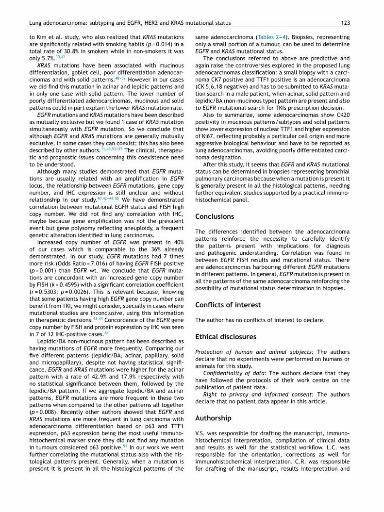
Although many studies demonstrated that EGFR muta-tions are usually related with an amplification in EGFRlocus, the relationship between EGFR mutations, gene copynumber, and IHC expression is still unclear and withoutrelationship in our study.40,42---44,58 We have demonstratedcorrelation between mutational EGFR status and FISH highcopy number. We did not find any correlation with IHC,maybe because gene amplification was not the prevalentevent but gene polysomy reflecting aneuploidy, a frequentgenetic alteration identified in lung carcinomas.
Increased copy number of EGFR was present in 40%of our cases which is comparable to the 36% alreadydemonstrated. In our study, EGFR mutations had 7 timesmore risk (Odds Ratio = 7.016) of having EGFR FISH positive(p = 0.001) than EGFR wt. We conclude that EGFR muta-tions are concordant with an increased gene copy numberby FISH (k = 0.4595) with a significant correlation coefficient(r = 0.5303; p = 0.0026). This is relevant because, knowingthat some patients having high EGFR gene copy number canbenefit from TKI, we might consider, specially in cases wheremutational studies are inconclusive, using this informationin therapeutic decisions.43,44 Concordance of the EGFR genecopy number by FISH and protein expression by IHC was seenin 7 of 12 IHC-positive cases.44
Lepidic/BA non-mucinous pattern has been described ashaving mutations of EGFR more frequently. Comparing ourfive different patterns (lepidic/BA, acinar, papillary, solidand micropapillary), despite not having statistical signifi-cance, EGFR and KRAS mutations were higher for the acinarpattern with a rate of 42.9% and 17.9% respectively withno statistical significance between them, followed by thelepidic/BA pattern. If we aggregate lepidic/BA and acinarpatterns, EGFR mutations are more frequent in these twopatterns when compared to the other patterns all together(p = 0.008). Recently other authors showed that EGFR andKRAS mutations are more frequent in lung carcinoma withadenocarcinoma differentiation based on p63 and TTF1expression, p63 expression being the most useful immuno-histochemical marker since they did not find any mutation
in tumours considered p63 positive.51 In our work we wentfurther correlating the mutational status also with the his-tological patterns present. Generally, when a mutation ispresent it is present in all the histological patterns of thearif
tional status 123
ame adenocarcinoma (Tables 2---4). Biopsies, representingnly a small portion of a tumour, can be used to determineGFR and KRAS mutational status.
The conclusions referred to above are predictive andgain raise the controversies explored in the proposed lungdenocarcinomas classification: a small biopsy with a carci-oma CK7 positive and TTF1 positive is an adenocarcinomaCK 5,6,18 negative) and has to be submitted to KRAS muta-ion search in a male patient, when acinar, solid pattern andepidic/BA (non-mucinous type) pattern are present and alsoo EGFR mutational search for TKIs prescription decision.
Also to summarize, some adenocarcinomas show CK20ositivity in mucinous patterns/subtypes and solid patternshow lower expression of nuclear TTF1 and higher expressionf Ki67, reflecting probably a particular cell origin and moreggressive biological behaviour and have to be reported asung adenocarcinomas, avoiding poorly differentiated carci-oma designation.
After this study, it seems that EGFR and KRAS mutationaltatus can be determined in biopsies representing bronchialulmonary carcinomas because when a mutation is present its generally present in all the histological patterns, needingurther equivalent studies supported by a practical immuno-istochemical panel.
onclusions
he differences identified between the adenocarcinomaatterns reinforce the necessity to carefully identifyhe patterns present with implications for diagnosisnd pathogenic understanding. Correlation was found inetween EGFR FISH results and mutational status. Therere adenocarcinomas harbouring different EGFR mutationsn different patterns. In general, EGFR mutation is present inll the patterns of the same adenocarcinoma reinforcing theossibility of mutational status determination in biopsies.
onflicts of interest
he author has no conflicts of interest to declare.
thical disclosures
rotection of human and animal subjects: The authorseclare that no experiments were performed on humans ornimals for this study.
Confidentiality of data: The authors declare that theyave followed the protocols of their work centre on theublication of patient data.
Right to privacy and informed consent: The authorseclare that no patient data appear in this article.
uthorship
.S. was responsible for drafting the manuscript, immuno-istochemical interpretation, compilation of clinical data
nd results as well for the statistical workflow. L.C. wasesponsible for the orientation, corrections as well formmunohistochemical interpretation. C.R. was responsibleor drafting of the manuscript, results interpretation and
1
ara
A
CG
R
1
1
1
1
1
1
1
1
1
1
2
2
2
2
2
2
2
2
2
2
3
3
3
3
3
24
lso for statistical works. M.S. carried out FISH assays andesults interpretation. A.M.A. carried out the immunoassaysnd mutational evaluation.
cknowledgement
IMAGO --- Centro de Investigacão em Meio Ambiente,enética e Oncobiologia.
eferences
1. Jemal A, Clegg LX, Ward E, et al. Annual report to the nationon the status of cancer, 1975---2001, with a special featureregarding survival. Cancer. 2004;101:3---27.
2. Kim YT, Kim TY, Lee DS, et al. Molecular changes of epidermalgrowth factor receptor (EGFR) and KRAS and their impact onthe clinical outcomes in surgically resected adenocarcinoma ofthe lung. Lung Cancer. 2008;59:111---8.
3. Shigematsu H, Gazdar AF. Somatic mutations of epidermalgrowth factor receptor signaling pathway in lung cancers. IntJ Cancer. 2006;118:257---62.
4. Uramoto H, Mitsudomi T. Which biomarker predicts benefit fromEGFR-TKI treatment for patients with lung cancer? Br J Cancer.2007;96:857---63.
5. Sharma SV, Bell DW, Settleman J, et al. Epidermal growthfactor receptor mutations in lung cancer. Nat Rev Cancer.2007;7:169---81.
6. Travis W.D., Brambilla E, Müller-Hermelink HK, Harris CC.Pathology genetics of tumours of the lung, pleura, thymus andheart. Lyon: IARC Press; 2004.
7. Stahel RA. Adenocarcinoma, a molecular perspective. AnnOncol. 2007;18 Suppl. 9:ix147---9.
8. Travis WD, Brambilla E, Noguchi M, et al. Internationalassociation for the study of lung cancer/American ThoracicSociety/European Respiratory Society International Multidisci-plinary Classification of lung adenocarcinoma. J Thorac Oncol.2011;6:244---85.
9. Gazdar AF. Activating and resistance mutations of EGFR in non-small-cell lung cancer: role in clinical response to EGFR tyrosinekinase inhibitors. Oncogene. 2009;28 Suppl. 1:S24---31.
0. Mitsudomi T, Yatabe Y. Mutations of the epidermal growth factorreceptor gene and related genes as determinants of epidermalgrowth factor receptor tyrosine kinase inhibitors sensitivity inlung cancer. Cancer Sci. 2007;98:1817---24.
1. Eberhard DA, Johnson BE, Amler LC, et al. Mutations in theepidermal growth factor receptor and in KRAS are predictiveand prognostic indicators in patients with non-small-cell lungcancer treated with chemotherapy alone and in combinationwith erlotinib. J Clin Oncol. 2005;23:5900---9.
2. Herbst RS, Fukuoka M, Baselga J. Gefitinib----a novel targetedapproach to treating cancer. Nat Rev Cancer. 2004;4:956---65.
3. Linardou H, Dahabreh IJ, Kanaloupiti D, et al. Assessmentof somatic k-RAS mutations as a mechanism associated withresistance to EGFR-targeted agents: a systematic review andmeta-analysis of studies in advanced non-small-cell lung cancerand metastatic colorectal cancer. Lancet Oncol. 2008;9:962---72.
4. Normanno N, De Luca A, Bianco C, et al. Epidermal growth fac-tor receptor (EGFR) signaling in cancer. Gene. 2006;366:2---16.
5. Rodenhuis S, van de Wetering ML, Mooi WJ, et al. Mutationalactivation of the K-ras oncogene. A possible pathogenetic factorin adenocarcinoma of the lung. N Engl J Med. 1987;317:929---35.
6. Suda K, Tomizawa K, Mitsudomi T. Biological and clinical signifi-cance of KRAS mutations in lung cancer: an oncogenic driverthat contrasts with EGFR mutation. Cancer Metastasis Rev.2010;29:49---60.
3
V. Sousa et al.
7. Kosaka T, Yatabe Y, Endoh H, et al. Mutations of the epider-mal growth factor receptor gene in lung cancer: biological andclinical implications. Cancer Res. 2004;64:8919---23.
8. Shigematsu H, Lin L, Takahashi T, et al. Clinical and biologicalfeatures associated with epidermal growth factor receptor genemutations in lung cancers. J Natl Cancer Inst. 2005;97:339---46.
9. Shigematsu H, Takahashi T, Nomura M, et al. Somatic mutationsof the HER2 kinase domain in lung adenocarcinomas. CancerRes. 2005;65:1642---6.
0. Endoh H, Yatabe Y, Kosaka T, et al. PTEN and PIK3CA expressionis associated with prolonged survival after gefitinib treat-ment in EGFR-mutated lung cancer patients. J Thorac Oncol.2006;1:629---34.
1. Varella-Garcia M, Diebold J, Eberhard DA, et al. EGFRfluorescence in situ hybridisation assay: guidelines for appli-cation to non-small-cell lung cancer. J Clin Pathol. 2009;62:970---7.
2. Inoue A, Suzuki T, Fukuhara T, et al. Prospective phase II study ofgefitinib for chemotherapy-naive patients with advanced non-small-cell lung cancer with epidermal growth factor receptorgene mutations. J Clin Oncol. 2006;24:3340---6.
3. Yoshida K, Yatabe Y, Park JY, et al. Prospective validation forprediction of gefitinib sensitivity by epidermal growth factorreceptor gene mutation in patients with non-small cell lungcancer. J Thorac Oncol. 2007;2:22---8.
4. Heatley M, Maxwell P, Whiteside C, et al. Cytokeratin intermedi-ate filament expression in benign and malignant breast disease.J Clin Pathol. 1995;48:26---32.
5. Obermuller S, Calegari F, King A, et al. Defective secretion ofislet hormones in chromogranin-B deficient mice. PLOS ONE.2010;5:e8936.
6. Pelosi G, Pasini F, Sonzogni A, et al. Prognostic implicationsof neuroendocrine differentiation and hormone production inpatients with Stage I nonsmall cell lung carcinoma. Cancer.2003;97:2487---97.
7. Ikeda S, Fujimori M, Shibata S, et al. Combined immunohisto-chemistry of beta-catenin, cytokeratin 7, and cytokeratin 20is useful in discriminating primary lung adenocarcinomas frommetastatic colorectal cancer. BMC Cancer. 2006;6:31.
8. Kubba LA, McCluggage WG, Liu J, et al. Thyroid transcriptionfactor-1 expression in ovarian epithelial neoplasms. Mod Pathol.2008;21:485---90.
9. Leech SN, Kolar AJ, Barrett PD, et al. Merkel cell carcinomacan be distinguished from metastatic small cell carcinoma usingantibodies to cytokeratin 20 and thyroid transcription factor 1.J Clin Pathol. 2001;54:727---9.
0. Woo T, Okudela K, Yazawa T, et al. Prognostic value of KRASmutations and Ki-67 expression in stage I lung adenocarcinomas.Lung Cancer. 2009;65:355---62.
1. Ikeda S, Fujimori M, Shibata S, et al. Combined immunohisto-chemistry of beta-catenin, cytokeratin 7, and cytokeratin 20is useful in discriminating primary lung adenocarcinomas frommetastatic colorectal cancer. BMC Cancer. 2006;2:6.
2. Sousa V, Espirito Santo J, Silva M, et al. EGFR/erB-1, HER2/erB-2, CK7, LP34, Ki67 and P53 expression in preneoplastic lesionsof bronchial epithelium: an immunohistochemical and geneticstudy. Virchows Arch. 2011;458:571---81.
3. Tsao SC, Su YC, Wang SL, et al. Use of caveolin-1, thy-roid transcription factor-1, and cytokeratins 7 and 20 indiscriminating between primary and secondary pulmonary ade-nocarcinoma from breast or colonic origin. Kaohsiung J Med Sci.2007;23:325---31.
4. Su YC, Hsu YC, Chai CY. Role of TTF-1, CK20, and CK7 immuno-histochemistry for diagnosis of primary and secondary lungadenocarcinoma. Kaohsiung J Med Sci. 2006;22:14---9.
5. Tsuta K, Ishii G, Yoh K, et al. Primary lung carcinoma with signet-ring cell carcinoma components: clinicopathological analysis of39 cases. Am J Surg Pathol. 2004;28:868---74.
muta
4
4
5
5
5
5
5
5
5
5
Lung adenocarcinoma: subtyping and EGFR, HER2 and KRAS
36. Rossi G, Murer B, Cavazza A, et al. Primary mucinous (so-called colloid) carcinomas of the lung: a clinicopathologic andimmunohistochemical study with special reference to CDX-2 homeobox gene and MUC2 expression. Am J Surg Pathol.2004;28:442---52.
37. Neves LR, Oshima CT, Artigiani-Neto R, et al. Ki67 and p53in gastrointestinal stromal tumours-GIST. Arq Gastroenterol.2009;46:116---20.
38. Scholzen T, Gerdes J. The Ki-67 protein: from the known andthe unknown. J Cell Physiol. 2000;182:311---22.
39. Gazdar AF. Activating and resistance mutations of EGFR in non-small-cell lung cancer: role in clinical response to EGFR tyrosinekinase inhibitors. Oncogene. 2009;28:24---31.
40. El-Zammar OA, Zhang S, Katzenstein AL. Comparison of FISH,PCR, and immunohistochemistry in assessing EGFR status inlung adenocarcinoma and correlation with clinicopathologicfeatures. Diagn Mol Pathol. 2009;18:133---7.
41. Mitsudomi T, Kosaka T, Yatabe Y. Biological and clinical impli-cations of EGFR mutations in lung cancer. Int J Clin Oncol.2006;11:190---8.
42. Mitsudomi T, Yatabe Y. Mutations of the epidermal growth factorreceptor gene and related genes as determinants of epidermalgrowth factor receptor tyrosine kinase inhibitors sensivity inlung cancer. Cancer Sci. 2007;98:1817---24.
43. Amann J, Kalyankrishna S, Massion PP, et al. Aberrant epidermalgrowth factor receptor signaling and enhanced sensitivity toEGFR inhibitors in lung cancer. Cancer Res. 2005;65:226---35.
44. Takano T, Ohe Y, Sakamoto H, et al. Epidermal growth factorreceptor gene mutations and increased copy numbers predictgefitinib sensitivity in patients with recurrent non-small-celllung cancer. J Clin Oncol. 2005;23:6829---37.
45. Cappuzzo F, Bemis L, Varella-Garcia M. HER2 mutation andresponse to trastuzumab therapy in non-small-cell lung cancer.N Engl J Med. 2006;354:2619---21.
46. Han SW, Kim TY, Jeon YK, et al. Optimization of patient selec-tion for gefitinib in non-small cell lung cancer by combinedanalysis of epidermal growth factor receptor mutation, K-rasmutation, and Akt phosphorylation. Clin Cancer Res. 2006;12:2538---44.
47. Wang SE, Narasanna A, Perez-Torres M, et al. HER2 kinasedomain mutation results in constitutive phosphorylation andactivation of HER2 and EGFR and resistance to EGFR tyrosinekinase inhibitors. Cancer Cell. 2006;10:25---38.
5
tional status 125
8. Finberg KE, Sequist LV, Joshi VA, et al. Mucinous differentia-tion correlates with absence of EGFR mutation and presence ofKRAS mutation in lung adenocarcinomas with bronchioloalveolarfeatures. J Mol Diagn. 2007;9:320---6.
9. Kobayashi T, Tsuda H, Noguchi M, et al. Association ofpoint mutation in c-Ki-ras oncogene in lung adenocarci-noma with particular reference to cytologic subtypes. Cancer.1990;66:289---94.
0. Marchetti A, Buttitta F, Pellegrini S, et al. Bronchioloalveolarlung carcinomas: K-ras mutations are constant events in themucinous subtype. J Pathol. 1996;179:254---9.
1. Sakuma Y, Matsukuma S, Yoshihara M, et al. Distinctiveevaluation of nonmucinous and mucinous subtypes of bron-chioloalveolar carcinomas in EGFR and K-ras gene-mutationanalyses for Japanese lung adenocarcinomas: confirmation ofthe correlations with histologic subtypes and gene mutations.Am J Clin Pathol. 2007;128:100---8.
2. Yatabe Y, Koga T, Mitsudomi T, et al. CK20 expression, CDX2expression, K-ras mutation, and goblet cell morphology in asubset of lung adenocarcinomas. J Pathol. 2004;203:645---52.
3. Pao W, Wang TY, Riely GJ, et al. KRAS mutations and primaryresistance of lung adenocarcinomas to gefitinib or erlotinib.PLoS Med. 2005;2:e17.
4. Gumerlock PH, Holland WS, Chen H, et al. Mutational analysisof K-RAS and EGFR implicates K-RAS as a resitance marker in theSouthwest Oncology Group (SWOG) trial S0126 of bronchioalve-olar carcinoma (BAC) patients (pts) treated with gefitinib. J ClinOncol. 2005;23:623s (suppl; abstr 7008).
5. Modjtahedi H, Essapen S. Epidermal growth factor receptorinhibitors in cancer treatment: advances, challenges and oppor-tunities. Anticancer Drugs. 2009;20:851---5.
6. Tam IY, Chung LP, Suen WS, et al. Distinct epidermal growthfactor receptor and KRAS mutation patterns in non-small celllung cancer patients with different tobacco exposure and clin-icopathologic features. Clin Cancer Res. 2006;12:1647---53.
7. Zhu CQ, da Cunha Santos G, Ding K, et al. Role of KRAS and EGFRas biomarkers of response to erlotinib in National Cancer Insti-tute of Canada Clinical Trials Group Study BR.21. J Clin Oncol.2008;26:4268---75.
8. Li AR, Chitale D, Riely GJ, et al. EGFR mutations in lung adeno-carcinomas: clinical testing experience and relationship to EGFRgene copy number and immunohistochemical expression. J MolDiagn. 2008;10:242---8.