lp (cmht0) -...
TRANSCRIPT
THE EFFECTS OF CHESTIRRADIATION ON PULMONARYFUNCTION* t
By STANFORDK. SWEANY,WILLIAM T. MOSSAND FRANCIS J. HADDYt WITHTHE TECHNICAL ASSISTANCEOF PATRICIA M. McKAYAND CLARICE B. BELL
(From the Departments of Medicine, Radiology and Physiology, Northwestern UniversityMedical School, and the Veterans Administration Research Hospital,
Chicago, Ill.)
(Submitted for publication June 19, 1958; accepted November 25, 1958)
Pathological changes in lung tissue followingchest wall irradiation have been adequately docu-mented in the past (1-5). However, pulmonaryfunction has not been systematically studied.Published studies (6-9) are incomplete and thefindings complicated by the original disease. Inview of this and because of a desire to determinethe amount of radiation which might safely beadministered to the chest, a systematic study wasundertaken in dogs. The results of this studyfollow.
METHODS
Pulmonary function and vascular resistance were evalu-ated in dogs before and after irradiation to the chest. Allstudies were performed under pentobarbital anesthesia.Two different irradiation schedules were utilized. Eightdogs received a calculated dose of 1,000 to 2,900 roentgens(r) to the mid-chest in a single exposure. No animalsurvived longer than two and one-half months. Forpractical reasons, it was not feasible to study resistanceand function in the same animal. Therefore, resistancewas evaluated in the first five animals within 24 hoursof irradiation. Diffusing capacity, functional residualvolume and compliance were determined in the remain-ing three animals at two to three week intervals. Inorder to effect a longer period of survival, seven animalswere irradiated under a second schedule consisting of adosage of 200 to 300 r to each side of the chest repeatedat weekly intervals up to a total calculated mid-chest doseof 3,000 to 4,800 r. Three of these animals survivedlonger than six months. Diffusing capacity, functionalresidual volume and compliance were determined at twoto three month intervals following onset of irradiation.Vascular studies were also conducted in four animals atthe time of the final evaluation.
* Reported in part before the Midwestern Section ofthe American Federation for Clinical Research, October31, 1957, Chicago, Ill.
t This investigation was supported in part by a Re-search Grant No. C-2656 (R) from the National In-stitute of Health, Public Health Service.
4 Clinical Investigator of the Veterans Administration.
All irradiation was given with 260 KV. peak equipment(filter, one-half mm. Cu, 1 mm. Al, half-value layer 1mm. Cu; target-skin distance, 50 to 70 cm.) with theanimals under pentobarbital anesthesia. Field size in-cluded the chest from xiphoid to lower neck.
Compliance. Values for total thorax, lung and chestwall compliance were obtained by the static method(10) with the animals anesthetized and curarized. Acardiac catheter, with a latex condom secured to theterminal 5 inches, was introduced well into the thoracicportion of the esophagus (11, 12). The catheter wasmarked at the level opposite the incisors, so that it couldbe placed in the same position in subsequent tests. Acuffed endotracheal tube was positioned within thetrachea and sealed by inflation of the cuff. Muscleparalysis was obtained by the intravenous injection ofapproximately 6 ml. of a 0.01 per cent solution of suc-cinyl-choline. Respiration was maintained artificiallywith a mechanical respirator, except during evaluationof the pressure-volume relationships. Air was intro-duced into the tracheal cannula until pressure in thecannula reached a pre-selected level. In order to mini-mize variations due to hysteresis, inflation time for agiven volume was maintained approximately equal ateach study. Tracheal and esophageal pressures were re-corded. The lungs were then allowed to deflate passivelyinto a recording spirometer. After 15 to 20 seconds theprocedure was repeated. Tracheal pressures of 10, 20,30, 40 and 50 cm. of H20 were utilized. Three determi-nations were made at each pressure. Compliance wascalculated as follows:
Volume (L. ATPS)Total thorax compliance =Tc lp e (cmHT0)Tracheal pressure (cm. H20)Thoracic cage compliance
Volume (L. ATPS)Esophageal pressure (cm. H20)
Lung complianceVolume (L. ATPS)
Tracheal-esophageal pressure (cm. H20).In order to determine the accuracy of values for lung
compliance calculated from the above formula, the chestwalls of three animals were opened and retracted, andcompliance of the lungs was determined directly. Valuesobtained with the chest open were compared with thosecalculated from data obtained with the chest closed.
587
STANFORDK. SWEANY, WILLIAM T. MOSSAND FRANCIS J. HADDY
Functional residual capacity (FRC). Functional re-sidual capacity was determined by the open circuit methodduring spontaneous breathing (13, 14). Respiration wasmonitored continuously during the procedure with a ni-trogen-analyzer 1 to detect completeness of washout.Washout of nitrogen was complete in three minutes.Gas collection was continued for two minutes longer.The sample volume was measured in a spirometer andthe per cent of nitrogen estimated by the analyzer.FRC was calculated according to the usual formula(14).
Diffusing capacity. Diffusing capacity was estimatedby the steady state carbon monoxide method duringspontaneous breathing (15). Equilibration and samplecollection times were two minutes each instead of thethree minutes suggested in the reference article. Arterialblood oxygen and carbon dioxide tensions were determinedby the bubble equilibrium technique (16). -Expired gassamples were analyzed for carbon monoxide and carbondioxide with infrared analyzers.2 The oxygen per-centage was determined with an oxygen analyzer.2 Totalgas volume, including portions analyzed, was spirometri-cally measured and converted to STPS. Minute ventila-tion was obtained by dividing the total gas volume bycollection time. Diffusing capacity (Dco) was calculatedfrom the original equations (15). Duplicate determina-
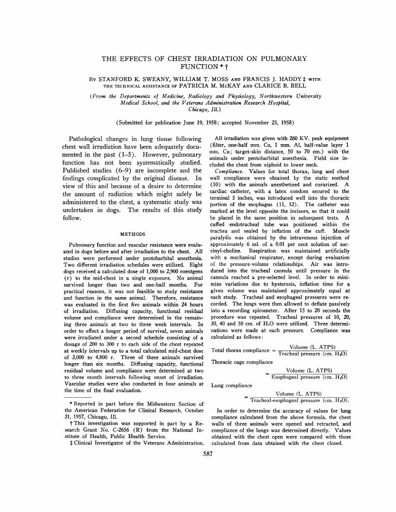
1600
m40
1200
1000
800
600
40C
2ao
*. Controlx-- 7 7 days0-c 133 days
0
Cage
0 10
Total Thorax Lung
AP Cm. H2010 20 30 40 50 10 20 30 40
FIG. 1. AVERAGE RELATION BETWEENPRESSUREAND
VOLUMEIN CAGE, LUNGS AND TOTAL THORAX BEFORE
AND AFTER FRACTIONAL IRRADIATION TO THE CHEST IN
Six ANESTHETIZEDCURARIZEDDOGSThree volume measurements were made at each pres-
sure in each animal.
1 Model A-5, Waters Conley Company, Rochester,
Minn.2 Liston-Becker Model 16, C02 Analyzer, Beckman
Instruments, Inc., Belmont, Cal. Model 15A, CO Ana-lyzer, Beckman Instruments, Inc., Springdale, Conn.
8 Model E-2, Arnold 0. Beckman, Inc., 1020 Mission
Street, South Pasadena, Cal.
tions were made in three animals in order to determinethe reproducibility of the method. The second determina-tion was 78, 80 and 103 per cent of the first in thethree animals, respectively. Single determinations weremade in all subsequent examinations.
Pulmonary vascular studies. Cardiac catheters wereinserted into the pulmonary artery and left atrium via thejugular vein and carotid artery, respectively. These tech-niques have been described elsewhere (17). Pressureswere measured with resistance wire pressure transducers.Cardiac output was calculated by the Fick principle.The per cent of oxygen in expired air was measured withthe oxygen analyzer. Oxygen consumption was ob-tained from the product of the percentage difference inoxygen between inspired and expired air and the cor-rected total one minute expired volume. Pulmonaryvascular resistance was calculated by dividing the dif-ference in pressure between the pulmonary artery andleft atrium by the cardiac output.
RESULTS
Compliance
The values for total thorax compliance increasedprogressively as the pressure was elevated to 30cm. H20 (Figure 1). Further pressure eleva-tion resulted in a reduction in compliance. Thesame relationship was apparent for the lung.These findings are similar to those reported byothers (10, 18).
Total compliance decreased following irradia-tion mainly as a result of a reduction in lungcompliance. Three animals were studied twoand four weeks following single dose irradiation.Changes were irregular at two weeks. By fourweeks, however, reduction in both total and lungcompliance was apparent in two of the threeanimals. Six dogs were studied an average of77 and 133 days following onset of fractional ir-radiation (Figure 1). Control values for totalthorax compliance at 20 cm. H2O averaged 0.034L. per cm. H2O. By 77 days, total compliancehad decreased in three of the six animals and theaverage value was 0.033. By 133 days, it haddecreased in all six and the average value was0.028 (t = 2.92, p = 0.05 to 0.01). The changesin lung compliance were similar. The averagecontrol value at 20 cm. H2O was 0.056. By 77days, lung compliance had decreased in four ofthe six animals and the average value was 0.052.By 133 days, it had decreased in all six and theaverage value was 0.045 (t = 3.36, p = 0.05 to0.01). Cage compliance did not change greatly
588
I.
1-

PULMONARYFUNCTION AND CHEST IRRADIATION
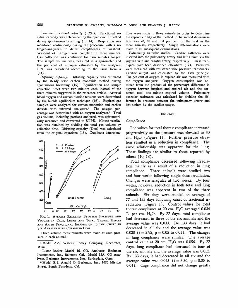
in any animal. These findings indicate that theincreased rigidity of the lungs-chest resultedfrom changes within the lung. Three of theseanimals were restudied 172, 228 and 273 days,respectively, following onset of irradiation. Totaland lung compliance was further decreased in allthree. Thoracic cage compliance was below thepre-irradiation value in only one of the three(Figure 2).
One nonirradiated and two irradiated dogs werestudied immediately before and after opening thechest. The values obtained with the chest opencompared favorably with those with the chestclosed (Figure 2).
Diffusing capacityDiffusing capacity did not change consistently
in three animals studied two weeks and four
TABLE I
Values for several measures of pulmonary function beforeand at various time intervals after the onset of
fractional irradiation to the chest
Weight TimeDog and of Min.Do. sex study FRC vent. pCO2 p02 Dco
mi. COL./min. min./ /
days ml. STPD mm.Hg mm.Hg mm.Hg1 38.5 lbs. C* 650 2.5 45.6 74.5 5.0
Male 80 609 4.2 51.9 71.1 2.3126 490 1.8 20.5 65.2 1.5
2 49 lbs. C 678 4.7 48.7 82.9 3.7Male 104 615 1.9 54.6 68.5 3.4
161 492 2.9 48.1 47.1 1.57 45 lbs. C 754 2.4 45.6 90.0 3.7
Male 97 269 2.0 38.3 84.2 3.7147 332 3.8 28.9 69.9 2.0
8 32 lbs. C 3.4 31.6 101.1 3.5Male 74 3.4 35.9 91.9 3.6
141 465 2.2 44.4 95.0 2.812 48 lbs. C 2.1 43.5 77.7 4.4
Male 87 566 3.9 30.3 86.2 3.1138 546 2.8 33.1 99.5 2.4273 344 3.9 33.5 81.7 3.6
13 46.5 lbs. C 7.2 38.3 84.4 5.7Male 64 545 2.3 41.6 77.7 3.1
104 509 1.9 47.9 80.7 2.0228 388 9.7 50.1 33.9 1.0
14 41 lbs. C 519 4.3 33.0 81.1 3.9Female 70 2.3 51.3 92.4 3.0
140 513 2.9 34.6 89.6 2.0172 418 3.1 53.0 80.6 4.1
Average C 3.8 40.9 84.5 4.382 2.9 43.4 78.9 3.2137 2.6 36.8 78.1 2.0
* C, control.
: 1'1600. 1
:461200 :e. 11000
600 ..'
400 1'
200. lxCage
0 10 1S
AP Cm.HsO Lung10 10 50 40 50550 0to 20 30 40
FIG. 2. RELATION BETWEENPRESSUREAND VOLUMEIN CAGE, LUNGSAND TOTAL THORAXOF DOGNo. 13 BE-FORE AND AT THREE PERIODS AFTER FRACTIONAL IRRADI-ATION TO THE CHEST
On the two hundred twenty-eighth day, values for thelung were also obtained immediately after opening thechest. Each point represents the average of three meas-urements.
weeks following irradiation with a single dose.It decreased progressively in those animals stud-ied more than two months following onset offractional irradiation (Table I, Figure 3). Controlvalues from seven animals averaged 4.3 ml. perminute per mm. Hg. By 82 days following theonset of irradiation, diffusing capacity was be-low the control value in five, above it in one andthe same in one. The average value was 3.2 ml.
_6.0
2.0-
40
103.
1.0
20 40 60 80 100 120 140 160Days
FIG. 3. CHANGEOF PULMONARYDIFFUSING CAPACrrYIN SEVEN DOGSFOLLOWINGFRACTIONAL IRRADIATION TOTHE CHEST
Each point represents one determination.
589

STANFORDK. SWEANY, WILLIAM T. MOSSAND FRANCIS J. HADDY
per minute per mm. Hg (t = 2.56, p = 0.05 to0.01). By 137 days, diffusing capacity was be-low the control value in all seven. The averagevalue was 2.0 ml. per minute per mm.Hg (t = 5.68,p = < 0.01). Three of these animals were stud-ied 172, 228 and 273 days, respectively, followingonset of fractional irradiation. Diffusing capacityfurther decreased from the preceding value inone, but increased toward normal in two.
A low diffusing capacity was frequently as-sociated with a low PO2, and in occasional in-stances this occurred despite a decrease in pCO2and minute ventilation. The changes in minuteventilation, pCO2 and PO2 were not regular, how-ever, and are only included for the sake of com-pleteness (Table I).
Functional residual capacity
There was no change in the functional residualcapacity in three animals studied two and fourweeks following irradiation with a single dose.Functional residual capacity decreased in all ani-mals studied more than 80 days following onsetof fractional irradiation (Table I). Pre-irradia-tion values from four dogs averaged 650 ml. Thisvalue, on a weight basis, is similar to that re-ported by Simmons and Hemingway (14).Eighty-one and 137 days following irradiation,average values were 521 and 478 ml. Functionalresidual capacity further decreased in three ani-mals restudied more than 172 days following onsetof fractional irradiation.
Pulmonary vascular resistance
Table II shows that pulmonary arterial pres-sure, pulmonary venous pressure, cardiac outputand pulmonary vascular resistance did not changesignificantly up to a period of five months follow-ing irradiation. In three animals studied aftersix months, pulmonary arterial pressures and re-sistances appeared to be elevated.
Values for resistance before and at three and24 hours after irradiation averaged 2.1, 3.0 and2.0 mm. Hg per L. per minute, respectively, infive animals. Re-evaluation of one animal 80days after irradiation revealed a value of 3.2 mm.Hg per L. per minute. Two additional dogswere studied 68 and 135 days after single andfractional dose irradiation, respectively. Resis-
TABLE II
Pulmonary vascular variables immediately before and atvarious time intervals following onset
of irradiation to chest
Mean MeanTime pul. pul. Pul.
Dog of Art. Ven. Cardiac art. ven. vas.no. study 02 02 output pres. pres. resist.
mm. Hg/days vol. % vol. % L./min. mm.Hg mm.Hg L./min.
lA C* 11.0 16.0 3.4 28.0 24.0 1.20.1t 13.1 16.3 6.5 19.0 6.0 2.0
2A C 14.5 17.7 5.7 17.5 10.8 1.20.1t 13.3 16.9 4.7 13.7 9.3 0.9
3A C 18.4 20.6 5.1 16.5 3.5 2.60.1t 20.3 24.7 2.7 20.0 3.0 6.30.9 17.3 20.0 6.2 15.5 2.0 2.2
5A C 13.2 15.9 4.3 12.5 3.5 2.10.1t 12.5 15.9 3.5 14.5 2.5 3.50.9 9.0 12.0 4.0 10.0 4.0 1.5
80.0 13.8 18.2 1.8 7.0 1.5 3.2
6A C 17.1 20.4 2.6 12.5 4.0 3.30.1t 16.6 20.3 2.4 8.5 3.5 2.10.9 16.8 20.3 3.0 12.5 5.5 2.4
11 68.0§ 14.7 17.7 5.8 9.0 4.5 0.8
8 135.011 10.5 17.6 2.5 11.0 4.5 1.0
12 273.0¶ 14.1 18.7 2.8 22.0 4.5 6.2
13 228.0¶ 12.1 18.5 0.8 30.8 1.9 38.0
14 172.0¶ 13.4 19.5 1.4 18.2 1.4 11.6
* Control.t Time after 2,000 r single dose.t Time after 3,000 r single dose.
Time after 2,900 r single dose.Time after starting 4,800 r fractional dose.Time after starting 3,000 r fractional dose.
tance values in these two animals were 0.8 and1.0 mm. Hg per L. per minute. Three dogswere studied more than six months following onsetof fractional irradiation. Comparison of thesevalues (Table II) with the control values, fromother animals in this study and with those re-ported elsewhere (19) indicate that two of thesevalues were questionably elevated and one wasdefinitely elevated. The animal with the highestvalue for pulmonary vascular resistance (No. 13)also had the greatest changes in compliance anddiffusing capacity as well as the most markedhistological changes.
Pathology
The lungs of 11 animals were examined micro-scopically. The findings four days after a single
590

PULMONARYFUNCTION AND CHEST IRRADIATION
dose of irradiation revealed little abnormality ex-cept for capillary dilatation. No changes wereseen in the larger vessels. Four to five monthsafter fractional irradiation, the histological find-ings demonstrated focal atelectasis with somefibrosis and hyperemia of the interstitial areas.Chest roentgenograms taken regularly during thestudy period failed to reveal any obvious abnor-malities.
In the three animals studied more than 172days following the onset of irradiation, obvious,interstitial fibrosis with paucity of cellular ele-ments and capillaries was apparent. The smallpulmonary arteries of Dog No. 13 revealed markednarrowing due to endothelial proliferation. Therewas also focal necrosis of the walls, and few areasof acute hemorrhage. The lymph vessels werenoticeably dilated.
DISCUSSION
The present study demonstrates that irradiationof the chest of the dog with the dosage employedis followed by progressive reduction in pulmonarydiffusing capacity, lung compliance and functionalresidual volume.
The study failed to demonstrate significantchanges in any of the physiological variables mea-sured during the first few weeks following ir-radiation. Pathological studies indicate that hy-peremia, edema and thick secretions occur withsome regularity during the early period follow-ing irradiation (2). This discrepancy betweenpathological and physiological findings might re-sult simply because measurements were made atinappropriate times or because the physiologicalchanges were too small to be detected by themethods utilized. On the other hand, the dis-crepancy may also be explained on physiologicalgrounds. For example, active constriction mightproduce changes in various resistance and diffu-sion components that would not be detected bymeasurements of total resistance and total diffu-sion. Pulmonary venular constriction is known tooccur following inhalation of steam (20) or in-jection of endotoxin (21). As a result of in-creased resistance to flow through venules, hy-drostatic pressures increase proximally. Theseelevated pressures passively distend capillariesand other small vessels, thereby reducing their
resistance to flow. Caliber changes which aredirectionally opposite might therefore account forthe absence of a significant change in total resis-tance. Venular constriction also increases capillaryblood volume and promotes edema formation (22).Edema may result from capillary hydrostatic pres-sures in excess of colloid osmotic pressures, in-creased capillary surface area or increased per-meability due to the noxious effect of irradiation oncapillary walls. A normal total diffusing capacitymight be expected since interstitial edema and in-creased blood volume have opposite and cancellingeffects upon total diffusing capacity (23, 24). Adifferential diffusion method such as the one de-cribed by Forster and associates (25, 26) mightdetect such changes. On the other hand, the ab-sence of measurable compliance and functionalresidual capacity changes during this early periodargues against venular constriction of a severedegree. Elevated blood volume and edema wouldbe expected to decrease compliance (27) and func-tional residual capacity.
All measured variables appeared to change sig-nificantly within six months following onset offractional irradiation. Compliance, diffusing ca-pacity and functional residual capacity probablydecreased by 80 days and definitely decreased by140 days. Pulmonary vascular resistance seemedto increase at about six months. These physio-logical changes apparently result from the inter-stitial fibrosis, cellular infiltration and reducedcross-sectional area of the pulmonary vesselswhich were observed at autopsy. Arguments forthis statement follow: It is possible that focalatelectasis may in part account for the reductionsin compliance and diffusion. That it is not theentire explanation is indicated by the fact thatthese functions were reduced in some anmials inwhich significant atelectasis was not observed.Variation in minute ventilation, due to differentdepths of anesthesia, was at first considered as apossible cause of the reduction in diffusing ca-pacity. It soon became apparent, however, thatthere was little correlation between the two param-eters (Table I). It is unlikely that the observedchanges in diffusing capacity are related to a fail-ure to correct for COHb in the blood. Such acorrection would significantly influence absolutevalues but could hardly account for changes withtime in a given animal. The changes in diffusing
591

STANFORDK. SWEANY, WILLIAM T. MOSSAND FRANCIS J. HADDY
capacity probably were not the result of changes inthe level of hemoglobin. The hemoglobin levels(mean + S. D.) before irradiation and at 2, 4 and7 month intervals after irradiation were 14.8 + 0.9,12.9 + 1.3, 12.5 ± 1.2 and 13.8 ± 1.3 Gm. per100 ml., respectively. Hence, the change in pul-monary function was probably the result of thepathological changes observed at autopsy.
The values for diffusing capacity reported inthis paper are considerably lower than those re-ported for four dogs by Otis and Jude (28). How-ever, their methods differed from those in thepresent study. Otis and Jude maintained respira-tion artificially during their measurements andused a different technique to sample expired vol-ume. These differences might tend to elevate dif-fusing capacity. The present study offers someevidence to support this statement. Diffusing ca-pacity was determined in two animals, each ven-tilated with tidal volumes of 250 and 500 ml.Diffusing capacity with the lower volume was 4.3and 4.0 ml. per minute per mm. Hg, respectively.With the higher volume the corresponding valueswere 14.0 and 10.3 ml. per minute per mm. Hg,respectively. These differences might be relatedto changes in diffusing area and vascular volumedue to differences in airway pressures.
Total compliance decreased almost entirely be-cause of decrease in lung compliance. Chest wallcompliance decreased measurably in only one ani-mal and its contribution to the change in totalcompliance was minimal. Increased rigidity ofthe lung may result from cellular infiltrate, fibro-sis and atelectasis. All were observed in the pres-ent study and have also been reported by others.Fibrosis, cellular infiltrate, edema, atelectasis andreduction of pulmonary capillary blood volume allprobably contributed to the progressive reductionin diffusing capacity. The latter most likely ac-counts for the tendency to a progressive decreasein arterial oxygen tension. The return of diffusioncapacity toward normal in two animals may berelated to clearing of cellular infiltrate or edemafluid. The most likely explanation of the progres-sive decrease in functional residual capacity isfibrous contracture of lungs and chest wall. How-ever, since fibrous contracture might be expectedonly after the passage of some time and sinceresidual capacity decreased even during the mod-erately early period following irradiation, other
factors might be involved. One such factor mightbe pulmonary congestion.
An increase in pulmonary vascular resistancecould result from passive reduction in vessel caliberby fibrosis and necrosis. Such an increase in re-sistance to flow would be manifested in an ele-vated pulmonary arterial pressure for a given rateof blood flow. Freid and Goldberg (7) havenoted elevated resistance to flow and right heartfailure in humans following irradiation. Thepresent study indicates that at least six monthsare necessary for these changes to become apparentin the dog. Studies during exercise might havedemonstrated vascular fixation earlier. The nor-mal pulmonary vascular bed responds to exercisewith a decrease in resistance due, at least in part,to passive distention as pressures increase sub-sequent to elevation of flow rate. In the presenceof fixation, pulmonary arterial pressure wouldrise to higher levels than normal because resistancewould remain constant. The fact that compliancedecreased before six months suggests that an in-crease in fibrous tissue was indeed present eventhough it was not demonstrated by pulmonaryvascular resistance studies made with the animalat rest and anesthetized. This fibrous tissue coulddecrease the distensibility of the vascular system.Therefore, studies during exercise might havedemonstrated fixation before six months. Pul-monary hypertension during exercise in irradiatedpatients has been reported by Whitfield, Bond andArnott (29).
SUMMARY
Pulmonary function has been studied in 15 dogsbefore and at intervals up to nine months follow-ing irradiation to the chest. Single doses of 1,000to 2,900 r and fractional doses of 3,000 to 4,800 rwere administered. Pulmonary diffusing capacity,lung compliance and functional residual volumedecreased progressively as time elapsed followingirradiation. Pulmonary vascular resistance re-mained normal for a period of five months fol-lowing irradiation. These physiological changeshave been discussed in relation to changes inhistology.
ACKNOWLEDGMENTS
The authors are indebted to Dr. Craig W. Borden forhis helpful suggestions during preparation of the manu-
592

PULMONARYFUNCTION AND CHEST IRRADIATION
script. Thanks are also due Dr. John Robinson for hisreview and evaluation of the histological sections andJoan Sweany for editorial suggestions.
REFERENCES
1. Groover, T. A., Christie, A. C., and Merritt, E. A.Intrathoracic changes following roentgen treat-ment of breast carcinoma. Amer. J. Roentgenol.1923, 10, 471.
2. Engelstad, R. B. Pulmonary lesions after roentgenand radium irradiation. Amer. J. Roentgenol.1940, 43, 676.
3. Bergmann, M., and Graham, E. A. Pneumonectomyfor severe irradiation damage of the lung. J.thorac. Surg. 1951, 22, 549.
4. Huguenin, R., Lemoine, J. M., and Fauvet, J. Effetsde la radiotherapie sur le poumon normal. J.franc. Med. Chir. Thor. 1949, 3, 54.
5. Warren, S., and Spencer, J. Radiation reaction inthe lung. Amer. J. Roentgenol. 1940, 43, 682.
6. Baldwin, E. de F., Cournand, A., and Richards, D.W., Jr. Pulmonary insufficiency. Study of 39cases of pulmonary fibrosis. Medicine 1949, 28, 1.
7. Freid, J. R., and Goldberg, H. Post-irradiationchanges in the lungs and thorax. Amer. J.Roentgenol. 1940, 43, 877.
8. Leach, J. E. Abnormal pulmonary physiology as a
result of chronic irradiation pleuropulmonitis; pre-
liminary report. Amer. J. Roentgenol. 1943, 50,772.
9. Stone, D. J., Schwartz, M. J., and Green, R. A. Fatalpulmonary insufficiency due to radiation effectupon the lung. Amer. J. Med. 1956, 21, 211.
10. Nims, R. G., Conner, E. H., and Comroe, J. H., Jr.The compliance of the human thorax in anesthe-tized patients. J. clin. Invest. 1955, 34, 744.
11. Mead, J., and Whittenberger, J. L. Physical proper-
ties of human lungs measured during spontaneousrespiration. J. appl. Physiol. 1953, 5, 779.
12. Mead, J., McIlroy, M. B., Selverstone, N. J., andKriete, B. C. Measurement of intraesophagealpressure. J. appl. Physiol. 1955, 7, 491.
13. Darling, R. C., Cournand, A., and Richards, D. W.,Jr. Studies on the intrapulmonary mixture ofgases. III. An open circuit method for measuringresidual air. J. clin. Invest. 1940, 19, 609.
14. Simmons, D. H., and Hemingway, A. Functionalresidual capacity and respiratory nitrogen excretionof dogs. J. appl. Physiol. 1955, 8, 95.
15. Filley, G. F., MacIntosh, D. J., and Wright, G. W.Carbon monoxide uptake and pulmonary diffusingcapacity in normal subj ects at rest and duringexercise. J. clin. Invest. 1954, 33, 530.
16. Brinkman, G. L., Johns, C. J., Donoso, H., andRiley, R. L. A modification of the method ofRiley, Proemmel and Franke for determination of
oxygen and carbon dioxide tensions in blood. J.appl. Physiol. 1954, 7, 340.
17. Haddy, F. J., Campbell, G. S., Adams, WV. L., andVisscher, M. B. A study of pulmonary venous andarterial pressures and other variables in the anes-thetized dog by flexible catheter techniques. Amer.J. Physiol. 1949, 158, 89.
18. Howell, J. B. L., and Peckett, B. W. Studies on theelastic properties of the thorax of supine anaes-thetized paralysed human subjects. J. Physiol.1957, 136, 1.
19. Haddy, F. J., and Campbell, G. S. Pulmonary vas-cular resistance in anesthetised dogs. Amer. J.Physiol. 1953, 172, 747.
20. Aviado, D. M., Jr., and Schmidt, C. F. Respiratoryburns with special reference to pulmonary edemaand congestion. Circulation 1952, 6, 666.
21. Kuida, H., Hinshaw, L. B., Gilbert, R. P., and Vis-scher, M. B. Effect of gram-negative endotoxinon pulmonary circulation. Amer. J. Physiol. 1958,192, 335.
22. Visscher, M. B., Haddy, F. J., and Stephens, G.The physiology and pharmacology of lung edema.Pharmacol. Rev. 1956, 8, 389.
23. Lewis, B. M., Lin, T., Komisaruk, R., and Noe, F. E.Diffusing capacity of pulmonary membrane andpulmonary capillary blood volume in normal sub-jects; the effects of exercise and body position;preliminary observations in cardiac patients. Clin.Res. Proc. 1957, 5, 309.
24. Rankin, J., McNeill, R. S., and Forster, R. E. Theinfluence of the pulmonary capillary blood volume,gas tensions, and hematocrit on diffusing capacityfor carbon monoxide. Clin. Res. Proc. 1957, 5,310.
25. Forster, R. E., Roughton, F. J. WV., Cander, L.,Briscoe, W. A., and Kreuzer, F. Apparent pul-monary diffusing capacity for CO at varying al-veolar 02 tensions. J. appl. Physiol. 1957, 11, 277.
26. Roughton, F. J. W., and Forster, R. E. Relativeimportance of diffusion and chemical reactionrates in determining rate of exchange of gases inthe human lung, with special reference to truediffusing capacity of pulmonary membrane andvolume of blood in the lung capillaries. J. appl.Physiol. 1957, 11, 290.
27. Bondurant, S., Hickam, J. B., and Isley, J. K. Pul-monary and circulatory effects of acute pulmonaryvascular engorgement in normal subj ects. J.clin. Invest. 1957, 36, 59.
28. Otis, A. B., and Jude, J. Effect of body temperatureon pulmonary gas exchange. Amer. J. Physiol.1957, 188, 355.
29. Wrhitfield, A. G. W., Bond, W. H., and Arnott, W. M.Pulmonary irradiation effects and their treatmentwith cortisone and ACTH. J. Fac. Radiol. (Lond.)1954, 6, 12.
593