low survival rates of oral and oropharyngeal...
TRANSCRIPT

Research ArticleLow Survival Rates of Oral and OropharyngealSquamous Cell Carcinoma
Anna Carolina Omena Vasconcellos Le Campion,1 Camila Maria Beder Ribeiro,1,2
Ronir Raggio Luiz,3 Francisco Feliciano da Silva Júnior,1
Herbert Charles Silva Barros,4 Karine de Cássia Batista dos Santos,1
Stefania Jeronimo Ferreira,1 Lucio Souza Gonçalves,5 and Sonia Maria Soares Ferreira1,2
1CESMAC University Center, Maceio, AL, Brazil2Federal University of Alagoas, Maceio, AL, Brazil3Federal University of Rio de Janeiro, Rio de Janeiro, RJ, Brazil4State Secretary of Health (SESAU), Alagoas, Brazil5Post-Graduate Program in Dentistry, Estacio de Sa University, Rio de Janeiro, RJ, Brazil
Correspondence should be addressed to Sonia Maria Soares Ferreira; [email protected]
Received 2 February 2017; Revised 3 April 2017; Accepted 27 April 2017; Published 30 May 2017
Academic Editor: Gilberto Sammartino
Copyright © 2017 Anna Carolina Omena Vasconcellos Le Campion et al. This is an open access article distributed under theCreative Commons Attribution License, which permits unrestricted use, distribution, and reproduction in any medium, providedthe original work is properly cited.
Aim. To assess the epidemiological and clinical factors that influence the prognosis of oral and oropharyngeal squamous cellcarcinoma (SCC).Methods. One hundred and twenty-one cases of oral and oropharyngeal SCC were selected. The survival curvesfor each variable were estimated using the Kaplan-Meier method. The Cox regression model was applied to assess the effect of thevariables on survival. Results. Cancers at an advanced stage were observed in 103 patients (85.1%). Cancers on the tongue were morefrequent (23.1%). The survival analysis was 59.9% in one year, 40.7% in two years, and 27.8% in 5 years. There was a significant lowsurvival rate linked to alcohol intake (𝑝 = 0.038), advanced cancer staging (𝑝 = 0.003), and procedures without surgery (𝑝 < 0.001).When these variables were included in the Cox regression model only surgery procedures (𝑝 = 0.005) demonstrated a significanteffect on survival. Conclusion. The findings suggest that patients who underwent surgery had a greater survival rate compared withthose that did not. The low survival rates and the high percentage of patients diagnosed at advanced stages demonstrate that oraland oropharyngeal cancer patients should receive more attention.
1. Introduction
The incidence of oral cancer in Brazil has been estimated ataround 15,290 new cases per year, putting oral cancer intothe seventh position of all malignant neoplasia [1]. Amongseveral histological types of oral neoplasia, the squamous cellcarcinoma (SCC) is the more frequent, representing 90% ofall cases of oral cancer [2].
Cancer is primarily a disease caused by genetic changesthat progress as sequential series of somatic mutations inspecific genes such as protooncogenes and tumor suppressorgenes, resulting in uncontrolled cancerous cell proliferation[3, 4]. These events can be triggered by extrinsic and/or
intrinsic factors, such as lifestyle, environmental, immuno-suppression, and individual susceptibility [5, 6]. Among theextrinsic factors, tobacco use and alcohol intake represent thehighest risk to the emergence of this malignant disease [7, 8].
Studies have shown that low social and economic statusand deprivation are significantly associated with an increasedrisk of oral cancer [9, 10].
The primary location of oral cancer is an importantprognostic factor because the affected anatomic area candetermine the accessibility and extension of surgery [11]. Inaddition, it can define the necessity of additional therapeuticprocedures, such as the prophylactic cervical ganglionectomyas well as radiotherapy and adjuvant chemotherapy [12].
HindawiInternational Journal of DentistryVolume 2017, Article ID 5815493, 7 pageshttps://doi.org/10.1155/2017/5815493

2 International Journal of Dentistry
Almost half of the cases of oral cancer worldwide havebeen diagnosed in stages III and IV [13, 14]. Early diagnosis oforal and oropharyngeal SCC is essential for a good prognosisand therefore dentists play a fundamental role in the earlydetection and prevention of this type of oral cancer [15].
Oral and oropharyngeal cancer has a survival rate ≤50%after 5 years. However, this time can be increased whenthe cancers are diagnosed at an early stage [16, 17]. Cancermortality is influenced by variations in quantity and qualityof the available health services, the delay in diagnosis,or diagnoses at advanced stages, beyond the sequelae oftreatment [18]. Oral cancer is a public health problem andearly diagnosis as well as assurance of appropriated andfast care is required in order to achieve a better quality oflife and lower morbidity/mortality for these patients. Thus,the aim of the current study was to assess the influenceof epidemiological and clinical factors on the prognostic oforal and oropharyngeal squamous cell carcinoma (SCC) ina group of patients from one referral center in the state ofAlagoas, Brazil.
2. Material and Methods
One hundred and twenty-one patients diagnosed in a refer-ence service in stomatology as having oral and oropharyngealsquamous cell carcinoma (SCC) and treated in an OncologyHigh Complexity Center (CACON) (Alagoas, Brazil) wereenrolled in the current study. As the inclusion criteria, allpatients must be diagnosed and treated only in these twocenters. All subjects were informed about the aims, risks, andbenefits of the study and signed a consent form. Patients olderthan 18 years were examined betweenMarch 2005 andMarch2013. The medical records whose lack of information madethe results of the study unfeasible were excluded.
The study protocol was conducted in full accordance withthe World Medical Association Declaration of Helsinki andwas approved by the Review Committee for Human Subjectsof the University Center of Research CESMAC (number367.585/2013).
2.1. Procedures and Instruments. Epidemiological data (age,gender, skin color, residence, education, and occupation), riskhabits (alcohol intake and tobacco use), clinical characteris-tics, treatment implemented for the oral cancer, date of thediagnosis, date of the last appointment, and date of deathwere collected from medical records and during the patient’sappointments with the examiner.
The date of death was confirmed using the MortalityInformation System applying the following filters: patient’sname, date of birth, and mother’s name. Date of death aswell as the underlying cause was searched according to theInternational Statistical Classification ofDiseases andRelatedHealth Problems-10th Revision (ICD-10).
The oral cancers were located on the hard palate, gingiva,anterior two-thirds of tongue, lips, jugal mucosa, floor ofmouth, and alveolar and retromolar mucosa, while theoropharyngeal cancers were on the soft palate, tongue base,tonsillar region, uvula, and posterior pharynx [2].The clinical
stage evaluation was carried out according to the Union forInternational Cancer Control [19].
2.2. Data Analysis. All analyses were performed using thesoftware SPSS�20.0 for Windows (Statistical Package forSocial Sciences, IBM,USA).Thedescriptive analysis includedthe absolute and relative frequency for categorical variables.Comparison between groups (death and survival) was carriedout using the Chi-square test or Fisher’s exact test.
For the inferential analysis with the outcome death, thefollowing variables were changed: the anatomical locationsof cancers were clustered as oral and oropharyngeal cancers;TNM system was categorized as early stage (I and II) andadvanced stage (III and IV); treatment was categorizedas no surgery [radiotherapy (RT), chemotherapy (CT), orcombination RT/CT] and surgery (surgery alone, surgery +RT, and surgery + RT + CT).
In order to assess the survival, all the deaths duringthe study were considered outcome. The variables for thesurvival analysis were age, gender, skin color, occupation,residence, education, tobacco use, alcohol intake, cancer loca-tion, cancer staging, and surgery procedures. The survivalcurve was estimated for each variable using the Kaplan-Meiermethod.The comparison between curves was obtained by thelong-rank test. The Cox regression model was used to assessthe effect of the variables on survival (multivariate analysisto calculate hazard ratios), which included variables withthe following characteristics according to the Kaplan-Meieranalysis: significant difference (𝑝 < 0.05) and no crossingbetween curves. The level of significance established for allanalyses was 5%.
3. Results
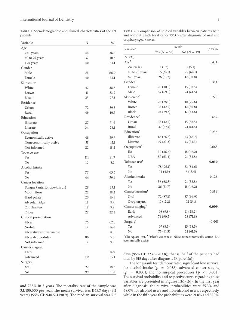
3.1. Characteristics of the Study Sample. The sociodemo-graphic and clinical characteristics of the 121 patients whoparticipated in the current study are presented in Table 1.The majority of the patients were male (𝑛 = 81; 66.9%),>40 years old (𝑛 = 77; 63.7%), and with white skin color(𝑛 = 47; 38.8%). The frequencies of tobacco use and alcoholintake were 111 (91.7%) and 77 (63.6%), respectively. Cancersat an advanced stage were observed in 103 patients (85.1%).Cancers on the tongue were the most frequent (23.1%),followed by mouth floor (18.2%) and hard and soft palate(16.5%). Of the patients who underwent surgery (𝑛 = 22;18.2%), 59% were diagnosed in advanced stages (III and IV).
Of the 121 patients, 82 (67.8%) died due to SCC and onlyone (1.2%) was <40 years old. The majority (𝑛 = 57; 69.5%)were male, illiterate (𝑛 = 63; 76.8%), noneconomically active(𝑛 = 52; 63.4%), smokers (𝑛 = 78; 95.1%), alcohol users (𝑛 =56; 68.3%), with advanced cancer staging (𝑛 = 74; 90.2%),and not undergoing any surgical procedures (𝑛 = 75; 91.5%).When the sociodemographic and clinical characteristicswere compared between dead and alive patients, significantdifference was found only in the cancer staging (𝑝 = 0.009)and surgery procedures (𝑝 < 0.001) (Table 2).
3.2. Survival Analysis. The survival analysis using Kaplan-Meier method was 59.9% in one year, 40.7% in two years,
International Journal of Dentistry 3
Table 1: Sociodemographic and clinical characteristics of the 121patients.
Variable 𝑁 %Age<40 years 44 36.340 to 70 years 37 30.6>70 years 40 33.1
GenderMale 81 66.9Female 40 33.1
Skin colorWhite 47 38.8Brown 41 33.9Black 33 27.3
ResidenceUrban 72 59.5Rural 49 40.5
EducationIlliterate 87 71.9Literate 34 28.1
OccupationEconomically active 48 39.7Noneconomically active 51 42.1Not informed 22 18.2
Tobacco useYes 111 91.7No 10 8.3
Alcohol intakeYes 77 63.6No 44 36.4
Cancer locationTongue (anterior two-thirds) 28 23.1Mouth floor 22 18.2Hard palate 20 16.5Alveolar ridge 12 9.9Oropharynx 12 9.9Other 27 22.4
Clinical presentationUlcer 76 62.8Nodule 17 14.0Ulcerative and verrucose 10 8.3Ulcerated nodules 06 5.0Not informed 12 9.9
Cancer stagingEarly 18 14.9Advanced 103 85.1
SurgeryYes 22 18.2No 99 81.8
and 27.8% in 5 years. The mortality rate of the sample was3.1/100,000 per year. The mean survival was 1165.7 days (3.2years) (95% CI: 940.5–1390.9). The median survival was 515
Table 2: Comparison of studied variables between patients withand without death (oral cancer/SCC) after diagnosis of oral andoropharyngeal cancer.
Variable Death𝑝 value
Yes (𝑁 = 82) No (𝑁 = 39)𝑁 (%)Age¶ 0.434<40 years 1 (1.2) 2 (5.1)40 to 70 years 55 (67.1) 25 (64.1)>70 years 26 (31.7) 12 (30.8)
Gender∗ 0.384Female 25 (30.5) 15 (38.5)Male 57 (69.5) 24 (61.5)
Skin color∗ 0.270White 23 (28.0) 10 (25.6)Brown 35 (42.7) 12 (30.8)Black 24 (29.3) 17 (43.6)
Residence∗ 0.659Urban 35 (42.7) 15 (38.5)Rural 47 (57.3) 24 (61.5)
Education∗ 0.236Illiterate 63 (76.8) 23 (66.7)Literate 19 (23.2) 13 (33.3)
Occupation∗ 0,665EA 30 (36.6) 18 (46.2)NEA 52 (63.4) 21 (53.8)
Tobacco use¶ 0.050Yes 78 (95.1) 33 (84.6)No 04 (4.9) 6 (15.4)
Alcohol intake 0.123Yes 56 (68.3) 21 (53.8)No 26 (31.7) 18 (46.2)
Cancer location¶ 0.334Oral 72 (87.8) 37 (94.9)Oropharynx 10 (12.2) 02 (5.1)
Cancer staging¶ 0.009Early 08 (9.8) 11 (28.2)Advanced 74 (90.2) 28 (71.8)
Surgery¶ <0.001Yes 07 (8.5) 15 (38.5)No 75 (91.5) 24 (61.5)
∗Chi-square test. ¶Fisher’s exact test. NEA: noneconomically active; EA:economically active.
days (95% CI: 323.3–703.8); that is, half of the patients haddied by 515 days after diagnosis (Figure 1(a)).
The long-rank test demonstrated significant low survivalfor alcohol intake (𝑝 = 0.038), advanced cancer staging(𝑝 = 0.003), and no surgical procedures (𝑝 < 0.001).The survival probability and respective curve regarding thesevariables are presented in Figures 1(b)–1(d). In the first yearafter diagnosis, the survival probabilities were 55.3% and68.0% for alcohol users and non-alcohol users, respectively,while in the fifth year the probabilities were 21.8% and 37.9%.

4 International Journal of Dentistry
Survival function
1000 2000 3000 40000Time (days)
0.0
0.2
0.4
0.6
0.8
1.0
Surv
ival
pro
babi
lity
Kaplan-Meier
(a)
Survival functions
Alcohol intake: yes
Alcohol intake: no
0.0
0.2
0.4
0.6
0.8
1.0
Surv
ival
pro
babi
lity p = 0.038
1000 2000 3000 40000Time (days)
Kaplan-Meier
(b)
Survival functions
Oral cancer: early stage
Oral cancer: advanced stage
0.0
0.2
0.4
0.6
0.8
1.0
Surv
ival
pro
babi
lity
1000 2000 3000 40000Time (days)
p = 0.003
Kaplan-Meier
(c)
Survival functions
Surgery: no
Surgery: yes
0.0
0.2
0.4
0.6
0.8
1.0Su
rviv
al p
roba
bilit
y
1000 2000 3000 40000Time (days)
p < 0.001
Kaplan-Meier
(d)
Figure 1: (a) Survival curve of patients with oral and oropharyngeal squamous cell carcinoma estimated by Kaplan-Meier method (Maceio,AL, Brazil, 2005–2013). (b) Survival curve for alcohol intake of patients with oral and oropharyngeal squamous cell carcinoma estimatedby Kaplan-Meier method (Maceio, AL, Brazil, 2005–2013). (c) Survival curve for cancer staging of patients with oral and oropharyngealsquamous cell carcinoma estimated by Kaplan-Meier method (Maceio, AL, Brazil, 2005–2013). (d) Survival curve for surgical procedure ofpatients with oral and oropharyngeal squamous cell carcinoma estimated by Kaplan-Meier method (Maceio, AL, Brazil, 2005–2013).
The median survival was 484 days (95% CI: 240.1–727.9) foralcohol users and 1095 (95% CI: 0–2646.8) for non-alcoholusers (Figure 1(b)). In the fifth year, the survival probabilitywas 51.6% and 25.3% for patients with early stage (I and II)and advanced stage (III and II) of cancer, respectively. Themedian survival was 1,900 days (95% CI: 1647.1–2152.9) forearly stage of cancer and 415 days (95% CI: 251.9–578.1) forthe advanced stage patients (Figure 1(c)). The death rate was1.1/100,000 subjects per year for early stage and 3.8/100,000subjects per year for advanced stage patients.
The survival probability of patients who underwent surgi-cal procedures (associated with chemotherapy, radiotherapy,or both) was 84.2% (second year) and 59.0% (fifth year).
On the other hand, the patients that did not receive anysurgical procedures presented lower survival probabilities:31.5% (second year) and 21.4% (fifth year) (Figure 1(d)).The median survival was 2,280 days for the patients whounderwent surgery, while for patients that did not receive anysurgical procedures the median survival was 395 days (95%CI: 326.3–703.8).
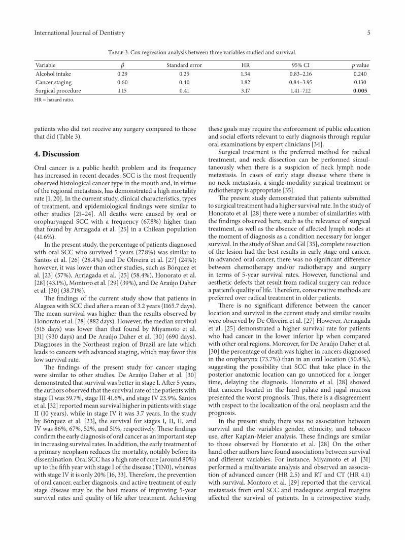
3.3. Multivariate Analysis. The variables alcohol intake, can-cer staging, and surgery were included in the Cox regressionmodel. Of the three variables, only surgical procedures (𝑝 =0.005) demonstrated significant effect on the survival. Inaddition, the outcome of death was 3 times faster for the
International Journal of Dentistry 5
Table 3: Cox regression analysis between three variables studied and survival.
Variable 𝛽 Standard error HR 95% CI 𝑝 valueAlcohol intake 0.29 0.25 1.34 0.83–2.16 0.240Cancer staging 0.60 0.40 1.82 0.84–3.95 0.130Surgical procedure 1.15 0.41 3.17 1.41–7.12 0.005HR = hazard ratio.
patients who did not receive any surgery compared to thosethat did (Table 3).
4. Discussion
Oral cancer is a public health problem and its frequencyhas increased in recent decades. SCC is the most frequentlyobserved histological cancer type in the mouth and, in virtueof the regional metastasis, has demonstrated a high mortalityrate [1, 20]. In the current study, clinical characteristics, typesof treatment, and epidemiological findings were similar toother studies [21–24]. All deaths were caused by oral ororopharyngeal SCC with a frequency (67.8%) higher thanthat found by Arriagada et al. [25] in a Chilean population(41.6%).
In the present study, the percentage of patients diagnosedwith oral SCC who survived 5 years (27.8%) was similar toSantos et al. [26] (28.4%) and De Oliveira et al. [27] (24%);however, it was lower than other studies, such as Borquez etal. [23] (57%), Arriagada et al. [25] (58.4%), Honorato et al.[28] (43.1%), Montoro et al. [29] (39%), andDe Araujo Daheret al. [30] (38.71%).
The findings of the current study show that patients inAlagoas with SCC died after a mean of 3.2 years (1165.7 days).The mean survival was higher than the results observed byHonorato et al. [28] (882 days). However, themedian survival(515 days) was lower than that found by Miyamoto et al.[31] (930 days) and De Araujo Daher et al. [30] (690 days).Diagnoses in the Northeast region of Brazil are late whichleads to cancers with advanced staging, which may favor thislow survival rate.
The findings of the present study for cancer stagingwere similar to other studies. De Araujo Daher et al. [30]demonstrated that survival was better in stage I. After 5 years,the authors observed that the survival rate of the patients withstage II was 59.7%, stage III 41.6%, and stage IV 23.9%. Santoset al. [32] reportedmean survival higher in patients with stageII (10 years), while in stage IV it was 3.7 years. In the studyby Borquez et al. [23], the survival for stages I, II, II, andIV was 86%, 67%, 52%, and 51%, respectively. These findingsconfirm the early diagnosis of oral cancer as an important stepin increasing survival rates. In addition, the early treatment ofa primary neoplasm reduces the mortality, notably before itsdissemination.Oral SCChas a high rate of cure (around 80%)up to the fifth year with stage I of the disease (T1N0), whereaswith stage IV it is only 20% [16, 33].Therefore, the preventionof oral cancer, earlier diagnosis, and active treatment of earlystage disease may be the best means of improving 5-yearsurvival rates and quality of life after treatment. Achieving
these goals may require the enforcement of public educationand social efforts relevant to early diagnosis through regularoral examinations by expert clinicians [34].
Surgical treatment is the preferred method for radicaltreatment, and neck dissection can be performed simul-taneously when there is a suspicion of neck lymph nodemetastasis. In cases of early stage disease where there isno neck metastasis, a single-modality surgical treatment orradiotherapy is appropriate [35].
The present study demonstrated that patients submittedto surgical treatment had a higher survival rate. In the study ofHonorato et al. [28] there were a number of similarities withthe findings observed here, such as the relevance of surgicaltreatment, as well as the absence of affected lymph nodes atthe moment of diagnosis as a condition necessary for longersurvival. In the study of Shan andGil [35], complete resectionof the lesion had the best results in early stage oral cancer.In advanced oral cancer, there was no significant differencebetween chemotherapy and/or radiotherapy and surgeryin terms of 5-year survival rates. However, functional andaesthetic defects that result from radical surgery can reducea patient’s quality of life. Therefore, conservative methods arepreferred over radical treatment in older patients.
There is no significant difference between the cancerlocation and survival in the current study and similar resultswere observed by De Oliveira et al. [27] However, Arriagadaet al. [25] demonstrated a higher survival rate for patientswho had cancer in the lower inferior lip when comparedwith other oral regions. Moreover, for De Araujo Daher et al.[30] the percentage of death was higher in cancers diagnosedin the oropharynx (73.7%) than in an oral location (50.8%),suggesting the possibility that SCC that take place in theposterior anatomic location can go unnoticed for a longertime, delaying the diagnosis. Honorato et al. [28] showedthat cancers located in the hard palate and jugal mucosapresented the worst prognosis. Thus, there is a disagreementwith respect to the localization of the oral neoplasm and theprognosis.
In the present study, there was no association betweensurvival and the variables gender, ethnicity, and tobaccouse, after Kaplan-Meier analysis. These findings are similarto those observed by Honorato et al. [28] On the otherhand other authors have found associations between survivaland different variables. For instance, Miyamoto et al. [31]performed a multivariate analysis and observed an associa-tion of advanced cancer (HR 2.5) and RT and CT (HR 4.1)with survival. Montoro et al. [29] reported that the cervicalmetastasis from oral SCC and inadequate surgical marginsaffected the survival of patients. In a retrospective study,
6 International Journal of Dentistry
Matos et al. [36] assessed 57 patients diagnosedwith oral SCC(except in lips) and found that tumoral thickness >10mmrepresents an independent risk factor for early progression oforal SCC after surgical treatment.
The epidemiological findings with respect to the deathswere similar to those found by Santos et al. [32].These authorsevaluated the profile of the patients who died between 2000and 2009 in Aracaju (Capital of Sergipe state, Brazil) andobserved that majority of the deaths (seventy-eight) weremales, 50–60 years old, with brown skin color, with loweducation, and from neighborhoods with low quality of life.In the current study, the comparison between survival andnonsurvival groups demonstrated significant difference forcancer staging (𝑝 = 0.009) and surgical procedures (𝑝 <0.001). The findings showed a high likelihood of death inpatients with advanced cancer staging and without surgicaltreatment. In the multivariate model, the risk factor wasconfirmed only for the absence of surgical procedures (𝑝 =0.005). However, Honorato et al. [28] observed associationof death with other variables, such as ethnicity (p = 0.033),cancer located in the lower gingiva (𝑝 = 0.048), andtreatment (𝑝 < 0.0001). On the present study surgicaltreatment increased survival even in the advanced stages.Better 5-year survival rate with patients who underwentsurgery was reported also in other studies [28, 37] evidencinggreater survival in those patients undergoing this procedure.
5. Conclusion
The low survival rates and the large percentage of patientswith an advanced cancer staging diagnosis, as well as thefindings that showed an improved survival of those patientswho underwent surgery, reinforce the need for greater atten-tion to be paid to oral and oropharyngeal cancer, especiallyamong high-risk populations (elderly, smokers, and alcoholusers). In order to increase the 5-year survival rate of oral andoropharyngeal carcinoma, it may be necessary to improvepublic education and social efforts relevant to early diagnosis.
Conflicts of Interest
The authors declare that they have no conflicts of interest.
Acknowledgments
This study was supported by Department of Science andTechnology, Ministry of Health (DECIT/SCTIE/MS), Na-tional Council for Scientific and Technological Develop-ment (CNPq), Foundation for Research Financial Supportin the State of Alagoas (FAPEAL), State Secretary of Health(SESAU) (Alagoas, Brazil), and Municipal Secretary ofHealth in Maceio, AL (Alagoas, Brazil). The manuscriptoriginated from the Research Program for SUS (PPSUS) no.60030 000713/2013.
References
[1] Instituto Nacional do Cancer (INCA), Incidencia de Cancer noBrasil, Estimativa 2014, INCA, Brasılia, 2014.
[2] B. Neville, D. D. Damm, andC.M. Allen,Oral andMaxillofacialPathology, Elsevier, St. Louis, 3rd edition, 2009.
[3] Z. Wang, C. Wang, Z. Zhao et al., “Association between−251A>T polymorphism in the interleukin-8 gene and oralcancer risk: a meta-analysis,” Gene, vol. 522, no. 2, pp. 168–176,2013.
[4] W. Li, J. Chen, and C. Liu, “Glutathione S-transferase P1Ile105Val polymorphism and oral cancer risk: a meta-analysis,”International Journal of Medical Sciences, vol. 10, no. 4, pp. 392–398, 2013.
[5] S. Petti, “Lifestyle risk factors for oral cancer,” Oral Oncology,vol. 45, no. 4-5, pp. 340–350, 2009.
[6] S. Warnakulasuriya, “Causes of oral cancer—an appraisal ofcontroversies,” British Dental Journal, vol. 207, no. 10, pp. 471–475, 2009.
[7] I. Hindle, M. C. Downer, D. R. Moles, and P. M. Speight, “Isalcohol responsible for more intra-oral cancer?”Oral Oncology,vol. 36, no. 4, pp. 328–333, 2000.
[8] J. L. Ferreira Antunes, T. N. Toporcov, M. G. H. Biazevic, A. F.Boing, C. Scully, and S. Petti, “Joint and independent effects ofalcohol drinking and tobacco smoking on oral cancer: a largecase-control study,” PLoS ONE, vol. 8, no. 7, Article ID e68132,2013.
[9] D. I. Conway, M. Petticrew, H. Marlborough, J. Berthiller, M.Hashibe, and L. M. D. Macpherson, “Socioeconomic inequali-ties and oral cancer risk: a systematic review and meta-analysisof case-control studies,” International Journal of Cancer, vol. 122,no. 12, pp. 2811–2819, 2008.
[10] M. A. F. Ferreira, M. N. Gomes, F. A. S. Michels, A. A. Dantas,and M. D. R. D. D. O. Latorre, “Social inequality in morbidityandmortality from oral and oropharyngeal cancer in the city ofSao Paulo, Brazil: 1997–2008,” Cadernos de Saude Publica, vol.28, no. 9, pp. 1663–1673, 2012.
[11] F. C. S. A. Almeida, C. Cazal, F. D. Nunes, M. E. Araujo, R. B.Dias, andD. P. Silva, “Prognostic factors in oral cancer,”Rev BrasCienc Saude, vol. 15, no. 4, pp. 471–478, 2011.
[12] K.-H. Fan, C.-Y. Lin, C.-J. Kang et al., “Combined-modalitytreatment for advanced oral tongue squamous cell carcinoma,”International Journal of Radiation Oncology Biology Physics, vol.67, no. 2, pp. 453–461, 2007.
[13] S. Warnakulasuriya, “Global epidemiology of oral and oropha-ryngeal cancer,” Oral Oncology, vol. 45, no. 4-5, pp. 309–316,2009.
[14] P. Guneri and J. B. Epstein, “Late stage diagnosis of oral cancer:components and possible solutions,”Oral Oncology, vol. 50, no.12, pp. 1131–1136, 2014.
[15] N. Akbulut, B. Oztas, S. Kursun, and S. Evirgen, “Delayeddiagnosis of oral squamous cell carcinoma: a case series,”Journal of Medical Case Reports, vol. 5, article 291, 2011.
[16] S. N. Rogers, J. S. Brown, J. A.Woolgar et al., “Survival followingprimary surgery for oral cancer,” Oral Oncology, vol. 45, no. 3,pp. 201–211, 2009, http://dx.doi.
[17] L. P. Kowalski, A. L. Carvalho, A. V. Martins Priante, and J.Magrin, “Predictive factors for distant metastasis from oral andoropharyngeal squamous cell carcinoma,” Oral Oncology, vol.41, no. 5, pp. 534–541, 2005.
[18] M. G. H. Biazevic, R. A. Castellanos, J. L. F. Antunes, and E.Michel-Crosato, “Trends in oral cancer mortality in the city ofSao Paulo, Brazil, 1980–2002,”Cad Saude Publica, vol. 22, no. 10,pp. 2105–2114, 2006.
International Journal of Dentistry 7
[19] L. H. Sobin, M. K. Gospodarowicz, and C.Wittekind,Union forInternational Cancer Control—UICC, Wiley-Liss, 7th edition.
[20] S. Warnakulasuriya, “Living with oral cancer: epidemiologywith particular reference to prevalence and life-style changesthat influence survival,” Oral Oncology, vol. 46, no. 6, pp. 407–410, 2010.
[21] L. C. O. dos Santos, O. de Medeiros Batista, and M. C. T.Cangussu, “Characterization of oral cancer diagnostic delay inthe state of Alagoas,” Brazilian Journal of Otorhinolaryngology,vol. 76, no. 4, pp. 416–422, 2010.
[22] A. L. R. O. Aui, H. M. Tanimoto, C. D. S. Queiroz et al.,“Oral and oropharynges neoplasm—a transversal study in PioXII Foundation—Cancer Hospital of Barretos,” Rev OdontolUNESP, vol. 41, no. 4, pp. 273–280, 2012.
[23] P. Borquez, F. Capdeville, A. Madrid, M. Veloso, and M.Cßrcamo, “Analysis of survival of 137 patients with oral cancer,”Rev Chil Cir, vol. 63, no. 4, pp. 351–355, 2011.
[24] L. G. Ferraz, E. Favero, A. S. Franzi, A. Rapoport, and A.O. Curioni, “Relationship between the clinical (TNM) andhistopathological (pTNM) staging with survival in advancedsquamous cell carcinoma of the mouth and oropharynx,”Revista Brasileira de Cirurgia de Cabeca e Pescoco, vol. 39, pp.48–56, 2010.
[25] O. C. Arriagada, B. R. Venegas, M. L. Cantın, D.M. Zavando, C.D. Manterola, and I. G. Suazo, “Oral squamous cell carcinoma:retrospective analysis of 36 cases.,” Revista chilena de cirugıa,vol. 62, pp. 441–448, 2010.
[26] M. A. Santos, C. C. Danesi, and B. H. Pinheiro, “Relationshipbetween survival of patients with squamous cell carcinoma ofthe oral cavity and pathological staging, operated at the Uni-versity Hospital of the Federal University of Santa RS.Maria,”Revista Brasileira de Cirurgia de Cabeca e Pescoco, vol. 43, no. 1,pp. 23–28, 2014.
[27] L. R. De Oliveira, A. Ribeiro-Silva, and S. Zucoloto, “Incidenceand survival profile of patients with oral squamous cell carci-noma in a Brazilian population,” Jornal Brasileiro de Patologia eMedicina Laboratorial, vol. 42, no. 5, pp. 385–392, 2006.
[28] J. Honorato, D. R. Camisasca, L. E. Silva, F. L. Dias, P. A. S.Faria, and S. Q. C. Lourenco, “Overall survival analysis in oralsquamous cell carcinoma patients diagnosed at the NationalCancer Institute,”Revista Brasileira de Epidemiologia, vol. 12, pp.69–81, 2009.
[29] J. R. D. M. C. Montoro, H. A. Hicz, L. De Souza et al.,“Prognostic factors in squamous cell carcinoma of the oralcavity,” Brazilian Journal of Otorhinolaryngology, vol. 74, no. 6,pp. 861–866, 2008.
[30] G. C. De Araujo Daher, G. De Araujo Pereira, and A. C. D.Oliveira, “Epidemiological characteristics of cases of mouthcancer registered in a hospital in the city of uberaba from1999–2003: a warning toward the need for early diagnosis,”Revista Brasileira de Epidemiologia, vol. 11, no. 4, pp. 584–596,2008.
[31] K. N. Miyamoto, R. F. Bruhn, D. S. Rosa, F. A. Capelli, and J.L. Kanda, “Treatment of oropharyngeal squamous cell cancerwith chemotherapy and radiotherapy.Rev Bras Cir Cabeca ePescoco,” inKanda JL. Treatment of oropharyngeal squamous cellcancer with chemotherapy and radiotherapy.Rev Bras Cir Cabecae Pescoco, p. 43, 43, 1-5, 2014.
[32] V. T. G. Santos, V. S. Santos, R. A. S. Cravalho, S. A. G. Guedes,and C. L. Trento, “Mortality from oral cancer in Aracaju/SE,” inAracaju/SE, vol. 42, pp. 204–210, Rev Odontol UNESP, Brazil,2013.
[33] I. van der Waal, R. de Bree, R. Brakenhoff, and J.-W. Coebergh,“Early diagnosis in primary oral cancer: is it possible?”MedicinaOral, Patologia Oral y Cirugia Bucal, vol. 16, no. 3, Article ID16788, pp. e300–e305, 2011.
[34] B. Seo, C. Lee, and J. Kim, “Changes in the management andsurvival rates of patients with oral cancer: a 30-year single-institution study,” Journal of Korean Association of Oral Max-illofacial Surgeons, vol. 42, pp. 31–37, 2016.
[35] J. P. Shan and Z. Gil, “Current concepts in management of oralcancer: surgery,” Oral Oncology, vol. 45, no. 4-5, pp. 394–401,2009.
[36] L. L.Matos, F. R. Pinto,M. A. V. Kulcsar et al., “Tumor thicknessas an independent risk factor of early recurrence in oral cavitysquamous cell carcinoma,” Rev Bras Cir Cabeca e Pescoco, vol.46, pp. 6–11, 2014.
[37] D. Geum, Y. Roh, S. Yoon et al., “he impact factors on 5-yearsurvival rate in patients operated with oral cancer,” J KoreanAssoc Oral Maxillofac Surg, vol. 39, no. 5, pp. 207–216, 2013.

Submit your manuscripts athttps://www.hindawi.com
Hindawi Publishing Corporationhttp://www.hindawi.com Volume 2014
Oral OncologyJournal of
DentistryInternational Journal of
Hindawi Publishing Corporationhttp://www.hindawi.com Volume 2014
Hindawi Publishing Corporationhttp://www.hindawi.com Volume 2014
International Journal of
Biomaterials
Hindawi Publishing Corporationhttp://www.hindawi.com Volume 2014
BioMed Research International
Hindawi Publishing Corporationhttp://www.hindawi.com Volume 2014
Case Reports in Dentistry
Hindawi Publishing Corporationhttp://www.hindawi.com Volume 2014
Oral ImplantsJournal of
Hindawi Publishing Corporationhttp://www.hindawi.com Volume 2014
Anesthesiology Research and Practice
Hindawi Publishing Corporationhttp://www.hindawi.com Volume 2014
Radiology Research and Practice
Environmental and Public Health
Journal of
Hindawi Publishing Corporationhttp://www.hindawi.com Volume 2014
The Scientific World JournalHindawi Publishing Corporation http://www.hindawi.com Volume 2014
Hindawi Publishing Corporationhttp://www.hindawi.com Volume 2014
Dental SurgeryJournal of
Drug DeliveryJournal of
Hindawi Publishing Corporationhttp://www.hindawi.com Volume 2014
Hindawi Publishing Corporationhttp://www.hindawi.com Volume 2014
Oral DiseasesJournal of
Hindawi Publishing Corporationhttp://www.hindawi.com Volume 2014
Computational and Mathematical Methods in Medicine
ScientificaHindawi Publishing Corporationhttp://www.hindawi.com Volume 2014
PainResearch and TreatmentHindawi Publishing Corporationhttp://www.hindawi.com Volume 2014
Preventive MedicineAdvances in
Hindawi Publishing Corporationhttp://www.hindawi.com Volume 2014
EndocrinologyInternational Journal of
Hindawi Publishing Corporationhttp://www.hindawi.com Volume 2014
Hindawi Publishing Corporationhttp://www.hindawi.com Volume 2014
OrthopedicsAdvances in