loss of mpc1 reprograms retinal metabolism to impair
TRANSCRIPT

Loss of MPC1 reprograms retinal metabolism to impairvisual functionAllison Grenella,b, Yekai Wanga,b, Michelle Yama,b, Aditi Swarupc, Tanya L. Dilana,b, Allison Hauera,b,Jonathan D. Lintond,e, Nancy J. Philpc, Elizabeth Gregora,b, Siyan Zhua,b, Quan Shia, Joseph Murphya,b, Tongju Guana,b,Daniel Lohnera,b, Saravanan Kolandaivelua,b, Visvanathan Ramamurthya,b, Andrew F. X. Goldbergf, James B. Hurleyd,e,and Jianhai Dua,b,1
aDepartment of Ophthalmology, West Virginia University, Morgantown, WV 26506; bDepartment of Biochemistry, West Virginia University, Morgantown,WV 26506; cDepartment of Pathology, Anatomy & Cell Biology, Thomas Jefferson University, Philadelphia, PA 19107; dDepartment of Biochemistry,University of Washington, Seattle, WA 98109; eDepartment of Ophthalmology, University of Washington, Seattle, WA 98109; and fEye Research Institute,Oakland University, Rochester, MI 48309
Edited by Matthew G. Vander Heiden, Koch Institute at Massachusetts Institute of Technology, and accepted by Editorial Board Member Jeremy NathansJanuary 14, 2019 (received for review July 26, 2018)
Glucose metabolism in vertebrate retinas is dominated by aerobicglycolysis (the “Warburg Effect”), which allows only a small frac-tion of glucose-derived pyruvate to enter mitochondria. Here, wereport evidence that the small fraction of pyruvate in photorecep-tors that does get oxidized by their mitochondria is required forvisual function, photoreceptor structure and viability, normal neu-ron–glial interaction, and homeostasis of retinal metabolism. Themitochondrial pyruvate carrier (MPC) links glycolysis and mito-chondrial metabolism. Retina-specific deletion of MPC1 results inprogressive retinal degeneration and decline of visual function inboth rod and cone photoreceptors. Using targeted-metabolomicsand 13C tracers, we found that MPC1 is required for cytosolic re-ducing power maintenance, glutamine/glutamate metabolism,and flexibility in fuel utilization.
MPC | retinal degeneration | mitochondrial metabolism | pyruvate |glutamine
Vertebrate retinas favor aerobic glycolysis, also called the“Warburg effect.” Retinas convert 80–96% of glucose into
lactate rather than fully oxidizing it to CO2 in their mitochondria(1, 2). The Warburg effect is critical to maintaining the integrityof the photoreceptor outer segment (OS) and promoting glucosetransport from retinal pigment epithelium (RPE) (3–5). Still,retinas are abundant with mitochondria and demonstrate activeoxidative phosphorylation (6). Dysfunctional mitochondrial me-tabolism is emerging as an important cause for retinal diseases(7–9). It remains to be elucidated the importance of mitochon-drial glucose oxidation in visual function and retinal energymetabolism in vivo.Pyruvate is the key intermediate linking glycolysis and mito-
chondrial metabolism. Glucose-derived pyruvate is mostly re-duced into lactate in the retina through lactate dehydrogenase(LDH). This reaction converts NADH into NAD+ to allowNAD+ recycling for glycolysis. Lactate could also be exported tofuel glial cells and RPE cells (5, 10). A small fraction of pyruvatefrom glycolysis enters mitochondria to be oxidized to acetyl-CoAthrough the pyruvate dehydrogenase (PDH) complex. The re-cently identified mitochondrial pyruvate carrier (MPC) trans-ports pyruvate into the mitochondrial matrix (11, 12). MPCexpression is associated with the Warburg effect and cell pro-liferation. In mammals, MPC has two subunits, MPC1 andMPC2, which form a multimeric complex of uncertain stoichi-ometry (11). Both subunits are required for the stability of thecomplex; loss of one subunit results in loss of the other (13–15).Deletion or inhibition of MPC increases proliferation of stemcells and cancers, while MPC overexpression increases pyruvateoxidation and suppresses cell growth (16, 17). In multiple can-cers, MPC1 is deleted or underexpressed (16). Global knockoutof either the MPC1 or the MPC2 subunit leads to embryonic
lethality, which can be rescued by a ketogenic diet (15, 18). Liver-specific knockout of MPC impairs gluconeogenesis and whole-body glucose homeostasis (13, 14). Inhibition of MPC in ex vivoretinal culture or in primary cortical neuron culture switches thepreferred substrate for the mitochondrial TCA cycle from glu-cose to glutamine (19, 20).In this study, we have generated a retina-specific knockout of
MPC1 to block mitochondrial pyruvate import specifically in theretina. We studied the impact of the retina-specific loss of MPC1on visual function, retinal viability, and retinal energy metabolism.Our results demonstrate that mitochondrial pyruvate transport inthe retina is essential for maintaining the integrity of photore-ceptors, regulating substrate utilization, and for neurotransmittersynthesis.
ResultsGeneration of Mice Lacking MPC1 in the Retina. To generate retina-specific knockout of MPC1 mice, we crossed homozygousMPC1flox/flox mice with homozygous Six3 Cre transgene mice(Fig. 1A). The Six3 Cre mice express Cre recombinases in thedeveloping neural retina at embryonic day 10 (21). The hetero-zygous MPC1flox/WT-Six3-Cre mice were bred with heterozygousMPC1flox/WT to generate MPCflox/flox-Six3-Cre (MPC KO) andtheir littermate controls MPCflox/flox (FL) or Six3 Cre (Cre).
Significance
Mitochondrial pyruvate carrier (MPC) is responsible for trans-porting pyruvate from glycolysis into mitochondria for oxida-tive phosphorylation. In the vertebrate retinas, the majority ofglucose is metabolized into lactate by glycolysis rather thanbeing oxidized in the mitochondria. Here, we show that al-though a small percentage of glucose is transported throughMPC, deletion of MPC impairs visual function, decreases theviability of photoreceptors, disrupts photoreceptor ultrastruc-ture, and causes glial activation. We find that MPC is requiredfor maintaining the retinal metabolism in amino acids, neuro-transmitters, and ketone bodies.
Author contributions: J.B.H. and J.D. designed research; A.G., Y.W., M.Y., A.S., T.L.D.,J.D.L., N.J.P., E.G., S.Z., J.M., T.G., D.L., S.K., V.R., A.F.X.G., and J.D. performed research;A.G., Y.W., M.Y., A.S., T.L.D., A.H., N.J.P., E.G., S.Z., Q.S., A.F.X.G., and J.D. analyzed data;and A.G., J.B.H., and J.D. wrote the paper.
The authors declare no conflict of interest.
This article is a PNAS Direct Submission. M.G.V.H. is a guest editor invited by theEditorial Board.
Published under the PNAS license.1To whom correspondence should be addressed. Email: [email protected].
This article contains supporting information online at www.pnas.org/lookup/suppl/doi:10.1073/pnas.1812941116/-/DCSupplemental.
Published online February 11, 2019.
3530–3535 | PNAS | February 26, 2019 | vol. 116 | no. 9 www.pnas.org/cgi/doi/10.1073/pnas.1812941116
Dow
nloa
ded
by g
uest
on
Apr
il 20
, 202
1
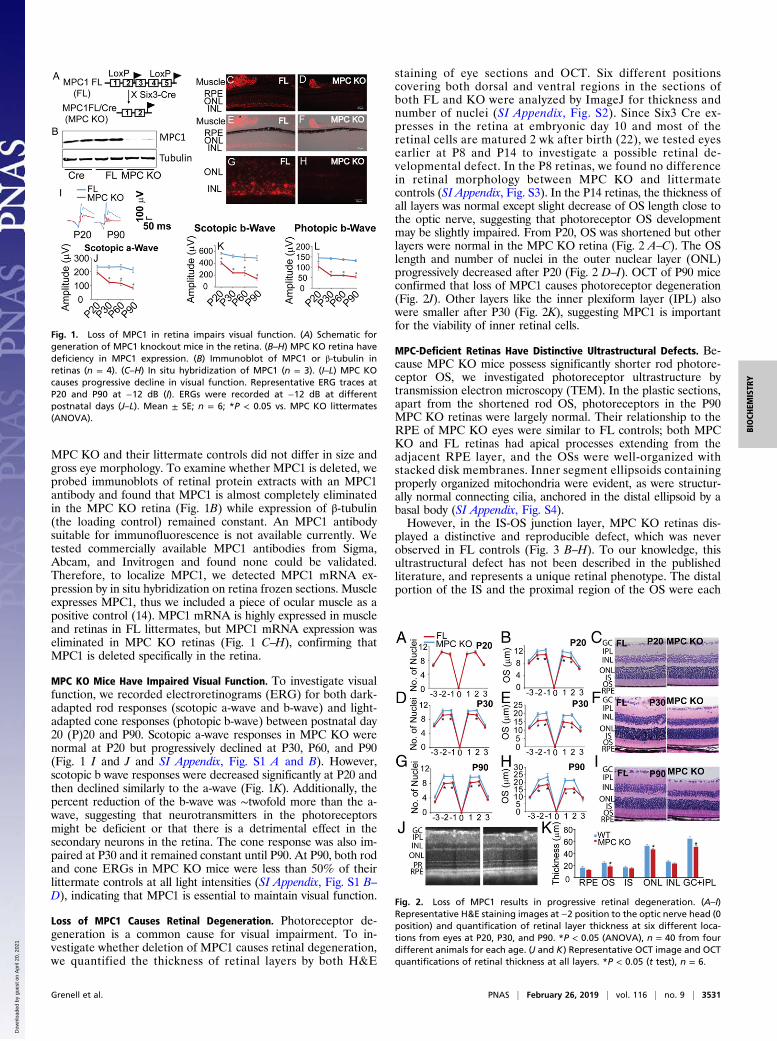
MPC KO and their littermate controls did not differ in size andgross eye morphology. To examine whether MPC1 is deleted, weprobed immunoblots of retinal protein extracts with an MPC1antibody and found that MPC1 is almost completely eliminatedin the MPC KO retina (Fig. 1B) while expression of β-tubulin(the loading control) remained constant. An MPC1 antibodysuitable for immunofluorescence is not available currently. Wetested commercially available MPC1 antibodies from Sigma,Abcam, and Invitrogen and found none could be validated.Therefore, to localize MPC1, we detected MPC1 mRNA ex-pression by in situ hybridization on retina frozen sections. Muscleexpresses MPC1, thus we included a piece of ocular muscle as apositive control (14). MPC1 mRNA is highly expressed in muscleand retinas in FL littermates, but MPC1 mRNA expression waseliminated in MPC KO retinas (Fig. 1 C–H), confirming thatMPC1 is deleted specifically in the retina.
MPC KO Mice Have Impaired Visual Function. To investigate visualfunction, we recorded electroretinograms (ERG) for both dark-adapted rod responses (scotopic a-wave and b-wave) and light-adapted cone responses (photopic b-wave) between postnatal day20 (P)20 and P90. Scotopic a-wave responses in MPC KO werenormal at P20 but progressively declined at P30, P60, and P90(Fig. 1 I and J and SI Appendix, Fig. S1 A and B). However,scotopic b wave responses were decreased significantly at P20 andthen declined similarly to the a-wave (Fig. 1K). Additionally, thepercent reduction of the b-wave was ∼twofold more than the a-wave, suggesting that neurotransmitters in the photoreceptorsmight be deficient or that there is a detrimental effect in thesecondary neurons in the retina. The cone response was also im-paired at P30 and it remained constant until P90. At P90, both rodand cone ERGs in MPC KO mice were less than 50% of theirlittermate controls at all light intensities (SI Appendix, Fig. S1 B–D), indicating that MPC1 is essential to maintain visual function.
Loss of MPC1 Causes Retinal Degeneration. Photoreceptor de-generation is a common cause for visual impairment. To in-vestigate whether deletion of MPC1 causes retinal degeneration,we quantified the thickness of retinal layers by both H&E
staining of eye sections and OCT. Six different positionscovering both dorsal and ventral regions in the sections ofboth FL and KO were analyzed by ImageJ for thickness andnumber of nuclei (SI Appendix, Fig. S2). Since Six3 Cre ex-presses in the retina at embryonic day 10 and most of theretinal cells are matured 2 wk after birth (22), we tested eyesearlier at P8 and P14 to investigate a possible retinal de-velopmental defect. In the P8 retinas, we found no differencein retinal morphology between MPC KO and littermatecontrols (SI Appendix, Fig. S3). In the P14 retinas, the thickness ofall layers was normal except slight decrease of OS length close tothe optic nerve, suggesting that photoreceptor OS developmentmay be slightly impaired. From P20, OS was shortened but otherlayers were normal in the MPC KO retina (Fig. 2 A–C). The OSlength and number of nuclei in the outer nuclear layer (ONL)progressively decreased after P20 (Fig. 2 D–I). OCT of P90 miceconfirmed that loss of MPC1 causes photoreceptor degeneration(Fig. 2J). Other layers like the inner plexiform layer (IPL) alsowere smaller after P30 (Fig. 2K), suggesting MPC1 is importantfor the viability of inner retinal cells.
MPC-Deficient Retinas Have Distinctive Ultrastructural Defects. Be-cause MPC KO mice possess significantly shorter rod photore-ceptor OS, we investigated photoreceptor ultrastructure bytransmission electron microscopy (TEM). In the plastic sections,apart from the shortened rod OS, photoreceptors in the P90MPC KO retinas were largely normal. Their relationship to theRPE of MPC KO eyes were similar to FL controls; both MPCKO and FL retinas had apical processes extending from theadjacent RPE layer, and the OSs were well-organized withstacked disk membranes. Inner segment ellipsoids containingproperly organized mitochondria were evident, as were structur-ally normal connecting cilia, anchored in the distal ellipsoid by abasal body (SI Appendix, Fig. S4).However, in the IS-OS junction layer, MPC KO retinas dis-
played a distinctive and reproducible defect, which was neverobserved in FL controls (Fig. 3 B–H). To our knowledge, thisultrastructural defect has not been described in the publishedliterature, and represents a unique retinal phenotype. The distalportion of the IS and the proximal region of the OS were each
Fig. 1. Loss of MPC1 in retina impairs visual function. (A) Schematic forgeneration of MPC1 knockout mice in the retina. (B–H) MPC KO retina havedeficiency in MPC1 expression. (B) Immunoblot of MPC1 or β-tubulin inretinas (n = 4). (C–H) In situ hybridization of MPC1 (n = 3). (I–L) MPC KOcauses progressive decline in visual function. Representative ERG traces atP20 and P90 at −12 dB (I). ERGs were recorded at −12 dB at differentpostnatal days (J–L). Mean ± SE; n = 6; *P < 0.05 vs. MPC KO littermates(ANOVA).
Fig. 2. Loss of MPC1 results in progressive retinal degeneration. (A–I)Representative H&E staining images at −2 position to the optic nerve head (0position) and quantification of retinal layer thickness at six different loca-tions from eyes at P20, P30, and P90. *P < 0.05 (ANOVA), n = 40 from fourdifferent animals for each age. (J and K) Representative OCT image and OCTquantifications of retinal thickness at all layers. *P < 0.05 (t test), n = 6.
Grenell et al. PNAS | February 26, 2019 | vol. 116 | no. 9 | 3531
BIOCH
EMISTR
Y
Dow
nloa
ded
by g
uest
on
Apr
il 20
, 202
1

involved. Higher-magnification views demonstrated that disrup-tions in normal cell structure might be caused by a shed portionof an ellipsoid region including isolated swollen mitochondria,adjacent to packets of disorganized OS disk membrane (Fig.3H). To estimate the frequency of shed ellipsoid regions, thenumber of such features observed was normalized to the numberof cell bodies present in a continuous ultrathin section. Thesedefects were observed at a frequency of roughly 4% relative tothe total rod photoreceptor population. To further examinethese defects, we stained IS membrane with Na/K ATPase (23,24) and OS with rhodopsin. MPC KO retinas formed smallvacuoles in the IS and IS-OS junction (Fig. 3 I–N) but not in theFL retinas, supporting the structural damage in the IS.
MPC KO Alters the Retinal Metabolic Profile.Next, we used targetedsteady-state metabolomics to investigate how loss of MPC1 in-fluences energy metabolism. We used LC MS/MS to target 93metabolites covering major pathways in the metabolism of glu-cose, amino acids, lipids, and nucleotides (SI Appendix, TableS1). Pyruvate was the most increased metabolite in the MPC KOretina at P20 in both volcano plot and t test (Fig. 4 A and B),confirming that MPC1 is deleted in the retina and blocks mito-chondrial pyruvate oxidation. Consistent with our previousanalysis of the effects of an MPC inhibitor ex vivo (19), aspartateaccumulated, while glutamate and glutamine were diminished, inthe MPC KO retina. Additionally, 3-hydroxybutyrate (3-HB),CoA, and glutathione (GSH) were diminished by ∼50% or morein the MPC KO retina (Fig. 4 A and B). Since RPE can be animportant source of 3-HB by oxidizing phagocytosed outer seg-ments (25), we also quantified metabolites in the RPE/choroid.We did not find differences in 3-HB, pyruvate, or other metab-olites in the RPE/choroid between MPC KO and their littermatecontrols (Fig. 4C), indicating that the metabolic changes arerestricted to the retina. We hypothesized that loss of MPC1blocks the entry of pyruvate into mitochondria and depletesα-ketoglutarate, a precursor for synthesis of glutamate, glutamine,
and GSH (Fig. 4D). To compensate for the mitochondrial energydeficit, the MPC KO retina may increase consumption of 3-HB toprovide acetyl-CoA for the TCA cycle (Fig. 4D).
MPC KO Impairs Retinal Glucose Metabolism. To test the hypothesisin Fig. 4D that glucose-derived pyruvate is critical for retinalamino acid metabolism, we incubated freshly isolated retinas atP20 with uniformly 13C-labeled glucose (U-13C glucose) for 1 h,and we used GC MS to quantify the labeled metabolites in gly-colysis, mitochondrial TCA cycle, and amino acid metabolism.The labeled carbons from each six-carbon molecule of labeledglucose (M6) that is metabolized through glycolysis can be foundin the three-carbon (M3) 3-phosphoglycerate (3PG), in the M3amino acid serine (Ser), in the M3 phosphoenolpyruvate (PEP),or in the M3 molecule pyruvate (Pyr). M3 pyruvate can entermitochondria through MPC1 to be oxidized to acetyl-CoA alongwith the loss of one carbon as CO2 (Fig. 5A). Therefore, theintermediates within the first round of the TCA cycle include twolabeled carbons from 13C glucose (M2). Consistent with ourexpectation that loss of MPC1 would block transport of pyruvateinto mitochondria for oxidation, M3 pyruvate increased ∼3.5fold in the MPC KO (Fig. 5F), similar to the effect MPC1 losshas on total pyruvate (Fig. 4B), but the glucose consumptionremained unchanged (SI Appendix, Fig. S5). Interestingly, MPCdeficiency increased the labeled serine and glycine, but not 3PG,PEP, and lactate. It has been reported that pyruvate can betransaminated into alanine to enter the TCA cycle when MPC1is deleted in the liver (14). However, in retina the labeled alaninewas not increased but decreased in the MPC KO. In the mito-chondrial intermediates, MPC KO decreased all of the labeledintermediates except M2 aspartate (Asp). M2 glutamine (Gln)decreased ∼5× while M2 Asp increased ∼3×.The amount of pyruvate increases while lactate remains con-
stant, so the retinal lactate/pyruvate ratio decreases in MPC KOretinas (Fig. 5Q). It is surprising that lactate does not increase, aspyruvate and lactate are interconverted through LDH. Becauselactate and pyruvate can easily be exported into the medium, we
Fig. 3. Loss of MPC1 disrupts the integrity of IS-OS junction layer. (A–H)TEM images of FL and MPC KO retinas in the IS-OS junction layer. The dis-rupted areas were marked by black arrowheads. n = 4. (I–N) Immunostainingshowed abnormal vacuoles marked by white arrowheads in the IS and IS-OSjunction in the MPC KO retinas. RHO, rhodopsin. (Scale bars: A–H, 500 nm; I–N, 20 μm.) n = 3.
Fig. 4. Loss of MPC1 alters the metabolic profile in the retina. (A) Mouseretinas at P20 were analyzed by LC MS/MS for metabolomics. Volcano plot(P < 0.05 and fold change > 1.5-fold were significant). y axis is the log10 of Pvalue, and x axis is the log2 of fold change. Significantly changed metabo-lites in mouse retina (B) and RPE/choroid (C). *P < 0.05 vs. FL (t test), n = 6.Relative abundance is the ion intensity relative to FL. (D) A proposed modelfor the metabolic change. Knockout of MPC1 accumulates cytosolic Pyr andaspartate (Asp) and depletes 3-HBA, glutamate (Glu), and glutamine (Gln).Mal, malate; Pro, proline.
3532 | www.pnas.org/cgi/doi/10.1073/pnas.1812941116 Grenell et al.
Dow
nloa
ded
by g
uest
on
Apr
il 20
, 202
1

examined the medium metabolites and found a parallel increaseof pyruvate and normal lactate (Fig. 5 R–T). Taken together,MPC KO blocks glucose oxidation in the mitochondria, accu-mulates pyruvate and aspartate, and reduces mitochondrialintermediates, especially glutamine.
MPC Deficiency Enhances Consumption of Ketone Bodies. Theamount of 3-HB, a ketone body, is substantially diminished inMPC KO retinas (Fig. 4 A and B). We hypothesized that loss ofMPC1 could cause a retina to oxidize more ketone bodies in itsmitochondria. To test this hypothesis, we incubated retinas with2,4-13C 3-HB in the presence of 5 mM unlabeled glucose andanalyzed the labeled metabolites by GC MS. 13C 3-HB first mustbe oxidized by NAD+ to form acetoacetate to supply acetyl-CoAfor the TCA cycle (SI Appendix, Fig. S6). After 1 h incubation,about 20–50% of mitochondrial intermediates were replacedwith 13C in the WT retinas (Fig. 6A), indicating that retinas canefficiently utilize ketone bodies as a fuel source. As expected,MPC deficiency significantly increases the percentage of labeledfraction (enrichment of 13C metabolite in total pool) for all ofthe mitochondrial intermediates (Fig. 6A). Since enrichment canbe influenced by pool size, we determined the relative amountsof labeled intermediates (abundance of 13C metabolites) (Fig.6B). The amounts of labeled α-ketoglutarate (αKG), fumarate,malate, proline, and aspartate are significantly enhanced in MPCKO retinas, while glutamine still is lower than in littermatecontrols. To confirm whether MPC KO retinas consume more 13
C 3-HB, we quantified medium metabolites and found thatMPC-KO retinas consumed more 13C 3-HB than FL retinas (Fig.6C). However, pyruvate still accumulated (produced from the5 mM unlabeled glucose) as it did with 13C glucose alone (SIAppendix, Fig. S6). Lactate was unchanged and the lactate/py-ruvate ratio was substantially diminished in MPC KO retinas.Ketone body is generated by fatty acids, which can also be oxi-dized directly in the mitochondria transported through acylcar-nitines. We found most of acylcarnitine are decreased in theMPC KO retinas (SI Appendix, Fig. S7), suggesting that fatty
oxidation is enhanced in the mitochondria. Overall, these resultsdemonstrate that loss of MPC1 enhances the use of ketonebodies to rescue the deficiency in TCA cycle intermediates.However, ketone bodies do not rescue the pyruvate accumula-tion and deficiency in glutamine.
MPC-Deficient Retinas Accumulate Aspartate at the Expense ofGlutamine. Glutamine can be a source of carbons for mitochon-drial intermediates once it has been converted to glutamate andthen to αKG. Glutamine also is a precursor for proline, gamma-aminobutyric acid (GABA) and 5-oxo-proline. To investigate theeffect of MPC deficiency on glutamine metabolism, we incubatedretinas with uniformly labeled 13C glutamine. In the first roundof the TCA cycle, M5 glutamine loses a carbon through αKGdehydrogenase to generate the M4 intermediates of the TCAcycle (Fig. 6D). M4 aspartate accumulates about fivefold and M4citrate is depleted by half in MPC KO retinas compared withcontrols. Aspartate accumulates because there is not enoughacetyl-CoA to convert oxaloacetate to citrate (Fig. 6E). In nor-mal mitochondria carbons that enter from M5 glutamine in thefirst cycle continue into subsequent cycles where they ultimatelyare fully oxidized to CO2. Intermediates in the second round ofthe cycle appear as M3 or M2 (Fig. 6D). These isotopomers arescarce in MPC-deficient retinas, indicating an overall slower rateof glutamine oxidation. M2 aspartate accumulates to higherlevels in MPC-deficient retinas because without pyruvate, abottleneck occurs at citrate synthesis. (Fig. 6F). Overall, gluta-mine oxidation appears to be slowed because of the limitedsupply of acetyl CoA required to catalyze complete oxidation ofthe carbons from glutamine. Consistent with this, we found thatconsumption of 13C glutamine in the medium by MPC-deficientretinas is diminished (SI Appendix, Fig. S8).Surprisingly, carbons from 13C glutamine appear in both M1
and M3 pyruvate in MPC-deficient retinas (SI Appendix, Fig. S9A–C). M3 pyruvate could be made by malic enzyme convertingM4 malate into pyruvate. The deficiency of mitochondrial pyruvatemay stimulate this pathway to generate pyruvate inside mitochondria.
Fig. 5. MPC1 knockout blocks the entry of 13C-glucose–derived pyruvateinto mitochondria. (A) A schematic of 13C glucose labeling. U-13C glucose wasmetabolized into M3 3PG, serine (Ser), PEP, Pyr, alanine (Ala), and lactate(Lac). Pyr enters the mitochondria through MPC and oxidized through TCAcycle. (B–P) Mouse retinas at P20 were incubated with 5 mM U-13C glucosefor 1 h. The y axis was 13C abundance relative to FL. (Q) The MPC knockoutreduced the Lac/Pyr ratio. (R–T) The efflux of Pyr into the medium was in-creased but the Lac remained unchanged. *P < 0.05 vs. FL (t test), n = 6. Fum,fumarate; Suc, succinate.
Fig. 6. The knockout of MPC1 enhances ketone body oxidation and accu-mulates Asp from Gln. (A–C) Mouse retinas were incubated with 13C 5 mM 3-HB and glucose for 1 h. 13C enrichment is the percentage of each 13C-labeledisotopomer out the total of all isotopomers. 13C abundance represents theabsolute abundance of each 13C-labeled isotopomer. 3-HB increased mito-chondrial intermediates except Gln. (D–F) Mouse retinas were incubatedwith 2 mM 13C Gln and glucose for 1 h. Incorporation of 13C from Gln wasblocked at the step of citrate synthesis and accumulated as 13C Asp. *P < 0.05vs. FL (t test), n = 6. Aco, aconitate; Cit, citrate; 5Oxo, 5-oxoproline.
Grenell et al. PNAS | February 26, 2019 | vol. 116 | no. 9 | 3533
BIOCH
EMISTR
Y
Dow
nloa
ded
by g
uest
on
Apr
il 20
, 202
1

Indeed, M6 citrate (two carbons from M3 pyruvate plus four carbonsfrom oxaloacetate) and M3 citrate (one carbon from M1 pyruvateand two carbons from the second cycle intermediates) accumulate tohigher levels in MPC KO retinas (SI Appendix, Fig. S9 C and E).However, this alternative pathway for pyruvate production accountsfor only ∼1% of total pyruvate (SI Appendix, Fig. S9D).Consistent with our previous finding (1), glutamine increases
the lactate/pyruvate ratio (SI Appendix, Fig. S9F) and it increases3-HB more than threefold in WT retinas. We found that thedecrease of 3-HB that occurs in MPC retinas can be preventedby supplementation with glutamine (SI Appendix, Fig. S9G).These results confirm that MPC-deficient retinas can use gluta-mine, but their inability to efficiently synthesize glutamine stillcauses an overall reduction of glutamine content.
MPC Deficiency Causes Mitochondrial Energy Deficit. To test mito-chondrial function, we measured oxygen consumption in isolatedmitochondria and levels of total ATP and NAD/NADH. Asexpected, the oxygen consumption rate was severely impaired inMPC KO retinal mitochondria (SI Appendix, Fig. S10A). Thelevels of ATP and NADH did not change at P20 retinas, mostprobably due to the compensational use of ketone bodies andfatty acid. However, the levels of ATP and NADH were de-creased in MPC KO retinas at P30 (SI Appendix, Fig. S10 B–D),indicating that mitochondrial dysfunction is critical for theretinal degeneration.
MPC Deficiency Activates Müller Glial Cells. Glutamine is synthe-sized by glutamine synthetase (GS), which is localized specificallyto Müller glial cells. Müller glia are hypersensitive to changes inthe retinal environment. To examine whether Müller glial cells wereactivated by MPC deficiency, we immunostained retinal sectionswith GS and glial fibrillary acidic protein (GFAP, a marker of glialactivation). In FL retinas, GS-stained Müller glial cells were wellorganized, and the minimal GFAP expression was restricted toMüller cell end feet, where the GFAP signal overlapped with GS (SIAppendix, Fig. S11 A–C). However, in MPC-deficient retinas, GSstaining was more irregular. GFAP was up-regulated, which reachedout to all layers where Müller glia are present including the IPL,inner nuclear layer (INL), and ONL (SI Appendix, Fig. S11 D–F).The Müller glia in the MPC KO retinas were disorganized (SIAppendix, Fig. S11 D–F), indicating activation and damage. Toquantify the protein expression, we tested the whole retinal proteinextracts with immunoblots. Consistently, the expression of GS wasdown-regulated while GFAP was up-regulated in the MPC KOretinas (SI Appendix, Fig. S11 G and H).
DiscussionRetina metabolism is dominated by aerobic glycolysis (theWarburg effect) with a small fraction of the pyruvate producedby glycolysis being oxidized in mitochondria. In this study, wereport that mitochondrial pyruvate transport is required fornormal retinal function and photoreceptor viability. MPC con-trols how a retina uses metabolic fuels. It influences retinal cy-tosolic reducing power, glutaminolysis, ketone body oxidation,and glutamine synthesis, which are critical for photoreceptorfunction and integrity.
Mitochondrial Pyruvate Transport in Visual Function and RetinalViability. Visual function declines progressively in retinas with-out MPC. The scotopic b-wave is affected early and severely. Theb-wave reflects glutamatergic transmission between photore-ceptors and biopolar cells. The depletion of glutamine/glutamateand accumulation of aspartate may be the major contributor tothe reduced b-wave response. Intraocular injection of a gluta-mine synthetase inhibitor in rats caused a similar suppression ofthe scotopic b-wave response (26). In isolated rat retinas, exog-enous glutamine or glutamate is required to maintain b-wave
responses and inhibition of glutamate transporters causes rapidloss of the b-wave (27). The monocarboxylate transporter in-hibitor, α-cyano-4-hydroxycinnamate (4-CIN), potently inhibitsMPC (28). Intravitreal injection 4-CIN suppresses the b-waveresponse, lowers glutamate, and raises aspartate (29), similarto the changes that occur in MPC KO mice.Shortening of outer segments occurs as early as P14 in MPC
KO retinas. The daily turnover of vertebrate outer segmentscreates a high demand for synthesis from lipids and amino acids.Pyruvate oxidation through the TCA cycle can be an importantsource for de novo lipid synthesis and nonessential amino acidsynthesis. We found that blocking import of pyruvate into mi-tochondria leads to oxidation of ketone bodies that are derivedfrom lipids. We speculate that the compensatory increase ofketone body and glutamine oxidation may not be fast enough tomeet the high demand for energy and biosynthesis, becauseglutamine/glutamate, glycine, and glutathione are significantlydepleted in the MPC KO retina (Fig. 4 A and B). Consistent withthis hypothesis, mutation of the gene encoding human isocitratedehydrogenase (IDH), a key enzyme in the TCA cycle, exclu-sively causes retinal degeneration (7). Loss of PDH or loss ofcitrate synthase causes light-induced photoreceptor degenera-tion in Drosophila (30) and pyruvate administration can protectmouse retinas from light damage (31). Furthermore, glial ac-tivation in response to diminished glutamine may aggravateretinal degeneration. GS expression is slightly up-regulated inMPC KO retinas, apparently by compensation, but its activityis substantially impaired based on our 13C labeling results witheither 13C glucose or 13C glutamine. GS is a potent neuro-protectant and inhibition of GS activity can lead to retinal celldeath (32).
MPC Influences Pyruvate Oxidation and Lactate/Pyruvate Ratio. Likecancer cells, photoreceptors express high levels of the M2 iso-form of pyruvate kinase (PKM2) and the LDH isoform A(LDHA) (3, 10, 33–35). These isoforms are generally associatedwith aerobic glycolysis in cancer cells and other proliferating celltypes, whereas MPC expression is negatively correlated withaerobic glycolysis in cancer cells (16). Consistent with this ob-servation, MPC expression is much lower in the photoreceptorlayer than in ocular muscle and in the inner retinal layers (Fig.1C). Knockout of MPC1 inhibits pyruvate oxidation. It alsocauses accumulation of serine and glycine (Fig. 5). Increased denovo serine synthesis could enhance phospholipid synthesis, butthis effect may be counteracted in the MPC KO retinas by en-hanced oxidation of fatty acids.There is no increase in lactate production from both the retina
and the RPE with MPC deficiency although pyruvate accumu-lates to a level substantially higher than normal (Fig. 5). Simi-larly, there was no increase in lactate in retinas cultured ex vivowith an MPC inhibitor (19). We showed previously that a highcytosolic lactate/pyruvate ratio in normal retinas can drivemalate aspartate shuttle activity, which diverts α-ketoglutarateaway from being oxidized by α-ketoglutarate dehydrogeanse (1).In MPC-deficient retinas, it is the accumulation of oxaloacetateand aspartate that drives malate aspartate shuttle activity. Becausetwo carbons from pyruvate are needed to make citrate, oxaloac-etate accumulates in MPC KO retinas. The oxaloacetate is con-verted by aminotransferase activity into aspartate (SI Appendix,Fig. S12), which is transported to the cytoplasm. There it is con-verted back to oxaloacetate, which forms malate while oxidizingNADH to NAD+. The malate exchanges with α-KG from thematrix, thereby diverting the α-KG from oxidation. The increasedamount of NAD+ in the cytoplasm contributes to the decreasedlactate/pyruvate ratio in MPC-deficient retinas. NAD+ also canoxidize 3-HB to acetoacetate to provide some matrix acetyl-CoA.The metabolic flexibility to use glutamine and lipids to compen-sate for MPC loss has been reported in other MPC-deficient cells
3534 | www.pnas.org/cgi/doi/10.1073/pnas.1812941116 Grenell et al.
Dow
nloa
ded
by g
uest
on
Apr
il 20
, 202
1

(14, 36). This may partially explain why retinal degeneration inMPC-deficient retinas is not very severe. However, despite thisadaptive metabolic pathway, the balance of retinal neurotrans-mitter and the integrity of photoreceptors still cannot be main-tained without MPC1. A ketogenic diet can rescue gestation ofglobal MPC1-deficient embryos, but the pups die within minutesafter birth (15).
MPC Influences Neuron–Glial Interaction. Photoreceptor neurons,Müller glial cells, and RPE cells have a symbiotic metabolic re-lationship. Disruption of these metabolic relationships can causephotoreceptor degeneration or gliosis (37). The up-regulatedGFAP and disorganized Müller glial cells in MPC-deficientretinas suggest that MPC is critical for neuron–glial interaction.Disruption of glutamine metabolism is likely to be a key factor
in glial dysfunction. Glutamine is synthesized in glial cells andtransported to photoreceptors where it is converted to glutamateto be used as a neurotransmitter or as fuel (SI Appendix, Fig.S12). We have reported evidence that Müller cells use lactateand aspartate from photoreceptors to synthesize glutamine (10).Why does MPC deficiency deplete glutamine? Müller cells lackthe mitochondrial aspartate-glutamate carrier (AGC) (10) that
diverts oxaloacetate away from the TCA cycle. This gives Müllercell mitochondria the ability to use aspartate to make glutamine(10) by adding carbons from acetyl-CoA to oxaloacetate to makecitrate. That citrate then is oxidized to α-KG for glutamate andglutamine synthesis. However, when MPC is deficient, acetyl-CoA from pyruvate is unavailable, thereby blocking this path-way for synthesis of glutamate and glutamine in Müller glia.
Materials and MethodsAll of the reagents, animals, and key resources were detailed in the keyresources form (SI Appendix). Mouse experiments were performed in ac-cordance with the National Institutes of Health guidelines and the protocolapproved by the Institutional Animal Care and Use Committee of WestVirginia University. MPC1 RNA in situ hybridization, metabolite analysis bymass spectrometry, EM, immunohistochemistry, and other methods wereperformed as reported (38, 39) and described in details in SI Appendix.
ACKNOWLEDGMENTS. We thank Dr. Jared Rutter for providing us theMPC1flox/flox mice and Dr. Victoria A. Kimler for technical assistance for elec-tron microscopy. This work was supported by NIH Grants EY026030 (to J.D.and Jennifer Chao, a multiple PI to this grant), EY06641 (to J.B.H.), EY017863(to J.B.H.), and EY025291 (to A.F.X.G.) and the Brightfocus Foundation(to J.D.).
1. Du J, et al. (2013) Cytosolic reducing power preserves glutamate in retina. Proc NatlAcad Sci USA 110:18501–18506.
2. Du J, et al. (2016) Reductive carboxylation is a major metabolic pathway in the retinalpigment epithelium. Proc Natl Acad Sci USA 113:14710–14715.
3. Chinchore Y, Begaj T, Wu D, Drokhlyansky E, Cepko CL (2017) Glycolytic reliancepromotes anabolism in photoreceptors. eLife 6:e25946.
4. Zhang L, et al. (2016) Reprogramming metabolism by targeting sirtuin 6 attenuatesretinal degeneration. J Clin Invest 126:4659–4673.
5. Kanow MA, et al. (2017) Biochemical adaptations of the retina and retinal pigmentepithelium support a metabolic ecosystem in the vertebrate eye. eLife 6:e28899.
6. Ames A, 3rd, Li YY, Heher EC, Kimble CR (1992) Energy metabolism of rabbit retina asrelated to function: High cost of Na+ transport. J Neurosci 12:840–853.
7. Hartong DT, et al. (2008) Insights from retinitis pigmentosa into the roles of isocitratedehydrogenases in the Krebs cycle. Nat Genet 40:1230–1234.
8. Brown EE, Lewin AS, Ash JD (2018) Mitochondria: Potential targets for protection inage-related macular degeneration. Adv Exp Med Biol 1074:11–17.
9. Han WH, et al. (2017) Modifications in retinal mitochondrial respiration precede type2 diabetes and protracted microvascular retinopathy. Invest Ophthalmol Vis Sci 58:3826–3839.
10. Lindsay KJ, et al. (2014) Pyruvate kinase and aspartate-glutamate carrier distributionsreveal key metabolic links between neurons and glia in retina. Proc Natl Acad Sci USA111:15579–15584.
11. Bricker DK, et al. (2012) A mitochondrial pyruvate carrier required for pyruvate up-take in yeast, Drosophila, and humans. Science 337:96–100.
12. Herzig S, et al. (2012) Identification and functional expression of the mitochondrialpyruvate carrier. Science 337:93–96.
13. McCommis KS, et al. (2015) Loss of mitochondrial pyruvate carrier 2 in the liver leadsto defects in gluconeogenesis and compensation via pyruvate-alanine cycling. CellMetab 22:682–694.
14. Gray LR, et al. (2015) Hepatic mitochondrial pyruvate carrier 1 is required for efficientregulation of gluconeogenesis and whole-body glucose homeostasis. Cell Metab 22:669–681.
15. Vanderperre B, et al. (2016) Embryonic lethality of mitochondrial pyruvate carrier 1deficient mouse can be rescued by a ketogenic diet. PLoS Genet 12:e1006056.
16. Schell JC, et al. (2014) A role for the mitochondrial pyruvate carrier as a repressor ofthe Warburg effect and colon cancer cell growth. Mol Cell 56:400–413.
17. Schell JC, et al. (2017) Control of intestinal stem cell function and proliferation bymitochondrial pyruvate metabolism. Nat Cell Biol 19:1027–1036.
18. Vigueira PA, et al. (2014) Mitochondrial pyruvate carrier 2 hypomorphism in miceleads to defects in glucose-stimulated insulin secretion. Cell Rep 7:2042–2053.
19. Du J, et al. (2013) Inhibition of mitochondrial pyruvate transport by zaprinast causesmassive accumulation of aspartate at the expense of glutamate in the retina. J BiolChem 288:36129–36140.
20. Divakaruni AS, et al. (2017) Inhibition of the mitochondrial pyruvate carrier protectsfrom excitotoxic neuronal death. J Cell Biol 216:1091–1105.
21. Christiansen JR, Kolandaivelu S, Bergo MO, Ramamurthy V (2011) RAS-convertingenzyme 1-mediated endoproteolysis is required for trafficking of rod phosphodies-terase 6 to photoreceptor outer segments. Proc Natl Acad Sci USA 108:8862–8866.
22. Morrow EM, Furukawa T, Cepko CL (1998) Vertebrate photoreceptor cell develop-ment and disease. Trends Cell Biol 8:353–358.
23. Plössl K, et al. (2017) Retinoschisin is linked to retinal Na/K-ATPase signaling and lo-calization. Mol Biol Cell 28:2178–2189.
24. Spencer M, Detwiler PB, Bunt-Milam AH (1988) Distribution of membrane proteins inmechanically dissociated retinal rods. Invest Ophthalmol Vis Sci 29:1012–1020.
25. Adijanto J, et al. (2014) The retinal pigment epithelium utilizes fatty acids for keto-genesis. J Biol Chem 289:20570–20582.
26. Barnett NL, Pow DV, Robinson SR (2000) Inhibition of Müller cell glutamine synthe-tase rapidly impairs the retinal response to light. Glia 30:64–73.
27. Winkler BS, Kapousta-Bruneau N, Arnold MJ, Green DG (1999) Effects of inhibitingglutamine synthetase and blocking glutamate uptake on b-wave generation in theisolated rat retina. Vis Neurosci 16:345–353.
28. Halestrap AP (1975) The mitochondrial pyruvate carrier. Kinetics and specificity forsubstrates and inhibitors. Biochem J 148:85–96.
29. Bui BV, Vingrys AJ, Wellard JW, Kalloniatis M (2004) Monocarboxylate transport in-hibition alters retinal function and cellular amino acid levels. Eur J Neurosci 20:1525–1537.
30. Jaiswal M, et al. (2015) Impaired mitochondrial energy production causes light-induced photoreceptor degeneration independent of oxidative stress. PLoS Biol 13:e1002197.
31. Ren H, Liu NY, Song XF, Ma YS, Zhai XY (2011) A novel specific application of pyruvateprotects the mouse retina against white light damage: Differential stabilization ofHIF-1α and HIF-2α. Invest Ophthalmol Vis Sci 52:3112–3118.
32. Gorovits R, Avidan N, Avisar N, Shaked I, Vardimon L (1997) Glutamine synthetaseprotects against neuronal degeneration in injured retinal tissue. Proc Natl Acad SciUSA 94:7024–7029.
33. Rueda EM, et al. (2016) The cellular and compartmental profile of mouse retinalglycolysis, tricarboxylic acid cycle, oxidative phosphorylation, and ∼P transferring ki-nases. Mol Vis 22:847–885.
34. Casson RJ, et al. (2016) M-type pyruvate kinase isoforms and lactate dehydrogenase Ain the mammalian retina: Metabolic implications. Invest Ophthalmol Vis Sci 57:66–80.
35. Rajala RV, Rajala A, Kooker C, Wang Y, Anderson RE (2016) The Warburg effectmediator pyruvate kinase M2 expression and regulation in the retina. Sci Rep 6:37727.
36. Yang C, et al. (2014) Glutamine oxidation maintains the TCA cycle and cell survivalduring impaired mitochondrial pyruvate transport. Mol Cell 56:414–424.
37. Shen W, et al. (2012) Conditional Müllercell ablation causes independent neuronaland vascular pathologies in a novel transgenic model. J Neurosci 32:15715–15727.
38. Swarup A, et al. (2018) Deletion of GLUT1 in mouse lens epithelium leads to cataractformation. Exp Eye Res 172:45–53.
39. Du J, Linton JD, Hurley JB (2015) Probing metabolism in the intact retina using stableisotope tracers. Methods Enzymol 561:149–170.
Grenell et al. PNAS | February 26, 2019 | vol. 116 | no. 9 | 3535
BIOCH
EMISTR
Y
Dow
nloa
ded
by g
uest
on
Apr
il 20
, 202
1