loss of expression of protein phosphatase magnesium ... · loss of expression of protein...
TRANSCRIPT
THE
JOURNAL • RESEARCH • www.fasebj.org
Loss of expression of protein phosphatasemagnesium-dependent 1A during kidney injurypromotes fibrotic maladaptive repairRohan Samarakoon,*,1 Alexandra Rehfuss,† Nidah S. Khakoo,* Lucas L. Falke,‡ Amy D. Dobberfuhl,§
Sevann Helo,† Jessica M. Overstreet,{ Roel Goldschmeding,‡ and Paul J. Higgins*,2
*Center for Cell Biology and Cancer Research and †Division of Urology, Albany Medical Center, Albany, New York, USA; ‡Department ofPathology, University Medical Center Utrecht, Utrecht, The Netherlands; §Department of Urology, Stanford University School of Medicine,Stanford, California, USA; and {Division of Nephrology and Hypertension, Vanderbilt Medical School, Nashville, Tennessee, USA
ABSTRACT: Protein phosphatase magnesium-dependent-1A (PPM1A) dephosphorylates SMAD2/3, which sup-presses TGF-b signaling in keratinocytes and during Xenopus development; however, potential involvementof PPM1A in chronic kidney disease is unknown. PPM1A expression was dramatically decreased in the tubu-lointerstitium in obstructive and aristolochic acid nephropathy, which correlates with progression of fibroticdisease. Stable silencing of PPM1A in human kidney-2 human renal epithelial cells increased SMAD3phosphorylation, stimulated expression of fibrotic genes, induced dedifferentiation, and orchestrated epithelialcell-cycle arrest via SMAD3-mediated connective tissue growth factor and plasminogen activator inhibitor-1 up-regulation. PPM1Astable suppression innormal rat kidney-49 renal fibroblasts, in contrast, promoted aSMAD3-dependent connective tissue growth factor and plasminogen activator inhibitor-1–induced proliferative response.Paracrine factors secretedbyPPM1A-depletedepithelial cells augmented fibroblastproliferation (>50%)comparedwith controls. PPM1A suppression in renal cells further enhanced TGF-b1–induced SMAD3phosphorylation andfibrotic gene expression, whereas PPM1A overexpression inhibited both responses. Moreover, phosphate tensinhomologon chromosome10depletion inhumankidney-2 cells resulted in lossof expressionanddecreasednuclearlevels of PPM1A,which enhanced SMAD3-mediated fibrotic gene induction and growth arrest that were reversedby ectopic PPM1Aexpression. Thus, phosphate tensin homolog on chromosome 10 is an upstream regulator of renalPPM1A deregulation. These findings establish PPM1A as a novel repressor of the SMAD3 pathway in renal fibro-sis and as a new therapeutic target in patients with chronic kidney disease.—Samarakoon, R., Rehfuss, A., Khakoo,N.S., Falke,L.L.,Dobberfuhl,A.D.,Helo,S.,Overstreet, J.M.,Goldschmeding,R.,Higgins,P. J. Lossof expressionofproteinphosphatasemagnesium-dependent 1Aduringkidney injurypromotes fibroticmaladaptive repair. FASEBJ.30, 3308–3320 (2016). www.fasebj.org
KEY WORDS: TGF-b • PAI-1 • CTGF • PTEN • renal fibrosis
Renal fibrotic disorders are generally refractive to cur-rent therapies. Chronic kidney disease (CKD) affects.12% of the U.S. population alone (1–4), with CKD riskfactors (e.g., diabetes and hypertension) likely to in-crease worldwide in the coming decades. Renal re-placement therapy, either dialysis or transplantation, isinadequate tomeet patient demand,which further addsto the increasing public health burden (1–4).
Diabetic, hypertensive, acute or toxic, and obstructivekidney injury result in maladaptive repair (i.e., epithelialcell-cycle arrest and death, secretion of fibrotic factors,persistent inflammation, and accumulation of extracellu-lar matrix–producing myofibroblasts), which eventuallyculminates in progressive fibrosis, tissue scarring, andend-stage renal disease (1–8). Regardless of the initial in-sult, activation of the TGF-b pathway is a prominentdriver of a dysfunctional repair response, which leads to
ABBREVIATIONS: AA, aristolochic acid; AAN, aristolochic acid nephrop-athy; BMP-6/7, bone morphogenic protein-6/7; CKD, chronic kidneydisease; CTGF, connective tissue growth factor; FBS, fetal bovine serum;GAPDH, glyceraldehyde 3-phosphate dehydrogenase; HK-2, human kidney 2;NRK-49F, normal rat kidney-49 fibroblast; PAI-1, plasminogen activator in-hibitor-1; PPM1A, protein phosphatase magnesium-dependent 1A; PTEN,phosphate tensin homolog on chromosome 10; shRNA, short hairpin RNA;siRNA, small interfering RNA; Ski, Sloan Kettering Institute proto-oncogene;a-SMA, a-smooth muscle actin; Sno, Ski-related novel gene; UUO, unilateralureteral obstruction1 Correspondence: Center for Cell Biology and Cancer Research, AlbanyMedical College, 47 New Scotland Ave., Albany, NY 12208, USA.E-mail: [email protected]
2 Correspondence: Center for Cell Biology and Cancer Research, AlbanyMedical College, 47 New Scotland Ave., Albany, NY 12208, USA.E-mail: [email protected]
doi: 10.1096/fj.201500105RThis article includes supplemental data. Please visit http://www.fasebj.org toobtain this information.
3308 0892-6638/16/0030-3308 © FASEB Vol.30, No.10 , pp:3308-3320, April, 2017The FASEB Journal. 171.65.89.189 to IP www.fasebj.orgDownloaded from

fibrosis (5–11). Binding of TGF-b1 ligands to the RI/RIIreceptor complex initiates both canonical SMAD2/3 andnoncanonical (e.g., reactive oxygen species, ataxia telan-giectasia mutated, p53, epidermal growth factor receptor,MAPK, Rho-GTPases) downstream signaling in kidneycells (9–15). Subsequent assembly of multimeric tran-scriptional complexes (e.g., SMADs, p53) leads to elevatedexpression of profibrotic target genes [e.g., plasminogenactivator inhibitor-1 (PAI-1), connective tissue growthfactor (CTGF), extracellular matrix proteins] and context-dependent phenotypic responses (e.g., cell-cycle arrest,proliferation, or apoptosis) (9–11, 13–15).
As amaster regulator of organ fibrosis and vasculardisease, TGF-b1 signal propagation is subjected toextensive negative control at the level of receptor ac-tivity, SMAD2/3 phosphorylation, SMAD2/3 nucleartranslocation or exit, transcriptional complex assem-bly, and target promoter engagement, thereby tightlyregulating associated transcriptional and biologicresponses (9–11, 16, 17). Deficiencies in key negativeregulators of the TGF-b pathway [e.g., bone mor-phogenic protein-6/7 (BMP-6/7), Sloan Kettering In-stitute proto-oncogene (Ski), Ski-related novel gene(Sno), and SMAD7] are evident during progressionof renal disease. BMP-6/7–mediated activation ofSMAD1/4/5, for example, antagonizes the TGF-b1–induced SMAD2/3 pathway (18, 19). Loss of BMP-6/7signaling, evident during kidney injury, leads to ex-acerbated TGF-b1 responses and renal disease (18, 19).SMAD2/3 activation is inhibited by SMAD7 and theSMAD2/3 corepressors, Ski and SnoN, which sup-presses target gene expression (16, 20). Progressionof renal disease is accompanied by loss—via ubiquitin-dependent degradation—of several negative regulators(e.g., SMAD7, Ski, SnoN), which leads to persistentTGF-b1 signaling in the failing kidney (20–22). Genetransfer of SMAD7 to the kidney dramatically reducedinterstitial fibrosis induced by unilateral ureteral ob-struction (UUO) (23).
Protein phosphatase magnesium-dependent 1A(PPM1A; also known as protein phosphatase 2Ca) hasbeen recently shown to have C-terminal SMAD2/3phosphatase activity, a critical event in the terminationof TGF-b1 signaling (24). We recently demonstratedthat TGF-b1 stimulation reduces the nuclear fraction ofPPM1A via Rho/ROCK-dependent mechanisms, therebyfurther enhancing SMAD3-dependent target gene (e.g.,PAI-1) expression in vascular smooth muscle cells (25).This study, to our knowledge, presents the first investi-gation of the potential deregulation and mechanistic in-volvement of PPM1A in progression of chronic kidneyinjury and details upstream and downstream effectors ofPPM1A in the context of renal pathology.
MATERIALS AND METHODS
Cell culture and creation of stable cell lines
Human kidney-2 (HK-2) proximal tubular epithelial cellsand normal rat kidney-49 fibroblast (NRK-49F) cells were
grown in DMEM that was supplemented with 10% fetalbovine serum (FBS). To generate stable cell lines, semi-confluent HK-2 and NRK-49F cells were treated with 5 mg/ml polybrene in 10% FBS/DMEM and infected with PPM1Aor control lentiviral particles (Santa Cruz Biotechnology,Santa Cruz, CA, USA) and incubated overnight. After in-cubation in fresh complete medium for 24 h, cells stablyexpressing PPM1A or control short hairpin RNA (shRNA)were selected with 5 mg/ml puromycin + 10% FBS/DMEMwith medium changes every 3 d. PPM1A knockdown wasconfirmed by Western blot analysis. To generate SMAD3-,PAI-1–, and CTGF-silenced HK-2 or NRK-49F cells in thecontext of a stable PPM1a knockdown, 60% confluent PPM1ashRNA-expressing cellswere reinfected (for 1 d)with controlor SMAD3, PAI-1, or CTGF shRNA lentiviral constructs(Santa Cruz Biotechnology), followed selection in puromy-cin. In transient PPM1A overexpression experiments, HK-2and NRK-49F cells or phosphate tensin homolog on chro-mosome 10 (PTEN) shRNA stably expressing cells were in-fected with PPM1A human ORF cDNA or control Lentifectlentiviral particles (GeneCopoeia, Rockville, MD, USA) for24–48 h. Cells were allowed to recover for 2–3 d before TGF-b1 stimulation or were subjected to puromycin selection. Fortransient PPM1A silencing, 50% confluent NRK-49F cellswere transfected with Accell control constructs or PPM1asmall interfering RNA (siRNA; 250–500 nM; Dharmacon,Lafayette, CO, USA) by using Accell siRNA delivery me-dium for 2–3 d, followed by a 2-d recovery in 5% FBS/DMEM.Western blot analysis confirmed PPM1A expressionlevels in both approaches.
Induction of renal fibrosis in response to UUO
Mice were anesthetized by isoflurane inhalation, and a smallincision was made in the flank under aseptic conditions. Theleft ureter was exposed and ligated with two 5-0 silk sutures.Control mice underwent sham procedures but not ureteralligation. At 7 and 14 d postsurgery, all animals were eutha-nized, and obstructed (UUO), contralateral, and sham kidneyswere harvested. This protocol was approved by the AnimalEthics Committee of the University of Utrecht (Utrecht, TheNetherlands).
Aristolochic acid–induced nephropathy
C57Bl/6 male mice received an i.p. injection of aristolochic acid(AA) sodium salt (5 mg/kg body weight dissolved in distilledwater; A9451; Sigma-Aldrich, St. Louis, MO, USA) once a dayconsecutively for 5 d or NaCl vehicle (control animals) alone.Urine creatinine levels were measured to confirm renal injury inresponse toAA (J2L Elitech, LabartheInard, France). At 25 d afterinitial injections,mice inbothgroupswereeuthanizedbyketamine-xylazine-atropine.
Plasma urea measurements
Plasma urea concentrations were determined by using a colori-metric assay (Diasys Diagnostic Systems, Holzheim, Germany).Standardcurvesweregeneratedbyusingcommercially availableurea (Diasys Diagnostic Systems).
Renal epithelial-fibroblast crosstalk studies
Conditioned medium that was isolated from PPM1A shRNA orcontrol shRNA HK-2 epithelial cells (at similar density) were
PPM1A DEREGULATION IN KIDNEY INJURY PROMOTES FIBROTIC PHENOTYPE 3309 Vol.30, No.10 , pp:3308-3320, April, 2017The FASEB Journal. 171.65.89.189 to IP www.fasebj.orgDownloaded from
directly added to identically confluent control shRNA stablyexpressing NRK-49F fibroblasts for 3 d. Fibroblast growth wasmeasured by using the Sceptor 2.0 Handheld Automated CellCounter (Millipore, Billerica, MA, USA) according to manufac-turer recommendations.
Immunoblotting
Western blots were performed as described (26). Specificantibodies used include rabbit anti-PPM1A (1:2000), mouseanti-PPM1A (1:1000), rabbit anti–phospho-SMAD3 (1:1000),rabbit anti-fibronectin (1:10,000), rabbit anti-CTGF (1:2000), rab-bit anti–a-smooth muscle actin (a-SMA) (1:2000), and rabbitanti–collagen IV (1:500; all fromAbcam, Cambridge, MA,USA).Rabbit anti-PTEN (1:2000), rabbit anti-vimentin (1:2000), rabbitanti-pSMAD2/3 (1:1000), rabbit anti–glyceraldehyde 3-phosphatedehydrogenase (GAPDH;1:5000), goat anti-CTGF (1:1000), andrabbit anti p21 (1:1000) were from Santa Cruz Biotechnology.Rabbit anti–PAI-1 (1:3000) was previously described (26).Membranes were incubated with appropriate secondary anti-bodies and immunoreactive proteinswere visualizedwith ECLreagent and quantitated by densitometry. GAPDH provided anormalization control.
Subcellular fractionation
PTEN shRNA- and control shRNA-expressing HK-2 cells werescraped in PBS, pelleted by centrifugation, and resuspended inhypertonic solution A (10 mM HEPES, pH 7.9, 10 mM KCl,0.1 mMEDTA, and 0.1mMEGTA) that was supplementedwithprotease and phosphatase inhibitor cocktail and shaken onice for 20 min. Solution B (3% NP-40) was added, followed bycentrifugation (8000 rpm, 60 s) to isolate cytoplasmic fraction.Remainingpelletwas resuspended in solutionC (20mMHEPES,pH7.9, 0.42MNaCl, 1mMEDTA, 1mMEGTA, and 1mMDTT)thatwas supplementedwith protease and phosphatase inhibitorcocktail, subjected to several freeze/thaw cycles, and incubatedovernight in the cold room. Nuclear (supernatant) fractionwas separated at 8000 rpm for 5 min. GAPDH and lamin A/Cprovided markers of the cytoplasmic and nuclear fractions,respectively.
Immunohistochemistry
Deparaffinized kidney sections were probed with primaryAbcamrabbit antibodies toPPM1A(1:500) andPTEN(1:500) andmorphometric analyzed as detailed (26).
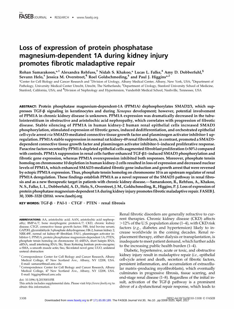
Figure 1. Tubular and interstitialloss of PPM1A expression in latestages of UUO and AA nephropa-thy (AAN). A) Immunoblotting forPPM1A (A, B), pSMAD3 (A, E),PAI-1 (A, F), a-SMA (A, G),fibronectin (A, H), and GAPDH(a loading and normalizationmarker) expression levels insham, contralateral (contra),and obstructed (UUO) mousekidneys at d 14 postsurgery (n =3–6 animals per experimentalcondition). Numbers 1–3 in panelA depict 3 individual mice for theindicated group. C) Immunohis-tochemistry images of paraffinsections of the contralateral orligated (UUO; d 14) kidneys de-rived from the same mice (A) byusing a rabbit anti-PPM1A anti-body. D) Quantitative image anal-ysis of diaminobenzidine (DAB)expression to assess differences inPPM1A expression between UUOand contralateral kidneys, settingstaining intensity of the contrakidney as 1. B, E–I) Histogramsdepict relative expression of in-dicated markers in UUO andcontralateral or sham kidneys. I)Western blot analysis for PPM1Aand GAPDH expression in lysatesderived from contralateral andUUO kidneys at d 7 postsurgery(n = 3 mice). J ) Immunostainingof kidney sections from micetreated with aristolochic acid(AAN; 5 mg/kg for 25 d) or NaClvehicle. K) Histogram illustratesrelative PPM1A levels in theAAN- and NaCl-administered kidneys, setting DAB intensities for NaCl-treated controls as 1. Data are given as means 6 SD. Scalebars, 60 mM (C), 90 mΜ (J). *P , 0.05; **P , 0.01; ***P , 0.001.
3310 Vol. 30 October 2016 SAMARAKOON ET AL.The FASEB Journal x www.fasebj.org Vol.30, No.10 , pp:3308-3320, April, 2017The FASEB Journal. 171.65.89.189 to IP www.fasebj.orgDownloaded from

Cell-cycle analysis
Control shRNA- and PPM1A shRNA-expressing HK-2 cellswere grown in serum-containing medium that was supple-mented with Puromycin for 2–3 d. Trypsin-harvested cellswere incubated with soybean trypsin inhibitor for 5 mins,washed twice in PBS, and fixed in suspension in 95% etha-nol for 1 h. Cells were washed in PBS 2 times and were in-cubated with RNaseA (20 ml/ml) and propidium iodide(2.5 mg/ ml) in PBS/Triton X-100 for 2 h in the dark. Cell-cycledistributions were measured by using a FACS Calibur flowcytometer (Becton Dickinson, Franklin Lakes, NJ, USA) and an-alyzed with FlowJo software (FlowJo, Ashland, OR, USA).
Statistical analysis
Two-tailed Student’s t test and ANOVAwith Tukey post hoc anal-ysis were used to assess statistical differences. A value of P, 0.05is considered significant.
RESULTS
Progressive loss of PPM1a expression in CKDinduced by UUO and AA nephropathy
Two well-established mouse models of tubulointer-stitial fibrosis were used to investigate potentialPPM1A deregulation in CKD. UUO is a highly re-producible model, with little interanimal variationfor inducing renal fibrosis, and it mimics obstructiveuropathy in children (27). AA nephropathy (AAN) isalso an established CKD model, and AA administrationin mice recapitulates certain features of human ne-phropathy that are evident in Chinese and Balkenpopulations as a result of consumption of this toxic herb(28). In late stages of UUO (d 14), renal PPM1A expres-sion is dramatically decreased by 90% as evident in im-mune blot analysis (Fig. 1A, B; P , 0.01, UUO vs.
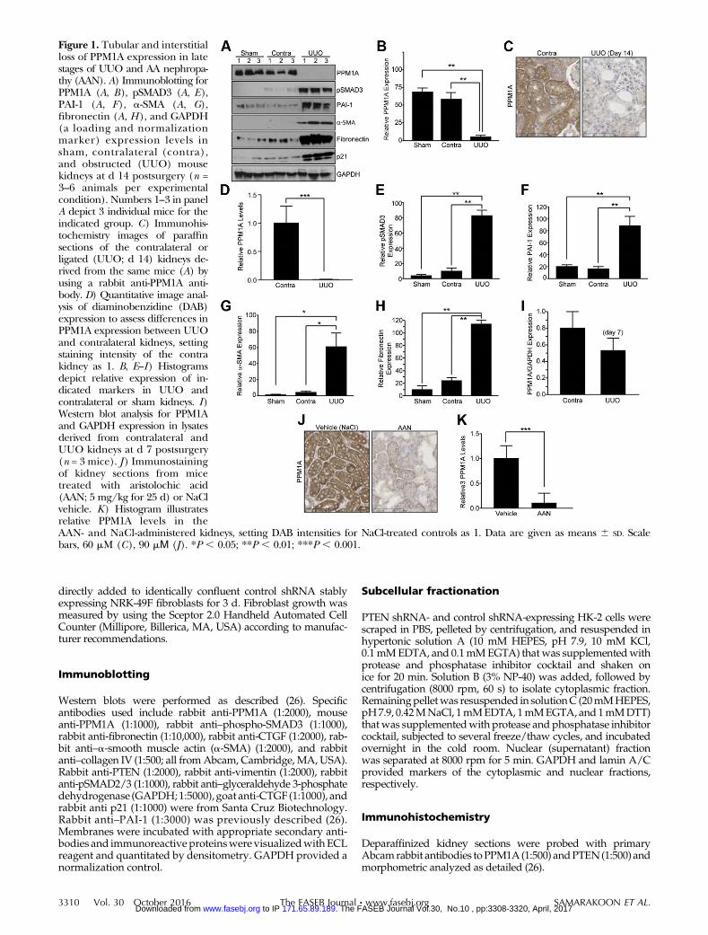
Figure 2. Prolonged silencing of PPM1a expression in HK-2 epithelial cells leads to fibrotic gene reprogramming. A) Subconfluentcontrol (con) shRNA and PPM1A shRNA stably expressing cell cultures were maintained in low serum (0.1%) medium for 5–7 d andcellular lysates that were immunoblotted with antibodies to PPM1A (A, B), fibronectin (A, C), PAI-1 (A, D), vimentin (A, E), p21(A, F), CTGF (A, G), collagen IV (Col-IV; A), pSMAD3 (A, H), and GAPDH (A). Numbers 1–3 in panel A represent 3 independentcultures of the indicated cell types. B–H) Plots summarize relative expression of indicated markers, setting levels in controls as 1.Data are given as means 6 SD. *P , 0.05; **P , 0.01; ***P , 0.001 vs. con shRNA.
PPM1A DEREGULATION IN KIDNEY INJURY PROMOTES FIBROTIC PHENOTYPE 3311 Vol.30, No.10 , pp:3308-3320, April, 2017The FASEB Journal. 171.65.89.189 to IP www.fasebj.orgDownloaded from

contralateralor shamcontrols) and immunohistochemistry(Fig. 1C, D; P , 0.001, UUO vs. contralateral kidneys),which exhibited prominent tubular expression of thisphosphatase (Fig. 1A–D). PPM1A reduction in theobstructed kidney correlated with elevated SMAD3 ac-tivation (Fig. 1A, E; P , 0.01) and expression of PAI-1(Fig. 1A, F; P , 0.01), a-SMA (Fig. 1A, G; P , 0.05),fibronectin (Fig. 1A, H; P , 0.01), and p21 (Fig. 1). Timecourse analysis indicates that renal expression of PPM1A ispartially decreased (35%) by d 7 after UUO compared withcontralateral controls (Fig. 1I), which suggests progressivePPM1A loss in chronic renal injury. A similar loss of PPM1Aexpression (.90%) was evident in the tubulointerstitialregions in AA-injured kidneys compared with NaClvehicle-treated controls (Fig. 1J, K; P , 0.001), which
indicated that PPM1A deregulation is common in bothmouse models of CKD. Furthermore, plasma urea levelswere elevated in mice that underwent renal obstruction(Supplemental Fig. 1A; P, 0.05 vs. sham control) and AAN(Supplemental Fig. 1B; P, 0.05 vs.NaCl vehicle), whichwas further indicative of the state of renalmalfunction indiseased mice in response to both types of renal injury.
Stable silencing of PPM1A expression inhuman renal epithelial cells promoted afibrogenic response
TomimicPPM1Aloss in renal tubules in response tokidneyinjury, stable silencing of PPM1A in HK-2 human renal
Figure 3. Stable silencing of PPM1A leads to epithelial dedifferentiation and cell-cycle arrest via SMAD3-dependent mechanisms. A)Stable PPM1A shRNA knockdown or control shRNA HK-2 cells (con; seeded at similar density) were grown for 7 d in 10% FBS-containing medium, then stained with Crystal Violet. B) Relative cell counts in triplicate cultures for each experimental conditionwere determined, setting values in controls as 1. **P , 0.01 vs. con shRNA cells. C) Flow cytometric analysis of propidiumiodide–stained PPM1A shRNA– and con shRNA–expressing cells grown in complete medium for 2–3 d provided assessments of cellcycle distribution. Insets in each histogram highlight percentages of G1, S, and G2/M phase cells. D) Histogram illustrates thefraction of PPM1A-knockdown or control cells exhibiting fibroblast morphology after 5 d growth as a percentage of the totalpopulation. ***P , 0.001 vs. con shRNA. E) Immunoblotting of lysates derived from stably expressing PPM1A + con, PPM1A +SMAD3, (PPM1A + PAI-1, and PPM1A + CTGF shRNA double-tranductant HK-2 cells for SMAD3, PAI-1, CTGF, and GAPDHexpression. F) Triplicate cultures of each of these 4, initially seeded equally (;25,000 cells), cell types were grown for 3 d before cellcount. Data are given as means 6 SD. Scale bars, 200 mM. **P , 0.01 vs. PPM1A + con stably expressing cells.
3312 Vol. 30 October 2016 SAMARAKOON ET AL.The FASEB Journal x www.fasebj.org Vol.30, No.10 , pp:3308-3320, April, 2017The FASEB Journal. 171.65.89.189 to IP www.fasebj.orgDownloaded from

epithelial cells via lentiviral transduction was used, whichresults in a .90% decrease in PPM1A protein expressioncompared with control shRNA-transduced cultures (Fig.2A, B; P , 0.001). Prolonged PPM1A suppression (main-tained for 5–7 d) increased expression of fibronectin (Fig.2A,C;10-fold,P, 0.01), PAI-1 (Fig. 2A,D; 5-fold,P, 0.05),vimentin (Fig. 2A, E; 3.7-fold, P , 0.05), p21 (Fig. 2A, F;4-fold,P,0.05),CTGF(Fig. 2A,G; 7-fold,P,0.05), collagenIV (Fig. 2A), and pSMAD3 (Fig. 2A, H; 7.5-fold, P , 0.01)expression compared with control shRNA-expressing cells.
Epithelial cell-cycle arrest upon PPM1Aknockdown is orchestrated via SMAD3-mediated CTGF and PAI-1 up-regulation
PPM1A depletion is also accompanied by decreased epi-thelial cell growth (;38%; Fig. 3A, B; P , 0.01) and G1cell-cycle arrest (Fig. 3C), which is consistent with p21 up-regulation, compared with control shRNA transductants
(Fig. 2). PPM1A silencing in HK-2 cells reflected a 5-foldincrease in the fraction of cells with mesenchymal mor-phology (Fig. 3D; P , 0.001 vs. control shRNA), whichsuggested that PPM1A deregulation in epithelial de-differentiation corresponded with the increased vimentinexpression (Fig. 2). To evaluate SMAD3 as a downstreamtransducer of PPM1A down-regulation (given the activa-tion of SMAD3 in PPM1A-depleted cells; Fig. 2), PPM1Astable knockdown HK-2 cells that were transducedwith SMAD3 or control shRNA lentiviral particles anddouble-deficient cells were subjected to Western blot anal-ysis to confirmSMAD3reduction (Fig. 3E).Cellswith stabledual PPM1A and SMAD3 silencing (PPM1A + SMAD3shRNA cells) exhibit reduced expression of both PAI-1and CTGF (Fig. 3E) compared with PPM1A cells that werestably infected with control shRNA lentivirus (PPM1A +control shRNA cultures). Growth inhibition evident in(PPM1A + control shRNA-expressing cultures was relievedin PPM1A + SMAD3 shRNA cells (Fig. 3F; P, 0.01), which
Figure 4. Silencing of PPM1A in NRK-49F renal fibroblasts promotes fibroproliferative phenotype. A) NRK-49F cells stably expressingPPM1A or control shRNA (con) were grown for 3–5 d and lysates were probed for PPM1A, pSMAD3, PAI-1, CTGF, and GAPDHexpression by Western blot analysis. B, C) Crystal Violet staining (B) and actual cell count analysis (C) of similarly seeded control andPPM1A shRNA fibroblasts after 3 d growth. Plot illustrates cell number (means 6 SD) in triplicate cultures for each condition at d 3.**P , 0.01 vs. con shRNA. D, E) Extracts of similarly seeded control siRNA and PPM1A siRNA transfected NRK-49F cells after 5 dgrowth were Western blotted for PPM1A, fibronectin, and GAPDH (D) and cell count analysis (E). Plot represents the means6 SD ofcell counts on triplicate cultures setting cell number in control siRNA–expressing fibroblasts as 1. *P , 0.05. F) Extracts of doubleshRNA construct NRK-49F cells PPM1A + con, PPM1A + SMAD3, PPM1A + PAI-1, and PPM1A + CTGF were blotted for SMAD3,PAI-1, CTGF, and GAPDH. G) These 4 double-transductant cell types cultures were seeded at similar densities and allowed to growfor 5 d before Crystal Violet staining and cell counting. Scale bar, 200 mM H) Histogram depicts means 6 SD of triplicate cellcounts, setting the relative cell number in PPM1A + con shRNA–expressing cultures as 1. **P , 0.01 vs. PPM1A + con 49F cells.
PPM1A DEREGULATION IN KIDNEY INJURY PROMOTES FIBROTIC PHENOTYPE 3313 Vol.30, No.10 , pp:3308-3320, April, 2017The FASEB Journal. 171.65.89.189 to IP www.fasebj.orgDownloaded from
suggested that SMAD3 activation in response to PPM1Aloss mediated not only fibrotic gene expression but alsosuppression of epithelial proliferation. Furthermore, si-lencing of PAI-1 or CTGF expression (PPM1A + PAI-1shRNA and PPM1A + CTGF shRNA cells) in stablyPPM1A-depleted cells similarly rescued proliferative re-sponse in PPM1a + control shRNA double transductants(Fig. 3F; P, 0.01 vs. PPM1A + PAI-1 shRNA; P, 0.01 vs.PPM1A + CTGF shRNA), which indicated novel roles forPAI-1 and CTGF induction in the growth-inhibited phe-notype associated with PPM1A deficiency.
PPM1A deregulation in NRK-49F cellspromoted a proliferative phenotype viaSMAD3-CTGF-PAI-1 up-regulation
Stable PPM1A knockdown in NRK-49F renal fibro-blasts increased pSMAD3 levels and expression of
SMAD3 target genes PAI-1 and CTGF relative to con-trol shRNA transductants (Fig. 4A). In sharp contrastto epithelial cells, PPM1A silencing in NRK-49F cellspromoted growth (.50%) compared with controlshRNA lentiviral-infected cultures (Fig. 4B, C; P ,0.01). Transient silencing of PPM1A expression viasiRNA approaches in NRK-49F cells similarly pro-duced a fibroproliferative response relative to controlsiRNA-expressing cells at d 5 after transfection (Fig.4D, E; P , 0.05 vs. control siRNA). SMAD3 knockdownin stable PPM1A shRNA-expressing renal fibroblasts(PPM1A + SMAD3 shRNA cells) markedly reduced levelsof PAI-1 and CTGF (Fig. 4F) and decreased (.50%) pro-liferation (Fig. 4G,H;P, 0.001) relative toPPM1AshRNA-expressing NRK-49F cells that were transduced with acontrol lentivirus (termed PPM1A + control shRNA cells).Similarly, dual stable silencing of PPM1A and PAI-1(PPM1A + PAI-1 shRNA cultures) or PPM1A and CTGF
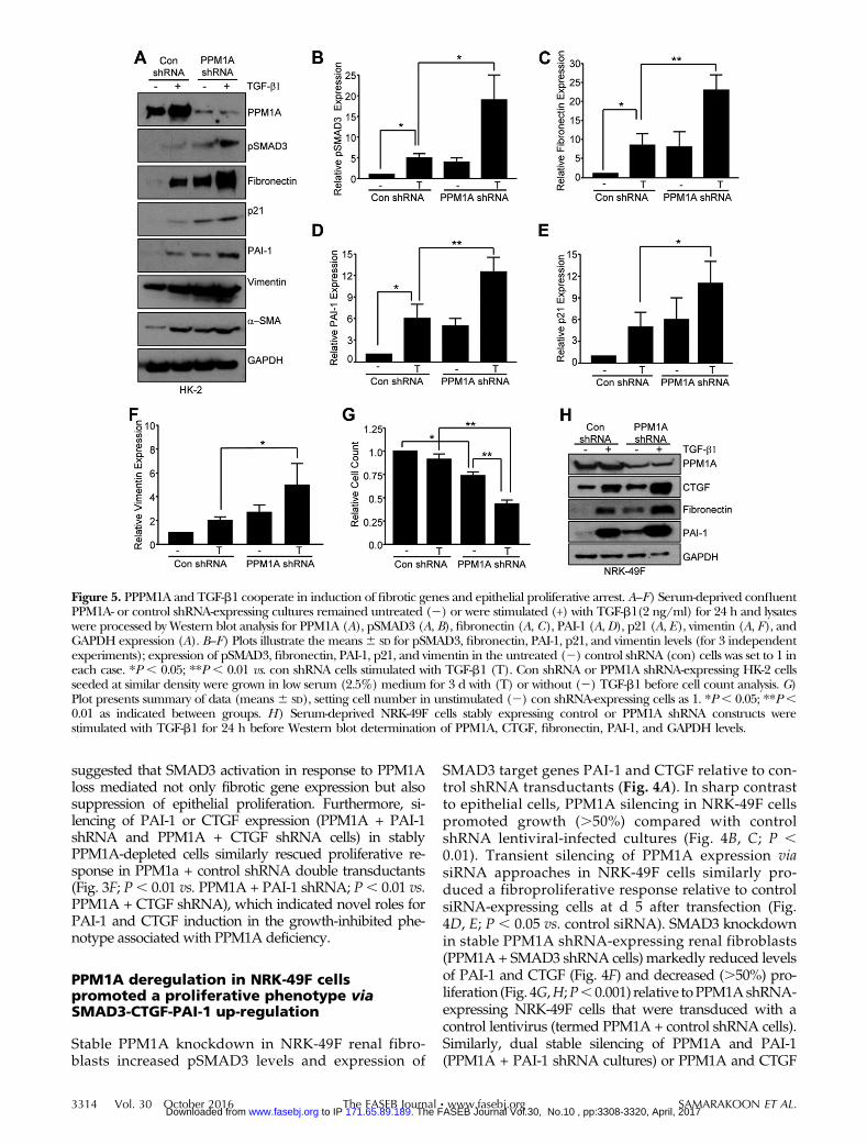
Figure 5. PPPM1A and TGF-b1 cooperate in induction of fibrotic genes and epithelial proliferative arrest. A–F) Serum-deprived confluentPPM1A- or control shRNA-expressing cultures remained untreated (2) or were stimulated (+) with TGF-b1(2 ng/ml) for 24 h and lysateswere processed by Western blot analysis for PPM1A (A), pSMAD3 (A, B), fibronectin (A, C), PAI-1 (A, D), p21 (A, E), vimentin (A, F), andGAPDH expression (A). B–F) Plots illustrate the means6 SD for pSMAD3, fibronectin, PAI-1, p21, and vimentin levels (for 3 independentexperiments); expression of pSMAD3, fibronectin, PAI-1, p21, and vimentin in the untreated (2) control shRNA (con) cells was set to 1 ineach case. *P , 0.05; **P , 0.01 vs. con shRNA cells stimulated with TGF-b1 (T). Con shRNA or PPM1A shRNA-expressing HK-2 cellsseeded at similar density were grown in low serum (2.5%) medium for 3 d with (T) or without (2) TGF-b1 before cell count analysis. G)Plot presents summary of data (means6 SD), setting cell number in unstimulated (2) con shRNA-expressing cells as 1. *P, 0.05; **P,0.01 as indicated between groups. H) Serum-deprived NRK-49F cells stably expressing control or PPM1A shRNA constructs werestimulated with TGF-b1 for 24 h before Western blot determination of PPM1A, CTGF, fibronectin, PAI-1, and GAPDH levels.
3314 Vol. 30 October 2016 SAMARAKOON ET AL.The FASEB Journal x www.fasebj.org Vol.30, No.10 , pp:3308-3320, April, 2017The FASEB Journal. 171.65.89.189 to IP www.fasebj.orgDownloaded from
(PPM1a + CTGF shRNA cells) suppressed growth relativeto PPM1A + control shRNA fibroblasts (Fig. 4F–H; P ,0.001 for both PPM1A + PAI-1 shRNA and PPM1A +CTGF shRNA). These findings established that SMAD3-dependent PAI-1 and CTGF induction in response toPPM1A silencing are critical regulators of NRK-49Fproliferation.
PPM1A depletion exacerbates TGF-b1–inducedfibrotic phenotype
Concurrent loss of PPM1A expression and TGF-b1/SMAD3 activation in fibrotic kidneys (Fig. 1) warrantedan investigation of possible cooperation betweenthese events in renal injury. TGF-b1–induced SMAD3phosphorylation (Fig. 5A, B; P , 0.05) as well as fi-bronectin (Fig. 5A, C; P , 0.01), PAI-1 (Fig. 5A, C; P ,0.01), p21 (Fig. 5A, D; P, 0.05), and vimentin (Fig. 5A, E;P , 0.05) expression in control shRNA transduc-tants was further enhanced in PPM1A-depleted HK-2cells. Moreover, TGF-b1–dependent reduction in cellnumber (;12%) in control conditions was further in-creased (;58%) by PPM1A silencing (Fig. 5F; P , 0.01).
TGF-b1–mediated fibronectin, PAI-1, and CTGF ex-pression in NRK-49F cells that were stably trans-duced with control shRNA was similarly furtheraugmented in stable PPM1A knockdown fibroblasts(Fig. 5H).
PPM1A ectopic expression inhibitsTGF-b1–induced fibrotic programming
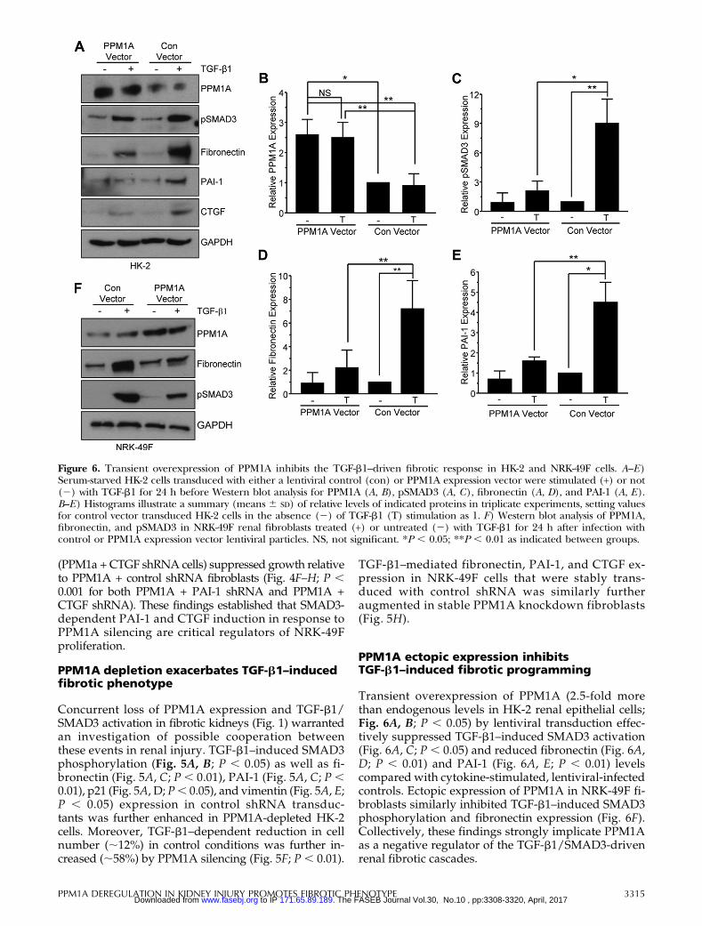
Transient overexpression of PPM1A (2.5-fold morethan endogenous levels in HK-2 renal epithelial cells;Fig. 6A, B; P , 0.05) by lentiviral transduction effec-tively suppressed TGF-b1–induced SMAD3 activation(Fig. 6A, C; P , 0.05) and reduced fibronectin (Fig. 6A,D; P , 0.01) and PAI-1 (Fig. 6A, E; P , 0.01) levelscompared with cytokine-stimulated, lentiviral-infectedcontrols. Ectopic expression of PPM1A in NRK-49F fi-broblasts similarly inhibited TGF-b1–induced SMAD3phosphorylation and fibronectin expression (Fig. 6F).Collectively, these findings strongly implicate PPM1Aas a negative regulator of the TGF-b1/SMAD3-drivenrenal fibrotic cascades.
Figure 6. Transient overexpression of PPM1A inhibits the TGF-b1–driven fibrotic response in HK-2 and NRK-49F cells. A–E)Serum-starved HK-2 cells transduced with either a lentiviral control (con) or PPM1A expression vector were stimulated (+) or not(2) with TGF-b1 for 24 h before Western blot analysis for PPM1A (A, B), pSMAD3 (A, C), fibronectin (A, D), and PAI-1 (A, E).B–E) Histograms illustrate a summary (means 6 SD) of relative levels of indicated proteins in triplicate experiments, setting valuesfor control vector transduced HK-2 cells in the absence (2) of TGF-b1 (T) stimulation as 1. F) Western blot analysis of PPM1A,fibronectin, and pSMAD3 in NRK-49F renal fibroblasts treated (+) or untreated (2) with TGF-b1 for 24 h after infection withcontrol or PPM1A expression vector lentiviral particles. NS, not significant. *P , 0.05; **P , 0.01 as indicated between groups.
PPM1A DEREGULATION IN KIDNEY INJURY PROMOTES FIBROTIC PHENOTYPE 3315 Vol.30, No.10 , pp:3308-3320, April, 2017The FASEB Journal. 171.65.89.189 to IP www.fasebj.orgDownloaded from
Epithelial paracrine factor secretionconsequent to PPM1A silencing promotesrenal fibroblast proliferation
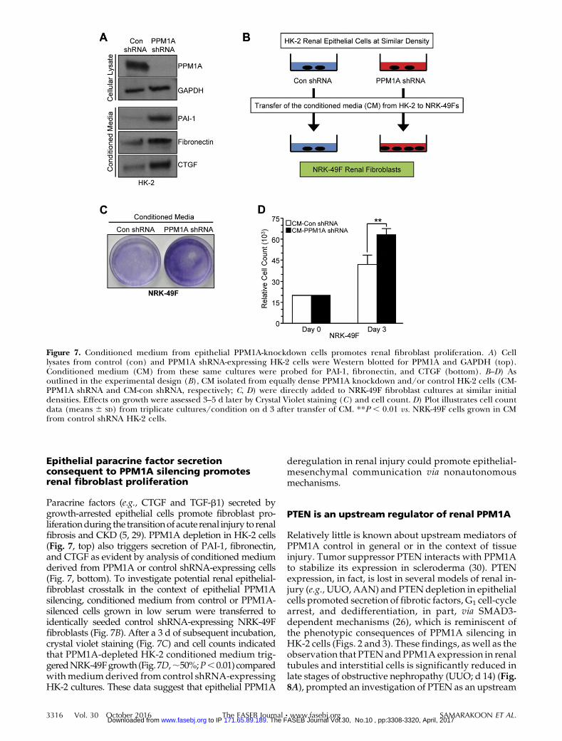
Paracrine factors (e.g., CTGF and TGF-b1) secreted bygrowth-arrested epithelial cells promote fibroblast pro-liferationduring the transitionof acute renal injury to renalfibrosis and CKD (5, 29). PPM1A depletion in HK-2 cells(Fig. 7, top) also triggers secretion of PAI-1, fibronectin,and CTGF as evident by analysis of conditioned mediumderived from PPM1A or control shRNA-expressing cells(Fig. 7, bottom). To investigate potential renal epithelial-fibroblast crosstalk in the context of epithelial PPM1Asilencing, conditioned medium from control or PPM1A-silenced cells grown in low serum were transferred toidentically seeded control shRNA-expressing NRK-49Ffibroblasts (Fig. 7B). After a 3 d of subsequent incubation,crystal violet staining (Fig. 7C) and cell counts indicatedthat PPM1A-depleted HK-2 conditioned medium trig-geredNRK-49Fgrowth (Fig. 7D,;50%;P,0.01) comparedwithmedium derived from control shRNA-expressingHK-2 cultures. These data suggest that epithelial PPM1A
deregulation in renal injury could promote epithelial-mesenchymal communication via nonautonomousmechanisms.
PTEN is an upstream regulator of renal PPM1A
Relatively little is known about upstream mediators ofPPM1A control in general or in the context of tissueinjury. Tumor suppressor PTEN interacts with PPM1Ato stabilize its expression in scleroderma (30). PTENexpression, in fact, is lost in several models of renal in-jury (e.g.,UUO, AAN) and PTENdepletion in epithelialcells promoted secretion of fibrotic factors, G1 cell-cyclearrest, and dedifferentiation, in part, via SMAD3-dependent mechanisms (26), which is reminiscent ofthe phenotypic consequences of PPM1A silencing inHK-2 cells (Figs. 2 and 3). These findings, as well as theobservation that PTENand PPM1A expression in renaltubules and interstitial cells is significantly reduced inlate stages of obstructive nephropathy (UUO; d 14) (Fig.8A), prompted an investigation of PTEN as an upstream
Figure 7. Conditioned medium from epithelial PPM1A-knockdown cells promotes renal fibroblast proliferation. A) Celllysates from control (con) and PPM1A shRNA-expressing HK-2 cells were Western blotted for PPM1A and GAPDH (top).Conditioned medium (CM) from these same cultures were probed for PAI-1, fibronectin, and CTGF (bottom). B–D) Asoutlined in the experimental design (B), CM isolated from equally dense PPM1A knockdown and/or control HK-2 cells (CM-PPM1A shRNA and CM-con shRNA, respectively; C, D) were directly added to NRK-49F fibroblast cultures at similar initialdensities. Effects on growth were assessed 3–5 d later by Crystal Violet staining (C) and cell count. D) Plot illustrates cell countdata (means 6 SD) from triplicate cultures/condition on d 3 after transfer of CM. **P , 0.01 vs. NRK-49F cells grown in CMfrom control shRNA HK-2 cells.
3316 Vol. 30 October 2016 SAMARAKOON ET AL.The FASEB Journal x www.fasebj.org Vol.30, No.10 , pp:3308-3320, April, 2017The FASEB Journal. 171.65.89.189 to IP www.fasebj.orgDownloaded from

regulator of PPM1A in renal pathology. PTEN-silencedHK-2 cells (Fig. 8B, C; 95% decrease; P , 0.001) wereused to mimic epithelial loss of PTEN expression inkidney injury. These cells, when maintained in lowserum–containing medium, not only have reduced totalPPM1A expression (;52%; Fig. 8B, D; P , 0.05) but alsodecreased nuclear PPM1A levels (.50%; Fig. 8E, F; P,0.01) as well as increased PPM1A in the cytoplasmicfraction (.2.4-fold; Fig. 8G, H; P , 0.05) comparedwith control shRNA-expressing epithelial cultures.
Decreased total PPM1A levels and its altered distri-bution (from nucleus to cytoplasm) evident in PTENknockdown cultures is associated with increased SMAD3activation compared with control shRNA transductants(Fig. 8E) (26).
Forced overexpression (.2.3-fold increase) ofPPM1A (Fig. 9A,B; P, 0.05) by lentiviral transductionin PTEN-depleted HK-2 cells (PTEN shRNA + PPM1Avector) indeed attenuated SMAD3 phosphorylation(.85%; Fig. 9A, C; P , 0.01), fibronectin expression
Figure 8. PTEN regulates PPM1A levels and intracellular localization in HK-2 cells. A) UUO and contralateral (contra) kidneys(d 14 after surgery) were immunostained with rabbit antibodies to PTEN and PPM1A. Scale bars, 60 mM. B) Cell extracts fromPTEN-knockdown and control shRNA-transduced HK-2 cells (maintained in low serum medium for 5 d) were Western blotted forPTEN, PPM1A, and GAPDH. C, D) Histograms illustrate relative PTEN and PPM1A levels (means6 SD) in PTEN-depleted HK-2 cellsfor 3 separate experiments vs. control shRNA (con) lentiviral-infected cells. E–H) Nuclear (E, F) and cytoplasmic fractions (G, H)isolated from parallel PTEN and control shRNA cultures as described in panel B were probed for PPM1A and pSMAD3 (E); laminA/C and GAPDH provided protein loading controls for the nuclear (E) and cytoplasmic (G) fractions, respectively. Plots illustratethe relative PPM1A nuclear (F) and cytoplasmic (H) levels (means6 SD) in 3 independent experiments, setting PPM1A expressionin control shRNA cultures as 1. *P , 0.05; **P , 0.01 vs. control shRNA cells; ***P , 0.001.
PPM1A DEREGULATION IN KIDNEY INJURY PROMOTES FIBROTIC PHENOTYPE 3317 Vol.30, No.10 , pp:3308-3320, April, 2017The FASEB Journal. 171.65.89.189 to IP www.fasebj.orgDownloaded from

(.53%; Fig. 9A, D; P, 0.05), and vimentin expression(Fig. 9A) relative to control vector–infected PTENshRNA-expressing cells (PTEN shRNA + control vec-tor cells; Fig. 9). Similarly, cell growth of PTEN shRNA +control vector HK-2 cultures was significantlyincreased by PPM1A ectopic expression (Fig. 9E;P , 0.01) at d 5. These findings demonstrate thatpSMAD3 inactivation by PPM1A overexpressionoverrides pSMAD3-mediated proliferative arrestimposed by epithelial PTEN depletion, which is con-sistent with previous observations (26). These dataclearly implicate PTEN as an upstream regulator ofPPM1A in the orchestration of epithelial dysfunctionin renal injury.
DISCUSSION
Loss of PPM1A expression—in both UUO and AANmodels of renal injury—initiates production of fibroticfactors, including PAI-1, CTGF, and fibronectin, and in-duces epithelial dedifferentiation,which is consistentwith
vimentin and a-SMA up-regulation, as well as epithelialG1 arrest, whereas PPM1A silencing in renal fibroblastsgenerates a proliferative phenotype. TGF-b1–stimulatedfibrotic gene expression in renal cells requires SMAD3activation (13–15) and, accordingly, ectopic expression ofPPM1A-attenuated TGF-b1–induced SMAD3 phosphor-ylation and subsequent induction of fibrotic genes in renalepithelial cells and fibroblasts (Fig. 6). Collectively, thesefindings establish PPM1A as a novel suppressor of TGF-b1/SMAD3 signaling and that prolonged PPM1A loss inrenal cells promotes a fibrotic phenotype.
PAI-1 and CTGF are known causative factors anddownstream targets of the TGF-b1/SMAD3 pathway inrenal fibrosis (10, 11, 31), yet the role CTGF and PAI-1induction in cell growth control during renal injury is lesswell understood.Our current findings establish PAI-1 andCTGF as important regulators of epithelial proliferativearrest initiated by PPM1A depletion as dual silencing ofPAI-1 or CTGF with PPM1A-rescued HK-2 cells from theproliferative inhibition imposed by PPM1A silencing(Fig. 3). In contrast, fibroblast growth in response toPPM1A silencing is blocked by SMAD3, PAI-1, or CTGF
Figure 9. Ectopic PPM1A ectopic expression reversesepithelial dysfunction induced by PTEN depletion.A–D) Extracts from PTEN-depleted HK-2 cells in-fected with lentiviruses engineered to deliver acontrol (con) vector or PPM1A expression construct(termed PTEN shRNA + con vector and PTENshRNA + PPM1A vector, respectively) were Westernblotted for PPM1A (A, B), pSMAD3 (A, C), fibronec-tin (A, D), vimentin, and GAPDH (A). Cell counts ofequally seeded PTEN shRNA + con vector– andPTEN shRNA + PPM1A vector–expressing cellswere done on d 3 and 5 of culture. E) Plot depicts cellnumber (means 6 SD) in triplicate cultures for eachcell type. *P # 0.05; **P , 0.01 vs. PTEN shRNA +con vector cells.
3318 Vol. 30 October 2016 SAMARAKOON ET AL.The FASEB Journal x www.fasebj.org Vol.30, No.10 , pp:3308-3320, April, 2017The FASEB Journal. 171.65.89.189 to IP www.fasebj.orgDownloaded from
shRNA lentiviral transduction in PPM1A-depletedNRK-49F cells (Fig. 4). PAI-1 and CTGF up-regulationdownstream of PPM1A loss, therefore, promotes renalfibroblast proliferation while imposing proliferative de-fects in epithelial cells, highlighting novel roles for PAI-1and CTGF in maladaptive cell cycle control in tubular-interstitial pathology. Moreover, loss of PPM1Aexpression in epithelial cells could promote epithelial-fibroblast communication during renal injury given thatcertain fibrotic factors (e.g., CTGF, PAI-1) secreted byPPM1A-depleted HK-2 cells promoted NRK-49F fibro-blast proliferation (Fig. 7) (29).
UUO- andAA-induced renal injury is accompanied byTGF-b1 up-regulation in the tubulointerstitium, and fi-brotic progression in bothmodels ismediated by SMAD3-dependent mechanisms (9–11, 13, 32). ConcurrentTGF-b1/SMAD3 activation and loss of PPM1A in renaltubular cells (Fig. 1) exacerbates TGF-b1–driven fibrosismarker gene expression and reduces epithelial cell growth.These studies identify PTEN as an upstream regulator ofPPM1A expression and establish that the PTEN loss-induced, SMAD3-mediated renal fibrotic phenotype islinked to PPM1A deficiency and cellular mislocalization(Fig. 8) (26). Accordingly, loss of PTEN in renal injury cor-related with decreased PPM1A expression in later stages ofrenal obstruction,which further highlights in vivo relevance.
Several recent studies suggest that PPM1A dereg-ulation in nonrenal pathologies is involved in tissueresponse to injury. Expression of VEGF and angiogen-esis, inflammation, and expression of TGF-b1 targetgenes during experimental ocular repair is further en-hanced in PPM1A-knockout mice compared with wild-type counterparts, which suggests that PPM1A alsoserves as an inhibitor of TGF-b1 signaling during eyeinjury (33). PPM1A expression is also decreased at sitesof experimental skin trauma, and PPM1A genetic
silencing in the skin in mice resulted in delayed dermalwound closure via SMAD2/3-dependent pathwayscompared with control counterparts that were identi-cally subjected to dermal injury (34).
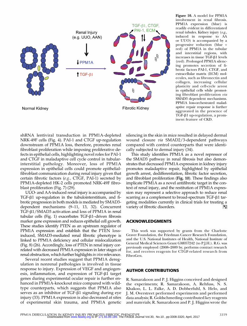
This study identifies PPM1A as a novel repressor ofthe SMAD3 pathway in renal fibrosis but also demon-strates that decreased PPM1A expression in kidney injurypromotes maladaptive repair, highlighted by epithelialgrowth arrest, dedifferentiation, fibrotic factor secretion,and fibroblast proliferation (Fig. 10). These findings alsoimplicate PPM1A as a novel antifibrotic target in the con-text of renal injury, and the restitution of PPM1A expres-sion may represent a selective approach to reduce renalscarring as a complement to broad-spectrum TGF-b1 tar-geting modalities currently in clinical trials for treating avariety of fibrotic disorders.
ACKNOWLEDGMENTS
This work was supported by grants from the CharlotteGraver Foundation, the Friedman Cancer Research Foundation,and the U.S. National Institutes of Health, National Institute ofGeneral Medical Sciences Grant GM057242 (to P.J.H.). R.G. waspreviously employed (2008–2009 by, performs contract researchfor, and receives reagents for CTGF-related research fromFibroGen.
AUTHOR CONTRIBUTIONS
R. Samarakoon and P. J. Higgins conceived and designedthe experiments; R. Samarakoon, A. Rehfuss, N. S.Khakoo, L. L. Falke, A. D. Dobberfuhl, S. Helo, andJ. M. Overstreet performed experiments and performeddata analysis; R. Goldschmeding contributed key reagentsand materials; R. Samarakoon and P. J. Higgins wrote the
Figure 10. A model for PPM1Ainvolvement in renal fibrosis.PPM1A expression (blue) isreadily evident in differentiatedrenal tubules. Kidney injury (e.g.,induced in response to AAor UUO) is accompanied by aprogressive reduction (blue +red) of PPM1A in the tubularand interstitial regions, withincreases in tissue TGF-b1 levels(red). Prolonged PPM1A silenc-ing promotes secretion of fi-brotic factors PAI-1, CTGF, andextracellular matrix (ECM) mol-ecules, such as fibronectin andcollagen, increasing cellularplasticity and cell-cycle arrestin epithelial cells while promot-ing fibroblast proliferation viaSMAD3 dependent mechanisms.PPM1A loss-orchestrated malad-aptive repair response is furtheraggravated in the presence ofTGF-b1 up-regulation, a prom-inent feature of CKD.
PPM1A DEREGULATION IN KIDNEY INJURY PROMOTES FIBROTIC PHENOTYPE 3319 Vol.30, No.10 , pp:3308-3320, April, 2017The FASEB Journal. 171.65.89.189 to IP www.fasebj.orgDownloaded from
paper; and all authors agreed on the manuscript prior tosubmission.
REFERENCES
1. Friedman, S. L., Sheppard, D., Duffield, J. S., and Violette, S. (2013)Therapy forfibroticdiseases: nearing the starting line.Sci.Transl.Med.5:167sr
2. Jha, V., Garcia-Garcia, G., Iseki, K., Li, Z., Naicker, S., Plattner, B.,Saran,R.,Wang,A. Y., andYang,C.W. (2013)Chronic kidneydisease:global dimension and perspectives. Lancet 382, 260–272
3. Perico,N., andRemuzzi,G. (2012)Chronickidneydisease: a researchand public health priority. Nephrol. Dial. Transplant. 27, iii19–iii26
4. Couser, W. G., Remuzzi, G., Mendis, S., and Tonelli, M. (2011) Thecontribution of chronic kidney disease to the global burden ofmajor noncommunicable diseases. Kidney Int. 80, 1258–1270
5. Yang, L., Humphreys, B. D., and Bonventre, J. V. (2011)Pathophysiology of acute kidney injury to chronic kidney disease:maladaptive repair. Contrib. Nephrol. 174, 149–155
6. Strutz, F., and Neilson, E. G. (2003) New insights intomechanisms offibrosis in immune renal injury. Springer Semin. Immunopathol. 24,459–476
7. Boor, P., Ostendorf, T., and Floege, J. (2010) Renal fibrosis: novelinsights intomechanisms and therapeutic targets.Nat. Rev. Nephrol. 6,643–656
8. Duffield, J. S., Lupher, M., Thannickal, V. J., and Wynn, T. A. (2013)Host responses in tissuerepair andfibrosis.Annu.Rev.Pathol.8, 241–276
9. Loeffler, I., and Wolf, G. (2014) Transforming growth factor-b andthe progression of renal disease. Nephrol. Dial. Transplant. 29, i37–i45
10. Samarakoon, R., Overstreet, J. M., and Higgins, P. J. (2013) TGF-bsignaling in tissue fibrosis: redox controls, target genes andtherapeutic opportunities. Cell. Signal. 25, 264–268
11. Samarakoon, R., Overstreet, J. M., Higgins, S. P., and Higgins, P. J.(2012) TGF-b1 → SMAD/p53/USF2 → PAI-1 transcriptional axis inureteral obstruction-induced renal fibrosis.Cell Tissue Res. 347, 117–128
12. Massague, J. (2012)TGFb signaling in context.Nat. Rev.Mol. Cell Biol.13, 616–630
13. Samarakoon, R., Dobberfuhl, A. D., Cooley, C., Overstreet, J. M.,Patel, S.,Goldschmeding,R.,Meldrum,K.K., andHiggins,P. J. (2013)Inductionof renalfibrotic genesbyTGF-b1 requiresEGFRactivation,p53 and reactive oxygen species. Cell. Signal. 25, 2198–2209
14. Overstreet, J.M., Samarakoon,R.,Gardona-Grau,D.,Goldschmeding,R., and Higgins, P. J. (2015) Tumor suppressor ATM functionsdownstream of TGF-b1 in orchestrating pro-fibrotic responses.FASEB J. 29, 1258–1268
15. Overstreet, J. M., Samarakoon, R., Meldrum, K. K., and Higgins, P. J.(2014)Redoxcontrolofp53 in the transcriptional regulationofTGF-b1target genes through SMAD cooperativity. Cell. Signal. 26, 1427–1436
16. Itoh, S., and ten Dijke, P. (2007) Negative regulation of TGF-betareceptor/Smad signal transduction. Curr. Opin. Cell Biol. 19, 176–184
17. Samarakoon, R., and Higgins, P. J. (2008) Integration of non-SMADand SMAD signaling in TGF-b1-induced plasminogen activator in-hibitor type-1 gene expression in vascular smooth muscle cells.Thromb. Haemost. 100, 976–983
18. Zeisberg, M., Hanai, J., Sugimoto, H., Mammoto, T., Charytan, D.,Strutz, F., and Kalluri, R. (2003) BMP-7 counteracts TGF-beta1-induced epithelial-to-mesenchymal transition and reverses chronicrenal injury. Nat. Med. 9, 964–968
19. Dendooven, A., van Oostrom, O., van der Giezen, D. M., Leeuwis,J.W., Snijckers,C., Joles, J.A.,Robertson,E. J.,Verhaar,M.C.,Nguyen,T. Q., and Goldschmeding, R. (2011) Loss of endogenous bonemorphogenetic protein-6 aggravates renal fibrosis. Am. J. Pathol. 178,1069–1079
20. Fukasawa,H., Yamamoto,T.,Togawa,A.,Ohashi,N., Fujigaki, Y.,Oda,T., Uchida, C., Kitagawa, K., Hattori, T., Suzuki, S., Kitagawa, M., andHishida, A. (2004) Down-regulation of Smad7 expression byubiquitin-dependent degradation contributes to renal fibrosis inobstructive nephropathy in mice. Proc. Natl. Acad. Sci. USA 101,8687–8692
21. Yang, J., Zhang, X., Li, Y., and Liu, Y. (2003)Downregulation of Smadtranscriptional corepressors SnoN and Ski in the fibrotic kidney: anamplificationmechanism for TGF-beta1 signaling. J. Am. Soc. Nephrol.14, 3167–3177
22. Fukasawa,H., Yamamoto,T.,Togawa,A.,Ohashi,N., Fujigaki, Y.,Oda,T., Uchida, C., Kitagawa, K., Hattori, T., Suzuki, S., Kitagawa, M., andHishida, A. (2006) Ubiquitin-dependent degradation of SnoN andSki is increased in renal fibrosis induced by obstructive injury. KidneyInt. 69, 1733–1740
23. Lan, H. Y., Mu, W., Tomita, N., Huang, X. R., Li, J. H., Zhu, H. J.,Morishita, R., and Johnson, R. J. (2003) Inhibition of renal fibrosis bygene transfer of inducible Smad7 using ultrasound-microbubble sys-tem in rat UUOmodel. J. Am. Soc. Nephrol. 14, 1535–1548
24. Lin,X.,Duan,X., Liang, Y. Y., Su, Y.,Wrighton,K.H., Long, J.,Hu,M.,Davis, C. M., Wang, J., Brunicardi, F. C., Shi, Y., Chen, Y. G., Meng, A.,and Feng, X. H. (2006) PPM1A functions as a Smad phosphatase toterminate TGFbeta signaling. Cell 125, 915–928
25. Samarakoon,R.,Chitnis, S. S.,Higgins, S.P.,Higgins,C.E., Krepinsky,J. C., andHiggins,P. J. (2011)Redox-inducedSrckinase and caveolin-1 signaling in TGF-b1-initiated SMAD2/3 activation and PAI-1 ex-pression. PLoS One 6, e22896
26. Samarakoon, R., Helo, S., Dobberfuhl, A. D., Khakoo, N. S., Falke, L.,Overstreet, J.M., Goldschmeding,R., andHiggins, P. J. (2015)Loss oftumour suppressor PTEN expression in renal injury initiates SMAD3-and p53-dependent fibrotic responses. J. Pathol. 236, 421–432
27. Chevalier, R. L., Forbes, M. S., and Thornhill, B. A. (2009) Ureteralobstruction as a model of renal interstitial fibrosis and obstructivenephropathy. Kidney Int. 75, 1145–1152
28. Gokmen,M. R., Cosyns, J. P., Arlt, V.M., Stiborova,M., Phillips, D.H.,Schmeiser,H.H., Simmonds,M.S.,Cook,H.T.,Vanherweghem, J.L.,Nortier, J. L., and Lord, G. M. (2013) The epidemiology, diagnosis,and management of aristolochic acid nephropathy: a narrativereview. Ann. Intern. Med. 158, 469–477
29. Yang,L., Besschetnova,T. Y., Brooks,C.R., Shah, J. V., andBonventre,J. V. (2010) Epithelial cell cycle arrest in G2/M mediates kidney fi-brosis after injury. Nat. Med. 16, 535–543, 1p, 143
30. Bu, S., Kapanadze, B.,Hsu, T., andTrojanowska,M. (2008)Oppositeeffects of dihydrosphingosine 1-phosphate and sphingosine1-phosphate on transforming growth factor-beta/Smad sig-naling are mediated through the PTEN/PPM1A-dependentpathway. J. Biol. Chem. 283, 19593–19602
31. Falke, L. L., Goldschmeding, R., and Nguyen, T. Q. (2014) Aperspective on anti-CCN2 therapy for chronic kidney disease.Nephrol.Dial. Transplant. 29, i30–i37
32. Zhou,L., Fu, P.,Huang,X.R., Liu, F.,Chung,A.C., Lai, K.N., andLan,H. Y. (2010) Mechanism of chronic aristolochic acid nephropathy:role of Smad3. Am. J. Physiol. Renal Physiol. 298, F1006–F1017
33. Dvashi, Z., Sar Shalom, H., Shohat, M., Ben-Meir, D., Ferber, S.,Satchi-Fainaro, R., Ashery-Padan, R., Rosner, M., Solomon, A. S., andLavi, S. (2014) Protein phosphatase magnesium dependent 1Agoverns the wound healing-inflammation-angiogenesis cross talk oninjury. Am. J. Pathol. 184, 2936–2950
34. Yang,X., Teng, Y., Hou,N., Fan, X., Cheng, X., Li, J., Wang, L.,Wang,Y.,Wu, X., and Yang,X. (2011)Delayed re-epithelialization in Ppm1agene-deficient mice is mediated by enhanced activation of Smad2.J. Biol. Chem. 286, 42267–42273
Received for publication January 29, 2016.Accepted for publication June 14, 2016.
3320 Vol. 30 October 2016 SAMARAKOON ET AL.The FASEB Journal x www.fasebj.org Vol.30, No.10 , pp:3308-3320, April, 2017The FASEB Journal. 171.65.89.189 to IP www.fasebj.orgDownloaded from
10.1096/fj.201500105RAccess the most recent version at doi:2016 30: 3308-3320 originally published online June 21, 2016FASEB J
Rohan Samarakoon, Alexandra Rehfuss, Nidah S. Khakoo, et al. maladaptive repair
fibroticmagnesium-dependent 1A during kidney injury promotes Loss of expression of protein phosphatase
Material
Supplemental
http://www.fasebj.org/content/suppl/2016/06/21/fj.201500105R.DC1
References
http://www.fasebj.org/content/30/10/3308.full.html#ref-list-1
This article cites 34 articles, 10 of which can be accessed free at:
Subscriptions
http://www.faseb.org/The-FASEB-Journal/Librarian-s-Resources.aspx
is online at The FASEB JournalInformation about subscribing to
Permissions
http://www.fasebj.org/site/misc/copyright.xhtmlSubmit copyright permission requests at:
Email Alerts
http://www.fasebj.org/cgi/alertsReceive free email alerts when new an article cites this article - sign up at
© FASEB
Vol.30, No.10 , pp:3308-3320, April, 2017The FASEB Journal. 171.65.89.189 to IP www.fasebj.orgDownloaded from