london cancer and macmillanlondoncancer.org/wp-content/uploads/2017/03/guide-to-coding-and... ·...
TRANSCRIPT
London Cancer and Macmillan: A guide to quality coding and safety netting in the context of Cancer
Feb 2017
EMIS Web v5
Dr Afsana Bhuiya

2
Contents
1. Foreword
2. Background to coding
3. What and how to code
a) Code the problem title
b) Use Symptom codes c) Symptom code within a consultation d) Ensuring the Problem code is saved under the most suitable category e) Cancer diagnosis f) Correctly read coding the Cancer diagnosis g) Patient history codes h) Coding urgent cancer referrals i) Screening engagement and non-attendance
i. Cervical screening ii. Bowel screening
iii. Breast screening
j) Using Templates to capture coded data k) Cancer care in the community: Living with and beyond cancer l) New registrants m) Docman coding n) Risk Prediction Algorithms o) Direct Access Investigations
4. Safety netting
a) Methods of safety netting b) Stages of safety netting
i. At the first consultation ii. With the same problem
iii. Investigations iv. Communications with the hospital v. Referrals
vi. Follow-up of patients vii. Locums and Leave
viii. Practice Administration team ix. Proactive safety netting
5. Medico-legal aspects to consider
6. Reasons to improve GP practice
7. Other systems
8. Additional information
9. Acknowledgements
3
1. Foreword
This guide to quality coding and safety netting aims to enhance patient safety by having the most up-to-date and connected information about your patient at your fingertips, regardless of how long you have known the patient. This quality improvement initiative will lead to more meaningful results from risk assessment tools, like QCancer®. Together, these processes will reduce misses, lead to earlier cancer referrals and early detection, as well as improved screening and care for patients who are with cancer and beyond. At the time of writing this guide, there are no standards to coding and safety netting. With these standards there is potential to reduce misses by tracking patients within the systems in primary care, leading to earlier cancer referrals and early detection. Late and emergency diagnosis of cancer is a significant causative factor in poor outcomes for patients. Improving earlier diagnosis of cancer, when treatments are more successful, has a positive impact on patient experience and survival. Asides from earlier detection, these skills could improve practice demographics and recalls of patient reviews and screening non-attenders. Higher quality information capturing would ensure cancer care reviews could be more relevant and person tailored. In primary care coding has been defined by the Quality and Outcome Framework (QOF), prompting usage of specific codes which in turn produce payments for the surgery. In this guide, I will look at coding beyond the financial implications but the numerous quality benefits it offers.
The quality of input – determines the quality of the output.
2. Background to coding
History of Read codes The Read code system was developed in the early 1980s by Dr James Read, who was a GP in Loughborough, to handle the problems of recording information in a way that could be retrieved from the computers available to GPs at the time. In 1989, as the importance of the system became clear and the number of codes rapidly expanded, it was purchased by the NHS Executive for further development, and to provide a uniform set of codes and terms to be used throughout the NHS. The Read codes have now been renamed the NHS Clinical Terms, although everyone still calls them Read codes or Read terms. What are Read codes? They are a comprehensive list of terms intended for use by all healthcare professionals to describe the care and treatment of their patients. They enable the capture and retrieval of patient centred information in a computer-based clinical language. What do Read codes cover? The Read code covers such topics as occupations, signs and symptoms, investigations, diagnoses, treatments and therapies, drugs and appliances (and more). The structure of the Read code system The Read codes exist in a hierarchical structure that looks like a family tree. They are arranged in chapters. Read codes in primary care electronic healthcare record software Free-typing the first few letters of a word, will auto-predict a drop-down list of words that may correspond to what the user is trying to free-type and these are Read-coded (it is similar to the predictive text on your mobile phone). The clinician can then select a Read coded word from the drop-down option. Sometimes there may be multiple Read

4
codes for a single problem and it can be confusing to decide which Read code to pick. This is historical and means the user will have to be more vigilant when choosing Read codes. I will elaborate on this further in the manual.
3. What and how to code?
Code the problem title This is putting a heading to your consultation entry, which captures the essence of the consultation. Headings act as signals for the reader and help them to organise and comprehend better what they are reading. To watch a video on how to code a problem title click here. Use Symptom codes Choosing the most relevant problem code is important to make data fit together, so patterns and connections can be made. Traditionally clinicians apply diagnosis codes to their consultations and this ties in with QOF payments too. The reality is that many interactions are likely to be around a symptom and the diagnosis comes later (or not). Cancer symptoms can be notoriously non-specific which is why it is difficult to pick up some cancers early (e.g. ovarian cancer). The NICE guidelines (revised in June 2015) regarding referrals for suspected cancer is primary care-focused and reveals a drive towards investigations and referrals for more non-specific symptoms (e.g. fatigue). Cancer risk assessment tools, like QCancer®, rely on symptoms to be coded over a period of time for the tool to be able to calculate a risk of cancer that is meaningful for the clinician. In order to improve the early detection of cancer in primary care, clinicians need to reflect more about the relevance of traditionally non-specific symptoms and read code them accordingly. This will assist to link clusters of symptoms together prompting more rapid investigations and cancer referrals. Not just diagnosis codes – but Symptom codes. As mentioned earlier, there are multiple codes for a seemingly simple problem. In general, it is advisable to pick the higher up code that fits the presenting problem. For example: Abdominal Pain – choose the first option Read code – 1969.
Figure 1. Screenshot of EMIS Web displaying the symptom code hierarchy for abdominal pain

5
Below is a list of prudent symptoms with their suggested Read code use. These codes are used in the QCancer® risk assessment toolkit. QCancer® relies on coded symptoms in order to calculate a risk.
Presenting problem Read code
Appetite loss 1612
Abnormal weight loss or unexplained weight loss 1625
Abdominal pain 1969
Abdominal swelling R0930
Difficulty swallowing liquids 1943
Difficulty in swallowing solids 1942
Indigestion symptoms 195
Heartburn 1955
Cough 171
Change in bowel habit 19EA
Constipation 19C
Painless rectal bleeding 196C
Painful rectal bleeding 196B
Blood in vomit – symptom 1994-1
Blood in sputum – haemoptysis 172
Blood in urine 1A45
Lump on neck 2I1A
Night sweats 1662-2
Spontaneous bruising 16B3
Male:
Testicular lump 2659
Pain in testicle 1A57
Cannot pass urine – retention 1A32
Urinary frequency 1A1-3
Nocturia 1A13
c/o erectile dysfunction 1D1B
Female:
Postmenopausal bleeding 1583
6
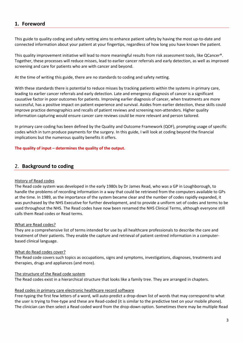
Irregular menstrual bleeding K594
Post-coital bleeding K597
Breast lump 1A8
Nipple discharge symptom 1A9
Deformation of breast 26BA
Persistent mastalgia 26BF
ADDITIONAL SYMPTOMS
Diarrhoea 1952
Diabetes (new onset) Use the usual codes. Link to pancreatic cancer in those with additional symptoms (diarrhoea, back pain,
abdominal pain, nausea etc.)
Table 1: List of top-tier symptom Read codes
To watch a video demonstrating how the integrated QCancer® risk assessment tool in EMIS Web operates and is dependent on symptoms codes click here. Symptom coding within a consultation The extent of Read coding within the consultation is not uniform and varies. Coding of physical parameters like blood pressure or weight would be coded by most physicians. What if we coded more data within the consultation? Coding the problem title has a clear advantage for users and is easy to do. Coding symptoms within the consultation may be perceived as more laborious and with less tangible gains. But coding within the consultation can be faster and easier, and the benefits could be dramatic, not only for the user but for researchers too. To watch a video demonstrating how to code symptoms within a consultation click here.
Ensuring the Problem code is saved under the most suitable category When saving a consultation there are options to categorise the entry. For example, shortness of breath symptom is saved as a clinical problem and is active and could be review. To watch a video demonstrating how to categorise consultation entries click here. Cancer diagnosis Diagnosis of cancer should always be an active problem indefinitely. This should be applied to patients who have cured cancer or have long periods of being cancer-free. This is because these patients have an increased risk of a recurrence or a second cancer. It also helps reduce the chance of the diagnosis of cancer being missed when a patient moves surgery and sees a new set of clinicians. Correctly read coding the Cancer diagnosis Coding diagnoses of cancer is not as easy as one would like. Emis Web will bring up certain codes that look acceptable, but often these are not picked up on the QOF register. Examples of unacceptable codes:
[RFC] Lung cancer (HNG0207) – NOT accepted in QOF (it is not a national code but an old EMIS LV code)
Carcinoma in situ of prostate (B834) - NOT accepted in QOF (it indicates where a cancer is, not an actual
diagnosis)
Examples of acceptable codes:
Malignant neoplasm of prostate (B46) - Accepted by QOF

7
Malignant neoplasm of bronchus or lung NOS (B22z) – Accepted by QOF
In general, if the user types in ‘malignant’ they will reach the correct code. These will start by B0, B1, B2, B3, B4, B5 or B6 are QOF codes. Codes starting with HNG, B8, B9, BA or BB are NOT QOF codes.
Type of Cancer Read Code ? Breast cancer B34 Malignant neoplasm of female breast
Ovarian cancer B440 Malignant neoplasm of ovary
Uterine cancer B43 Malignant neoplasm of body of uterus
Cervical cancer B41 Malignant neoplasm of cervix uteri
Prostate cancer B46 Malignant neoplasm of prostate
Testicular cancer B47 Malignant neoplasm of testis
Stomach cancer B11 Malignant neoplasm of stomach
Oesophageal cancer B10 Malignant neoplasm of oesophagus
Colon cancer B13 Malignant neoplasm of colon
Rectal cancer B14 Malignant neoplasm of rectum, rectosigmoid junction and anus
Bladder cancer B49 Malignant neoplasm of urinary bladder
Lung cancer B22 Malignant neoplasm of trachea, bronchus and lung
Brain cancer B51 Malignant neoplasm of brain
Skin cancer B32 Malignant melanoma of skin
Sarcoma B3 Malignant neoplasm of bone, connective tissue, skin and beast
Lymphoma B62y Malignant lymphoma NOS
Leukaemia B64
Lymphoid leukaemia
Table 2: QOF compliant common cancer diagnosis codes
Patient history codes Patient history codes which are recommended at new patient checks or ad hoc in the consultation, if it has not been captured before:
Code smoking history
Code weight – as only way to audit it and see if accurate weight drop – use electronic scales if possible
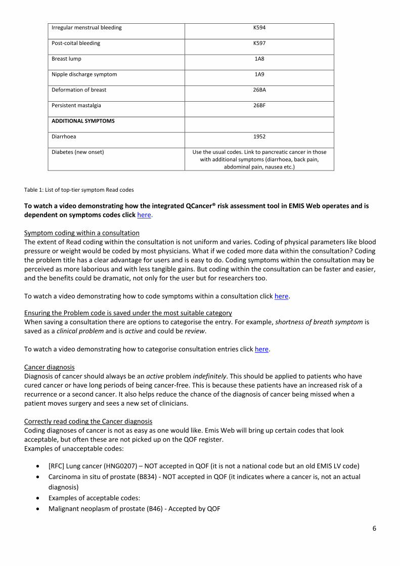
Code any family history of cancer – either by typing into consultation or using the QCancer® template
8
Figure 2. EMIS Web screenshot demonstrating how to code family history of cancer in the consultation entry
Figure 3. EMIS Web screenshot demonstrating how to code family history of cancer in the QCancer® template
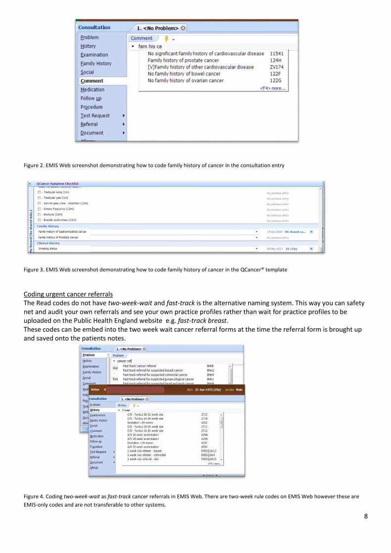
Coding urgent cancer referrals The Read codes do not have two-week-wait and fast-track is the alternative naming system. This way you can safety net and audit your own referrals and see your own practice profiles rather than wait for practice profiles to be uploaded on the Public Health England website e.g. fast-track breast. These codes can be embed into the two week wait cancer referral forms at the time the referral form is brought up and saved onto the patients notes. Figure 4. Coding two-week-wait as fast-track cancer referrals in EMIS Web. There are two-week rule codes on EMIS Web however these are
EMIS-only codes and are not transferable to other systems.
9
Screening engagement and non-attendance Screening engagement and non-attendance can be captured through appropriate coding which would enable tracing of non-attenders. The national programmes include cervical, bowel and breast screening. The variation in how screening data is processed varies much in general practice. It depends on who is responsible for coding and what systems are in place. Cervical screening:
Smear results can be sent as a paper result which gets scanned. They should be coded appropriately in order to have accurate data on non-attenders.
Smear results that come through electronically still require coding as the code applied is only to say smear was done.
An appropriate comment should be applied to the result (e.g.: abnormal smear), so this will sit in the consultation page and can be more easily picked up by any clinician seeing the patient.
A diary entry should be created or modified to ensure robust follow-up and it should reflect the correct follow-up depending on the patient’s background (e.g. once a year or every three years).
Patients with abnormal cervical cytology results are automatically contacted by the local trust for further action, but if coded in timely manner patients who have been missed can be contacted. This is particularly prudent to be able to capture transitional patients who move GP surgeries frequently.
Reminder letters are sent to patients from the screening service. GP surgeries also send reminders.
By Read coding and commenting on results, then clinicians can act ad hoc to prompt patients or actively run more accurate searches on non-attenders.
Cervical smear taken 7E2A2
Cervical smear: inadequate 4K21
Cervical smear: negative 4K22
Cervical smear: HPV positive 4K2R
Cervical smear due 685F
Cervical screen: action req. 4K4
Table 3: Relevant codes related to cervical screening.
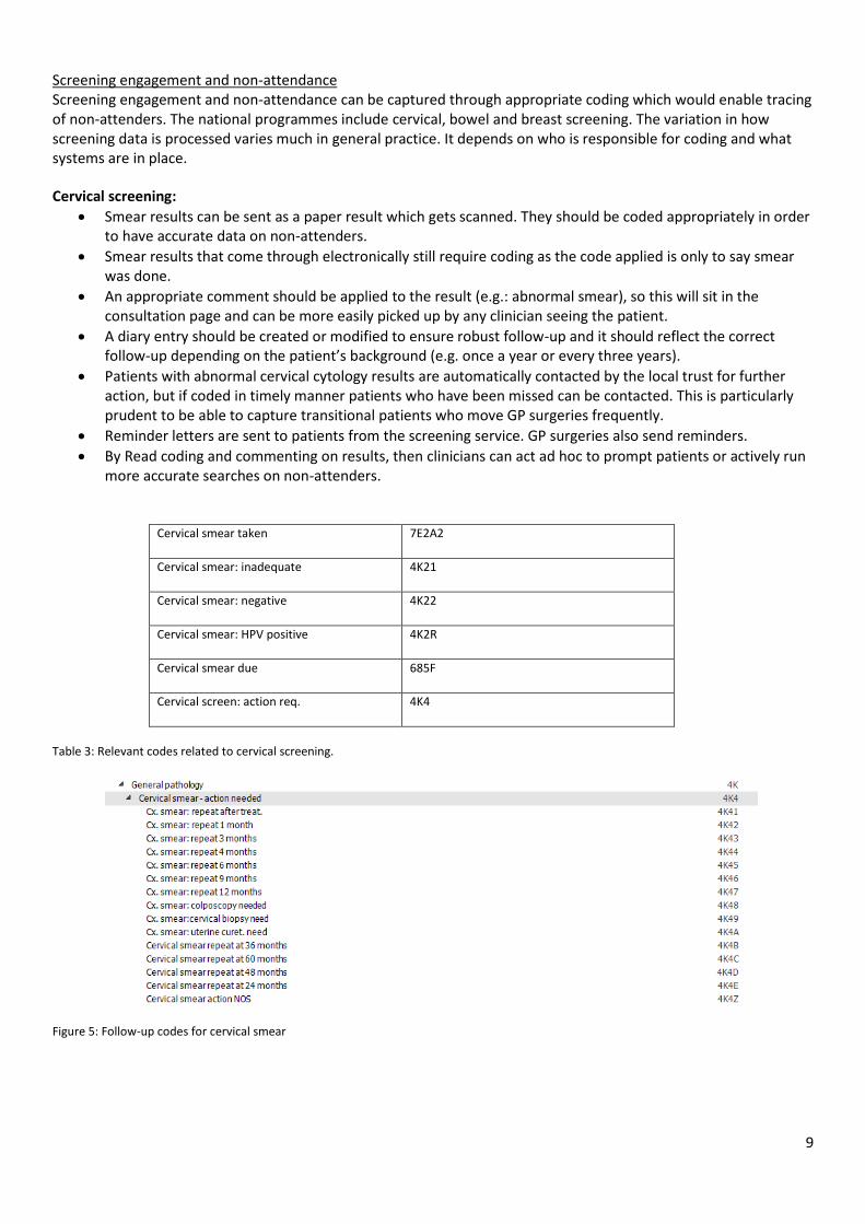
Figure 5: Follow-up codes for cervical smear

10
Bowel screening:
The majority of GP surgeries should receive bowel screening data electronically (through the same system as the pathology links). The faecal occult blood test (FOBt) result is therefore already attached to an appropriate Read code.
Some CCG areas have non-responders flagged up automatically in the EMIS pop-up alerts on the bottom right hand corner of the screen.
If the surgery receives paper notification of bowel screening results, then these should be scanned and coded appropriately in order to have accurate data on non-attenders. Electronic screening notification is clearly preferable; to enable this, the surgery could contact the local screening hub to check if this is available.
An appropriate comment should be applied to the result (e.g.: abnormal smear), so this will sit in the consultation page and can be more easily noticed by any clinician seeing the patient.
By Read coding and commenting on results then clinicians can act ad hoc to prompt patients or actively run more accurate searches on non-attenders.
The Bowel screening programme in England currently invites men and women every two years from 60 to 75 years old (extended from 70 to 75 years). GPs could play a role in advising above the invitation age to self-refer.
BCSP: Faecal occult blood test normal 686A
No response to bowel screen invitation 9Ow2
Bowel scope (flexi-sig) screen: normal – no
further action
68W21
Table 4: Relevant codes related to bowel screening
Breast screening:
Most practices receive paper notification of mammography results; these should be scanned and Read coded appropriately in order to have accurate data on non-attenders.
An appropriate Read code should be applied to the result so that it sits within the entry and can be easily noticed by any clinician seeing the patient.
By Read coding and commenting on results clinicians can either counsel patients within the consultation for screening or run more regular searches on non-attenders.
The breast screening programme in England currently invites women for screening every three years up to the age of 70 years old. The NHS cancer screening programme will extend this to 73 years by 2016. GPs could play a role in advising women above the invitation age to self-refer to the breast screening programmes.
Breast screen non-attender 9OHC
Breast screening offered 9OHF
Table 5: Relevant codes related to breast screening
Using Templates to capture coded data Templates within Emis Web can be created to make it much easier for GP’s to code relevant data without having to give much thought about recalling the correct codes. This saves time for the GP and it ensures the use of the correct read codes are used within a surgery or CCG area or wider. The coded data can be extracted from the GP surgery to ensure more accurate payment or analysis of data had occurred. Templates can be produced and used for a vast array of clinical and non-clinical scenarios within a GP practice. In the context of cancer they can be used to embed cancer care reviews or safety netting.

11
Cancer care in the community: Living with and beyond cancer Patients are gradually surviving cancer or living with stable cancer. Living with cancer is now considered a chronic disease, much like diabetes or heart failure. In this climate of disease management change, general practice will be required to adapt to manage cancer patients in the community. The care provided should align with other chronic disease management and managed holistically. Work-streams to develop support and education for primary care will be required, and coding of this information will form the foundation of good practice and ensure that the relevant information about the patient’s condition is captured within the system. Such information would include coding of cancer treatment and side effects of treatment (see table 6). There are a number of cancer care review templates present, but the amount of data and relevant data captured is very limited (see figures 6 and 7).
Radiotherapy NEC 7M371
Chemotherapy 8BAD
Hormone Therapy 5A87-1
No evidence of recurrence of cancer 1I20
Radiation proctitis J574E
Postmastectomy lymphoedema G860
Table 6: Cancer treatment and complication codes.
Figure 6: Cancer templates in EMIS Web by searching in templates

12
Figure 7: Macmillan cancer care review and GP contract cancer care review template
At London Cancer we have developed our own Cancer Care Review Template that we have found captures the holistic needs assessment and treatment summary data. It is a comprehensive template that can be covers medical and social aspects of the patient’s life and allows for care planning to be captured and shared. To watch the video demonstrating its use click here. The template file can be found the London Cancer website here. New registrants When a patient registers with a GP surgery their electronic and paper notes are transferred to the new surgery. Electronic transferring (known as GP2GP) works well when the exchange is between similar systems because the notes integrate as a seamless set of electronic notes. But if data is being transferred from Vision to EMIS, then notes get filed as a large attachment which results in a loss of previous Read codes. The exchange of paper notes wholly or partly requires summarising. There is a lot of data, including cancer data that could be missed or misfiled or not coded. Having a standard for summarizing this data may be useful (in the additional section you can find an example of this). Docman coding Intelli Sense is a function on Docman which can be used to Read code and these are compatible with EMIS. Many surgeries in London are using Docman to scan documents. This is another area where there is much variability on what data is coded. Having a protocol or a designated pathway for coding through Docman would ensure any important information about patients is Read coded. Risk Prediction Algorithms In General Practice the use of risk prediction algorithms are well established. GPs will have used tools like QRISK®2 (cardiovascular risk) and QDiabetes® (risk of developing diabetes) to help identify risk of a disease. The algorithm can be used within a consultation or be applied on a population list and used to identify patients to screen for further investigation or management. In order to maximise the accuracy of a risk predictor calculation the data the system holds on a patient should be coded as far as possible. ClinRisk has produced QCancer® which has been discussed above.
Direct Access Investigations Direct access is when a test is performed and primary care retains clinical responsibility throughout, including acting on the result. This can also be referred to as Straight to Test. Coding a Direct Test would ensure it could be tracked for better safety netting.

13
Coding is much faster and easier than it is perceived to be.
4. Safety Netting
The concept of safety netting was introduced to general practice by Roger Neighbour who considered it a core component of the GP consultation. He defined safety netting as encompassing three questions:
• If I'm right what do I expect to happen? • How will I know if I'm wrong? • What would I do then?
Safety netting is a strategy to help manage diagnostic uncertainty. It helps ensure patients undergoing investigations for, or presenting with symptoms which could indicate serious disease, are followed up in a timely and appropriate manner. The aim is to ensure patients do not drop out of the primary care net but are ‘monitored’ until their symptoms have been explained or resolved. Good safety netting is dependent on good continuity of information and record keeping/coding. In the context of cancer, NICE defines safety netting as:
1. Ensuring that the results of investigations are reviewed and acted upon appropriately, with the healthcare
professional who ordered the investigation taking or explicitly passing on responsibility for this. Being aware of
the possibility of false‑negative results for chest X‑rays and tests for occult blood in faeces. a. Considering a review for people with any symptom that is associated with an increased risk of cancer,
but who do not meet the criteria for referral or other investigative action. The review may be planned within a time frame agreed with the person or
b. Patient-initiated if new symptoms develop, the person continues to be concerned, or their symptoms recur, persist or worsen.
SUMMARY OF GOOD CODING PRACTICES IN CANCER • PROBLEM CODE all consultation entries making them relevant and capturing symptoms
as well as disease. • SYMPTOM CODING would lead to more meaning data and risk scoring. • Consultation title codes should be CATEFGORISED and LINKED accurately eg. Breast
Cancer diagnosis remains an active problem indefinitely. • Patient HISTORY Codes are captured (family history of cancer) and smoking and weight
are refreshed. • Coding REFRRALS from urgent cancer referrals, direct access and follow up plans, which
can be audited and traced. • Use TEMPLATES to capture the relevant read codes • Cancer is a long term condition and treatment and its consequences should be coded. • NEW PATIENTS should have a standard way of ensuring their history is captured
accurately. • Use risk predictors like QCancer® to assist decision making.

14
Whilst the Independent Cancer Taskforce report says: ‘It is important that GP practices continue to monitor those patients sent for an investigative test. This will ensure test results are reported and communicated, and that any abnormal results are followed up appropriately. This is especially the case if symptoms persist despite a negative test, as further testing or follow up may be required.’ And it goes on to recommend that ‘NHS England should incentivise the establishment of processes by GP practices to ensure ‘safety netting’ of patients, including adequate support for training’.
Safety netting is considered to be a core aspect to the GP consultation, yet there is virtually no guidance and very few formal documents on it. In 2011, the Oxford Department of Primary Health wrote a report entitled Cancer safety netting report. It was comprehensive and they recommended a strategy for safety netting. The highlights have been collated here. The theory and models behind safety netting are well established, and most clinicians are aware of these (e.g. Calgary-Cambridge model for consultations). Any specific recommendations of what exactly to do have been lacking.
This guide arranges and prioritises the different components of safety netting, and the methods used to safety-net in relation to cancer diagnosis in primary care (though these can be very easily applied to other diseases). The guide sets out practical steps which can be adopted very easily into daily practice. The ideal aim is to ensure safety netting has occurred and documented through coding or other electronic means.

15
Methods of safety netting
Figure 8: Highlights a range of examples and methods of safety netting used within a consultation
Electronic safety netting is the method recommended that provides practices with a rigorous, robust, traceable and auditable pro-active approach to tracking patients where needed, for example suspected cancer referrals. Examples of functions using the patient healthcare record include alerts, tasks, read codes, electronic referrals, diary entries, follow up codes and text messaging (Bhuiya, Patel).
Verbal •This is the least stringent form of safety netting. It
should be used if the clinical level of concern is minor, but the GP must be clear on their instructions. The GP should state a specific time period that the patient should return if they're not better.
•Issues: No documentation, so no auditable way to chase patients if you were concerned.
Written •Better than verbal. •This can be in the form of written records for yourself (paper
diary) or a written plan given to the patient on a piece of paper.
•Not ideal for managing systems like cancer referral lists. •Issues: Paper can go missing, be misplaced or get destroyed
more easily.
Electronic •Alert function: This can relay important information on
opening a patient record. They should be dated and deleted once the information is no longer valid.
•Tasks: Sent to GP or others.
•Electronic referrals: Use mail-merged referral letters and take advantage of emailing referrals over fax.
•Electronic pathology/imaging requests (over hand written) •Text messaging (SMS): Results, reminders etc. •Diary function: Coding actions like referrals and tagging to a
diary date means it is easier to track actions and follow up to see if they were carried out or not.

16
Stages of safety netting The below actions are recommended for a patient when the GP has reasonable concerns. These actions should be undertaken by either the GP, patient or support staff at different levels of the patient’s journey. At the first consultation the GP should:
Give the patient clear oral and written instructions.
Book the follow-up appointment.
Ensure that the patients contact details are correct, and that their mobile number is documented.
Send him/herself a patient task to remember to follow-up with the patient.
Ensure that the patient understands how and where to go for investigations, and how to get any results.
Document and code follow-up (9N7).
Review notes briefly prior to consultation to check for significant disease or if unknown patient to the doctor.
With the same problem after several consultations the GP should:
Implement investigations for recurring and/or unresolved problems.
Clear follow up plan to follow results During the investigations process the GP should:
Tell the patient to chase results within a reasonable timeframe and told how to do so.
Not rely on patient calling - significant result recall should be in place.
Relay significant results urgently and in person or telephone.
Document their recalls and any failed recalls.
Keep electronic list of worrying results
Ensure pathology comments are suitable, and that reception staff can understand them. During communication with the hospital, the GP should:
Check their local hospital pathology and radiology policies regarding how urgent results are communicated.
Phone through urgent results (this is ideal but not universal)
Ensure new clinical colleagues have pathology codes set-up so results are not sent elsewhere.
Review near misses/SEA.
Advocate that the hospital communicates new cancer diagnoses in a timely fashion.
Advocate that the hospital sends up-to-date diagnoses and treatment plans in a timely fashion.
Ask the hospital to clarify follow-up plans if there are spurious (sometimes the clinician can be contacted on NHS.net).
During the referral process the GP should:
Communicate to patient what to expect, and give them the cancer referral leaflet.
Recommend using electronic methods to send cancer referrals (many sites now have dedicated email referral).
Keep an electronic list of cancer referrals (this is made easier if referrals are coded). During follow-up of the patients:
Proactively chase non-attenders by calling or writing to them.
Consider that vulnerable patients will require more flexibility (i.e.: elderly, illicit drug user, and alcohol dependent patients etc.).
Ensure locums use electronic methods of relaying concern (i.e. tasks/alerts for patient with concerns).
Administrative staff should also document their attempts to follow-up with the patient. Strategies for locums and when you are on leave:
Give locums a Locum Pack with information on how to refer and code.

17
Ensure that the locum uses the right pathology code so results come back to the regular doctor.
Ensure results and/or letters are buddied up with another colleague if you are away.
Ensure any concerns are relayed to a colleague before taking leave. This should be documented in notes.
Functions for the Practice Administration Team
Front of house to check with existing patients and new patients their contact details are correct, in particular
telephone numbers.
Ensure systems are activated to enable patient’s opportunities to update details including online.
Take opportunities to learn from Cancer Significant Event Analysis (SEA).
Proactive safety netting (tasks to be completed by various practice staff):
QCancer QCancer®® risk stratification alerts turned on
Cancer Significant Event Analysis (SEA)performed
Cancer Audits – National and Local
Table 7: Safety netting codes which can be used
Figure 9: Summary of stages of safety netting at the practice level
Safety netting code Read code
Follow-up visit 9N7B
Follow-up appointment offered 9n7M
Asked to come, investigate result 9N75
Follow-up 9c0H
Follow-up arranged 8H8
Patient asked to make an appointment 9N7C
Active monitoring 9Ok4
Stages of safety netting 1. At the first consultation 2. With the same problem 3. Investigations – inc. DIRECT ACCESS to investigations 4. Communications with the hospital 5. Referrals 6. Follow-up of patients 7. Locums and leave 8. Practice Administration team 9. Proactive safety netting
GP/Patient
System Processes Practice/Hospital

18
5. Medico-legal aspects to consider
QCancer® can be used during a consultation with patients to calculate the absolute risk of a patient having a current as yet undiagnosed cancer, with separate risks for each type of cancer. It is a risk assessment tool and cannot diagnose cancer. The results of the calculation can then be shared with the patient and used to inform the decision to undertake further investigation or referral. Litigation concerns may be a barrier to utilisation of the calculator. I have made an enquiry with the Medical Defence Union, MDDUS and with the LMC. They have advised that transparency and cancer risk should be discussed with the patient. The extracts from MMDUS and the LMC are included here, consecutively: “I think there might be criticism of a GP who was aware that a patient had cancer or potential cancer and was also aware that that patient had failed to engage with clinical input about the matter. You would need to be very careful to make it clear that you had taken steps to contact the patient and to make the patient aware of the potential risk they were running by not engaging with clinical care. Without this, I suspect that you would find yourself open to criticism and possibly legal action. As I say, the LMC may be able to advise you more accurately in this matter but I do urge you to be cautious.”
- MDDUS “Risks to GPs include a civil action for negligence, referral to the regulator (GMC) for impaired fitness to practice by virtue of poor performance and action under the performers’ list regulations (suitability and/or efficiency). All of these may run in parallel and the criteria for action under these mechanisms are all different! “Given that using the QCancer® toolkit is prompted by symptoms, I think that failing to act if the risk were raised would certainly give rise to criticism, and have the potential for an action if the patient suffered harm as a result of failing to act in a timely way. “Non-response to a screening initiative is slightly more problematic, but my advice would be that at the very least, this should be noted on an easily visible area of the patient record; in my practice, we would also check the patient’s address and send a reminder. There is a legal precedent for requiring action on patients who do not attend an appointment for considering further investigation of a potential cancer, but I am not aware (yet!) of any action taken for not following up a screening DNA. There is an argument to be made that any such follow-up is actually the responsibility of the screening organisation. The GMC’s Good Medical Practice does not address this area specifically.”
- Dr Tony Grewal Medical Director, Londonwide LMCs
6. Reasons to improve GP practice
The reasons to improve GP practice are:
These skills will benefit all chronic and acute diseases, not just cancer patients.
These skills will help the GP to identify learning, better knowledge of patient symptom patterns, and opportunities for intervention in the patient’s behaviour.
Better coding will help those diagnosed with cancer so they are not missed. It will also aid the patients care plans, and set a foundation for tailored care plans for patients who have survived a disease and require community follow-up or awareness.
It will reduced misses
The research benefits could be immense because it could generate richer, more detailed and more accurate data. This data could be used to study and understand the patterns in disease and progression.

19
Learning these skills will improve quality in the practice (a key area for GP appraisal and revalidation).
It would improve financial measures. QOF and enhanced payments rely on accurate coding. Consequently payments are made based on the coded data. Savings may be made from less appropriate referrals because the patient’s histories are easier to follow and read.
7. Other systems
Since Read coding is a universal phenomenon, the remit of this document is for EMIS Web but the principle extends to other systems. In the UK we have a variety of electronic patient record holding systems. Three common systems used in London are Emis Web, System One and Vision. Each system has its own way of carrying out particular functions and sometimes there will not be similar functionalities across them.
8. Additional information
Presentations Coding and safety netting project presentation Papers Coding and safety netting references Coding and summarising protocol Electronic Safety Netting definition by Dr A Bhuiya and Dr I Patel.
9. Acknowledgements
Dr Terry Bowley, Macmillan GPA Dr Tania Anastasiadis, GP Tower Hamlets Dr Ishani Patel, TSCT Dr Pawan Randhev, TCST Dr Lucia Grun, Camden GP Sharon Cavanagh, London Cancer/UCLH Cancer collaborative