local hand flaps - northwestern...
TRANSCRIPT

LOCAL HAND FLAPS
BY JEROME D. CHAO, MD, JOSEPHINE M. HUANG, MD, AND THOMAS A. WIEDRICH, MD
Wounds of the hand continue to be a challenge for the hand surgeon. The repairof hand wounds has evolved from the simple, such as allowing primary closure,to the complex, such as free tissue transfer. This evolution has occurred because ofbetter understanding of the vascular supply to the skin of the hand along with thedevelopment of improved, more sophisticated surgical technique. Our review of theliterature has shown numerous local hand flaps that have stood the test of time.Though lower on the reconstructive ladder, these flaps continue to aid the handsurgeon in dealing with soft tissue losses of the hand. It would be impossible tocatalogue all local hand flaps in this article. Thus, we present many of the mostuseful and historic flaps, which we feel should be a part of every hand surgeon’sarmamentarium.
Copyright © 2001 by the American Society for Surgery of the Hand
The practitioner of hand surgery usually has analgorithm for the closure of hand wounds thatinvolve soft tissue losses. That algorithm in-
cludes the use of a combination of primary woundhealing, grafts, local flaps, distant flaps, and free tissuetransfers. Many hand wounds can be closed by simplemeasures. When more complex wounds present them-selves, hand surgeons generally rely on a few favoritelocal tissue coverage options. The advent of free tissuetransfer has made this means an attractive way to closethe most complicated wounds. A review of the liter-ature has shown a great number of local hand flapsthat have stood the test of time and can be called onas effective tools to aid the hand surgeon in dealingwith soft tissue losses of the hand. These flaps can
generally be divided into flaps for the fingertips, dig-its, thumb, palmar hand, and dorsal hand.
RANDOM FLAPS
The principles that govern the use of random flapscan be used throughout the hand and fingers.
Generally these flaps are of greater use over the dorsalsurfaces, but in the correct circumstances they can beapplied to the palmar aspect of the hand as well. Aform of transposition flap with which the hand sur-geon is familiar is the Z-Plasty (Fig 1). The Z-plastyis of great value when dealing with skin contracturesover the volar aspects of the hand, especially contrac-tures that cross the normal volar skin creases. Whenthe Z-plasty technique is used, all the limbs of theZ-plasty should be of equal length. The most commonangle Z-plasty is the 60° Z-plasty. Theoretically,there will be a 75% increase in length of the long axisof the Z-plasty. The greatest theoretical gain mathe-matically comes from a 90° Z-plasty, but this is notclinically feasible.
In the first web space, a 4-flap Z-plasty can be used.Four 60° equilateral triangles are arranged as shown
From the Division of Hand Surgery, Northwestern University Med-ical School, Chicago, IL.Address reprint requests to Thomas A. Wiedrich, MD, 448 EastOntario Street, Suite 500, Chicago, IL 60611.
Copyright © 2001 by the American Society for Surgery of the Hand1531-0914/01/0101-0003$35.00/0doi:10.1053/jssh.2001.21783
JOURNAL OF THE AMERICAN SOCIETY FOR SURGERY OF THE HAND z VOL. 1, NO. 1, FEBRUARY 2001 25
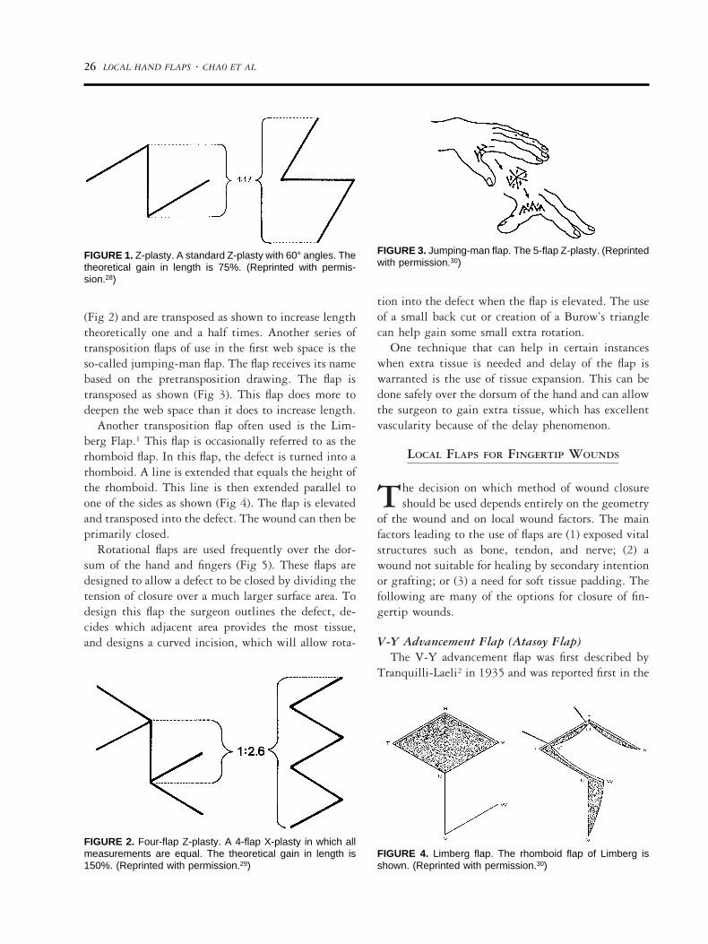
(Fig 2) and are transposed as shown to increase lengththeoretically one and a half times. Another series oftransposition flaps of use in the first web space is theso-called jumping-man flap. The flap receives its namebased on the pretransposition drawing. The flap istransposed as shown (Fig 3). This flap does more todeepen the web space than it does to increase length.
Another transposition flap often used is the Lim-berg Flap.1 This flap is occasionally referred to as therhomboid flap. In this flap, the defect is turned into arhomboid. A line is extended that equals the height ofthe rhomboid. This line is then extended parallel toone of the sides as shown (Fig 4). The flap is elevatedand transposed into the defect. The wound can then beprimarily closed.
Rotational flaps are used frequently over the dor-sum of the hand and fingers (Fig 5). These flaps aredesigned to allow a defect to be closed by dividing thetension of closure over a much larger surface area. Todesign this flap the surgeon outlines the defect, de-cides which adjacent area provides the most tissue,and designs a curved incision, which will allow rota-
tion into the defect when the flap is elevated. The useof a small back cut or creation of a Burow’s trianglecan help gain some small extra rotation.
One technique that can help in certain instanceswhen extra tissue is needed and delay of the flap iswarranted is the use of tissue expansion. This can bedone safely over the dorsum of the hand and can allowthe surgeon to gain extra tissue, which has excellentvascularity because of the delay phenomenon.
LOCAL FLAPS FOR FINGERTIP WOUNDS
The decision on which method of wound closureshould be used depends entirely on the geometry
of the wound and on local wound factors. The mainfactors leading to the use of flaps are (1) exposed vitalstructures such as bone, tendon, and nerve; (2) awound not suitable for healing by secondary intentionor grafting; or (3) a need for soft tissue padding. Thefollowing are many of the options for closure of fin-gertip wounds.
V-Y Advancement Flap (Atasoy Flap)The V-Y advancement flap was first described by
Tranquilli-Laeli2 in 1935 and was reported first in the
FIGURE 1. Z-plasty. A standard Z-plasty with 60° angles. Thetheoretical gain in length is 75%. (Reprinted with permis-sion.28)
FIGURE 2. Four-flap Z-plasty. A 4-flap X-plasty in which allmeasurements are equal. The theoretical gain in length is150%. (Reprinted with permission.29)
FIGURE 3. Jumping-man flap. The 5-flap Z-plasty. (Reprintedwith permission.30)
FIGURE 4. Limberg flap. The rhomboid flap of Limberg isshown. (Reprinted with permission.30)
26 LOCAL HAND FLAPS z CHAO ET AL

United States by Atasoy and colleagues in 1970.3 Theindications for this flap are transverse or dorsal obliqueamputations with exposed bone and sufficient nail bedsupport and length. This flap is contraindicated involar oblique tip amputations.
This flap is generally described as having the base ofthe volar triangle as the distal edge of the amputationand the apex occurring at the distal interphalangeal(DIP) crease (Fig 6). It has also been described toextend proximal to the DIP crease without sequelae.Many drawings show the base of the flap to be narrow,but it is helpful and in fact desirable to make the baseextend from radial midaxial line to ulnar midaxialline. This will increase the number of vessels flowinginto the flap. The flap is mobilized by gently dividingthe fibrous septa, which attach the skin to the deeperstructures. The deep margin of the flap is cut directlyoff the periosteum and the flexor sheath. The neuro-vascular structures should be preserved. With carefuland complete mobilization, approximately 1 cm ofadvancement can be obtained. After advancement thewound is closed as a Y; hence the name V-Y advance-ment. This flap provides excellent sensation as well assoft tissue coverage.
Bilateral Triangular Advancement Flap(Kutler Flap)
Kutler4 described a double lateral advancement flapin 1947. Shepard5 has modified this original tech-
nique. This flap has been included in most treatisesdescribing fingertip coverage. Although it is probablydescribed far more than it is actually used, there is theoccasional patient in whom this flap is useful. TheKutler flap has been condemned primarily becausegenerally the flap does not allow for advancement ofmore than 3 to 4 mm, and there is a sagittal scarplaced on the tip of the finger. Classically, this flap isindicated in patients with transverse or volar obliqueamputations. In actuality, the patient in whom thisflap is useful generally will have an amputation wherethere is more tissue on the radial and ulnar margins ofan amputation and exposed distal phalanx.
As in the V-Y advancement flap, the bases of thetriangles are the cut edges of the wound, this time theradial and ulnar aspects of the wound (Fig 7). Thedorsal edges of the 2 flaps begin 1 to 2 mm volar tothe edge of the fingernail and extend volarly 7 to 8mm. The apex of the flap sits just distal to the DIPcrease. Soft tissues are mobilized as with the V-Yadvancement flap. It is not necessary to close the skincentrally over the fingertip as long as there is good softtissue closure over the exposed distal phalanx. Thewound is then closed as a Y, again as in the V-Yadvancement flap.
Oblique Triangular FlapWith the description of the volar V-Y advancement
flap and the lateral V-Y advancement flap, Ven-
FIGURE 5. (A) Rotational flap and outline of planned skin incisions. (B) Rotational flap. Excision of Burrow’s triangle facilitatessliding of the flap toward the defect. (Reprinted with permission.28)
LOCAL HAND FLAPS z CHAO ET AL 27

kataswami and Subramanian described the obliquetriangular flap (Fig 8) in 1980.6 As suggested in itsname, establishing an oblique triangle in essence cre-ates this flap. This flap has the advantage in that it canbe used for palmar/oblique amputations and it can beconverted to a neurovascular island pedicle flap ifmore advancement is required than can be obtainedwith a standard elevation of the flap.
The flap is outlined beginning at the midlateralline and is extended proximally 2.5 times the diam-eter of the wound. The opposite midlateral line is
identified, and an oblique incision is made from thedistal midlateral wound edge to the proximal mar-gin of the midlateral incision. Care is taken topreserve the neurovascular bundle near the straightmidlateral incision. With elevation, the flap is ad-vanced, inset, and then closed in a V-Y fashion. Ifadditional mobilization is necessary, the flap is thenconverted to a neurovascular island flap (describedbelow) and advanced further into the defect. Clo-sure of the donor site with a full-thickness skingraft may be necessary.
FIGURE 6. V-Y advancement flap. (A) Skin incision andmobilization of triangular flap. (B) Advancement of flap. (C)Closure of defect with V-Y technique. (D) V-Y advancementflap. Advancement of triangular flap. (Parts A, B, and Creprinted with permission.31)
28 LOCAL HAND FLAPS z CHAO ET AL

Cross-Finger Flap (Transdigital Flap)When there is a loss of greater that one third of the
volar tissue of the fingertip—especially with exposedflexor tendon, joint, or bone—more tissue is requiredthan with advancement-type flaps. The cross-fingerflap is a popular option under these circumstances (Fig9). This technique was first described by Cronin7 in1951 and is still widely used. Multiple fingertips canbe covered simultaneously. Diseases that limit jointmotion (such as arthritis and Dupuytren’s disease) arecontraindications. Patients with impaired digital cir-culation are also considered contraindications.
There are multiple donor choices for this flap. Themiddle finger can be used to cover the thumb, index,or ring fingers. The ring finger can be used to coverthe long or small fingers. Once the donor digit hasbeen selected, the recipient finger is debrided. Theflap from the donor digit should be designed slightlylarger than the recipient defect. The flap is rectangularand is based on the midlateral line closest to therecipient digit. The plane of dissection is just dorsal tothe paratenon. Care must be taken not to injure theparatenon during elevation. The donor site is coveredwith a full-thickness skin graft, and the flap is sutured
to the recipient site. The digit is held together eitherwith a splint or with Kirschner wires. At 10 days to3 weeks, the flap is separated from the donor site andthe remainder of the flap is inset.
Variations of the cross-finger flap have been de-scribed in which dorsal nerves have been included andanastomosed to the digital nerve stump with reportedimprovement in sensation. A reverse cross-finger flaphas also been described by Atasoy8 in which theepidermis and papillary dermis are divided and thereticular dermis and subcutaneous tissue have beenused to cover the dorsum of an adjacent digit (Fig 10).The skin flap is laid back into place over the donorsite. A full-thickness graft is then placed on thereverse flap.
Hueston FlapHueston9 described a lateral palmar advancement
flap to cover the tip of the amputated finger (Fig 11).Souquet10 described a similar flap (Fig 12). The dif-ference between the 2 flaps is that the Hueston flapincludes only the neurovascular bundle at the base ofthe flap, whereas Souquet’s flap includes both neuro-vascular bundles. Both flaps are technically rotationadvancement flaps. In the Hueston flap the longitu-
FIGURE 7. Bilateral triangular advancement flap (Kutler). (A)Designing and developing 2 triangular flaps on the midlateralaspect of the distal phalanx. (B) Incisions are deepened intothe subcutaneous tissue. (C) Flaps are advanced and su-tured by converting V to Y. (Redrawn from Germann.32)
FIGURE 8. Oblique triangular flap. (A, B) Designing the flapon the side of the finger and incision. (C) Raising the flapbased on a digital neurovascular bundle. (D, E) Advancingthe flap and closing the defect with the V to Y technique.(Redrawn from Germann.32)
LOCAL HAND FLAPS z CHAO ET AL 29

dinal incision is made volar to the neurovascular bun-dle, and in the Souquet flap the incision is made dorsalto the bundle. The incision is generally 2 to 3 cmlong. A transverse back cut is made across the volaraspect of the finger, and the flap is elevated off theflexor tendon sheath. Next, the flap is elevated androtated into the defect. There will generally be atriangular defect proximally, and this must be closedwith a skin graft.
Thenar Flap (Thenar H-Flap)The thenar flap is used to cover defects and preserve
length in tip injuries to the index and long fingers(Fig 13). This flap is not used to cover defects in thering and small fingers. Contraindications to this flapare similar to that for the cross-finger flaps.
The donor site is found by taking the tip of theinjured index or ring finger and placing it against thethenar eminence. It is helpful to draw a circle around
FIGURE 9. Standard cross-finger flap. (A) The donor site is grafted. (B) Flaps raised on hinges. (C) Flaps inset and donordefects covered with full-thickness skin graft. (D) Divided and healed result. (Part A reprinted with permission.32)
30 LOCAL HAND FLAPS z CHAO ET AL

the area of contact. An H is drawn that is slightlylarger than the outlined circle. The transverse portionof the H is made at the distal-most portion of thecircle. The H is incised, and flaps in the subcutaneousplane are elevated. The ends of the flaps are sutured tothe respective dorsal and volar fingertips and to eachother along the lateral margins. This method effec-tively advances the edges of the donor defects to oneanother. The digit is left attached to the donor site forapproximately 2 weeks, and the flap is then divided byusing the proximal flap to cover the fingertip and thedistal flap to fill in the donor site.
Dorsal Middle Phalangeal Finger FlapHirase and colleagues11 described a potentially sen-
sate flap to cover the fingertip from the dorsum of thelong finger (Fig 14). The dorsal middle phalangealfinger flap has been described to be used for the tips ofany finger except the thumb. This flap requires thatdigital Allen’s test be normal in the donor digit.
The flap is outlined exactly as would be a cross-finger flap. However, the flap is incised as an island,and the base is taken volar enough to include avascular bundle. The dorsal sensory branch of the longfinger is taken with enough length proximally toperform an anastomosis with the recipient digitalnerve. The vascular pedicle is dissected proximallyeither to the level of the common digital artery oreven to the superficial arch if needed. Once the dis-section is complete, the island flap is transferred to therecipient site by means of an incision from the originof the pedicle to the recipient defect. The wound isclosed after the neural anastomosis is carried out.Finally, a full-thickness skin graft is used to close thedonor defect. Joshi12 describes a similar flap but usesit exclusively on the donor digit.
Homodigital Bipedicle Island Advancement FlapThe homodigital bipedicle island advancement flap
is useful for defects over the volar aspect of the fingerin the area of the middle phalanx. O’Brien13 andSnow14 originally described an advancement flap forinjuries to the tip of the thumb. This flap was alsoapplied to the other digits. After a wave of initialenthusiasm, the use of the flap decreased because ofproblems with flexion contractures of the digits andthe risk of dorsal skin loss. Whereas the thumb hasrelatively independent volar and dorsal circulation,the dorsum of the fingers have circulation more de-pendent on the volar blood supply. That said, there is
FIGURE 10. Reverse cross-finger flap. (A) Design of the flap.(B) Raising a full-thickness skin flap. (C) Raising a full-thick-ness subcutaneous flap in the opposite direction. (D) Suturingthe reversed flap along the defect. (Redrawn from Ger-mann.32)
FIGURE 11. Hueston flap. (A) Outline on the radial border ofthe thumb. (B) Dissection anterior to pedicle. (C) Flap inplace. (Reprinted with permission.34)
FIGURE 12. Souquet flap. (A) Outline. (B) Dissection. (C)Flap in place. (Reprinted with permission.34)
LOCAL HAND FLAPS z CHAO ET AL 31

still some value for the neurovascular island flap in thedigits. Although the flap is generally described tocover fingertip injuries, it can also be used to coverdefects of the volar middle phalanx. Damage to thedigital arteries is a contraindication to the use of thisflap. Advancement of 10 to 15 mm can usually beachieved.
A rectangular incision is made from midlateral lineto midlateral line (Fig 15). The flap is elevated, andthe radial and ulnar neurovascular bundles in the flapare preserved. The flap is elevated off the flexor sheathwith care not to expose the flexor tendons. By usinggentle traction, the neurovascular bundles are pre-served, and the flap is advanced with the aid of gentle
FIGURE 13. Thenar flaps can be based (A) proximally, (B) distally, or (C) both. The donor site is closed primarily. (D) A Thenarflap. (Parts A, B, and C reprinted with permission.32)
FIGURE 14. Dorsal middle phalangeal finger flap. Illustration of the steps for elevation. (Reprinted with permission.35)
32 LOCAL HAND FLAPS z CHAO ET AL

dissection of the neurovascular bundles. The flap isthen inset, and the donor defect is covered with afull-thickness skin graft.
Turkisk Flap (C-Ring Cross-Finger Flap)The Turkisk flap can be used to cover relatively
large defects of the volar and dorsal skin of the fingers(Fig 16). It is sometimes used to cover degloved fingerstumps. An abnormal digital Allen’s test is a contra-indication for the use of this flap.
The flap is based on one of the digital vascularbundles and can be based distally or proximally. Thedigital nerve is not included in the flap. Although theflap is based on the dorsum of the finger, it can includevolar skin of the area of the middle phalanx. As withmany of these flaps, the plane of dissection is justsuperficial to the paratenon, with care taken to leavethe paratenon intact. Although a small skin bridge isleft intact to protect the digital artery, the flap can bemade into an island flap to allow for more flap mo-
FIGURE 15. Homodigital bipedicle island advancement flap (O’Brien flap). (A) Outline. (B) Elevation of flap and proximaldissection of the pedicle. (C) Flap in place. (D) Outline of flap drawn adjacent to defect. (E) Flap sutured in place distally withfull-thickness skin graft covering secondary defect. (Parts A, B, and C reprinted with permission.34)
LOCAL HAND FLAPS z CHAO ET AL 33

bility. The flap is then inset into the recipient defect.For a degloving injury the flap can be sewn to itself tocreate a cap over the degloved segment defect. Afull-thickness skin graft is used to cover the donorsite. Flap division is carried out at 10 to 14 days.
Reverse Vascular Pedicle Digital Island FlapThe reverse vascular pedicle digital island flap was
described in 1989 by Lai et al15 and then in 1990 byKojima et al.16 Although most local coverage of thefinger relies on antegrade arterial flow, this flap func-tions on the digit much as the reverse radial forearmflap does on the hand and arm. A positive digitalAllen’s test is a contraindication to the use of this flap.
An island of skin is outlined at the base of theaffected finger (Fig 17). The flap is centered on one ofthe digital arteries. The flap is elevated from a prox-imal to distal direction and the digital vessel is ligatedproximally. Elevation of the flap continues in a distaldirection. Digital vessels are kept in the soft tissues ofthe flap. Once the island has been elevated, the digitalvessel along with a cuff of perivascular fat is taken toinclude the venae commitantes. The vessels are dis-sected distally to the level of the mid–middle phalanx.The flap is then rotated into the recipient site andsutured in place. The donor site is skin grafted.
FLAPS FOR COVERAGE OF THE VOLAR THUMB
Many of the flaps that have been described for usein covering the thumb were originally described
to aid in thumb reconstruction. With the advent ofmicrovascular toe-to-thumb transfer, some of theseflaps are used with less frequency. There still arespecific indications, however, for which knowledge ofthese flaps is of value.
Moberg Volar Advancement FlapMoberg17 first described the thumb advancement
flap in 1964 as a means to preserve length in thedistally amputated thumb. There are certain reasonswhy this flap is an attractive option for the closure ofwounds for the tip of the thumb. First, the flapvascular supply is easily identified at the end of thewound. Second, the blood supply to the dorsal aspectof the thumb is quite independent of the volar bloodsupply. Third, as opposed to the fingers, a resultantflexion contracture of the thumb generally leaves thethumb in a functional position.FIGURE 16. Steps for elevation and inset of C-ring cross-finger
flap (Turkisk flap.) (Reprinted with permission from the ChristineM. Kleinert Institute for Hand and Microsurgery, Inc.32)

Moberg’s thumb advancement flap is used whenthere are amputations through the distal phalanx. Theneurovascular bundles are identified and incisions aremade on either side of the thumb, dorsal to thebundles (Fig 18). Dissection is carried down to theflexor sheath, and the flap is elevated off the flexorsheath. Usually the proximal metacarpal phalangealcrease of the thumb is the proximal margin of thedissection. The flap is then advanced to cover thedefect. If necessary, the interphalangeal joint of thethumb can be flexed. This flap can be converted intoa bipedicle island flap to gain greater length if neces-sary. The resulting defect can be skin grafted with afull-thickness skin graft.
Neurovascular Island Pedicle FlapBefore free tissue transfer for thumb reconstruction,
the neurovascular island pedicle flap was used to at-tempt to provide sensation to an insensate thumbreconstruction. There were those surgeons who werevery enthusiastic about the flap, but there was diffi-culty in that the brain would still interpret sensory
information as coming from the donor digit. A po-tential solution was to suture the digital nerve fromthe proximal thumb to the divided distal nerve in theflap. The problems with this, however, are incompletesensory recovery and possible damage to the vascularstructures in the pedicle. Currently, the only indica-tion for the use of this flap is the scarred and sensitivethumb with ischemic pain.
The donor site is usually the ulnar aspect of thelong or ring finger. Most surgeons prefer the longfinger, because the pedicle is longer. It is essential toperform a digital Allen’s test to the donor finger aswell as to the adjacent finger before considering thisflap. Attention is first turned to the recipient site. Thescarred and sensitive area of the thumb tip is excised,and the defect is created and measured. The ulnardigital nerve is found and is prepared for anastomosis.The flap is then outlined on the donor digit and isincised, preserving the neurovascular bundle (Fig 19).The bundle is traced proximally as far as the superficialarch. As much perivascular fat as possible is kept withthe bundle. The common digital vessel with the adjacent
FIGURE 17. Steps for elevation and inset of reverse vas-cular pedicle digital island flap. (Reprinted with permis-sion.36)
LOCAL HAND FLAPS z CHAO ET AL 35

finger is divided, and the vessel is taken back to thesuperficial arch. A subcutaneous tunnel is created to therecipient site, and the island flap is passed into therecipient site. The flap should not be twisted or be tightin any way. The nerve transposition is then performed. Afull-thickness graft is used to cover the donor site.
Holevich Racquet FlapHolevich18 described the racquet flap in 1963 as a
means of restoring sensibility to the thumb by usingtissue from the dorsum of the hand, especially forchronic median nerve lesions. His flap used portions ofthe superficial branch of the radial nerve to substitute
for the lost sensation over the volar thumb. This flapcan be used to add soft pliable tissue to contractures ordefects of the first web space.
A proximally based flap is created over the base ofthe second metacarpal (Fig 20). The flap includes 2 to3 branches of the dorsal sensory branch of the radialnerve. The flap is extended distally to the level of themetacarpal head. Because the second dorsal intermeta-carpal artery is included in the flap, the flap can bemade quite narrow (2 to 3 cm) relative to its length.The flap can be easily elevated from the extensortendons. An incision is made from the donor site tothe defect on the thumb. The flap is then inset into
FIGURE 18. Moberg flap. (A) A Z-plasty at the base of the flap facilitates mobilization. (B) Volar flap advanced distally to coverdefect. (C) Advancement of flap. (D) Clinical appearance after graft has healed. (Part A reprinted with permission.28)
36 LOCAL HAND FLAPS z CHAO ET AL

the recipient site. The donor defect can sometimes beclosed primarily, but if tension of the closure is toogreat, a full-thickness graft can be used to close thedonor site.
Foucher Kite FlapAs a logical extension of the previous flap, Foucher
and Braun19 in 1979 improved on the Holevich flapby undertaking a detailed description of the anatomyof the first dorsal metacarpal artery. By defining theanatomy so precisely, they were able to devise anisland flap from the dorsum of the index finger to thethumb. This flap is an excellent choice when there isa need for soft tissue coverage on the thumb. The flaphas also been used when there is trophic scarring ofthe thumb or a sensory deficit of the thumb thatcannot be managed in other ways. The flap is contra-
indicated when the soft tissue tunnel is so scarred thatthe resulting tight tunnel would jeopardize circula-tion of the flap.
The flap is outlined over the dorsum of the proxi-mal phalanx of the index finger (Fig 21). The flap caninclude the skin overlying the metacarpophalangealjoint if necessary. An incision is then made in thedorsal first web space, and dissection is carried downto the first dorsal metacarpal artery, which arises fromthe radial artery. With a very complete dissection, apedicle of 7 to 8 cm can be created. The flap is thenincised and elevated at the level of the paratenon froma distal to proximal direction. Fascia adjacent to thesecond metacarpal along with adjacent fat are keptintact along the course of the vessels. A subcutaneoustunnel is created from the donor site to the recipientsite, and the flap is brought through, with care takennot to kink or twist the pedicle. The dorsal proximalphalanx is covered with a full-thickness skin graft.
Annular FlapGoumain et al20 described the tetrapedicled ho-
modigital island flap in 1972 (Fig 22). This flap isused especially when sensory tissue is required tocover defects of the thumb, particularly at the level ofthe proximal phalanx, but occasionally it has beenused also for the ring finger. The flap is contraindi-cated when there has been disruption of one or bothneurovascular bundles.
A circular area is outlined approximately 2 cmproximal to the edge of the soft tissue defect. Only theskin and subcutaneous tissue are dissected. The neu-rovascular bundles are freed proximally to allow the
FIGURE 19. Neurovascular island pedicle flap. The island isoutlined to include the blood supply and nerve. (Reprintedwith permission.37)
FIGURE 20. Holevich flap. (A) Skin defect on thumb. (B)Elevation of flap. (C) Flap in place with donor defect skingrafted. (Reprinted with permission.34)
FIGURE 21. Foucher kite flap. (A) Skin defect on thumb. (B)Elevation of flap. (C) Placement of flap throught subcutane-ous tunnel. (Reprinted with permission.34)
LOCAL HAND FLAPS z CHAO ET AL 37

annular flap to advance distally 10 to 12 mm. Thedistal wound can thus be closed, and the proximalwound is either grafted or allowed to heal by second-ary intention.
COVERAGE OF DEFECTS OF THE HAND AND
PROXIMAL FINGERS
W ith defects of the hand, often the tissues im-mediately adjacent to the wound are of insuf-
ficient quantity to close the wound. In some instancesrecruitment of the local tissues of the forearm arenecessary to gain adequate coverage of the wound. Theskin and subcutaneous tissues of the forearm are anexcellent match in quality and depth to cover woundsof the hand.
Dorsal Island Digital Flap (Axial Flap)Lister21 has advocated and described a flap from the
dorsal proximal phalanx of a digit that can be used tocover adjacent defects of the finger and hand. This flapusually covers defects of the ipsilateral or adjacentdigit to the level of the proximal interphalangealjoint. This flap is based on the dorsal digital artery ofthe adjacent finger.
The flap is outlined over the dorsum of the proxi-mal phalanx of the donor digits (Fig 23). Flaps up to3 3 3 cm can easily be raised. The dorsal digital arteryis identified. It generally arises as a branch of theproper digital artery but may also arise from the dorsalmetacarpal artery. When the vessel is identified, it ispreserved with the dorsal regional venous drainage.The flap is then elevated as an island from distal to
proximal, preserving the extensor paratenon. Withfull elevation of the flap, the flap is rotated intoposition, with care taken to avoid kinking of thepedicle. The donor defect is closed with a full-thick-ness skin graft.
Fillet FlapThe fillet flap should always be considered when
one is dealing with injuries requiring amputation of adigit. The soft tissues of a digit can sometimes besalvaged by fillet of the bony structures from the softtissue and skin. The flap is useful for covering palmaror dorsal hand wounds. In designing the fillet flap, thepulp tissue is usually discarded (Fig 24). A longitu-dinal incision is made according to the location of thedefect to be closed. An incision is made approximately0.5 cm proximal to the nail fold. The phalanges andtendons are excised. The flap is placed into the defectwith the base of the flap as a hinge point.
Radial Forearm Flap/Radial Forearm FascialFlap
Chang and Wang22 published their results with aretrograde flap based on the radial artery in 1980. Thisflap has proven to have wide use in the hand and evenfingers to cover relatively large defects. The flap has alsobeen popular as a free tissue transfer. Occasionally, theflap can be used as a fascial flap as well if skin andsubcutaneous tissue are not needed at the recipient site.The flap is contraindicated in instances where, eithercongenitally or because of trauma, the palmar arch isincomplete. There is discussion as to whether or not toreconstruct the radial artery after flap transfer, but thisdoes not appear to be necessary most of the time.
The flap is designed along the longitudinal axis ofthe radial artery (Fig 25). There is some variation indesigning the flap to allow for varying lengths of the
FIGURE 22. Annular flap. (A) Amputation with protrudingbone. (B) Dissection and advancement. (C) Distal closure.(Reprinted with permission.34)
FIGURE 23. Dorsal island digital flap (axial). The dorsaldigital artery may arise either from the proximal digital arteryor from the dorsal interosseous metacarpal artery. (Reprintedwith permission.38)
38 LOCAL HAND FLAPS z CHAO ET AL

pedicle. Thus, the flap can be designed to cover eitherproximal or distal hand wounds. More distal defectswill require more proximal skin paddles. Dissection isfirst carried out through the distal forearm to exposethe radial artery and the venae comitantes. When thedistal border of the flap is encountered, the flap is
dissected first from its ulnar border. The dissectioncontinues to the pedicle. The plane of dissection issuperficial to the palmaris longus and flexor carpiradialis. Just radial to the flexor carpi radialis, thepedicle will be found lying within a septum. Thepedicle and the perforators, which supply the flap, are
FIGURE 24. Fillet flap. (A) Third metacarpal and digit with extensive injury. (B, C) Dissection of soft tissue from skin. (D, E) Useof skin for coverage of defect on hand dorsum. (Reprinted with permission.39)
LOCAL HAND FLAPS z CHAO ET AL 39

kept contiguous with the flap. Dissection is thenbegun over the radial aspect of the flap, and thedissection continues radially until the septum isreached. The dissection then proceeds under the radialvascular bundle, and muscular branches are ligatedand divided. The radial artery is divided at the level ofthe proximal margin of the flap, and the dissectioncontinues distally. When the flap and pedicle areadequately dissected distally, the flap is rotated, withcare taken not to kink the radial artery. The flap andpedicle can be brought into a defect either by gener-ous tunneling or by using an incision to connect thedistal pedicle incision to the recipient site. A tunnel is
generally preferred over the radial wrist because of thesuperficial radial nerve. Care should be taken over theulnar wrist in the area of the dorsal sensory branch ofthe ulnar nerve. The donor site is then covered with asplit-thickness skin graft.
When a radial forearm fascial flap is to be used, alongitudinal or zigzag incision is used throughout theforearm. The skin and subcutaneous tissue are ele-vated, leaving the antebrachial fascia intact. The flapis designed on the fascia, and the dissection proceedsas with the standard reverse radial forearm flap. Thedonor site is closed primarily, and the fascia is coveredwith a split-thickness skin graft at the recipient site.
FIGURE 25. Radial forearm flap. (A) Flap outlined. (B) Dissection. (C) Inset. (D) Flap covering traumatic amputations with lossof soft tissue, syndactylizing the long and ring fingers. (E) After maturation of graft and division of long and ring fingers. (F) Thekey point in raising a radial forearm flap is recognizing the level of the septum containing the radial artery and its septocutaneousperforators. BR, brachioradialis; FCR, flexor carpi radialis; FDS, flexor digitorum superficialis; PL, palmaris longus; R, radius;RA, radial artery; U, ulna. (Reprinted with permission.40)
40 LOCAL HAND FLAPS z CHAO ET AL

Posterior Interosseous Forearm FlapThe posterior interosseous forearm flap was de-
scribed by Penteado, Masquelet and Chevrel23 as wellas by Zancolli and Angrigiani.24 This flap is anotherretrograde flap that is based on the posterior interosse-ous artery (PIA). Defects of the first web space,thumb, dorsal hand to the level of the proximal in-terphalangeal joints, palm, and anterior wrist are po-tentially covered by this flap. Significant injuries tothe wrist are contraindications to this flap as is anyother condition that may cause PIA thrombosis.
Flap design is carried out by drawing a line fromthe lateral epicondyle to the ulnar styloid with theelbow held at 90° of flexion (Fig 26). This line ap-proximates the septocutaneous axis of the flap. ThePIA arises at the junction of the proximal to middlethirds of this line. Along the course of the PIA thereare 7 to 14 cutaneous perforators, which supply theskin and fascia. The most proximal perforator arisesclose to the proximal/middle third junction. The cen-ter of the flap should be located distal to this perfo-rator. The PIA anastomoses with the dorsal wristarcade 2 cm proximal to the end of the drawn axis
line. Dissection is begun at this point to ascertainintegrity of the PIA. Technically, a flap 6 to 7 cmwide can be raised, but defects greater than 4 cm inwidth are difficult to close primarily.
The radial incision is created first, and a subfascialdissection is carried ulnarly. By retracting the extensordigiti quinti proprius, extensor indicis proprius, andextensor digitorum communis radially, as well as theextensor carpi ulnaris ulnarly, the septum containingthe PIA and venae comitantes can be identified. Mus-cular branches of the PIA are ligated and divided. Caremust be taken to avoid injury to the posterior in-terosseous nerve, which lies radial to the vessels. Dis-section continues proximally to the main septal per-forator (most proximal perforator). The artery isseparated from the posterior interosseous nerve. Thevessels are ligated proximal to the main perforator tothe flap. Next, the ulnar border of the flap is elevated,and the pedicle including the septum is released fromthe ulnar shaft. The flap and pedicle are dissectedproximally until enough length of the pedicle is ob-tained to rotate into the defect. The vessels cannot bekinked at the rotation point, and the most distal
FIGURE 26. (A) Posterior interosseous forearm flap. Land-marks: the epicondyle, the distal radioulnar articulation, andthe straight line that joins them. The origin of the posteriorinterosseous artery is located at the junction of the proximaland middle third of this line. The center of the flap should bebased distal to this point. (B) Loss of small finger and ulnarside of hand because of injury. (C) Flap inset into defect.(Part A reprinted with permission.34)
LOCAL HAND FLAPS z CHAO ET AL 41

vessels can be rotated 2 cm proximal to the distal endof the initial axis line.
Dorsal Ulnar Artery FlapBecker and Gilbert25 described a flap based on the
dorsal ulnar artery in 1992 (Fig 27). This flap can beused to cover defects of the dorsal or palmar hand.
To construct this flap, an incision is made 2 cmproximal to the pisiform. The flexor carpi ulnaris isretracted to reveal the origin of the dorsal branch ofthe ulnar artery, which arises 2 to 5 cm proximal tothe pisiform. The flap is then centered along the axisof the ulna with the palmaris longus as the volar limitand the extensor digitorum communis to the ringfinger as the dorsal limit of the dissection. The lengthof the flap is variable and designed to fit the defect.The rotation point of the flap is 2 to 4 cm from thepisiform, depending on the origin of the dorsal branchof the ulnar artery. The flap is elevated from proximalto distal. When the dorsal branch of the ulnar arteryis reached, the flap can be rotated and inset. The donorsite is covered with a split-thickness skin graft.
Retrograde Radial Forearm Fascial FlapWeinzweig et al26 described the retrograde radial
forearm fascial flap in 1994 (Fig 28). Some additional
clinical applications were discussed by Braun et al27 in1995. This flap is unlike the other radial forearm flapin that the radial artery is left in situ. Essentially, thisflap is a distally based turnover flap of the volarforearm fascia. Vascularity of the flap comes fromdistal perforating vessels of the radial artery. This flapcan be used to cover defects of the volar and dorsalhand.
The design of the flap is such that the flap shouldminimally be 2 to 3 cm wide. The pivot point isgenerally 5 to 8 cm proximal to the radial styloid. Inaddition, the flap needs to be designed to be largerthan the defect it is to fill. A gentle S-shaped incisionis made over the volar forearm. Skin flaps are elevatedjust deep to the hair follicles. Branches of the radialand lateral antebrachial cutaneous nerve are identifiedand freed from the subcutaneous tissue. Once the skinflaps are elevated to expose the area of flap desired, thesubcutaneous tissue and fascia are incised and elevatedin a proximal to distal direction. Dissection ends atthe pivot point, and the flap is turned over or rotatedinto position. Care needs to be taken to avoid unduetension on the flap.
There are many different problems in woundcoverage because of the many different wounds that
FIGURE 27. Dorsal ulnar artery flap. (A) Anatomy of the dorsal ulnar artery: 1, muscular branch; 2, osseous branch to thepisiform; 3, cutaneous branches (ascending and descending). (B) Elevation of the flap. (Reprinted with permission.34)
42 LOCAL HAND FLAPS z CHAO ET AL

the hand surgeon will encounter. Working know-ledge of a number of local coverage options will aid the
hand surgeon in dealing with these sometimes vexingwounds.
REFERENCES
1. Limberg A. Mathematical principles of local plastic proce-dures on the surface of the human body. Leningrad: Medgis,1946.
2. Tranquilli-Leali L. Ricostruzione dell’apice delle falangi un-gueali medianti autoplastica volare peduncolata per scorri-mento. Infort Traum Lavoro 1935;1:186-193.
3. Atasoy E, Ioakimidis E, Kasdan ML, et al. Reconstruction ofthe amputated finger tip with a triangular volar flap. A newsurgical procedure. J Bone Joint Surg Am 1970;52A:921-926.
4. Kutler W. A new method for fingertip amputation. J AmMed Assoc 1947;133:29-30.
5. Shepard GH. The use of lateral V-Y advancement flaps forfingertip reconstruction. J Hand Surg [Am] 1983;8A:254-259.
6. Venkataswami R, Subramanian N. Oblique triangular flap: anew method of repair for oblique amputations of the fingertipand thumb. Plast Reconstr Surg 1980;66:296-300.
7. Cronin T. The cross finger flap: a new method of repair. AmSurg 1951;17:419-425.
8. Atasoy E. Reversed cross-finger subcutaneous flap. J HandSurg [Am] 1982;7A:481-483.
9. Hueston J. Local flap repair of fingertip injuries. Plast Re-constr Surg 1966;37:349-350.
10. Souquet R. The asymmetric arterial advancement flap indistal pulp loss (modified Hueston’s flap). Ann Chir Main1985;4:233-238.
11. Hirase Y, Kojima T, Matsuura S. A versatile one-stage neu-rovascular flap for fingertip reconstruction: the dorsal middle
phalangeal finger flap. Plast Reconstr Surg 1992;90:1009-1015.
12. Joshi BB. A local dorsolateral island flap for restoration ofsensation after avulsion injury of fingertip pulp. Plast Recon-str Surg 1974;54:175-182.
13. O’Brien B. Neurovascular island pedicle flaps for terminalamputations and digital scars. Br J Plast Surg 1968;21:258-261.
14. Snow JW. The use of a volar flap for repair of fingertipamputations: a preliminary report. Plast Reconstr Surg 1967;40:163-168.
15. Lai CS, Lin SD, Yang CC. The reverse digital artery flap forfingertip reconstruction. Ann Plast Surg 1989;22:495-500.
16. Kojima T, Tsuchida Y, Hirase Y, et al. Reverse vascularpedicle digital island flap. Br J Plast Surg 1990;43:290-295.
17. Moberg E. Aspects of sensation in reconstructive surgery ofthe upper extremity. J Bone Joint Surg Am 1964;46A:817-825.
18. Holevich J. A new method of restoring sensibility to thethumb. J Bone Joint Surg Br 1963;45B:496-502.
19. Foucher G, Braun JB. A new island flap transfer from thedorsum of the index to the thumb. Plast Reconstr Surg1979;63:344-349.
20. Goumain AJ, Baudet J, Massard JF. Our experience withrecent thumb mutilations. Revue de Chirurgie Orthopediqueet Reparatrice de l’Appareil Moteur 1972;58:563-574.
21. Lister G. The theory of the transposition flap and its practicalapplication in the hand. Clin Plast Surg 1981;8:115-127.
22. Chang TS, Wang W. Application of microsurgery in plastic
FIGURE 28. Radial forearm fascial flap. (A) Skin and tissueloss over dorsum of forearm and hand. (B) Identification offascial radial forearm flap on volar surface of forearm. (C)Fascial flap tunneled under skin, set into defect and coveredwith a split-thickness skin graft.
LOCAL HAND FLAPS z CHAO ET AL 43

and reconstructive surgery. J Reconstr Microsurg 1984;1:55-63.
23. Penteado CV, Masquelet AC, Chevrel JP. The anatomic basisof the fascio-cutaneous flap of the posterior interosseous ar-tery. Surg Radiol Anat 1986;8:209-215.
24. Zancolli EA, Angrigiani C. Posterior interosseous island fore-arm flap. J Hand Surg [Br] 1988;13B:130-135.
25. Becker C, Gilbert A. Lambeau antebrachial des branchesdistales de l’artere cubital. Paris: Expansion Scientifique Fran-caise, 1990.
26. Weinzweig N, Chen L, Chen ZW. The distally based radialforearm fasciosubcutaneous flap with preservation of the ra-dial artery: an anatomic and clinical approach. Plast ReconstrSurg 1994;94:675-684.
27. Braun RM, Rechnic M, Neill-Cage DJ, et al. The retrograderadial fascial forearm flap: surgical rationale, technique, andclinical application. J Hand Surg [Am] 1995;20A:915-922.
28. Germann G. Principles of flap design for surgery of the hand.Atlas Hand Clin 1998;3(2):33.
29. Green DP, Hotchkiss R, Pederson WC. Green’s operativehand surgery. Vol 2. London: Churchill-Livingstone, 1998.
30. Chase RA. Historical review of skin and soft tissue coverageof the upper extremity. Hand Clin 1985;1:599-608.
31. Atasoy E, Ioakimidis E, Kasdan ML, Kutz JE, Kleinert HE.J Bone Joint Surg Am 1970;52:921-926.
32. Atasoy E, O’Neill E. Local flap coverage about the hand. AtlasHand Clin 1998;3(2):179.
33. McCarthy JG, ed. Plastic Surgery. Vol 7. Philadelphia: Saun-ders, 1989.
34. Gilbert A. Pedicle flaps of the upper limb. Philadelphia:Lippincott, 1992.
35. Hirase Y, Kojima T, Matsuura S. A versatile one-stage neu-rovascular flap for fingertip reconstruction: the dorsal middlephalangeal finger flap. Plast Reconstr Surg 1992;20:1006-1015.
36. Kojima T. Reverse vascular pedicle digital island flap. Br JPlast Surg 1990;43:290-295.
37. Marks MW, Marks C. Fundamentals of plastic surgery. Phil-adelphia: Saunders, 1997.
38. Lister G. The theory of the transposition flap and its practicalapplication in the hand. Clin Plast Surg 1981;1:115-127.
39. Chase RA, ed. Atlas of Hand Surgery. Philadelphia: Saunders,1970.
40. Martin D, Bakhach J, Casoli V, Pellisier P, Ciria-Llorens G,Khouri RK, et al. Reconstruction of the hand with forearmisland flaps. Br J Plast Surg 1990;43:290-295.
44 LOCAL HAND FLAPS z CHAO ET AL