local calcium signals induced by hyper-osmotic stress in ... · pdf filelocal calcium signals...
TRANSCRIPT

ORIGINAL PAPER
Local calcium signals induced by hyper-osmotic stressin mammalian skeletal muscle cells
Simona Apostol Æ Daniel Ursu ÆFrank Lehmann-Horn Æ Werner Melzer
Received: 18 April 2009 / Accepted: 27 April 2009
� Springer Science+Business Media B.V. 2009
Abstract Strenuous activitiy of skeletal muscle leads to
temporary osmotic dysbalance and isolated skeletal muscle
fibers exposed to osmotic stress respond with characteristic
micro-domain calcium signals. It has been suggested that
osmotic stress targets transverse tubular (TT) dihydropyr-
idine receptors (DHPRs) which normally serve as voltage-
dependent activators of Ca release via ryanodine receptor
(RyR1s) of the sarcoplasmic reticulum (SR). Here, we
pursued this hypothesis by imaging the response to
hyperosmotic solutions in both mouse skeletal muscle
fibers and myotubes. Ca fluctuations in the cell periphery of
fibers exposed to osmotic stress were accompanied by a
substantial dilation of the peripheral TT. The Ca signals
were completely inhibited by a conditioning depolarization
that inactivates the DHPR. Dysgenic myotubes, lacking the
DHP-receptor-alpha1-subunit, showed strongly reduced,
yet not completely inhibited activity when stimulated with
solutions of elevated tonicity. The results point to a mod-
ulatory, even though not essential, role of the DHP receptor
for osmotic stress-induced Ca signals in skeletal muscle.
Keywords Mouse muscle fibers � Myotubes �Transverse tubular system � Dihydropyridine receptor �Confocal calcium imaging � Osmotic stress
Introduction
Skeletal muscle cells respond to action potential (AP)
depolarization with a rapid, almost uniform increase of
their cytoplasmic calcium concentration. The release of
Ca stored in the sarcoplasmic reticulum (SR) is controlled
by voltage-sensitive L-type Ca channels (dihydropyridine
receptors, DHPRs) located in the membrane of the
transverse tubules (TTs) that conduct the AP from the
surface to the center of the cell. The DHPRs trigger the
opening of ryanodine receptors (RyR1) in the SR mem-
brane by means of a direct mechanical link (for reviews
see Melzer et al. 1995; Dulhunty 2006). Experiments on
frog skeletal muscle revealed a quantal substructure of the
voltage-controlled Ca transients as an unexpected and
important feature of the excitation–contraction (EC)
coupling mechanism (Klein et al. 1996). The quanta,
termed ‘‘sparks’’ can be studied in isolation at rest or at
low activation levels (Shirokova et al. 1999a; Klein and
Schneider 2006). In contrast to amphibian skeletal muscle
fibers, intact mammalian fibers normally show no spon-
taneous Ca sparks and depolarization recruits elementary
events of smaller amplitude, named ‘‘embers’’ that prob-
ably result from the opening of individual ryanodine
receptors (Zhou et al. 2003; Csernoch et al. 2004). They
do, however, produce sparks when the intracellular space
is exposed to artificial solutions after mechanical ablation
of the plasma membrane or permeabilizing plasma
membrane and TTs (Kirsch et al. 2001). Studies on
immature myocytes in culture showed that sparks occur-
red preferentially in TT-free regions where the voltage-
controlled Ca release was not functional, indicating a role
of the TT system and the DHPR voltage sensors in
silencing spontaneous Ca sparks (Shirokova et al. 1999b;
Zhou et al. 2006).
S. Apostol � D. Ursu � F. Lehmann-Horn � W. Melzer (&)
Institute of Applied Physiology, Ulm University,
Albert-Einstein-Allee 11, 89069 Ulm, Germany
e-mail: [email protected]
S. Apostol
Physics Department, Faculty of Sciences and Arts,
Valahia University, 24 Bd. Unirii, 0200 Targoviste, Romania
123
J Muscle Res Cell Motil
DOI 10.1007/s10974-009-9179-8

In frog muscle, Ca phenomena ranging from spark-like
spatially restricted events to oscillations and waves could
be demonstrated in response to solutions with increased
osmolarity (Chawla et al. 2001; Martin et al. 2003). Later
Wang et al. (2005) showed that enzymatically dissociated
mouse muscle fibers likewise exhibit local Ca signals under
these conditions and that fibers isolated from the mdx
mouse, an animal model for muscular dystrophy, show
dramatically enhanced activity. Both application of hyper-
osmotic solution and the return to iso-osmotic solution
after a brief hypo-osmotic challenge induced similar local
signals (Wang et al. 2005). The authors proposed a
mechanism in which osmotic stress weakens a constitutive
inhibitory control of the DHPR on the ryanodine receptor
(RyR1) thus leading to spark activity. In a subsequent
study, Martins et al. (2008) presented evidence for an
involvement of reactive oxygen species generated by
NADPH oxidase in the formation of the tonicity-induced
Ca signals. Based on experiments investigating the tonic-
ity-induced response in muscle fibers of the dystrophic mdx
mouse Teichmann et al. (2008) suggested mechano-sensi-
tive ion channels interacting with the DHP receptors within
a common macromolecular signaling complex that is sta-
bilized by cytoskeletal elements including dystrophin.
The specific aims of the present study were (1) to
characterize the tonicity-induced Ca signals by comparing
their spatio-temporal parameters under different triggering
conditions, (2) to investigate structural changes that
accompany the local Ca signals, (3) To compare mature
muscle fibers with developing myocytes regarding the
response to osmotic stress, and (4) to test the hypothesis of
DHPR involvement by studying chronically depolarized
muscle fibers and DHPR-deficient myocytes.
Materials and Methods
Muscle cell preparations
Balb/c and Sv129/J mice (age 2–6 months) were sacrificed
by exposure to CO2, followed by cervical dislocation, in
agreement with the guidelines of the local Animal Care
Committee. The interosseus muscles were excised from the
hindlimbs, and subjected to enzymatic dissociation at 37�C
for 60 min using a Krebs–Ringer solution containing
2 mg/ml type I collagenase (Sigma-Aldrich). Fibers
loosely attached to the cover slip bottom of the recording
chamber were loaded using 5 lM Fluo-4-AM (60 min,
room temperature). Experiments commenced after washing
out the dye from the chamber with isotonic Ringer’s
solution followed by 15 min of equilibration. In some
experiments, the impermeable and hydrophobic dye FM
4–64 was added to the bathing solution (5 lg/ml) to label
the plasma membrane and TT system (Vida and Emr
1995). Both dyes were purchased from Invitrogen-Molec-
ular Probes. In addition, muscle fibers cut at both ends were
manually dissected from the extensor digitorum longus
(EDL) in relaxing solution using fine scissors and tweezers
and fixed in the recording chamber by metallic clips cov-
ered with silicone. Fibers were stretched to a sarcomere
length of 3–3.7 lm and were permeabilized by a 2 min
exposure to internal solution containing 0.01% saponin
followed by washing with internal solution containing the
potassium salt of the fluorescent dye Fluo-4 (100 lM). For
experiments on myotubes we used two murine cell lines:
(1) C2C12 which show normal voltage-activated L-type Ca
inward current and Ca release (Schuhmeier et al. 2003;
Schuhmeier and Melzer 2004) and (2) GLT (Powell et al.
1996), derived from homozygous dysgenic (mdg) mice,
which lack the dihydropyridine receptor a1S subunit and
show no Ca response to depolarization. Myotubes were
cultured as described (Schuhmeier et al. 2003, 2005). Cells
were plated on carbon- and gelatin-coated coverslips loa-
ded with 5 lM Fluo-4-AM in isotonic Ringer solution
(20 min; 37�C). The coverslips with the adherent cells
were then attached to the bottom of a Plexiglas chamber
and equilibrated for at least 15 min in isotonic Ringer’s
solution.
Solutions
Cells were bathed in isotonic Ringer’s solution containing
(mM) 140 NaCl, 2 MgCl2, 2.5 CaCl2, 10 HEPES, 5 KCl,
pH 7.4, *290 mOsm. During experiments the bath solu-
tion was refreshed every 30 min using a home-made per-
fusion system. For local stimulation, solutions were
pressure-ejected from a micropipette positioned close to
the cell surface under the control of computer-gated mag-
netic valves. Pipettes were pulled from borosilicate glass
(GB150TF10, Science Products, Hofheim, Germany) using
a commercial patch pipette puller (DMZ, Zeitz Instru-
ments, Munchen, Germany). Hypertonic solutions were
prepared by increasing the osmolarity to *420 mOsm
using either 50 mM CaCl2 (Ca-HyO), as described by
Wang et al. (2005) or by adding 120 mM sucrose (S-HyO).
In other experiments mannitol or glucose were used instead
of sucrose. The osmolality was checked with a cryoscopic
osmometer (Osmomat 030, Gonotec, Berlin, Germany). In
experiments to depolarize the membrane to close to 0 mV,
an isotonic solution (High-K) with the following constitu-
ents was used (mM): 16 KCl, 92 K2SO4, 7.6 CaSO4,
1 MgSO4, 11 Glucose, 40 Sucrose, 2 TES, pH 7.4,
*290 mOsm. Solutions used for experiments with per-
meabilized fibers had the following constitution: Relaxing
solution (mM): 140 K glutamate, 10 MgCl2, 0.3 CaCl2, 10
HEPES, 1 EGTA, 5 glucose, pH 7.0 balanced with KOH.
J Muscle Res Cell Motil
123

Internal solution (mM): 140 K glutamate, 4.5 MgCl2(0.6 mM free Mg2?), 0.096 CaCl2 (100 nM free Ca2?), 10
HEPES, 0.5 EGTA, 5 glucose, 5 Na2ATP, 5 Na2 creatine
phosphate, 0.1 K5-fluo-4, pH 7.0 balanced with KOH.
Confocal microscopy
Experiments were performed using a Radiance 2000 con-
focal scanner (Bio-Rad Cell Science Division, Hemel
Hempstead, UK) adapted to an Eclipse T300 inverted
microscope (Nikon, Tokyo, Japan). For Ca imaging with
Fluo-4 we used a 609 oil immersion objective (PlanApo,
609, 1.4 N.A.; Nikon). The spatial resolution was esti-
mated as 0.25 lm in the x and y dimensions, and 0.5 lm in
the z dimension. Fluorescence was excited by the 488 nm
laser line and collected using a bandpass HQ520/30 or a
longpass HQ500LP emission filter. For membrane staining
with FM 4-64 we used a HQ600LP emission filter. In the
xy-scanning mode a series of 100–200 images (512 9 512
pixels, 750 lines s-1) was acquired at about 1.46 Hz rep-
etition frequency. Generally, transmission images were
recorded simultaneously with the confocal fluorescence
images. Line scans (xt-images) contained 512 pixels in
space (*94.9 lm) and 1,024 pixels in time (repetition
intervals 1.333, 2.000 or 6.024 ms). Within a pre-defined
region of the fiber, the position of the line was randomly
changed after each image acquisition to avoid photo-
dynamic damage. All experiments were conducted at room
temperature (21 ± 1�C).
Image analysis
Lines showing the lowest fluctuations in intensity were
automatically selected and their average used for back-
ground subtraction. The difference image was normalized
by the background (DF/F0). After filtering and setting a
threshold criterion of 0.6 times the standard deviation of
the normalized raw image, a binary image was generated
highlighting the detected signals (super-threshold events
covering at least 50 pixels). The parameters amplitude (DF/
F0), full width at half-maximum (FWHM) and full duration
at half-maximum (FDHM) were evaluated for the detected
events using the normalized difference image. For myo-
tubes a semi-automatic detection, guided by visual
inspection, was applied. In some figures we present space-
time images that were constructed from a series of xy-scans
using a specified rectangular region of interest (ROI). The
pixel intensities in one dimension of the ROI were aver-
aged thus condensing the rectangle to a line exhibiting the
averaged intensity profile. The lines were then assembled
in chronological order producing the final space-time
image (crop image). To determine global fluorescence
increase, we averaged the pixel intensities in large ROIs
covering cytoplasmic regions (excluding nuclei). The
average from 10 frames recorded before the stimulus was
used for normalization. To determine the cytoplasmic area
free of nuclei in xy images of myotubes we used a com-
bination of Matlab (The MathWorks, Natick, Massachu-
setts, USA) and Adobe Photoshop (Adobe Systems,
Munchen, Germany) tools.
Statistics
Unless otherwise stated, averaged data are presented and
plotted as means ± SEM (n = number of experiments).
Student’s two-sided t test was used to test for significant
differences of mean values. DF/F0 and FDHM were also
tested using the non-parametric Mann–Whitney U-test.
Results
Triggering local Ca signals by osmotic stress
To investigate osmotic stress-induced calcium signals, we
used local perfusion from a micropipette positioned close to
the muscle fiber as demonstrated in Fig. 1A. The pressure-
ejected hyperosmotic solution caused a decrease in fiber
diameter which could be determined from a series of
transmission images as shown in Fig. 1A and B. Simulta-
neously fluo-4 fluorescence images were recorded to
determine myoplasmic changes of the Ca concentration
(Fig. 1C–E). As described by Wang et al. (2005), spark-like
events appeared in the periphery of the fiber in a region of
10–15 lm from the surface (Fig. 1D). Kirsch et al. (2001)
termed the local Ca events identified by them in skinned
mammalian skeletal muscle fibers ‘‘elementary calcium
release events’’ (ECRE) to distinguish them from the brief
and stereotyped sparks of amphibian muscle. We adopt this
terminology for the heterogeneous signals described here
acknowledging that their mechanism of formation may be
different. Figure 1E is a space-time image constructed from
crops of a series of xy-frames and shows the temporal
evolution of fluorescence intensity (z-axis) at the border of
the fiber segment. Figure 1F exemplifies the quantification
of the changes observed during exposure to a hyperosmotic
solution according to Wang et al. (2005). The fiber diameter
change in the transmission images (Fig. 1B) was recorded
using an automatic edge detector and was converted to the
fractional change in volume (trace a) by normalizing to the
averaged values obtained from 10 images before stimula-
tion. The fluorescence images were analyzed in two ways:
(1) by determining the global increase in fluorescence
within a rectangular ROI covering almost the entire visible
fiber image (trace b) and (2) by counting the ECRE per
frame (diagram c).
J Muscle Res Cell Motil
123
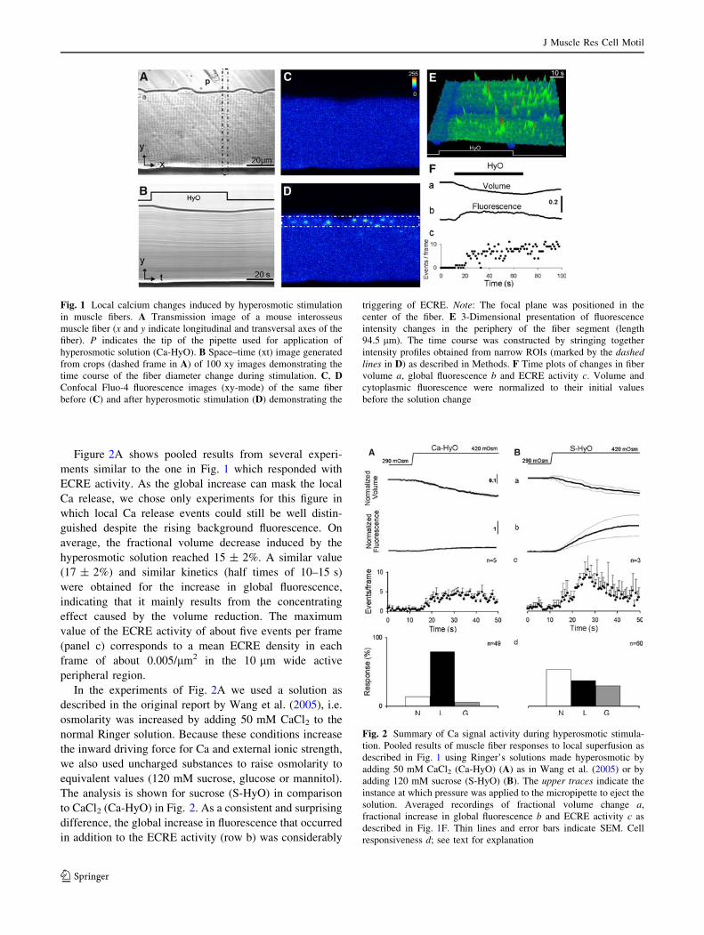
Figure 2A shows pooled results from several experi-
ments similar to the one in Fig. 1 which responded with
ECRE activity. As the global increase can mask the local
Ca release, we chose only experiments for this figure in
which local Ca release events could still be well distin-
guished despite the rising background fluorescence. On
average, the fractional volume decrease induced by the
hyperosmotic solution reached 15 ± 2%. A similar value
(17 ± 2%) and similar kinetics (half times of 10–15 s)
were obtained for the increase in global fluorescence,
indicating that it mainly results from the concentrating
effect caused by the volume reduction. The maximum
value of the ECRE activity of about five events per frame
(panel c) corresponds to a mean ECRE density in each
frame of about 0.005/lm2 in the 10 lm wide active
peripheral region.
In the experiments of Fig. 2A we used a solution as
described in the original report by Wang et al. (2005), i.e.
osmolarity was increased by adding 50 mM CaCl2 to the
normal Ringer solution. Because these conditions increase
the inward driving force for Ca and external ionic strength,
we also used uncharged substances to raise osmolarity to
equivalent values (120 mM sucrose, glucose or mannitol).
The analysis is shown for sucrose (S-HyO) in comparison
to CaCl2 (Ca-HyO) in Fig. 2. As a consistent and surprising
difference, the global increase in fluorescence that occurred
in addition to the ECRE activity (row b) was considerably
Fig. 1 Local calcium changes induced by hyperosmotic stimulation
in muscle fibers. A Transmission image of a mouse interosseus
muscle fiber (x and y indicate longitudinal and transversal axes of the
fiber). P indicates the tip of the pipette used for application of
hyperosmotic solution (Ca-HyO). B Space–time (xt) image generated
from crops (dashed frame in A) of 100 xy images demonstrating the
time course of the fiber diameter change during stimulation. C, DConfocal Fluo-4 fluorescence images (xy-mode) of the same fiber
before (C) and after hyperosmotic stimulation (D) demonstrating the
triggering of ECRE. Note: The focal plane was positioned in the
center of the fiber. E 3-Dimensional presentation of fluorescence
intensity changes in the periphery of the fiber segment (length
94.5 lm). The time course was constructed by stringing together
intensity profiles obtained from narrow ROIs (marked by the dashedlines in D) as described in Methods. F Time plots of changes in fiber
volume a, global fluorescence b and ECRE activity c. Volume and
cytoplasmic fluorescence were normalized to their initial values
before the solution change
Fig. 2 Summary of Ca signal activity during hyperosmotic stimula-
tion. Pooled results of muscle fiber responses to local superfusion as
described in Fig. 1 using Ringer’s solutions made hyperosmotic by
adding 50 mM CaCl2 (Ca-HyO) (A) as in Wang et al. (2005) or by
adding 120 mM sucrose (S-HyO) (B). The upper traces indicate the
instance at which pressure was applied to the micropipette to eject the
solution. Averaged recordings of fractional volume change a,
fractional increase in global fluorescence b and ECRE activity c as
described in Fig. 1F. Thin lines and error bars indicate SEM. Cell
responsiveness d; see text for explanation
J Muscle Res Cell Motil
123

larger when the uncharged osmolytes were used. The mean
fractional increase in global fluorescence in the case of
sucrose (S-HyO) was 110 ± 40% compared to a mean
decrease in volume by 12 ± 2%.
Further differences can be noticed from the panels in
row d of this figure in which the responsiveness of the cells
is detailed. The columns labeled N, L and G represent the
percentage of cells responding not at all (N), with local Ca
ECRE (L) and with a clearly noticeable global increase in
fluorescence (G), respectively. Here, a global increase in
fluorescence of more than 50% of the initial value was
considered a positive response. Cells showing no ECRE
and a global fluorescence increase of less than 20% were
regarded as non-responsive. The result shown in the panels
of row d confirms that a higher number of cells responded
with a strong global increase in fluorescence to S-HyO
application. On the other hand, a lower percentage of cells
showed ECRE responses and the fraction of non-respond-
ers was considerably larger.
In a separate series of experiments we applied solutions
of different osmolarities ranging from 330 to 440 mOsm
(adjusted with sucrose). The results are summarized in
Fig. 3. They show that osmolarities above 400 mM are
required to induce a robust response, but a certain, rela-
tively small percentage of the cells showed ECRE already
at the lowest osmolarity increase.
Figure 4 shows representative line scan recordings and
frequency histograms for the parameters DF/F0, FWHM
and FDHM. Figure 4A shows results from permeabilized
fibers treated as described in Kirsch et al. (2001), whereas
Fig. 4B and C present data from isolated intact interosseus
fibers stimulated with Ca-HyO and S-HyO, respectively.
To better resolve also extremely long lasting events which
were rarely observed in the permeabilized fibers but were
characteristic of fibers exposed to osmotic stress, we
increased the recording time by lowering the scanning
frequency from 750 lines/s to 500 and 166 lines/s (number
of lines per frame kept constant at 1,024; note the different
time scales in the xt-images). The histograms correspond-
ing to the fast, medium and slow scanning modes are dis-
played in white, gray and black, respectively. The
parameter values determined in these experiments are
summarized in Table 1.
Morphological changes of the TT system during
osmotic stress
A likely reason for the restriction of ECRE to the periphery
of the muscle fibers is that the tonicity-induced deforma-
tion remains confined to this region. Therefore, we sear-
ched for local structural changes occurring in parallel with
the local calcium signals. Transmission images of osmot-
ically challenged muscle fibers showed a conspicuous
‘‘graininess’’. In confocal images of fluo-4-loaded fibers
this structural change could be attributed to the appearance
of vacuoles within the fiber devoid of dye fluorescence.
Staining the plasma membrane and its transverse tubular
invaginations with the dye FM4-64 (Fig. 5A, red color)
revealed that the vacuoles resulted from alterations in the
TT system. Figure 5B shows a sequence of image crops
from xy-scans recorded at different times within a one
minute interval after application of the hyperosmotic
solution. The arrows point to the fluorescently labeled
tubules. It can be noticed that the regular double row pat-
tern of the TT system gets deranged by growing dark areas.
Applying a non-permeant fluorescent dye (fluo-4 salt) to
the extracellular space (green color, Fig. 5C) showed that
the empty spaces became filled with the dye demonstrating
their continuity with the extracellular space and identifying
them as dilated T-tubules. Like the ECRE, the TT swelling
events were most evident in the peripheral regions of the
fibers. Furthermore, the simultaneous recordings of chan-
ges in membrane morphology and intracellular Ca are
compatible with a causal relationship between the two
events. Figure 5D shows two xy-scans of a series of
recordings from a FM4-64-stainded fiber loaded with fluo-
4-AM (merged images) that contains examples of tonicity-
induced ECRE at locations which half a minute later (panel
b) showed strong TT dilation. The time course of the
changes in the region indicated with the arrow in panel a is
depicted in Fig. 5E. It was constructed by using narrow
crops from a sequence of 80 consecutive recordings. Panel
Fig. 3 Response of muscle fibers to different extracellular tonicities
Percentage of fluo-4-loaded cells showing ECRE (upper panel) and
fractional increase in global fluorescence (lower panel) in response to
Ringer’s solutions with different osmolarity (ranging from 330 to
440 mOsm). Numbers of cells tested are indicated. Asterisks indicate
a significant difference to the highest concentration (* P \ 0.05,
** P \ 0.01)
J Muscle Res Cell Motil
123

a is the fluo-4 image showing the Ca increase at the onset
of the emerging empty space and panel b the FM4-64
image demonstrating that the empty space originates from
one of the double bands representing transverse tubules.
Global and local Ca response to osmotic stress
are suppressed by fiber depolarization
To explain the osmotic stress-induced Ca signals, Wang
et al. (2005) suggested that mechanical perturbation caused
relief from a constitutive inhibition of the RyR1 Ca release
channels by their DHPR voltage sensor leading to local
release of Ca from the SR. A goal of our study was to
obtain experimental evidence for or against this hypothesis.
A long lasting depolarization converts the DHPR to an
inactivated state and prevents voltage-sensitive Ca release.
We tested whether a depolarization that produces DHPR
inactivation suppressed tonicity-induced local Ca signals.
For this purpose, we applied a modified Ringer’s solution
(High-K) in which the major cation was potassium and the
major anion sulfate (Methods, Dulhunty 1991). After
20 min in the depolarizing solution, fibers were focally
Fig. 4 Comparison of ECRE morphology in permeabilized fibers
under isosmotic conditions and in intact fibers subjected to hyperos-
motic solutions Line scan images and frequency histograms of ECRE
parameters (see text). Note: Events reaching beyond the image
boundaries were not counted. Records are from a saponin-
permeabilized EDL fiber (A) and from intact interosseus muscle
fibers stimulated with Ca-HyO (B) and S-HyO (C). The traces on the
right of each panel show the time course of the fluorescence changes
at the positions indicated by the dashed lines (numbers 1–7)
J Muscle Res Cell Motil
123

stimulated by hyperosmotic solution. Only a small increase
in basal fluorescence could be observed during the chal-
lenge (Fig. 6, lower right panel), probably caused by con-
centrating the dye due to fiber shrinkage. Importantly, none
of the fibers tested responded with ECRE (upper right
panel). In contrast, fibers in the same series of experiments
bathed in Ringer’s solution responded with a more than
100% increase in global fluorescence and showed a high
percentage of ECRE responsiveness. Thus, conditions that
chronically inactivate the DHPR appear to also make the
Ca release system refractory to osmotic stress. Revers-
ibility after the long lasting depolarization was tested in
five fibers. Three fibers responded again with ECRE on
return to Ringer’s solution.
Hypertonicity-induced TT swelling and Ca signals
in myotubes
A second approach to gain information about the role of the
DHPR is studying muscle cells deficient of this protein. As
Table 1 Morphology of ECRE in muscle fibers
Condition Event parameters (xt-images) Event density (xy-images)
Speed (lps) Amplitude (DF/F0) FWHM (lm) FDHM (ms) N Events/frame N
Ca-HyO 750 0.88 ± 0.01 1.64 ± 0.02 26.10 ± 1.23 951 4.87 ± 0.04 5,485
500 0.75 ± 0.01 1.72 ± 0.01 45.61 ± 0.83 2,091
166 0.58 ± 0.01 1.84 ± 0.01 119.83 ± 3.58 1,955
S-HyO 750 1.03 ± 0.03 1.43 ± 0.04 15.52 ± 0.75 189 4.16 ± 0.05 2,943
500 0.81 ± 0.02 1.66 ± 0.03 31.97 ± 2.53 221
166 0.58 ± 0.01 1.94 ± 0.04 144.91 ± 9.52 388
Permeabilized 750 0.56 ± 0.04 2.05 ± 0.02 17.29 ± 0.56 3,729 – –
Summary of spatio-temporal parameters obtained from permeabilized fibers under isosmotic conditions and in intact fibers subjected to
hyperosmotic conditions. See text for further explanation
Fig. 5 T-tubular dilation accompanying ECRE under hyperosmotic
conditions. A Section of an isolated muscle fiber showing plasma
membrane and transverse tubules stained with the fluorescent dye
FM4-64. B Magnified ROI near the surface of a fiber at different
times after exposure to hyperosmotic solution (Ca-HyO). Arrowsindicate T-tubules undergoing dilation. C Peripheral region of a
muscle fiber stained with FM4-64 (red) and exposed to a hyperos-
motic Ringer’s solution (S-HyO) containing 100 lM mM fluo-4
(green). The appearance of areas stained green (from fluo-4
fluorescence) within the fiber indicate dilated T-tubules with free
access to the extracellular space. D Muscle fiber section stained with
extracellular FM4-64 and intracellular fluo-4-AM at two times (see E)
after application of hyperosmotic solution (S-HyO). The arrowsindicate an ECRE in a and T-tubule dilation at the same location in b,
respectively. E Reconstructed line scan images obtained from crops
of xy-images that contained the region labelled with the arrow in (D);
panels a and b display the fluo-4 and the FM4-64 channel,
respectively. Note the Ca elevation at the beginning of the T-tubule
swelling. Black arrows and the vertical lines indicate the times when
images Da and Db were taken
J Muscle Res Cell Motil
123

DHPR-deficiency is not compatible with postnatal survival
(Adams and Beam, 1990), we studied cultured skeletal
myotubes with and without DHPR. Figure 7 depicts
experimental results from a myotube cultured from the
mouse-derived myogenic cell line C2C12 (Blau et al.
1983). Myotubes of this line show a well-developed
DHPR-mediated Ca release (Schuhmeier et al. 2003). The
procedure was similar to the one described for fibers: The
membrane portions with access to the extracellular solution
were first labeled with externally applied FM4-64; then an
external solution containing 0.1 mM fluo-4 was applied
and the extracellular osmolarity was rapidly increased by
superfusion from a micropipette. Figure 7A and B show
xy-scans of a region of the myotube before and 150 s after
raising the osmolarity to 420 mOsm. As in the isolated
muscle fibers, plasma membrane and T-tubules are stained
red by FM4-64 and the appearance of green domains in B
indicates the dilation of T-tubules with free access to the
extracellular space. The time course of the tonicity-induced
dilation of two selected tubules of the same cell is dem-
onstrated in the crop image shown in C which was con-
structed from lines (indicated in A) taken from the
individual frames of the sequence of xy-scans. This mock
line scan image also demonstrates, that the swelling is
reversible after returning from the high osmolarity back to
normal (290 mOsm).
Figure 8 shows that C2C12 myotubes responded with
local spike-like Ca events and with a concomitant more
widespread global fluorescence change as observed in
muscle fibers. Figure 8A, B and C are frames obtained before
(A) and after commencement of the hyperosmotic stimulus.
Panel D is a xt-plot constructed from crops of the xy-scans as
described before, demonstrating a pattern of localized Ca
elevations that merge into a more continuous fluorescence
increase. Figure 8E and F present a quantification of the
signals (as shown in Fig. 2) using pooled data from cells
responding to the stimulus with ECRE. Global fluorescence
intensity (panel a) was determined from a selected
Fig. 6 Local Ca response suppressed by depolarization Response of
local (upper panel) and global (lower panel) fluo-4 signals to
hyperosmotic stress (420 mOsm) in isolated muscle fibers under
conditions that favor a normal resting potential (left and middlecolumns) and under depolarizing conditions (right columns). At
normal polarization, the responsiveness was high (regardless whether
osmolarity was raised by sucrose, mannitol or glucose). However,
depolarization strongly suppressed the response. ** indicates signif-
icant difference (P \ 0.01)
Fig. 7 TT swelling in a myotube exposed to hyperosmotic stimula-
tion. A, B Two confocal xy-images of a time series showing
fluorescence from a C2C12 myotube labeled with FM4-64 (red) to
stain membranes in contact with the extracellular solution. The
solution in the extracellular space contained the cell-impermeant
variant of Fluo-4 (green). The myotube was stimulated by raising the
osmolarity to 420 mOsm (S-HyO). A Before stimulation. B 150 s
after application of hyperosmotic solution. Note the green circular
areas that appear as the result of T-tubular dilation and entry of Fluo-
4. C xt-image (crops) constructed from single lines (indicated in A)
showing the time course of TT dilation
J Muscle Res Cell Motil
123

representative ROI of nuclei-free cytoplasm. The pixel
intensities within the ROI were averaged and normalized to
the intensity average of the initial 15 frames in the series.
Local events per frame were counted and normalized by the
area of the optical section of the cell (panel b). There were
only gradual differences regarding the type of stimulus used,
i.e. whether Ca-HyO or S-HyO was applied. A characteristic
of the Ca signaling in C2C12 myotubes that differed from the
mature fibers was the occurrence of periodic oscillations of
large amplitudes. The percentage of cells exhibiting these
oscillations was similar to the fraction of cells that showed
local ECRE events (Fig. 8E and F, panel c).
Comparison of tonicity-induced local Ca signals
in myotubes and muscle fibers
Although the Ca responses elicited by hyperosmotic solu-
tion in myotubes resembled those observed in fibers, their
spatio-temporal characteristics showed differences. Fig-
ure 9A and B show line scan recordings at high time res-
olution (750 lines/s) from C2C12 myotubes and from a
muscle fiber, respectively. The scans were recorded when
the changes in diameter, induced by the hyperosmotic
solution had settled. In both preparations, short events,
lasting less then 100 ms and longer events, lasting several
100 ms, could be noticed. Figure 9C and D compare events
in myotubes and fibers at a 100-fold slower time scale.
These are ‘‘line’’-scans constructed from crops of xy-ima-
ges as described earlier. Comparing A with B and C with D
it is evident that both the spatial extent of the local events
and their duration are higher in myotubes (C) compared to
fibers (D). It should be noted that in Fig. 9C and D, the
time resolution is not sufficient to resolve the very short
events imaged in panels A and B.
Tonicity-induced Ca signals in DHP-receptor deficient
myotubes
Next, we studied DHPR-deficient myotubes. We used
myotubes of the cell line GLT (Powell et al. 1996) which
was derived from myoblasts of mice exhibiting the mus-
cular dysgenesis mutation (mdg). These mice harbor a
natural deletion mutation that eliminates expression of the
Fig. 8 Global and local Ca activity during hyperosmotic stimulation
in C2C12 myotubes. A–C Confocal xy-images of a time series
showing fluo-4 fluorescence from a C2C12 myotube stimulated with
hyperosmotic solution (Ca-HyO). Before (A) and 33 (B) and 38 s (C)
after onset of solution application. D xt-image constructed from crops
(ROI indicated in A). E, F Pooled results of myotube responses to
local superfusion using Ca-HyO and S-HyO, respectively. Averaged
recordings of fractional increase in global fluorescence (a, normalized
to the initial values before the solution change) and ECRE activity
(b). Thin lines and error bars indicate SEM. c Cell responsiveness
expressed as percentage of cells showing either no detectable
response (N), local ECRE signals (L) global increase in fluorescence
(G) or oscillations (O). Acquisition conditions: 750 lines/s, 1.46
frames/s)
c
J Muscle Res Cell Motil
123

alpha1S subunit of the L-type Ca channel (Beam et al.
1986; Knudson et al. 1989; Chaudhari 1992).
The xy-scans of Fig. 10 were obtained from a GLT
myotube before (A) and 12 (B), 16 (C) and 68 s (D) after
the onset of the solution ejection. ECRE responses can be
observed at different locations of the cell combined with a
gradual global rise in flurorescence. Figure 10E and F
quantify the responses as shown in Fig. 8E and F. When
the high-Ca solution used by Wang et al. (2005) was
applied, local events emerged in about half of the cells
tested. However the ECRE activity found in these cells was
lower than in C2C12 cells under identical conditions. A
strongly distinct response pattern was obtained when
sucrose was used to raise the osmolarity. Whereas every
other C2C12 myotube showed ECRE under these condi-
tions, only a minor fraction of the GLT myotubes respon-
ded. Together with the ECRE responsiveness, the number
of cells showing oscillations was strongly reduced. Thus,
like DHP receptor inactivation in myofibers, the lack of
DHP receptors in myotubes appears to exert a suppressing
effect on local Ca transients during osmotic stress.
Discussion
Tonicity-induced Ca signals in mouse muscle fibers
In the first part of this study, we focused on the conditions
to elicit microdomain calcium signals by hyperosmotic
stress in mouse muscle fibers (Wang et al. 2005) and on
their spatio-temporal characteristics. The extent of fiber
shrinkage and the parameters of the local calcium signals
that appeared in the cell periphery were not substantially
different when comparing the trigger conditions of Wang
et al. (2005) (Ca-HyO) with those avoiding ionic strength
changes by using sugars (predominantly sucrose, S-HyO)
in an otherwise identical experimental setting. Yet, the
ECRE responsiveness (Fig. 2) and the event density
(Table 1) were higher in Ca-HyO compared to S-HyO
indicating a contribution of the high extracellular calcium
concentration to the efficiency of the osmotic stress. A
surprising feature of muscle fibers stimulated with S-HyO
vs. Ca-HyO was an increase in the global Ca concentration
(corresponding to a uniform increase in fluorescence by
110 ± 40%) that occurred in addition to the local ECRE.
Depolarization of the fiber membrane, a possible reason for
a widespread elevation in basal Ca, seems unlikely as an
explanation because careful measurements by Teichmann
et al. (2008) showed a slight hyperpolarization rather than a
depolarization under similar conditions.
Comparing ECRE elicited by hyper-osmotic stress in
intact fibers with those appearing spontaneously in perme-
abilized fibers using the procedure of Kirsch et al. (2001), a
large part of the events were found to be short and spark-like
in both cases. However, a hallmark of the Ca activity induced
by osmotic stress both with Ca-HyO and S-HyO was the
additional frequent presence of signals with similar ampli-
tudes but much longer durations reaching several hundreds
of milliseconds (see Fig. 4B and C). Similar characteristics
were reported for the local Ca signals observed after the
return to iso-osmotic conditions after a short strong hypo-
osmotic challenge (Weisleder et al. 2007).
Alterations in transverse tubular morphology
by hypertonic solutions
Our finding of TT dilations in mouse fibers are in accord
with ultra-structural investigations in frog muscle fibers
Fig. 9 Comparison of tonicity-induced local Ca signals in myotubes
and muscle fibers. A xt-images (750 lines/s) of fluo-4-loaded C2C12
myotubes during stimulation with hypertonic solution (Ca-HyO).
B xt-image of a fluo-4-loaded muscle fiber recorded under identical
conditions. C, D Space-time images of a myotube and a fiber,
respectively, constructed from sequences of xy-images (750 lines/s)
using frame crops (each line is the average of 15 adjacent lines in the
original xy image)
J Muscle Res Cell Motil
123

exposed to hyper-osmotic solutions (Martin et al. 2003).
Under highly hyper-tonic conditions (addition of 350 mM
sucrose for 30 min) electron micrographs showed marked
dilation and vacuolation of the T-tubules combined with
SR shrinkage, whereas milder conditions (100 mM
sucrose) similar to ours caused less dilated T-tubules and
smaller reductions in SR volume. Our local perfusion
experiments traced the dynamic onset of the structural
alterations in the TT. Apparently, the hyper-osmotic solu-
tion, when entering the tubules, causes a rapid water out-
flow, predominantly from the near-surface intracellular
space, leading to tubule dilation in the peripheral fiber
regions. Our finding of ECRE co-localized with TT dilation
(Fig. 5D and E) is further evidence that the deformations of
the T-tubular membrane and the Ca signals are causally
related. Even though the expansion of the TT showed a
rapid onset, it lagged behind the ECRE activity. Never-
theless, we think that volume changes are the initial
mechanical event for the generation of the local Ca signals.
Probably the initial phase of dilation, below the resolution
of the optical microscope, is sufficient to induce transient
ECRE activity. Figure 2 (row c) shows that the mean
ECRE activity reaches its maximal value before the cor-
responding volume change approaches its final value. This
likewise indicates that relatively small volume changes are
sufficient to induce local Ca transients. In fact, we found
that also considerably lower osmolarities were sufficient to
induce ECRE, yet at a much reduced success rate.
Osmotic stress situations occur during physiological
conditions in skeletal muscle. Strenuous activity leads to the
extrusion of osmotically active substances. Vacuole forma-
tion, caused by local deletions of the TT system has been
observed in Xenopus skeletal muscle following fatiguing
activity (reviewed by Lannergren et al. 2002) and has been
attributed to the accumulation of osmolytes, in particular
lactate, in the TT lumen (Lannergren et al. 2000). Mamma-
lian unlike amphibian muscle showed no vacuole formation
under identical conditions. TT dilations were, however, no-
ticable when external lactate was applied during the fatigu-
ing activity indicating that normally lactate may be too
rapidly extruded from the TT lumen for vacuolation to occur
(Lannergren et al. 2000). Still, volume changes too subtle to
be detected in the confocal images may cause alterations of
the triadic micro-structure. Such changes have been sug-
gested by Lannergren et al. (2002) to influence excitation-
contraction coupling explaining the loss of force in low
frequency fatigue. Experiments assessing ultra-structural
changes of frog muscle triads after fatiguing activity, that
revealed increases in TT diameter below the resolution of the
light microscope and an increase in TT-SR distance, support
this view (Usher-Smith et al. 2007).
Role of the DHP receptor in tonicity-induced Ca signals
The large changes in transverse tubular morphology
observed here might explain the local Ca fluctuations by
Fig. 10 Ca signals in response to hyperosmotic stimulation in
myotubes deficient in the DHP receptor alpha1-subunit. A–DConfocal xy-scans of a DHPR-deficient GLT myotube before and
12, 16, and 68 s after the onset of superfusion with Ca-HyO,
respectively. E, F Pooled data of myotube response using Ca-HyO
and sucrose S-HyO, respectively. See description of Fig. 8. Thin lines
and error bars indicate SEM
J Muscle Res Cell Motil
123

unspecific membrane damage. Several lines of previous
evidence, however, point to the involvement of specific
signaling mechanisms in their generation. A participation of
cytoskeletal elements is indicated by the studies of Wang
et al. (2005) and Teichmann et al. (2008) showing much
stronger responses in dystrophin-deficient mdx muscle
fibers. ECRE persisted in Ca-free external solution, making
the SR the likely source of Ca for these fluctuations
(Teichmann et al. 2008; Weisleder and Ma 2006). Experi-
ments by Martins et al. (2008) point to the participation of
reactive oxygen species (ROS) resulting from NADPH-
oxidase (NOX) activity in the generation of tonicity-induced
ECRE. NOX is expressed in the T-tubular region of skeletal
muscle (Hidalgo et al. 2006). Finally, using a specific
blocker, Teichmann et al. (2008) provided evidence for a role
of mechano-sensitive channels as part of the transduction
process that leads to ECRE in mdx fibers. They suggested a
model incorporating all these elements and proposed the
DHPR-RyR1 connection as the final target for the mechan-
ical response. The DHPR (L-type Ca channel) normally
serves as the voltage sensor for the depolarization-activated
Ca release and is thought to be mechanically coupled to the
RyR1. Consistent with the hypothesis that the physical
contact between DHPR and RyR forms also the basis for the
tonicity-induced local Ca signals (Wang et al. 2005, Teich-
mann et al. 2008), we found that chronic depolarization,
which converts the DHPR to an inactivated conformational
state, reversibly prevented the Ca response to osmotic stress.
According to the experiments in frog muscle performed by
Martin et al. (2003) the ultra-structure of the T-SR junction
remains unchanged during even much higher hypertonicity
(350 mM sucrose) despite obvious alterations in TT and SR
volume. Therefore, it seems feasible that the DHPR-RyR1
contact is not lost during osmotic stress and that the tonicity-
induced SR Ca release remains under partial control of the
DHPR and can be suppressed by DHPR inactivation.
A further piece of evidence in favor of DHPR involve-
ment in the osmotic stress response is our finding that
DHPR-deficient cells show lower ECRE activity than
normal myotubes when challenged by hyper-osmotic
solutions. GLT myotubes, unlike normal myotubes, were
almost completely unresponsive to hyperosmotic stimula-
tion using S-HyO. On the other hand, they showed ECRE
when stimulated with Ca-HyO, indicating that the DHPR
alpha1 subunit is, even though apparently important, not
essential for the mechanism evoking these signals. Other
structural links between TT and SR obviously exist
because triad formation between these two membrane
compartments is maintained in the absence of the DHPR in
GLT-myotubes (Powell et al. 1996). These structures may
convey a mechanical gating of Ca release caused by the
volume increase of the TT system that we see under
hyperosmotic conditions.
Conclusions
In summary, we investigated properties and prerequisites
of local Ca signals (ECRE) in mammalian muscle cells
induced by hypertonic stress. Sites of ECRE were associ-
ated with a substantial dilation of nearby transverse tubules
and ECRE activity was eliminated by long-lasting depo-
larization that inactivates the DHPR and were strongly
reduced in DHPR-deficient myotubes. Yet, the incomplete
inhibition of ECRE in these myotubes (in particular under
high-Ca conditions) suggests that the specific interaction
between DHPR and ryanodine receptors is not absolutely
necessary for the tonicity-induced Ca response to occur.
The effect may be mediated by other protein links between
the T-system and the SR but modulated by the DHPR
voltage-dependent states.
Acknowledgments We thank R. Khan and H. Neumann for support
regarding the image analysis, Z. Andronache for helpful discussions
and assistance with the manuscript, M. Orynbayev for providing
dissociated muscle fibers and K. Fuchs, A. Riecker and E. Schoch for
expert technical help. W. Melzer received funding from the Deutsche
Forschungsgemeinschaft (DFG) (ME-713/19-1).
References
Adams BA, Beam KG (1990) Muscular dysgenesis in mice: a model
system for studying excitation-contraction coupling. FASEB J
4:2809–2816
Beam KG, Knudson CM, Powell JA (1986) A lethal mutation in mice
eliminates the slow calcium current in skeletal muscle cells.
Nature 320:168–170. doi:10.1038/320168a0
Blau HM, Chiu CP, Webster C (1983) Cytoplasmic activation of
human nuclear genes in stable heterocaryons. Cell 32:1171–
1180. doi:10.1016/0092-8674(83)90300-8
Chaudhari N (1992) A single nucleotide deletion in the skeletal
muscle-specific calcium channel transcript of muscular dysgen-
esis (mdg) mice. J Biol Chem 267:25636–25639
Chawla S, Skepper JN, Hockaday AR, Huang CL (2001) Calcium
waves induced by hypertonic solutions in intact frog skeletal
muscle fibres. J Physiol 536:351–359. doi:10.1111/j.1469-
7793.2001.0351c.xd
Csernoch L, Zhou J, Stern MD, Brum G, Rios E (2004) The
elementary events of Ca2? release elicited by membrane
depolarization in mammalian muscle. J Physiol 557:43–58. doi:
10.1113/jphysiol.2003.059154
Dulhunty AF (1991) Activation and inactivation of excitation-
contraction coupling in rat soleus muscle. J Physiol 439:605–626
Dulhunty AF (2006) Excitation-contraction coupling from the 1950s
into the new millennium. Clin Exp Pharmacol Physiol 33:763–
772. doi:10.1111/j.1440-1681.2006.04441.x
Hidalgo C, Sanchez G, Barrientos G, Aracena-Parks P (2006) A
transverse tubule NADPH oxidase activity stimulates calcium
release from isolated triads via ryanodine receptor type 1 S -
glutathionylation. J Biol Chem 281:26473–26482. doi:10.1074/
jbc.M600451200
Kirsch WG, Uttenweiler D, Fink RH (2001) Spark- and ember-like
elementary Ca2? release events in skinned fibres of adult
mammalian skeletal muscle. J Physiol 537:379–389. doi:10.1111/
j.1469-7793.2001.00379.x
J Muscle Res Cell Motil
123

Klein MG, Schneider MF (2006) Ca2? sparks in skeletal muscle. Prog
Biophys Mol Biol 92:308–332. doi:10.1016/j.pbiomolbio.2005.
05.016
Klein MG, Cheng H, Santana LF, Jiang YH, Lederer WJ, Schneider
MF (1996) Two mechanisms of quantized calcium release in
skeletal muscle. Nature 379:455–458. doi:10.1038/379455a0
Knudson CM, Chaudhari N, Sharp AH, Powell JA, Beam KG,
Campbell KP (1989) Specific absence of the alpha 1 subunit of
the dihydropyridine receptor in mice with muscular dysgenesis.
J Biol Chem 264:1345–1348
Lannergren J, Bruton JD, Westerblad H (2000) Vacuole formation in
fatigued skeletal muscle fibres from frog and mouse: effects of
extracellular lactate. J Physiol 526(Pt 3):597–611. doi:10.1111/
j.1469-7793.2000.00597.x
Lannergren J, Westerblad H, Bruton JD (2002) Dynamic vacuolation
in skeletal muscle fibres after fatigue. Cell Biol Int 26:911–920.
doi:10.1006/cbir.2002.0941
Martin CA, Petousi N, Chawla S, Hockaday AR, Burgess AJ, Fraser
JA, Huang CL, Skepper JN (2003) The effect of extracellular
tonicity on the anatomy of triad complexes in amphibian skeletal
muscle. J Muscle Res Cell Motil 24:407–415. doi:10.1023/A:
1027356410698
Martins AS, Shkryl VM, Nowycky MC, Shirokova N (2008) Reactive
oxygen species contribute to Ca2? signals produced by osmotic
stress in mouse skeletal muscle fibres. J Physiol 586:197–210.
doi:10.1113/jphysiol.2007.146571
Melzer W, Herrmann-Frank A, Luttgau HC (1995) The role of Ca2?
ions in excitation-contraction coupling of skeletal muscle fibres.
Biochim Biophys Acta 1241:59–116
Powell JA, Petherbridge L, Flucher BE (1996) Formation of triads
without the dihydropyridine receptor alpha subunits in cell lines
from dysgenic skeletal muscle. J Cell Biol 134:375–387. doi:
10.1083/jcb.134.2.375
Schuhmeier RP, Melzer W (2004) Voltage-dependent Ca2? fluxes in
skeletal myotubes determined using a removal model analysis.
J Gen Physiol 123:33–51. doi:10.1085/jgp.200308908
Schuhmeier RP, Dietze B, Ursu D, Lehmann-Horn F, Melzer W
(2003) Voltage-activated calcium signals in myotubes loaded
with high concentrations of EGTA. Biophys J 84:1065–1078.
doi:10.1016/S0006-3495(03)74923-6
Schuhmeier RP, Gouadon E, Ursu D, Kasielke N, Flucher BE,
Grabner M, Melzer W (2005) Functional interaction of CaV
channel isoforms with ryanodine receptors studied in dysgenic
myotubes. Biophys J 88:1765–1777. doi:10.1529/biophysj.104.
051318
Shirokova N, Gonzalez A, Kirsch WG, Rios E, Pizarro G, Stern MD,
Cheng H (1999a) Calcium sparks: release packets of uncertain
origin and fundamental role. J Gen Physiol 113:377–384. doi:
10.1085/jgp.113.3.377
Shirokova N, Shirokov R, Rossi D, Gonzalez A, Kirsch WG, Garcia J,
Sorrentino V, Rios E (1999b) Spatially segregated control of
Ca2? release in developing skeletal muscle of mice. J Physiol
521(Pt 2):483–495. doi:10.1111/j.1469-7793.1999.00483.x
Teichmann MD, Wegner FV, Fink RH, Chamberlain JS, Launikonis BS,
Martinac B, Friedrich O (2008) Inhibitory control over Ca(2?)
sparks via mechanosensitive channels is disrupted in dystrophin
deficient muscle but restored by mini-dystrophin expression. PLoS
ONE 3:e3644. doi:10.1371/journal.pone.0003644
Usher-Smith JA, Fraser JA, Huang CL, Skepper JN (2007) Altera-
tions in triad ultrastructure following repetitive stimulation and
intracellular changes associated with exercise in amphibian
skeletal muscle. J Muscle Res Cell Motil 28:19–28. doi:
10.1007/s10974-007-9100-2
Vida TA, Emr SD (1995) A new vital stain for visualizing vacuolar
membrane dynamics and endocytosis in yeast. J Cell Biol
128:779–792. doi:10.1083/jcb.128.5.779
Wang X, Weisleder N, Collet C, Zhou J, Chu Y, Hirata Y, Zhao X,
Pan Z, Brotto M, Cheng H, Ma J (2005) Uncontrolled calcium
sparks act as a dystrophic signal for mammalian skeletal muscle.
Nat Cell Biol 7:525–530. doi:10.1038/ncb1254
Weisleder N, Ma JJ (2006) Ca2? sparks as a plastic signal for skeletal
muscle health, aging, and dystrophy. Acta Pharmacol Sin
27:791–798. doi:10.1111/j.1745-7254.2006.00384.x
Weisleder N, Ferrante C, Hirata Y, Collet C, Chu Y, Cheng H,
Takeshima H, Ma J (2007) Systemic ablation of RyR3 alters
Ca2? spark signaling in adult skeletal muscle. Cell Calcium
42:548–555. doi:10.1016/j.ceca.2007.01.009
Zhou J, Brum G, Gonzalez A, Launikonis BS, Stern MD, Rıos E
(2003) Ca2? sparks and embers of mammalian muscle. Proper-
ties of the sources. J Gen Physiol 122:95–114. doi:10.1085/
jgp.200308796
Zhou J, Yi J, Royer L, Launikonis BS, Gonzalez A, Garcia J, Rios E
(2006) A probable role of dihydropyridine receptors in repression
of Ca2? sparks demonstrated in cultured mammalian muscle. Am
J Physiol Cell Physiol 290:C539–C553. doi:10.1152/ajpcell.
00592.2004
J Muscle Res Cell Motil
123