local and regional anesthesia - illinois state veterinary ... · local anesthesia defined as the...
TRANSCRIPT

Local and Regional Anesthesia
Jessica Antonicic CVT, VTS (anesthesia/analgesia)

Nervous system
• Is an organ system that contains a network of specialized
cells called neurons
These neurons transmit signals between different parts of the body resulting in action
• There are 2 main parts to the system-central and
peripheral
Central-brain and spinal cord
Peripheral-sensory neurons, ganglia, and nerves
1. Somatic-innervate skin, joints and muscles
2. Autonomic-innervate internal organs, blood vessels, and glands (sympathetic/parasympathetic)


3 Peripheral Nerve typesA-large, myelinated fibers
4 types of A nerve cells
Alpha-carry motor, touch, and pressure information
Beta-carry motor, touch, and pressure information
Gamma-carry motor, touch, and pressure information
Delta-mediates pain and temperature sensations
B-slightly smaller myelinated fibers-motor only
C-small unmyelinated fibers-mediate pain sensation

Types of Neurons

Neuron Function Nerve impulses are
electrochemical currents that pass along the axon to the presynaptic membrane
The resting potential is -90 to -45.
Depolarization is when the sodium ions flow inward and the potassium ions flow outward resulting in a decrease in electronegativity within the postjunctional membrane

Action Potential
• Action potential is a rapid change in membrane potential when a nerve cell is stimulated, i.e. going from negative to positive in milliseconds
• Reaches up to +30mv
• Depolarization requires 0.2-0.4ms then repolarization returns the cell to resting potential
• Muscle contraction is delayed 2-3ms


Local Anesthesia
Defined as the use of a chemical agent on sensory neurons to produce a disruption of nerve impulse transmission, leading to temporary loss of sensation
Motor neurons may be affected leading to loss of voluntary motor control


How do locals work?• Local anesthetics produce conduction blockade by
inhibiting voltage-gated sodium channels located on
nerve cell membranes
• Impulses are slowed so that the threshold potential is
not reached and the action potential is not propagated
• Nerve size is critical factor in determining sensitivity to
local blocks
• The smaller unmyelinated C fibers are blocked before
the myelinated A fibers
How do locals work?
• Locals are absorbed over mucosal, pleural,
interpleural, epidural and peritoneal surfaces
with high bioavailability
• Systemic absorption is determined by dose,
volume, protein binding ability, lipid solubility,
injection site blood flow, and use of a
vasoconstrictor (epinephrine)
• All locals cause vasodilation-which
accelerates systemic absorption
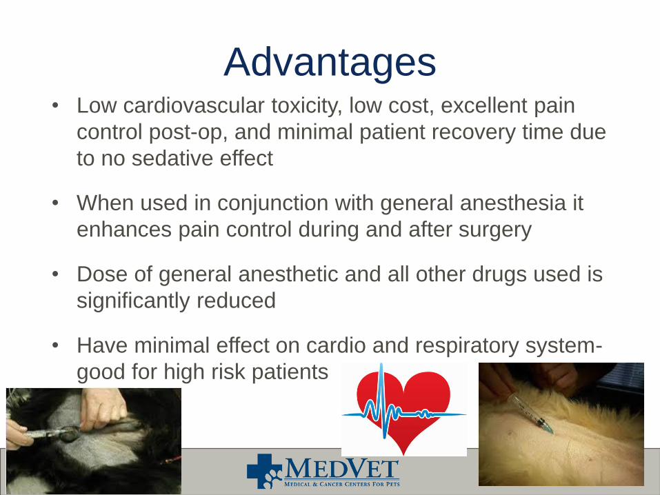
Advantages• Low cardiovascular toxicity, low cost, excellent pain
control post-op, and minimal patient recovery time due
to no sedative effect
• When used in conjunction with general anesthesia it
enhances pain control during and after surgery
• Dose of general anesthetic and all other drugs used is
significantly reduced
• Have minimal effect on cardio and respiratory system-
good for high risk patients


Toxicity Tissue toxicity can occur from administering a
local Using a vasoconstrictor (epinephrine) can contribute to delayed
wound healing and promote tissue necrosis
Systemic toxicity can result from accidental IV administration
Usually manifests in CNS with symptoms of restlessness, muscle tremors, sedation, disorientation, and ataxia
Can progress to seizures, unconsciousness, respiratory arrest or cardiac complications
Degrees of toxicity depend on dose, drug, and route administered

When to use locals
A veterinarian determines when to use a local solo, or in conjunction with general anesthesia (GA) Factors include: temperament, age, physical status of patient, cost,
nature of operation, and anesthetist’s skill in performing procedure
Some procedures that might require a local alone include: eye exam, urinary catheter placement, foreign body removal in eye, conjunctival scraping, small biopsy of skin, through chest tube, repair minor lacerations, etc
Some procedures that might be tied with GA: declaws, orthopedic procedures, thoracotomies, dental extractions, arthroscopy, etc

Local Anesthetics

Local Anesthetics Lidocaine-most commonly used due to
fast onset 3-5 min
moderate potency
short duration 60-90min
versatility
Bupivacaine-is also common but for the opposite
reasons
Slower onset:15-20min
Longer duration: 180-480min
High potency

Local Adjuncts Pure mu opioids: can be used to increase the efficacy
and extend the duration of local blocks Mostly used in epidurals and intra-articular blocks
Can be used on its own or in conjunction with local anesthetics
Preservative free morphine is the typical drug of choice
Minimal adverse effects are seen with peripheral administration
α₂ agonists can also be used
Mostly used in epidurals
Causes vasoconstriction which decreases systemic absorption
Acts at the dorsal horn of the spinal cord

Types of Blocks Topical
Infiltration/Continuous infiltration
Dental
Eye and Orbit
Intercostal
Intraperitoneal
Ring/Line
Intraarticular
Brachial plexus/Paravertebral
IV regional anesthesia/Bier
Epidural/Continuous Epidural

Topical• Local anesthetics used topically can be used to: relieve pain
during cleaning/dressing of wounds, examine of a painful eye,
desensitize the larynx, minimize chest tube discomfort, place
urinary catheters, etc
• Drugs used for this technique: EMLA cream, lidocaine,
bupivacaine, lidocaine gel, proparacaine, tetracaine, etc.
• EMLA cream is typically used to desensitize intact skin for
superficial minor procedures
A thick layer is applied to skin and covered with a bandage 10 minutes. Duration is 1-2 hours
• Application modalities: injectables used topically, creams,
ointments, gels, powders, aerosols, and patches.

Topical

Infiltration Direct injection of local anesthetics is the most
reliable and safest of all the local anesthetic
techniques
Lidocaine is the drug of choice
Used mainly to anesthetize an area for small
laceration repairs, small skin mass removals or
biopsies, and incisional line blocks
Lidocaine volume can be diluted with 0.9% NaCl if a
larger volume is needed Sodium Bicarbonate can be added to help reduce the pain at a 1:9 ratio

Infiltration Use aseptic technique; everything
should be sterile, and use a small needle like a 22 or 25 gauge
Needle should be placed just under the skin, aspirate (no blood), then slowly inject while advancing the needle(line)
For a mass, the local can be placed in a circle around the mass by inject a bleb under the skin then inserting the needle in to the previous anesthetized area and creating a circle of anesthetized skin, aspirating between each injection
Do not inject local into infected tissue, and is not likely to be as effective in inflamed tissue
Duration of effectiveness depends on drug used
Adding epinephrine helps prolong the effect of the drug by vasoconstriction

Continuous Infiltration via Soaker
CatheterProvides analgesia directly to the affected tissues through a fenestrated catheter placed prior to incisional closure
Can be placed on a infusion pump as a CRI, a manufacturer supplied reservoir, or intermittent injections can be administered
Provides excellent analgesia for leg amputations, large tumor resections, TECA’s, and thoracotomies

Dental Contraindications for block
Coagulopathies
Infection at site of injection
Anatomic malformation-results in uncertainty of needle placement
Sensitivity to local anesthetic used
Can be difficult to perform in brachycephalics and obese
patients due to inability to palpate landmarks
Possible complications: trauma to nerve, inadvertent IV or intra-
arterial injection, retrobulbar hemorrhage
Drugs of choice: Lidocaine and Bupivacaine

Infraorbital Block Block will affect first, second, and third premolars,
incisors, K9’s, and tissue rostral to the upper 4th premolars
of the side being anesthetized
The foramen is palpable inside the upper lip just rostral to
the fourth premolar
Should be used in caution with cats and brachycephalics
due to the foramen’s proximity to the eyeball
To perform block: retract upper lip dorsally and palpate the
foramen, then insert needle through buccal mucosa close
to the maxilla in a caudal direction into the foramen
without hitting bone. If you hit bone withdraw the needle,
redirect and advance the needle. ASPIRATE then inject

Infraorbital Block

Maxillary Block Block will affect all the structures affected by the
infraorbital block as well as the maxillary fourth
premolar, all molars, and the tissue caudal to the
fourth premolar
To perform this technique: Open the patient’s mouth
wide and retract the lips caudally, then advance the
needle in the dorsal direction perpendicular to the
palate just behind the second molar. The needle
should only be advanced 2-5mm beyond the mucosa
(depending on size of patient). ASPIRATE then inject

Maxillary Block

Mental Block Block will affect first three premolars, canines,
incisors and tissues of mandible rostral to the foramen
In dogs, the middle foramen is the largest and the one most commonly used.
Can be palpated ventral to first root of second premolar
In cats, middle mental foramen is difficult to palpate
It is located ventral and slightly caudal to the K9s about midway down the jaw

Mental Block To perform block in dogs-retract the bottom lip
ventrally, palpate foramen, insert needle into the
mucosa aiming caudally and advance 1-2mm
into the foramen, ASPIRATE and inject
To perform in small dogs and cats-retract the lip
ventrally, palpate foramen, insert needle into the
mucosa at the border of foramen halfway down
the mandible, advance needle slightly, cats (do
not advance into canal), ASPIRATE and inject
Digital pressure can be used to facilitate
movement of the local into the foramen

Mental Block

Mandibular Block
Block will affect all teeth in the mandible including all
tissue; if infiltrates more caudal can anesthetize
tongue
The nerve is blocked before its entry into the
mandibular canal
Foramen is located on lingual surface of mandible, is
2/3 of the distance from the last molar to the angular
process.
In dogs- it is ½ to 1” from ventral surface of mandible
In cats- it is ¼” from ventral surface of mandible

Mandibular Block Extra-oral procedure
Locate notch under the mandible just before the angular process
Use the lateral canthus of the eye as a plumb line
Slide thumb under jaw toward lingual surface of caudal mandible and palpate foramen
Insert needle next to finger, parallel to lingual aspect, stop when the needle tip is about 1/3 the distance from the ventral to the dorsal aspect of the mandible
With tip of finger, feel for tip of needle, ASPIRATE and inject just caudal to foramen opening
Intra-oral procedure
Palpate angular process extra-orally and mandibular foramen intra-orally
Insert needle just caudal to last molar toward angular process
Advance needle along lingual surface so that it is adjacent to the foramen
ASPIRATE and inject slowly

Intra oral mandibular blockExtra oral mandibular block

Mandibular block cat

Intra-articular Block Defined as an injection into a joint
Joints like: shoulders, stifles, elbows, hips, hocks, etc
Common in humans and horses
Not so common in small animal general practice due to lack of knowledge
If used properly is a great addition to multimodal analgesia
Providing pain relief right at the site of injury
Can be used before, during, or after surgery or not during surgery at all
A variety of drugs can be used for IA injections:
Hyaluronic acid
Corticosteroids
Stem cells
Platelet rich plasma
Local anesthetics
Opioids

Intercostal Block Used for analgesia for a
thoracotomy, pleural drainage, and rib fracture
Not recommended in animals with pulmonary disease
Need to be observed for several hours after block-potential for delayed pneumothorax
Location is caudal border of rib near intervertebral foramen
Minimum of 2 adjacent intercostal spaces both cranial and caudal to incision/injury should be blocked

Ring Block/Digital Nerve Block• Blocks the superficial branches of the radial, median, and the dorsal
and palmar branches of the ulnar nerve
• Provides effective analgesia to the distal extremity for onychectomies,
toe amputations, etc
• No epinephrine due to potential for ischemic injury in the extremity
• Bupivacaine is the drug of choice and can be combined with lidocaine
for faster onset
• To perform the ring block: inject 0.2-0.4mg/kg of local subcutaneously
in three sites
• Lateral and proximal to the accessory carpal pad
• Medial to the accessory carpal pad
• Dorsal-medial aspect of the proximal carpus
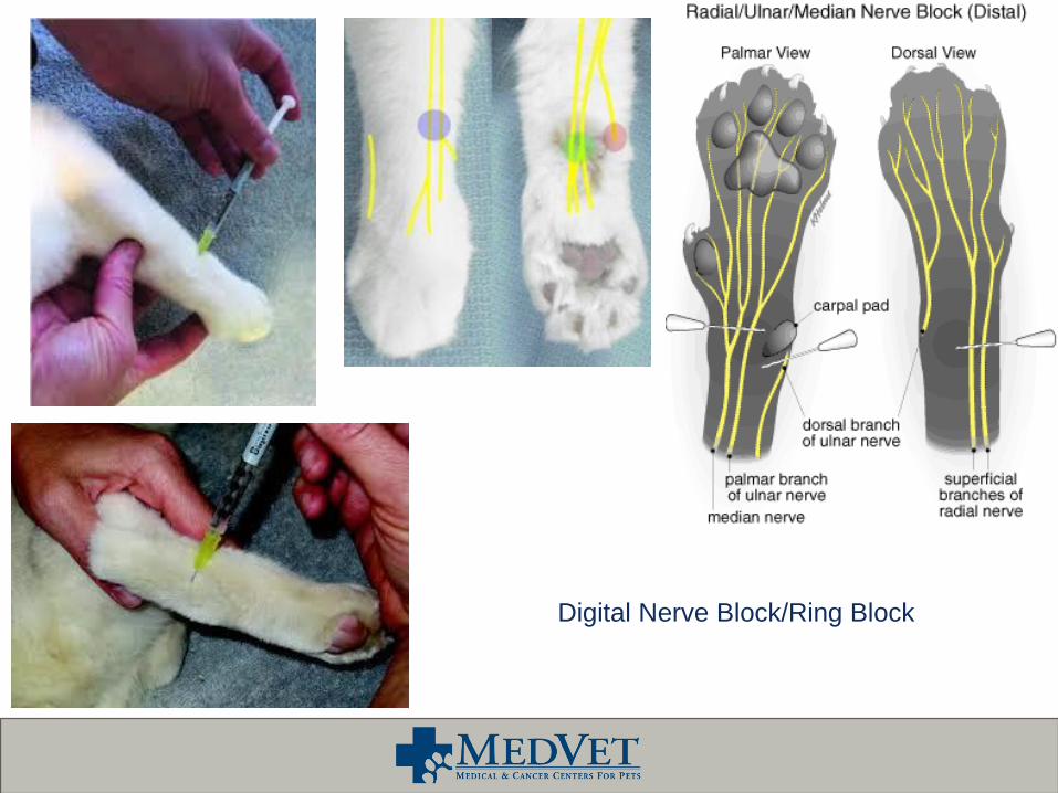
Digital Nerve Block/Ring Block

IV regional Anesthesia/Bier Block Rapid, reliable method for producing short term anesthesia of the extremities with minimal
systemic absorption of the local anesthetic
Block good for distal limb mass removals, wound management, surgical biopsy or foreign body removal
Provides 60-90 minutes of regional anesthesia distal to tourniquet
Place IV catheter in the distal limb (cephalic, saphenous)
Place a rubber tourniquet or blood pressure cuff (with sphygmomanometer) around the limb proximal to the IV catheter and tighten to occlude blood flow
It is important that the tourniquet does not come off early due to potential risk of rapid plasma uptake of local
An Esmarch bandage can be placed to help exsanguinate the limb
Inject Lidocaine or Mepivacaine into the catheter
Never use Bupivacaine
Anesthesia will take about 5-10 min to achieve and will last about 90 min distal to the tourniquet then remove tourniquet
Sensation should return in 5-10 min after removing the tourniquet with residual analgesia lasting another 20-30 min


Brachial Plexus Blocko Provides analgesia distal to the elbow and possibly including the
elbow by blocking
o Drugs that can be used to perform this block are: local anesthetics, opioids, α₂ agonists or any combination
o Patient should be heavily sedated or anesthetized
o Spinal needles are typically used to perform this block because normal needles are too short
o How to perform: patient should be in lateral recumbency, then insert the needle into the axillary region medial to and at the level of the shoulder joint, direct it parallel to the vertebral column- the needle’s distal end should lie just caudal to the spine of the scapula. ASPIRATE. Then slowly inject 1/3 of the volume, then slowly inject the remaining volume as you are removing the needle
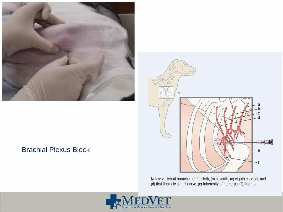
Brachial Plexus Block

Paravertebral Brachial Plexus Block
Provides analgesia to the entire forelimb
Drugs that can be used are locals, opioids, and α₂ agonists
Animal should be heavily sedated or anesthetized
The cervical spinal nerves C6, C7 and C8 and thoracic spinal
nerve T1 are blocked as close to the intervertebral foramen as
possible
This block is technically more difficult to perform vs the brachial
plexus block
Potential complications: blockade of the phrenic nerve,
pneumothorax, and inadvertent injection into the thoracic dural
sheath could result in systemic hypotension and respiratory
depression

Paravertebral Brachial Plexus Block

Epidural Anesthesia One of the most frequently used regional anesthetic technique
described for surgical procedures caudal to umbilicus
Is recommended for C-sections since does not depress puppies
Epidural space is located between the inner and outer layers of the dura mater in the lumbosacral intervertebral space
Animal should be sedated, tranquilized, or under GA to perform procedure
Can be placed in sternal recumbency with legs folded cranially under animal or placed in lateral recumbency with legs pulled forward
This technique helps open up vertebral space
Contraindications: increased intracranial pressure, clotting disorders, uncorrected hypovolemia, degenerative central or peripheral axonal diseases, anatomical abnormalities (broken pelvis), or skin infection

Epidural Anesthesia• Clip hair over iliac wings of pelvis from L5-S3 and surgically prepare area thoroughly-sterile gloves should be used
• Spinal needle of the appropriate size should be used
• 22g x 1.5” for <10kg patients
• 22g x 2.5” for 10-45kg patients
• 20g x 3.5”for >45kg patients
• Palpate iliac wings on either side of spine using thumb and middle finger of one hand, locate spinous process of L7 with
index finger-can do this prior to scrub to get an idea of where the area is prior to performing block
• Move finger back and forth to find L7-S1 interspace-should feel like a divot
• Midline positioning is critical to avoid contact with the L7 vertebra
• Insert spinal needle, bevel pointing cranially and stylet in place, just caudal to L7 on midline until a popping sensation is felt-
stop advancing the needle
• Loss of resistance test should properly identify space-air-filled or saline-filled syringe should be used or hanging drop
technique-the stylet needs to be removed first
• Blood and/or CSF should not be present if they are remove needle and try again
• Should elevate head for ~5min to prevent excessive cranial advancement of anesthetic
• Bevel orientation makes a difference-so bevel should face the head so it split rather than cuts dural fibers

Epidural AnesthesiaDrugs
Depends on size, desired extent of anesthesia, and desired onset and duration of effect
There are so many combinations-local, opioid, alpha 2, ketamine and combinations of them all
Most common is local alone, opioid alone or combination of local and opioid
Talk with DVM to get combo and dosages that they like to use
To properly prepare the syringe after a drug protocol is established, draw a small volume of air into the syringe then attach the syringe to the spinal needle-leaving a bubble on top of the fluid
If the needle is in the right place the fluid should flow with no resistance
Observe the patient after injection to see if the epidural was successful: relaxation of the anal sphincter and tail should be observed, a lack of response to surgical stimulus, and a need for less gas anesthesia


