load response functions in the human spatial working ... · load response functions in the human...
TRANSCRIPT
www.elsevier.com/locate/ynimg
NeuroImage 35 (2007) 368–377Load response functions in the human spatial working memory circuitduring location memory updating
Hoi-Chung Leung,⁎ Hwamee Oh, Jamie Ferri, and Yuji Yi
Department of Psychology, State University of New York at Stony Brook, Stony Brook, NY 11794-2500, USA
Received 15 September 2006; revised 11 November 2006; accepted 4 December 2006Available online 19 December 2006
Previous studies have emphasized that the dorsolateral prefrontalcortex is important for manipulating information in working memory,although activations in other frontal and parietal areas are commonlyobserved under the same conditions. We conducted an fMRIexperiment to examine brain responses as a parametric function ofmemory updating, which is considered as an elemental process inworking memory. In a variant spatial delayed-response task, humansubjects performed updating operations over a 9-second delay period,during which they mentally transform the location of a memorizedtarget in a 4 by 4 grid according to 3 to 12 instruction cues. Activityincreased monotonically with increasing updating load in numerouscortical and subcortical regions including the rostrodorsal premotor(rdPM), lateral precentral sulcus, lateral prefrontal, posteriorassociative, striatal and cerebellar areas. The rdPM and superiorparietal were particularly sensitive to the updating manipulation.There were several main findings. First, updating spatial workingmemory involved mostly the same cortical and subcortical regions thatwere activated during maintenance of spatial information. Second, theupdating load response functions of regions in the spatial workingmemory circuit showed a strong linear component. However, noneshows significant increases in activity from 9 to 12 updating operations.Third, activity in the right rdPM and anterior inferior frontal gyruscorrelated positively with working memory performance in the highupdating load condition. Our findings suggest that updating andmaintenance of spatial information may share similar processes andthat the rostrodorsal premotor cortex and anterior inferior frontalgyrus may be important for the success of tracking spatial informationin working memory.© 2006 Elsevier Inc. All rights reserved.
Keywords: Prefrontal cortex; Premotor cortex; Information processing;Central executive; Cognition; fMRI
⁎ Corresponding author. Fax: +1 631 632 7876.E-mail address: [email protected] (H.-C. Leung).Available online on ScienceDirect (www.sciencedirect.com).
1053-8119/$ - see front matter © 2006 Elsevier Inc. All rights reserved.doi:10.1016/j.neuroimage.2006.12.012
Introduction
Working memory is commonly modeled as a system thatsupports the temporary maintenance and manipulation of informa-tion during complex cognitive tasks (Baddeley, 1986). It has beenproposed that the underlying working memory network can bedivided into specialized functional units. In particular, thedorsolateral prefrontal cortex (PFC) has been associated withmanipulation processes of working memory and the ventrolateralPFC with maintenance and retrieval processes (D'Esposito et al.,1998; Owen et al., 1999; Petrides, 1994). Other cortical regions,especially the dorsal premotor and posterior parietal cortices, arealso involved in working memory (see reviews by Collette and Vander Linden, 2002; Wager and Smith, 2003). It remains unclearwhether these regions serve similar functions in working memory orwhether they each have a specific function, such as maintenance,updating, shifting, inhibition, etc.
One useful approach to study the underlying neural substratesof working memory is to vary the task load parametrically.Numerous neuroimaging studies have used the n-back task, inwhich subjects are required to make judgments as to whether thecurrent task item is the same as an item n back in the sequence.Various load response functions have been observed for the PFCincluding step (Cohen et al., 1997), linear (e.g., Braver et al.,1997), and invert-U shaped functions (Callicott et al., 1999;Jansma et al., 2004). However, the n-back paradigm is a complexworking memory task involving not only manipulation processes(e.g., updating, reordering) but also maintenance processes (e.g.,rehearsal, storage). Recent studies have examined the effect ofload on simple maintenance by varying only the number of itemsto be remembered in delayed-recognition tasks. Some have founda linear increase in activity in the dorsolateral prefrontal cortexwith increasing maintenance load during the delay period (Lindenet al., 2003; Rypma et al., 2002), while others have founddecreased or leveled activity at high maintenance load (Leung etal., 2004). Similar patterns of load-dependent activity for workingmemory maintenance have also been observed in the dorsalpremotor and posterior associative areas (Leung et al., 2004;Linden et al., 2003; Todd and Marois, 2004; Xu and Chun,2006).
369H.-C. Leung et al. / NeuroImage 35 (2007) 368–377
The present study aimed to examine responses in the spatialworking memory circuit as a parametric variation in memoryupdating load while keeping the maintenance load minimum andunchanged. Memory updating has been considered an elementalexecutive operation in working memory (Baddeley, 1996; Miyakeand Shah, 1999). It refers to the operation of adjusting the currentcontents in working memory to adapt new information (Morris andJones, 1990), for example, updating the current product during asequence of mental arithmetic operations. We were particularlyinterested in the lateral PFC, posterior parietal cortex, and dorsalpremotor cortex as these regions have been implicated to playimportant roles in spatial working memory (e.g., Awh et al., 1999;Courtney et al., 1998; Leung et al., 2002). We expected activity toincrease with the number of updating operations in brain areas thatare sensitive to manipulation processes in working memory.Conversely, we expected to find no or minimum variation ofactivity in areas that are specialized for maintaining information inworking memory since only one item was required to beremembered at any point of time in the updating conditions.
Methods
Subjects
Fourteen right-handed healthy adults (6 females and 8 males, aged21–32 years, mean age=24) were recruited from the Yale Universitycommunity, none with a history of drug abuse and psychiatric andneurological disorder according to self-report. All subjects gaveinformed consent to the protocol that was reviewed and approved bythe Institution Review Boards of both Yale University School ofMedicine and State University of New York at Stony Brook.
Data from one subject were excluded due to excessive motionduring scanning. A total of 13 data sets were used in the final dataanalysis.
Working memory updating task
We implemented a variant of the spatial delayed-response taskto incorporate the updating manipulation. Our design was modified
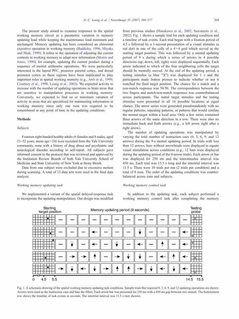
Fig. 1. A schematic drawing of the spatial working memory updating task conditionArrows were used as the instruction cues and bars the fillers. Each arrow/bar was prrow shows the timeline of task events in seconds. The intertrial interval was 11.5
from previous studies (Hanakawa et al., 2002; Sawamoto et al.,2002). Fig. 1 shows a sample trial for each updating condition anda timeline of task events. Each trial began with a fixation period of4.5 s followed by a 1-second presentation of a visual stimulus (ared dot) in one of the cells of a 4×4 grid which served as thestarting target position. This was followed by a mental updatingperiod of 9 s during which a series of arrows in 4 possibledirections (up, down, left, right) were displayed sequentially. Eacharrow indicated to which of the four neighboring cells the targetshould be mentally moved. At the end of the updating period, atesting stimulus (a blue "X") was displayed for 1 s and theparticipants made button presses to indicate whether or not itmatched the final target position. The chance for a match and anon-match response was 50/50. The correspondence between thetwo fingers and match/non-match responses was counterbalancedacross participants. The initial target stimulus and the testingstimulus were presented in all 16 possible locations at equalchance. The arrow series were generated pseudorandomly with noregular patterns, repeating patterns, or patterns that would confinethe mental target within a local area. Only a few series containedthree arrows of the same direction in a row. There were also noimmediate back and forth arrows (e.g., a left arrow right after aright arrow).
The number of updating operations was manipulated byvarying the total number of instruction cues (0, 3, 6, 9, and 12arrows) during the 9-s mental updating period. In trials with lessthan 12 arrows, bars without arrowheads were displayed to equatevisual stimulation across conditions (e.g., 12 bars were displayedduring the updating period of the 0-arrow trials). Each arrow or barwas displayed for 250 ms and the interstimulus interval was450 ms. Each trial was 15.5 s long and the intertrial interval was11.5 s. There were 10 trials per run (2 trials per condition) and atotal of 8 runs. The order of the updating conditions was counter-balanced across runs and subjects.
Working memory control task
In addition to the updating task, each subject performed aworking memory control task after completing the memory
s. Sample trials that required 0, 3, 6, 9, and 12 updating operations are shown.esented for 250 ms with a 450 ms gap between two stimuli. The bottommosts (not shown).
370 H.-C. Leung et al. / NeuroImage 35 (2007) 368–377
updating runs. At the beginning of each trial, 4 dots (targets) weresimultaneously displayed on the grid at 4 different locations toserve as the working memory set. Following a 9-s blank delayperiod (without grid, arrows, or bars), a testing stimulus wasdisplayed at either one of the four target locations or a newlocation. The subjects made button presses to indicate whether ornot the testing dot matched the location of one of the four targetlocations. The timing of task events and other parameters was thesame as the updating task (see above). Each subject completed onerun with 12 trials. This task was used for comparison purposes andto determine brain activations in response to a challenging workingmemory task without any explicit updating or other manipulationdemands.
Imaging procedures
Whole brain images were acquired using a 3 T system (Trio,Siemens AG, Erlangen, Germany). Conventional T1-weightedsagittal images were collected for slice localization. Twenty-fouraxial-oblique slices (5 mm) were prescribed parallel to theanterior–posterior commissural line. T1-weighted structural imageswere obtained with a 300 ms repetition time (TR), a 2.5 ms echotime, a 60° flip angle (FA), a 22 cm×22 cm field of view (FOV),and a matrix size of 256×256. Functional images were acquiredwith the same slice selection and a TR of 1500 ms, TE of 30 ms,FA of 80°, matrix size of 64×64, and a FOV 22×22 cm using T2*-sensitive gradient-recalled single shot echo-planar pulse sequence.Each subject was scanned for 8 functional runs of the updating task(180 image volumes/run) and 1 run of the memory control task(216 image volumes). High-resolution anatomical images werealso obtained (MPRAGE sequence, 176 sagittal 1 mm thick slices,TR=2530 ms, TE=3.52 ms, FA=7°, matrix=256×256).
Image processing and analysis
SPM2 was used for image processing and constructingindividual and group contrast maps (Welcome Department ofImaging Neuroscience, University College London, London, UK).The first 8 images were discarded from each functional run.Functional images were corrected for different times of sliceacquisition followed by a 6-parameter rigid body motion cor-rection. Runs with images that have motion greater than 3 mm inthe x, y or z direction or more than 1.5° of pitch, yaw or roll wereeliminated from further analysis. Images were realigned withreference to the first image of the middle run. Functional imageswere co-registered with in-plane anatomical as well as high-resolution anatomical images, segmented (gray and white matter),and were then normalized to a Montreal Neurological Institute(MNI) gray matter template using a 12-parameter affine registra-tion following by nonlinear transformation (Friston et al., 1995a).The image volumes were resampled to 3×3×3 mm voxel size.Images were subsequently smoothed in the spatial domain with aGaussian filter of 8 mm at full-width at half maximum. The datawere also high-pass filtered with 1/128 Hz cutoff frequency toremove low-frequency signals (e.g., linear drifts).
Two kinds of statistical design were applied to analyze datafrom each individual. First, a standard design was constructed foreach data set, using the general linear model (GLM). For eachupdating condition, the onset times of the cue, updating, and probeevents were defined and the durations of the events at 1, 6, and 2scan steps, respectively. Each event was convolved with a canonical
hemodynamic response function and entered as regressors in themodel (Friston et al., 1995b). For each individual, t-tests wereperformed to examine both simple main effects of each updatingload condition and differences between the higher updating loadconditions and the 0-arrow (0-load) condition. Beta weights (orestimated parameters) of each condition of interest were used in thet-test calculations. Second, a parametric design was constructed foreach data set, including unweighted covariates for the onset times ofthe task events of each condition and covariates weighted by thecorresponding updating load levels (0, 3, 6, 9, 12) as linearregressors. Both correct and incorrect trials were included in theseanalyses to determine the overall effect of updating load.
For the whole group, random effects analyses were conductedto test for statistical differences between conditions of interest (e.g.,6-arrow vs. 0-arrow) using the corresponding contrast values fromeach individual. The contrast values of a condition were theweighted sum of the beta weights from the single subject analysis.T-values for group comparisons were calculated using one-samplet-test. Final group statistics were corrected by false discovery rate(FDR) (Genovese et al., 2002).
Regions of interest (ROIs) were defined as spheres (radius=10 mm) with the center at the peak coordinates of the activationclusters obtained from the group composite maps of updating loadusing the parametric analysis. Two control regions (primary motorand visual cortices) were selected anatomically. We used theMarsBar Matlab toolbox (Brett et al., 2002) (http://marsbar.courceforge.net) to extract ROI data from each individual. Time-courses were collapsed across trials for each task condition, ofwhich the average percent signal change for each time point wascalculated relative to the baseline. Baseline was the average of thefirst three images of a trial. Repeated measures analysis ofvariance (ANOVA) was applied to determine the updating loadeffect and linearity of load response function for each ROI. Errortrials were removed from these tests. Linear regression wasapplied to determine the correlation between the average percentsignal change of an ROI during the updating period and workingmemory performance (hit rate–false alarm rate) in the 12-arrowcondition.
Results
Behavioral results
The average accuracy for each updating conditionwas 99, 92, 82,88, and 78% and reaction time was 987, 1017, 1006, 1103, and1098 ms from the 0- to 12-arrow conditions. The average accuracywas 92% and reaction time was 1128 ms for the memory controlcondition. Difference in performance between the updating condi-tions was significant for accuracy (F(4,48)=9.60, p<0.001), but notfor reaction time (F(4,48)=1.25, p>0.05). Post hoc t-tests showedthat accuracy in conditions that require 3 or more updatingoperations was lower than the condition with 0 number of updatingoperations (all comparisons with p<0.05). The recognition rate (hitrate–false alarm rate) decreased from 0.97 to 0.58 (F(4,48)=8.75,p<0.0001) as false alarm rate increased with increasing number ofupdating operations.
Spatial working memory circuit: maintenance versus updating
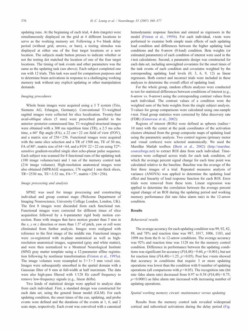
Results from the memory control task revealed widespreadcortical and subcortical activations during the delay period (Fig.
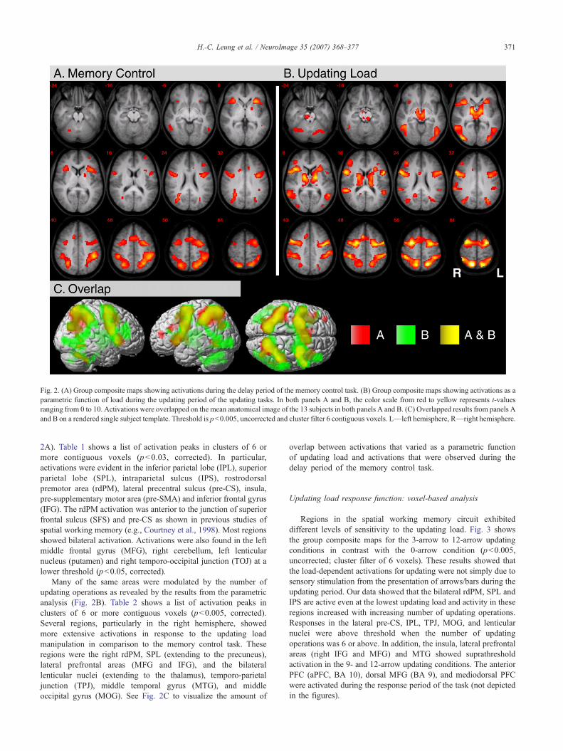
Fig. 2. (A) Group composite maps showing activations during the delay period of the memory control task. (B) Group composite maps showing activations as aparametric function of load during the updating period of the updating tasks. In both panels A and B, the color scale from red to yellow represents t-valuesranging from 0 to 10. Activations were overlapped on the mean anatomical image of the 13 subjects in both panels A and B. (C) Overlapped results from panels Aand B on a rendered single subject template. Threshold is p<0.005, uncorrected and cluster filter 6 contiguous voxels. L—left hemisphere, R—right hemisphere.
371H.-C. Leung et al. / NeuroImage 35 (2007) 368–377
2A). Table 1 shows a list of activation peaks in clusters of 6 ormore contiguous voxels (p<0.03, corrected). In particular,activations were evident in the inferior parietal lobe (IPL), superiorparietal lobe (SPL), intraparietal sulcus (IPS), rostrodorsalpremotor area (rdPM), lateral precentral sulcus (pre-CS), insula,pre-supplementary motor area (pre-SMA) and inferior frontal gyrus(IFG). The rdPM activation was anterior to the junction of superiorfrontal sulcus (SFS) and pre-CS as shown in previous studies ofspatial working memory (e.g., Courtney et al., 1998). Most regionsshowed bilateral activation. Activations were also found in the leftmiddle frontal gyrus (MFG), right cerebellum, left lenticularnucleus (putamen) and right temporo-occipital junction (TOJ) at alower threshold (p<0.05, corrected).
Many of the same areas were modulated by the number ofupdating operations as revealed by the results from the parametricanalysis (Fig. 2B). Table 2 shows a list of activation peaks inclusters of 6 or more contiguous voxels (p<0.005, corrected).Several regions, particularly in the right hemisphere, showedmore extensive activations in response to the updating loadmanipulation in comparison to the memory control task. Theseregions were the right rdPM, SPL (extending to the precuneus),lateral prefrontal areas (MFG and IFG), and the bilaterallenticular nuclei (extending to the thalamus), temporo-parietaljunction (TPJ), middle temporal gyrus (MTG), and middleoccipital gyrus (MOG). See Fig. 2C to visualize the amount of
overlap between activations that varied as a parametric functionof updating load and activations that were observed during thedelay period of the memory control task.
Updating load response function: voxel-based analysis
Regions in the spatial working memory circuit exhibiteddifferent levels of sensitivity to the updating load. Fig. 3 showsthe group composite maps for the 3-arrow to 12-arrow updatingconditions in contrast with the 0-arrow condition (p<0.005,uncorrected; cluster filter of 6 voxels). These results showed thatthe load-dependent activations for updating were not simply due tosensory stimulation from the presentation of arrows/bars during theupdating period. Our data showed that the bilateral rdPM, SPL andIPS are active even at the lowest updating load and activity in theseregions increased with increasing number of updating operations.Responses in the lateral pre-CS, IPL, TPJ, MOG, and lenticularnuclei were above threshold when the number of updatingoperations was 6 or above. In addition, the insula, lateral prefrontalareas (right IFG and MFG) and MTG showed suprathresholdactivation in the 9- and 12-arrow updating conditions. The anteriorPFC (aPFC, BA 10), dorsal MFG (BA 9), and mediodorsal PFCwere activated during the response period of the task (not depictedin the figures).
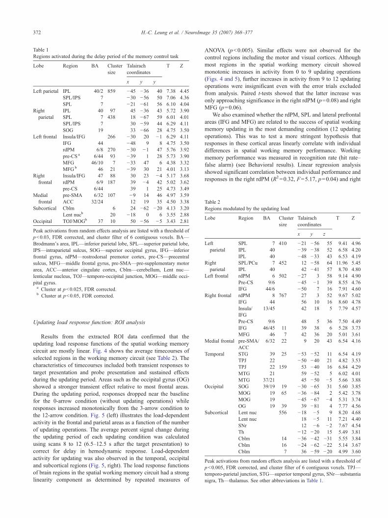
Table 1Regions activated during the delay period of the memory control task
Lobe Region BA Clustersize
Talairachcoordinates
T Z
x y y
Left parietal IPL 40/2 859 −45 −36 40 7.38 4.45SPL/IPS 7 −30 −56 50 7.06 4.36SPL 7 −21 −61 56 6.10 4.04
Rightparietal
IPL 40 97 45 −36 43 5.72 3.90SPL 7 438 18 −67 59 6.01 4.01SPL/IPS 7 30 −59 44 6.29 4.11SOG 19 33 −66 28 4.75 3.50
Left frontal Insula/IFG 266 −30 20 −1 6.29 4.11IFG 44 −48 9 8 4.75 3.50rdPM 6/8 270 −30 −1 47 5.76 3.92pre-CS a 6/44 93 −39 1 28 5.73 3.90MFG 46/10 7 −33 47 6 4.38 3.32MFGb 46 21 −39 30 21 4.01 3.13
Rightfrontal
Insula/IFG 47 88 30 23 −4 5.17 3.68rdPM 6/9 187 39 −4 42 5.02 3.62pre-CS 6/44 39 1 25 4.73 3.49
Medialfrontal
pre-SMA 6/32 107 −9 14 46 4.97 3.59ACC 32/24 12 19 35 4.50 3.38
Subcortical Cblm 6 24 −62 −20 4.13 3.20Lent nucb 20 −18 0 6 3.55 2.88
Occipital TOJ/MOGb 37 10 50 −56 −5 3.43 2.81
Peak activations from random effects analysis are listed with a threshold ofp<0.03, FDR corrected, and cluster filter of 6 contiguous voxels. BA—Brodmann's area, IPL—inferior parietal lobe, SPL—superior parietal lobe,IPS—intraparietal sulcus, SOG—superior occipital gyrus, IFG—inferiorfrontal gyrus, rdPM—rostrodorsal premotor cortex, pre-CS—precentralsulcus, MFG—middle frontal gyrus, pre-SMA—pre-supplementary motorarea, ACC—anterior cingulate cortex, Cblm—cerebellum, Lent nuc—lenticular nucleus, TOJ—temporo-occipital junction, MOG—middle occi-pital gyrus.a Cluster at p<0.025, FDR corrected.b Cluster at p<0.05, FDR corrected.
Table 2Regions modulated by the updating load
Lobe Region BA Clustersize
Talairachcoordinates
T Z
x y z
Leftparietal
SPL 7 410 −21 −56 55 9.41 4.96IPL 40 −39 −38 52 6.58 4.20IPL 40 −48 −33 43 6.53 4.19
Rightparietal
SPL/PCu 7 452 12 −58 64 11.96 5.45IPL 40 42 −41 57 8.70 4.80
Left frontal rdPM 6 502 −27 3 58 9.14 4.90Pre-CS 9/6 −45 −1 39 8.55 4.76IFG 44/6 −50 7 16 7.91 4.60
Right frontal rdPM 8 767 27 3 52 9.67 5.02IFG 44 56 10 16 8.60 4.78Insula/IFG
13/45 42 18 5 7.79 4.57
Pre-CS 9/6 48 5 36 7.50 4.49IFG 46/45 11 39 38 6 5.28 3.73MFG 46 7 42 36 20 5.01 3.61
Medial frontal pre-SMA/ACC
6/32 22 9 20 43 6.54 4.16
Temporal STG 39 25 −53 −52 11 6.54 4.19TPJ 22 −50 −40 21 4.82 3.53TPJ 22 159 53 −40 16 6.84 4.29MTG 21 59 −52 5 6.02 4.01MTG 37/21 45 −50 −5 5.66 3.88
Occipital SOG 39/19 19 −30 −65 31 5.60 3.85MOG 19 65 −36 −84 2 5.42 3.78MOG 19 −45 −67 −4 5.31 3.74OG 19 39 39 −81 4 7.77 4.56
Subcortical Lent nuc 556 −18 −5 9 8.20 4.68Lent nuc 18 −5 11 7.21 4.40SNr 12 −6 −2 7.67 4.54Th −12 −20 15 5.49 3.81Cblm 14 −36 −42 −31 5.55 3.84Cblm 16 −24 −62 −22 5.14 3.67Cblm 7 36 −59 −20 4.99 3.60
Peak activations from random effects analysis are listed with a threshold ofp<0.005, FDR corrected, and cluster filter of 6 contiguous voxels. TPJ—temporo-parietal junction, STG—superior temporal gyrus, SNr—substantianigra, Th—thalamus. See other abbreviations in Table 1.
372 H.-C. Leung et al. / NeuroImage 35 (2007) 368–377
Updating load response function: ROI analysis
Results from the extracted ROI data confirmed that theupdating load response functions of the spatial working memorycircuit are mostly linear. Fig. 4 shows the average timecourses ofselected regions in the working memory circuit (see Table 2). Thecharacteristics of timecourses included both transient responses totarget presentation and probe presentation and sustained effectsduring the updating period. Areas such as the occipital gyrus (OG)showed a stronger transient effect relative to most frontal areas.During the updating period, responses dropped near the baselinefor the 0-arrow condition (without updating operations) whileresponses increased monotonically from the 3-arrow condition tothe 12-arrow condition. Fig. 5 (left) illustrates the load-dependentactivity in the frontal and parietal areas as a function of the numberof updating operations. The average percent signal change duringthe updating period of each updating condition was calculatedusing scans 8 to 12 (6.5–12.5 s after the target presentation) tocorrect for delay in hemodynamic response. Load-dependentactivity for updating was also observed in the temporal, occipitaland subcortical regions (Fig. 5, right). The load response functionsof brain regions in the spatial working memory circuit had a stronglinearity component as determined by repeated measures of
ANOVA (p<0.005). Similar effects were not observed for thecontrol regions including the motor and visual cortices. Althoughmost regions in the spatial working memory circuit showedmonotonic increases in activity from 0 to 9 updating operations(Figs. 4 and 5), further increases in activity from 9 to 12 updatingoperations were insignificant even with the error trials excludedfrom analysis. Paired t-tests showed that the latter increase wasonly approaching significance in the right rdPM (p=0.08) and rightMFG (p=0.06).
We also examined whether the rdPM, SPL and lateral prefrontalareas (IFG and MFG) are related to the success of spatial workingmemory updating in the most demanding condition (12 updatingoperations). This was to test a more stringent hypothesis thatresponses in these cortical areas linearly correlate with individualdifferences in spatial working memory performance. Workingmemory performance was measured in recognition rate (hit rate–false alarm) (see Behavioral results). Linear regression analysisshowed significant correlation between individual performance andresponses in the right rdPM (R2=0.32, F=5.17, p=0.04) and right
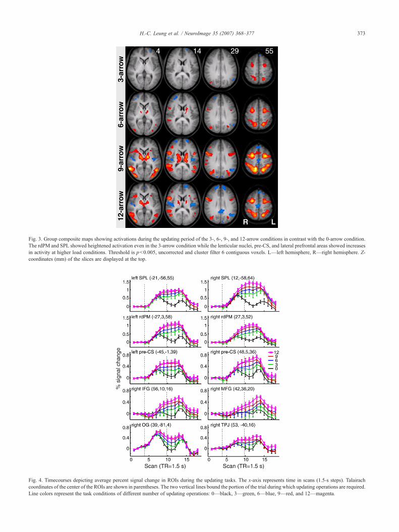
Fig. 3. Group composite maps showing activations during the updating period of the 3-, 6-, 9-, and 12-arrow conditions in contrast with the 0-arrow condition.The rdPM and SPL showed heightened activation even in the 3-arrow condition while the lenticular nuclei, pre-CS, and lateral prefrontal areas showed increasesin activity at higher load conditions. Threshold is p<0.005, uncorrected and cluster filter 6 contiguous voxels. L—left hemisphere, R—right hemisphere. Z-coordinates (mm) of the slices are displayed at the top.
Fig. 4. Timecourses depicting average percent signal change in ROIs during the updating tasks. The x-axis represents time in scans (1.5-s steps). Talairachcoordinates of the center of the ROIs are shown in parentheses. The two vertical lines bound the portion of the trial during which updating operations are required.Line colors represent the task conditions of different number of updating operations: 0—black, 3—green, 6—blue, 9—red, and 12—magenta.
373H.-C. Leung et al. / NeuroImage 35 (2007) 368–377
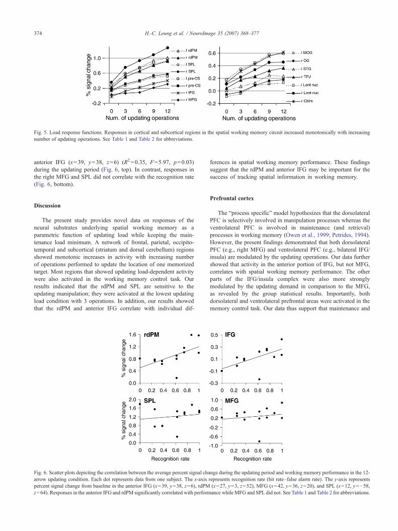
Fig. 5. Load response functions. Responses in cortical and subcortical regions in the spatial working memory circuit increased monotonically with increasingnumber of updating operations. See Table 1 and Table 2 for abbreviations.
374 H.-C. Leung et al. / NeuroImage 35 (2007) 368–377
anterior IFG (x=39, y=38, z=6) (R2=0.35, F=5.97, p=0.03)during the updating period (Fig. 6, top). In contrast, responses inthe right MFG and SPL did not correlate with the recognition rate(Fig. 6, bottom).
Discussion
The present study provides novel data on responses of theneural substrates underlying spatial working memory as aparametric function of updating load while keeping the main-tenance load minimum. A network of frontal, parietal, occipito-temporal and subcortical (striatum and dorsal cerebellum) regionsshowed monotonic increases in activity with increasing numberof operations performed to update the location of one memorizedtarget. Most regions that showed updating load-dependent activitywere also activated in the working memory control task. Ourresults indicated that the rdPM and SPL are sensitive to theupdating manipulation; they were activated at the lowest updatingload condition with 3 operations. In addition, our results showedthat the rdPM and anterior IFG correlate with individual dif-
Fig. 6. Scatter plots depicting the correlation between the average percent signal chaarrow updating condition. Each dot represents data from one subject. The x-axispercent signal change from baseline in the anterior IFG (x=39, y=38, z=6), rdPMz=64). Responses in the anterior IFG and rdPM significantly correlated with perform
ferences in spatial working memory performance. These findingssuggest that the rdPM and anterior IFG may be important for thesuccess of tracking spatial information in working memory.
Prefrontal cortex
The “process specific” model hypothesizes that the dorsolateralPFC is selectively involved in manipulation processes whereas theventrolateral PFC is involved in maintenance (and retrieval)processes in working memory (Owen et al., 1999; Petrides, 1994).However, the present findings demonstrated that both dorsolateralPFC (e.g., right MFG) and ventrolateral PFC (e.g., bilateral IFG/insula) are modulated by the updating operations. Our data furthershowed that activity in the anterior portion of IFG, but not MFG,correlates with spatial working memory performance. The otherparts of the IFG/insula complex were also more stronglymodulated by the updating demand in comparison to the MFG,as revealed by the group statistical results. Importantly, bothdorsolateral and ventrolateral prefrontal areas were activated in thememory control task. Our data thus support that maintenance and
nge during the updating period and working memory performance in the 12-represents recognition rate (hit rate–false alarm rate). The y-axis represents(x=27, y=3, z=52), MFG (x=42, y=36, z=20), and SPL (x=12, y=−58,ance while MFG and SPL did not. See Table 1 and Table 2 for abbreviations.
375H.-C. Leung et al. / NeuroImage 35 (2007) 368–377
manipulation processes are integrated within the lateral prefrontalareas (Goldman-Rakic, 1987). Similar results were reported inneuroimaging studies using a running span design in the verbaldomain (Postle et al., 2001; Salmon et al., 1996). However,previous studies have found greater activations in the dorsolateralPFC relative to the ventrolateral PFC in an alphabetization task(Postle et al., 1999) and in a continuous attentional switch task(Garavan et al., 2000). The additional dorsolateral PFC activitymay be a result of the greater mental demand since these lattertasks required additional cognitive processes (e.g., retrieval andreordering) and manipulation of multiple items in workingmemory. A recent study also did not find functional dissociationalong the dorsoventral axis of PFC, but did find scattereddissociation in responses to manipulation (anterior MFG andinferior frontal junction) in comparison to maintenance (dorsalpremotor cortex) of target color and orientation in workingmemory (Mohr et al., 2006). The apparent discrepancy in findingsbetween studies may be due to differences in maintenance loadand, in addition, the type and number of manipulation operationsrequired by the task.
The right MFG showed a linear load response function forupdating in the present study. This was different from our previousspatial workingmemory study, in which wemanipulated the numberof spatial locations to be remembered and found poor recognitionperformance (~66% accuracy) and a reduction in MFG activity athigh maintenance load (4 locations) (Leung et al., 2004). It islikely that our subjects may have just reached their limit inupdating capacity since their performance was lower in the 12-arrow condition than the other conditions. In addition, it isunlikely that the activity will increase any further even with ahigher updating load since the increases in activity from 9 to 12updating operations were not significant for the prefrontal areas andother areas that we have examined. Nevertheless, the activity inseveral regions appeared to have reached an asymptote earlier thansome other regions (e.g., compare right MFG with right OG inFigs. 4 and 5). This potentially reflects that the right lateral PFCtogether with the right rdPM and SPL (see below) is involved insupporting spatial working memory of larger processing loadsthan other regions (e.g., OG and TPJ). Previous studies haveimplicated that the dorsolateral PFC is involved in executiveprocesses that help holding more information in verbal workingmemory (Rypma et al., 2002) and object working memory (Lindenet al., 2003).
Dorsal premotor cortex
While the premotor cortex has been traditionally considered as ahigher order motor region for motor preparation, planning andsequencing, more recent studies have consistently demonstrated thatportions of the dorsal premotor cortex are activated in cognitivetasks (see Picard and Strick, 2001). It has been demonstrated that thepremotor cortex shows functional specialization along the rostro-caudal axis, where cognitive operations (e.g., attention) involve therostral portion and motor operations (e.g., preparation) involve thecaudal portion (see Boussaoud, 2001). The dorsal premotoractivation associated with spatial working memory is near thejunction of SFS and pre-CS (BA 6/8) (Courtney et al., 1998; Rowe etal., 2000; Simon et al., 2002), which is usually rostral and dorsal tothe functionally defined frontal eye fields (FEF) (Koyama et al.,2004; Paus, 1996). In contrast, the caudal premotor area includingFEF has been related to motor preparation in several fMRI studies of
spatial workingmemory (Curtis et al., 2004; Simon et al., 2002; Toniet al., 1999).
Our findings corroborate the notion that the rdPM is involved inspatial working memory. In an elegant experiment, Tanaka et al.(2005) have demonstrated that the dorsal premotor cortex (here wecalled rdPM) has a critical role in updating spatial information. Theyfound that repetitive transcranial magnetic stimulation (rTMS) of therdPM interrupted updating operations in the spatial domain but notin the verbal domain (Tanaka et al., 2005). It has been shown thatactivations in the rdPM are not due to motor control (Hanakawa etal., 2002) or motor preparation per se (Simon et al., 2002). However,others have found that the rdPM was predominantly involved inmaintenance rather than manipulation of working memory in a taskrequiring reordering of spatial memoranda (Postle et al., 2000).Since eye movements were not monitored in the present study, itcannot be ruled out that the premotor activations may also reflectsmall gaze shifts during the updating period. However, distinguish-able activations in the rdPM and FEF have been identifiedrespectively with spatial working memory and saccadic eyemovements in previous fMRI studies that directly compared thetwo (see Courtney et al., 1998; Hanakawa et al., 2002).
Attentional load-dependent activity has been observed in therdPM during visual tracking of multiple moving targets in an fMRIstudy (Culham et al., 2001). Although the targets were alwaysvisible in the visual tracking task, this task may require spatialworking memory to distinguish and monitor the motion of multipletargets among non-target distractors. Alternatively, our updatingtask may modulate the attentional network as visual cues (arrows)were presented to guide updating the mental representation of thetarget location in working memory. It has been demonstrated that thetwo cognitive systems may share underlying neural circuits (e.g.,LaBar et al., 1999). Covert spatial attention has been implicated toplay a role in the maintenance of spatial information (Awh andJonides, 2001) and target selection in working memory (Griffin andNobre, 2003; Lepsien et al., 2005).
Other cortical and subcortical areas in the spatial workingmemory network
Besides rdPM and lateral PFC, updating load-dependentfunctions were observed in many other regions including theposterior parietal cortex (SPL and IPS) and the lateral pre-CS. Bothregions are frequently reported in neuroimaging studies of attention(Corbetta, 1998) and working memory (Smith and Jonides, 1999).Aside from a role in spatial attention, the involvement of posteriorparietal cortex in cognitive control has been recognized (seereview by Corbetta et al., 2002). In a set of experiment,Medendorp and colleagues show that the posterior parietal cortexis involved in monitoring target locations (Medendorp et al.,2003) and, perhaps more importantly, in integrating target andeffector information for guiding movements (e.g., reaching atarget) (Beurze et al., 2006; Medendorp et al., 2005). Indeed,studies showed that right parietal neglect patients have selectivedeficits in remembering target locations (Husain et al., 2001;Pisella et al., 2004), which is needed for the later preparation andplanning of delayed responses.
The pre-CS activation in the present study is in the inferior frontaljunction (IFJ), an area considered to serve as a general mechanism incognitive control by recent neuroimaging studies (Derrfuss et al.,2004). IFJ and anterior insula are commonly activated during tasksinvolving cognitive control, especially during response inhibition
376 H.-C. Leung et al. / NeuroImage 35 (2007) 368–377
(Konishi et al., 1999) and mental set switch (Nagahama et al., 2001).Both the posterior parietal cortex and pre-CS showed greateractivation in correspondence to rejecting negative probes at morefamiliar locations than the less familiar locations (Leung and Zhang,2004). A recent study also showed that the pre-CS is activated duringface/house memory updating (Roth et al., 2006). Perhaps memoryupdating is the shared basic process among the more complexcognitive control tasks (e.g., the Stroop task).
Substantial load-dependent activations for updating wereobserved in subcortical regions (e.g., lenticular nucleus of the basalganglia and the dorsal cerebellum). Accumulating evidencedemonstrates that the basal ganglia and cerebellum are activelyinvolved in cognitive processes (see reviews by Houk and Wise,1995; Middleton and Strick, 2000). For example, neuroimagingstudies have shown that the caudate nucleus is active duringtemporary maintenance of spatial information beyond motorpreparation in spatial working memory tasks (Simon et al., 2002).Others have also found activations in the lenticular nucleus duringcovert shifts of spatial attention (Gitelman et al., 1999). Furthermore,recent neurocomputational models have implicated that a network offrontal and striatal regions forms the neural basis of workingmemory and that a key function of basal ganglia is working memoryupdating (O'Reilly and Frank, 2006). Our results thus providedempirical evidence supporting that the basal ganglia (lenticularnuclei) are involved in updating spatial working memory. Since eyemovements were not monitored in the present study, additionalevidence is clearly needed to delineate the functional relationshipbetween the subcortical systems and the cortical systems duringworking memory and eye movement control.
In summary, findings from the present study extended previousresearch by showing load-dependent activity in a widespreadnetwork of cortical and subcortical regions in correspondence withupdating spatial representations in working memory. In addition, wefound that responses of the right rdPM and anterior inferior frontalgyrus are closely related to individual differences in spatial workingmemory performance. While the switch between visual cues andtheir representations may contribute to some of the observed effects,we show that, by keeping the maintenance load minimum, theregions in the spatial working memory circuit are modulated by thenumber of updating operations in a rather linear manner. The closecorrespondence between our findings using a working memoryupdating task and previous findings using an attentional trackingtask (Culham et al., 2001) provides additional evidence that spatialattention and working memory may share underlying neuralsubstrates. Future experiments will need to differentiate betweenmemory updating, attentional/visual tracking, and reorderingoperations.
Acknowledgments
We thank Dr. Todd Constable for providing NMR imagingresources and Terry Hickey, Hedy Sarofin, and other members at theYale MRRC center for their technical assistance. Our research issupported by State University of New York at Stony Brook and aDrescher Award.
References
Awh, E., Jonides, J., 2001. Overlapping mechanisms of attention and spatialworking memory. Trends Cogn. Sci. 5, 119–126.
Awh, E., Jonides, J., Smith, E.E., Buxton, R.B., Frank, L.R., Love, T.,Wong, E.C., Gimeindl, L., 1999. Rehearsal in spatial working memory:evidence from neuroimaging. Psychol. Sci. 10, 433–437.
Baddeley, A.D., 1986. Working Memory. Clarendon, Oxford.Baddeley, A., 1996. Exploring the central executive. Q. J. Exp. Psychol.
49A, 5–28.Beurze, S.M., De Lange, F.P., Toni, I., Medendorp, W.P., 2006. Integration
of target and effector information in the human brain during reachplanning. J. Neurophysiol. 23, 23.
Boussaoud, D., 2001. Attention versus intention in the primate premotorcortex. NeuroImage 14, S40–S45.
Braver, T.S., Cohen, J.D., Nystrom, L.E., Jonides, J., Smith, E.E., Noll, D.C.,1997. A parametric study of prefrontal cortex involvement in humanworking memory. NeuroImage 5, 49–62.
Brett, M., Anton, J.-L., Valabregue, R., Poline, J.-P., 2002. Region of interestanalysis using an SPM toolbox. Paper presented at Eighth InternationalConference on Functional Mapping of the Human Brain, Sendai, Japan,June.
Callicott, J.H.,Mattay, V.S., Bertolino, A., Finn, K., Coppola, R., Frank, J.A.,Goldberg, T.E., Weinberger, D.R., 1999. Physiological characteristics ofcapacity constraints in working memory as revealed by functional MRI.Cereb. Cortex 9, 20–26.
Cohen, J.D., Perlstein, W.M., Braver, T.S., Nystrom, L.E., Noll, D.C.,Jonides, J., Smith, E.E., 1997. Temporal dynamics of brain activationduring a working memory task. Nature 386, 604–608.
Collette, F., Van der Linden, M., 2002. Brain imaging of the centralexecutive component of working memory. Neurosci. Biobehav. Rev. 26,105–125.
Corbetta, M., 1998. Frontoparietal cortical networks for directing attentionand the eye to visual locations: identical, independent, or overlappingneural systems? Proc. Natl. Acad. Sci. U. S. A. 95, 831–838.
Corbetta, M., Kincade, J.M., Shulman, G.L., 2002. Neural systems for visualorienting and their relationships to spatial working memory. J. Cogn.Neurosci. 14, 508–523.
Courtney, S.M., Petit, L., Maisog, J.M., Ungerleider, L.G., Haxby, J.V.,1998. An area specialized for spatial working memory in human frontalcortex. Science 279, 1347–1351.
Culham, J.C., Cavanagh, P., Kanwisher, N.G., 2001. Attention responsefunctions: characterizing brain areas using fMRI activation duringparametric variations of attentional load. Neuron 32, 737–745.
Curtis, C.E., Rao, V.Y., D'Esposito, M., 2004. Maintenance of spatial andmotor codes during oculomotor delayed response tasks. J. Neurosci. 24,3944–3952.
Derrfuss, J., Brass, M., von Cramon, D.Y., 2004. Cognitive control in theposterior frontolateral cortex: evidence from common activations in taskcoordination, interference control, and working memory. NeuroImage23, 604–612.
D'Esposito, M., Aguirre, G.K., Zarahn, E., Ballard, D., Shin, R.K., Lease, J.,1998. Functional MRI studies of spatial and nonspatial workingmemory. Brain Res. Cogn. Brain Res. 7, 1–13.
Friston, K.J., Ashburner, J., Frith, C.D., Poline, J.-B., Heather, J.D.,Frackowiak, R.S.J., 1995a. Spatial registration and normalization ofimages. Hum. Brain Mapp. 3, 165–189.
Friston, K.J., Holmes, A.P., Worsley, K.J., Poline, J.-B., Frith, C.D.,Frackowiak, R.S.J., 1995b. Statistical parametric maps in functionalimaging: a general linear approach. Hum. Brain Mapp. 2, 189–210.
Garavan, H., Ross, T.J., Li, S.J., Stein, E.A., 2000. A parametricmanipulation of central executive functioning. Cereb. Cortex 10,585–592.
Genovese, C.R., Lazar, N.A., Nichols, T., 2002. Thresholding of statisticalmaps in functional neuroimaging using the false discovery rate.NeuroImage 15, 870–878.
Gitelman, D.R., Nobre, A.C., Parrish, T.B., LaBar, K.S., Kim, Y.H., Meyer,J.R., Mesulam, M., 1999. A large-scale distributed network for covertspatial attention: further anatomical delineation based on stringentbehavioural and cognitive controls. Brain 122, 1093–1106.
Goldman-Rakic, P.S., 1987. Circuitry of primate prefrontal cortex and
377H.-C. Leung et al. / NeuroImage 35 (2007) 368–377
regulation of behavior by representational memory. In: Mountcastle,V.B., Plum, F. (Eds.), Handbook of Physiology: The Nervous System,Higher Functions of the Brain, vol. 5. American Physiological Society,Bethesda, pp. 373–417.
Griffin, I.C., Nobre, A.C., 2003. Orienting attention to locations in internalrepresentations. J. Cogn. Neurosci. 15 (8), 1176–1194.
Hanakawa, T., Honda, M., Sawamoto, N., Okada, T., Yonekura, Y.,Fukuyama, H., Shibasaki, H., 2002. The role of rostral Brodmann area 6in mental-operation tasks: an integrative neuroimaging approach. Cereb.Cortex 12, 1157–1170.
Houk, J.C., Wise, S.P., 1995. Distributed modular architectures linking basalganglia, cerebellum, and cerebral cortex: their role in planning andcontrolling action. Cereb. Cortex 5, 95–110.
Husain, M., Mannan, S., Hodgson, T., Wojciulik, E., Driver, J., Kennard, C.,2001. Impaired spatial working memory across saccades contributes toabnormal search in parietal neglect. Brain 124, 941–952.
Jansma, J.M., Ramsey, N.F., van der Wee, N.J., Kahn, R.S., 2004. Workingmemory capacity in schizophrenia: a parametric fMRI study. Schizophr.Res. 68, 159–171.
Konishi, S., Nakajima, K., Uchida, I., Kikyo, H., Kameyama, M., Miyashita,Y., 1999. Common inhibitory mechanism in human inferior prefrontalcortex revealed by event-related functional MRI. Brain 122, 981–991.
Koyama, M., Hasegawa, I., Osada, T., Adachi, Y., Nakahara, K., Miyashita,Y., 2004. Functional magnetic resonance imaging of macaque monkeysperforming visually guided saccade tasks: comparison of cortical eyefields with humans. Neuron 41, 795–807.
LaBar, K.S., Gitelman, D.R., Parrish, T.B., Mesulam, M., 1999. Neuroa-natomic overlap of working memory and spatial attention networks: afunctional MRI comparison within subjects. NeuroImage 10 (6),695–704.
Lepsien, J., Griffin, I.C., Devlin, J.T., Nobre, A.C., 2005. Directing spatialattention in mental representations: interactions between attentionalorienting and working-memory load. NeuroImage 26 (3), 733–743.
Leung, H.-C., Zhang, J.X., 2004. Interference resolution in spatial workingmemory. NeuroImage 23, 1013–1019.
Leung, H.-C., Gore, J.C., Goldman-Rakic, P.S., 2002. Sustained mnemonicresponse in the human middle frontal gyrus during online storage ofspatial memoranda. J. Cogn. Neurosci. 14, 659–671.
Leung, H.-C., Seelig, D., Gore, J.C., 2004. The effect of memory load oncortical activity in the spatial working memory circuit. Cognitive,Affective and Behavioral Neuroscience 4, 553–563.
Linden, D.E., Bittner, R.A., Muckli, L., Waltz, J.A., Kriegeskorte, N.,Goebel, R., Singer, W., Munk, M.H., 2003. Cortical capacity constraintsfor visual working memory: dissociation of fMRI load effects in afronto-parietal network. NeuroImage 20, 1518–1530.
Medendorp, W.P., Goltz, H.C., Vilis, T., Crawford, J.D., 2003. Gaze-centered updating of visual space in human parietal cortex. J. Neurosci.23, 6209–6214.
Medendorp, W.P., Goltz, H.C., Crawford, J.D., Vilis, T., 2005. Integration oftarget and effector information in human posterior parietal cortex for theplanning of action. J. Neurophysiol. 93, 954–962.
Middleton, F.A., Strick, P.L., 2000. Basal ganglia and cerebellar loops: motorand cognitive circuits. Brain Res. Brain Res. Rev. 31, 236–250.
Miyake, A., Shah, P., 1999. Models of Working Memory: Mechanisms ofActive Maintenance and Executive Control. Cambridge Univ. Press,Cambridge; New York.
Mohr, H.M., Goebel, R., Linden, D.E., 2006. Content-and task-specificdissociations of frontal activity during maintenance and manipulation invisual working memory. J. Neurosci. 26, 4465–4471.
Morris, R.G., Jones, D.M., 1990. Memory updating in working memory.The role of the central executive. Br. J. Psychol. 81, 111–121.
Nagahama, Y., Okada, T., Katsumi, Y., Hayashi, T., Yamauchi, H., Oyanagi,C., Konishi, J., Fukuyama, H., Shibasaki, H., 2001. Dissociablemechanisms of attentional control within the human prefrontal cortex.Cereb. Cortex 11, 85–92.
O'Reilly, R.C., Frank, M.J., 2006. Making working memory work: acomputational model of learning in the prefrontal cortex and basalganglia. Neural Comput. 18, 283–328.
Owen, A.M., Herrod, N.J., Menon, D.K., Clark, J.C., Downey, S.P.,Carpenter, T.A., Minhas, P.S., Turkheimer, F.E., Williams, E.J., Robbins,T.W., Sahakian, B.J., Petrides, M., Pickard, J.D., 1999. Redefining thefunctional organization of working memory processes within humanlateral prefrontal cortex. Eur. J. Neurosci. 11, 567–574.
Paus, T., 1996. Location and function of the human frontal eye-field: aselective review. Neuropsychologia 34, 475–483.
Petrides, M., 1994. Frontal lobes and working memory: evidence frominvestigations of the effects of cortical excisions in nonhuman primates.In: Boller, F., Grafman, J. (Eds.), Handb. Neuropsychol., vol. 9. Elsevier,Amsterdam, pp. 59–82.
Picard, N., Strick, P.L., 2001. Imaging the premotor areas. Curr. Opin.Neurobiol. 11, 663–672.
Pisella, L., Berberovic, N., Mattingley, J.B., 2004. Impaired workingmemory for location but not for colour or shape in visual neglect: acomparison of parietal and non-parietal lesions. Cortex 40, 379–390.
Postle, B.R., Berger, J.S., D'Esposito, M., 1999. Functional neuroanatomicaldouble dissociation of mnemonic and executive control processescontributing to working memory performance. Proc. Natl. Acad. Sci.U. S. A. 96, 12959–12964.
Postle, B.R., Berger, J.S., Taich, A.M., D'Esposito, M., 2000. Activity inhuman frontal cortex associated with spatial working memory andsaccadic behavior. J. Cogn. Neurosci. 12, 2–14.
Postle, B.R., Berger, J.S., Goldstein, J.H., Curtis, C.E., D'esposito, M.,2001. Behavioral and neurophysiological correlates of episodiccoding, proactive interference, and list length effects in a runningspan verbal working memory task. Cogn. Affect. Behav. Neurosci. 1,10–21.
Roth, J.K., Serences, J.T., Courtney, S.M., 2006. Neural system forcontrolling the contents of object working memory in humans. Cereb.Cortex. 16, 1595–1603.
Rowe, J.B., Toni, I., Josephs, O., Frackowiak, R.S., Passingham, R.E., 2000.The prefrontal cortex: response selection or maintenance within workingmemory? Science 288, 1656–1660.
Rypma, B., Berger, J.S., D'Esposito, M., 2002. The influence of working-memory demand and subject performance on prefrontal cortical activity.J. Cogn. Neurosci. 14, 721–731.
Salmon, E., Van der Linden, M., Collette, F., Delfiore, G., Maquet, P.,Degueldre, C., Luxen, A., Franck, G., 1996. Regional brain activityduring working memory tasks. Brain 119, 1617–1625.
Sawamoto, N., Honda, M., Hanakawa, T., Fukuyama, H., Shibasaki, H.,2002. Cognitive slowing in Parkinson's disease: a behavioral evaluationindependent of motor slowing. J. Neurosci. 22, 5198–5203.
Simon, S.R., Meunier, M., Piettre, L., Berardi, A.M., Segebarth, C.M.,Boussaoud, D., 2002. Spatial attention and memory versus motorpreparation: premotor cortex involvement as revealed by fMRI. J. Neuro-physiol. 88, 2047–2057.
Smith, E.E., Jonides, J., 1999. Storage and executive processes in the frontallobes. Science 283, 1657–1661.
Tanaka, S., Honda, M., Sadato, N., 2005. Modality-specific cognitivefunction of medial and lateral human Brodmann area 6. J. Neurosci. 25,496–501.
Todd, J.J., Marois, R., 2004. Capacity limit of visual short-term memory inhuman posterior parietal cortex. Nature 428, 751–754.
Toni, I., Schluter, N.D., Josephs, O., Friston, K., Passingham, R.E., 1999.Signal-, set- and movement-related activity in the human brain: an event-related fMRI study [published erratum appears in Cereb Cortex 1999Mar;9(2):196]. Cereb. Cortex 9, 35–49.
Wager, T.D., Smith, E.E., 2003. Neuroimaging studies of working memory:a meta-analysis. Cogn. Affect. Behav. Neurosci. 3, 255–274.
Xu, Y., Chun, M.M., 2006. Dissociable neural mechanisms supportingvisual short-term memory for objects. Nature 440, 91–95.