liver–lung interactions in acute respiratory distress syndrome
TRANSCRIPT
RESEARCH Open Access
Liver–lung interactions in acute respiratorydistress syndromeRaquel Herrero1,2,3* , Gema Sánchez3,4, Iris Asensio5,6, Eva López3, Antonio Ferruelo2, Javier Vaquero5,6,Laura Moreno2,7, Alba de Lorenzo3, Rafael Bañares5,6 and José A. Lorente1,2,3,8
From 4th International Symposium on Acute Pulmonary Injury and Translational Research - INSPIRES 2019Dresden, Germany. 25-26 November 2019
* Correspondence: [email protected] of Critical CareMedicine, Hospital Universitario deGetafe, Madrid, Spain2CIBER de EnfermedadesRespiratorias, Instituto deInvestigación Carlos III, Madrid,SpainFull list of author information isavailable at the end of the article
Abstract
Patients with liver diseases are at high risk for the development of acute respiratorydistress syndrome (ARDS). The liver is an important organ that regulates a complexnetwork of mediators and modulates organ interactions during inflammatorydisorders. Liver function is increasingly recognized as a critical determinant of thepathogenesis and resolution of ARDS, significantly influencing the prognosis of thesepatients. The liver plays a central role in the synthesis of proteins, metabolism oftoxins and drugs, and in the modulation of immunity and host defense. However,the tools for assessing liver function are limited in the clinical setting, and patientswith liver diseases are frequently excluded from clinical studies of ARDS. Therefore,the mechanisms by which the liver participates in the pathogenesis of acute lunginjury are not totally understood. Several functions of the liver, including endotoxinand bacterial clearance, release and clearance of pro-inflammatory cytokines andeicosanoids, and synthesis of acute-phase proteins can modulate lung injury in thesetting of sepsis and other severe inflammatory diseases. In this review, wesummarized clinical and experimental support for the notion that the liver criticallyregulates systemic and pulmonary responses following inflammatory insults.Although promoting inflammation can be detrimental in the context of acute lunginjury, the liver response to an inflammatory insult is also pro-defense and pro-survival. A better understanding of the liver–lung axis will provide valuable insightsinto new diagnostic targets and therapeutic strategies for clinical intervention inpatients with or at risk for ARDS.
Keywords: Liver–lung interaction, Acute respiratory distress syndrome, Liverdysfunction, Mechanisms, Immunomodulation, Acute-phase response, Critical illness
© The Author(s). 2020 Open Access This article is licensed under a Creative Commons Attribution 4.0 International License, whichpermits use, sharing, adaptation, distribution and reproduction in any medium or format, as long as you give appropriate credit to theoriginal author(s) and the source, provide a link to the Creative Commons licence, and indicate if changes were made. The images orother third party material in this article are included in the article's Creative Commons licence, unless indicated otherwise in a creditline to the material. If material is not included in the article's Creative Commons licence and your intended use is not permitted bystatutory regulation or exceeds the permitted use, you will need to obtain permission directly from the copyright holder. To view acopy of this licence, visit http://creativecommons.org/licenses/by/4.0/.
Intensive Care MedicineExperimental
Herrero et al. Intensive Care Medicine Experimental 2020, 8(Suppl 1):48https://doi.org/10.1186/s40635-020-00337-9
BackgroundAcute respiratory distress syndrome (ARDS) is a severe respiratory failure, due to non-
cardiogenic pulmonary edema [1, 2], associated with a hospital mortality between 35%
and 46% [1, 3, 4]. The pathology of ARDS involves diffuse alveolar damage (DAD),
which comprises severe alveolar epithelial cell damage, neutrophil infiltration, activa-
tion of alveolar macrophages, production of cytokines and chemokines, plasma extrava-
sation, procoagulant activity with fibrin deposition, hyaline membrane formation,
myofibroblast proliferation, and fibrosis in the intra-alveolar spaces [2, 4]. Formation of
protein-rich edema in the airspaces due to the disruption of the alveolar–capillary
membrane is one of the main factors that contributes to the severe impairment of
blood and tissue oxygenation early in the evolution of DAD [2, 4]. The DAD occurs
not only in response to a direct injury to the lung (e.g., pneumonia), but it may also
represent a pulmonary manifestation of diverse systemic immunoregulatory disorders,
such as sepsis [4]. The pathogenesis of ARDS, therefore, is linked to changes in local
and systemic host defense and immune responses [5], in which the liver plays an
important role (Fig. 1).
The liver has unique anatomic, cellular, and physiological characteristics that enable
the clearance of circulating microbial products, tissue debris, altered platelets, products
of intravascular coagulation, and different bioactive molecules (Fig. 1) [6–10]. Also, the
liver has a key role in the synthesis of proteins, metabolism of toxins and drugs, and in
the modulation of systemic inflammatory responses and host defense (Fig. 1). It is
becoming more evident that normal liver function exerts lung protection and is
necessary for recovery from lung damage [11, 12]. In this line, it has been observed that
established ARDS during acute liver allograft rejection is resolved within hours of
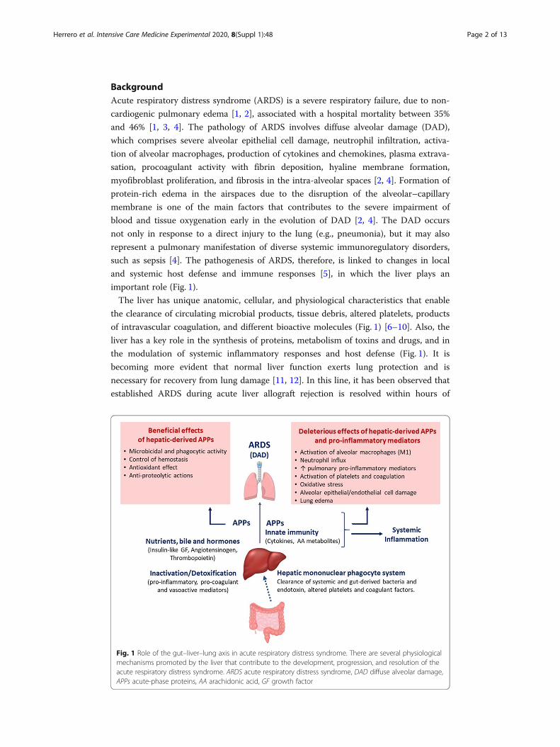
Fig. 1 Role of the gut–liver–lung axis in acute respiratory distress syndrome. There are several physiologicalmechanisms promoted by the liver that contribute to the development, progression, and resolution of theacute respiratory distress syndrome. ARDS acute respiratory distress syndrome, DAD diffuse alveolar damage,APPs acute-phase proteins, AA arachidonic acid, GF growth factor
Herrero et al. Intensive Care Medicine Experimental 2020, 8(Suppl 1):48 Page 2 of 13

hepatic re-transplantation [13]. On the other hand, experimental studies suggest that
the presence of the liver is also absolutely necessary for inducing lung injury in rats
[14]. These apparently paradoxical observations highlight the relevant crosstalk
between lung and liver in ARDS.
Despite the well-recognized liver–lung interaction in the pathogenesis of ARDS, its
underlying mechanisms and its effects on the outcome of these patients have been
barely studied due to several reasons. First, patients with liver diseases are frequently
excluded from studies of ARDS. In addition, liver function is not precisely reflected by
the standard liver function tests in the clinical setting, and the liver is not as accessible
as other organs such as the lung, making liver dysfunction not as evident as dysfunc-
tions of other organs. Finally, its clinical consequences are also heterogeneous in critic-
ally ill patients [15]. The present work reviews the important role of the liver on the
development and resolution of ARDS and aims to provide an integrated view of the
underlying mechanisms that support the liver–lung interaction in critically ill patients.
The reciprocal impacts of lung and liver dysfunctionsFollowing hepatocellular damage, the liver may reduce its clearance function, increase
the synthesis of deleterious substances, and dysregulate immune responses, leading to
systemic complications such as coagulopathy, elevated risk of infection, hypoglycaemia,
exacerbated inflammatory responses, encephalopathy, and damage of other extrahepatic
organs, including lung injury [16–22]. In critically ill patients, hepatic dysfunction is
recognized as a relevant clinical condition that influences the development, severity,
and progression of ARDS [5, 11, 19, 23–27]. In ARDS patients, liver dysfunction is a
major determinant of mortality [24–26]. It is well known that cirrhosis and other
chronic liver diseases make the patients more susceptible for developing ARDS, which
adversely affects patient outcomes [24–26, 28]. A growing body of evidence suggests
that liver damage activates and enhances inflammation in the pulmonary intravascular
compartment and lower respiratory tract, leading to important changes in the structure
and/or functions of the lung [29, 30]. Although all these observations indicate that liver
function is an important factor for the development and resolution of ARDS, there is
also evidence that such interorgan communication is bidirectional. Thus, acute lung
injury is known to impair hepatic function and to aggravate liver diseases by
mechanisms involving hypoxemia, activation of systemic inflammatory responses, and
cardiovascular changes [24, 31, 32].
Liver dysfunction is common in critical care patientsThe frequency of liver damage in critical illness has considerably increased over
the last decades [23, 33–35], reaching up to 20% of ICU patients in some series
and elevating their morbi-mortality [33, 34, 36]. In critically ill patients, liver
dysfunction usually occurs after inflammatory insults such as sepsis and trauma
[15, 23, 33, 37], and the underlying interactive mechanisms are complex. The
mechanisms of liver dysfunction in critically ill patients implicate microbial
products, the paracrine action of cytokines and other inflammatory mediators,
hypoxemia, oxidative stress, toxic compounds, hypoperfusion, passive congestion,
and effect of nutrition support, among others [34, 38–40].
Herrero et al. Intensive Care Medicine Experimental 2020, 8(Suppl 1):48 Page 3 of 13
Liver dysfunction can be manifested by plasma elevation of liver enzymes (aspartate
aminotransferase (AST), alanine aminotransferase (ALT), alkaline phosphatase (ALP),
γ-glutamyl transpeptidase) and bilirubin, decreased plasma levels of albumin and
coagulation factors, and/or increased international normalized ratio (INR) [33, 36]. The
clearance rate of indocyanine green has been used as a dynamic test to assess the
functional capacity of the liver. Although the indocyanine green test has shown to
reflect better the excretory and/or microvascular dysfunction of the liver, its clinical
use has certain limitations [41]. Increased plasma levels of bilirubin are associated with
high mortality in critically ill patients [39, 42]. Furthermore, hyperbilirubinemia has
been proposed as a biomarker of ARDS and found to be an independent factor of
mortality in patients with ARDS [26, 35, 39, 43]. Unfortunately, neither bilirubinemia
nor other hepatic parameters routinely measured in the clinical setting have the
sensitivity and specificity required for an early identification of hepatic injury in
critically ill patients [23, 33, 36, 40].
Mechanisms of liver–lung interactions in ARDSThe mechanisms by which the liver modulates lung injury involve interrelated elements
of systemic and pulmonary host defense, inflammatory responses, and metabolism and
include the following (see Fig. 1).
Clearance by the hepatic mononuclear phagocyte system of systemic endotoxemia,
bacteremia, vasoactive by-products, and procoagulant factors
The mononuclear phagocyte system located in the liver, spleen, lung, and bone marrow
constitutes the major mechanism to uptake and detoxify bacteria, fungi, viruses, and
dying cells, limiting the magnitude and duration of infections [10, 44, 45]. Although
these mononuclear phagocytic cells can exert this function in all these locations, their
major mass is in the hepatic sinusoids [10, 46]. The hepatic sinusoid is a unique
vascular structure with highly specialized endothelial cells (liver sinusoidal endothelial
cells) and liver macrophages (Kupffer cells) that reside within the lumen. The cells of
the hepatic sinusoid are constantly exposed to gut-derived bacteria, microbial debris,
and bacterial endotoxins. Kupffer cells, which line the extensive sinusoidal network,
constitute nearly 80–90% of the tissue macrophages present in the body and exert an
important role in host defense through phagocytosis and a multitude of secretory
functions [46]. The hepatic mononuclear phagocyte system acts as a first line of defense
in clearing bacteria and their products. Besides uptake of microbial pathogens and
products, Kupffer cells also protect the lung and other extrahepatic organs by removing
altered platelets and intravascular coagulation products (Fig. 1) [8–10, 46].
Dysfunction of the reticuloendothelial system of the liver allows bacterial and micro-
bial products, including the so-called pathogen-associated molecular patterns (PAMs)
[47], to reach the lung and the systemic circulation, where they activate pulmonary and
systemic inflammatory responses (Fig. 1) [29, 47–49]. Indeed, increased plasma levels of
endotoxin, probably of intestinal origin, along with increased levels of some cytokines
have been found in the blood of patients with acute and chronic liver diseases [50–54].
Pulmonary deposition of intravascular bacteria, and their products alter the structure
and function of the lung by different mechanisms including (i) direct cytotoxic effect
Herrero et al. Intensive Care Medicine Experimental 2020, 8(Suppl 1):48 Page 4 of 13
on alveolar epithelial and endothelial cells, (ii) modulation of local innate immune re-
sponses in the lung via activation of toll-like receptors (TLRs), resulting in activation of
resident alveolar macrophages and neutrophil influx and in the production of reactive
oxygen species, (iii) activation of the coagulation cascades and platelet aggregation,
leading to pulmonary microvascular thrombosis [29, 48, 49, 55–59], and (iv) a sustain-
able increase in pulmonary vascular resistance [22, 30] (Fig. 2). All these mechanisms
alter the alveolar endothelial and epithelial cell functions and enhance barrier perme-
ability leading to the formation pulmonary alveolar edema and respiratory failure [14,
60], the two main characteristics of ARDS (Fig. 2).
Metabolic inactivation and detoxification of endogenous inflammatory mediators
The hepatobiliary system has an important capacity to inactivate and detoxicate pro-
inflammatory cytokines, vasoactive mediators, and eicosanoids from the systemic circu-
lation. Removal of all these mediators constitutes a critical element of systemic and
pulmonary host defense, protecting the lung and other extrahepatic organs from injury
(Fig. 1) [8–10, 46]. Like endotoxin, increased levels of cytokines (such as IL-8, IL-1β,
ENA-78, TNF-α, MCP-1, MIP-1α,…) and arachidonic acid-derived eicosanoids
(thromboxane, leukotrienes) not cleared by the liver have been shown to exert a direct
cytotoxic effect on alveolar epithelial and endothelial cells, to activate local innate
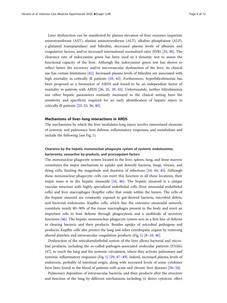
Fig. 2 Liver damage contributes to the development of acute respiratory distress syndrome. Liver injuryleads to changes in the expression of acute-phase proteins (APPs) and to an increase in plasma levels ofbacteria/bacterial products, pro-inflammatory cytokines, and pro-coagulant and vasoactive factors in thelung and systemic circulation. These mediators generate deleterious effects on the lung (passage ofbacteria /bacterial products and inflammation) and on the gut (intestinal dysbiosis, impairment of gutbarrier integrity, leakage of bacteria/bacterial products into the portal circulation and into the mesentericlymph), resulting in relevant changes in the hepatic and pulmonary microbiota and promotinginflammation and oxidative stress in liver and lung tissues. In addition, lung-derived cytokines promote thesynthesis of APPs and activation of inflammatory responses in the liver. All these responses mediated by thegut–liver–lung axis contribute to lung injury and multiple organ dysfunction in critical illness. IL interleukin,TNF tumor necrosis factor, INF interferon
Herrero et al. Intensive Care Medicine Experimental 2020, 8(Suppl 1):48 Page 5 of 13
immune responses and to promote platelet aggregation in the lung, contributing to the
development of diffuse alveolar damage (DAD) [55–59].
Hepatic synthesis of inflammatory mediators that can activate pulmonary alveolar
macrophages and, consequently, increase inflammation in the lung
Hepatic mononuclear cells include a heterogeneous population of lymphocytes,
Kupffer cells (hepatic resident macrophages), monocytes, and granulocytes that
perform vital functions for the innate and adaptive immune system. In response to
injury, activation of these hepatic mononuclear cells enhances the production and
release of inflammatory mediators, such as IL-1, IL-6, TNF-α, platelet-activating
factor (PAF), and leukotrienes, into the systemic circulation [61], where they play
an important role in the lung–liver interaction [18, 31, 51, 61–64]. These liver-
derived inflammatory mediators alter lung structure function early in acute inflam-
matory diseases (such as sepsis) and contribute to some extent to lung damage
upon activation of pulmonary alveolar macrophages (Fig. 1] [17, 65]. In this line,
elevated levels of TNF-α and IL-1β, two cytokines that are mainly synthetized by
alveolar macrophages, have been found in the lungs of rats with carbon tetrachlor-
ide (CCl4)-induced cirrhosis, along with an increase in lipid peroxidation (TBARS)
and antioxidant enzymes (superoxide dismutase and catalase) in the liver and lung
tissues. These events are also associated with altered gas exchange and changes in
the size of pulmonary vessels in these rats [66, 67]. Besides high levels of endo-
toxin [50, 68], patients with liver disorders also have high circulating levels of
TNF-α, IL-1, and IL-6 [51–54] because of the altered capacity for inactivation and
detoxification and the increased synthesis of pro-inflammatory mediators by the
liver [9, 44, 46, 61]. These specific cytokines have been shown to modulate
systemic inflammatory responses and participate in the development of lung
damage [69–72]. Therefore, it is possible that cytokines of hepatic origin may
control and modulate the local host defense and immune system of the lung,
contributing to lung injury (Figure 2).
The liver is the main organ responsible for the acute-phase response
The organism responds to tissue injury or infection by local changes such as those
associated to inflammation and by a coordinated sequence of systemic and metabolic
process, known as the acute-phase response, aimed to restore homeostasis and recover
from injury [63, 73–76]. One of the mayor characteristics of this acute-phase response
is a change in plasma concentration (either increase or decrease) of the acute-phase
proteins (APPs) expressed in the liver [74]. Cytokine-driving synthesis of acute-phase
proteins in the liver modulates the systemic and pulmonary host inflammatory
responses and intermediary metabolism (Fig. 1) [22, 30, 48]. The hepatic APPs have a
variety of functions that include microbicidal and phagocytic activity (e.g., LPS binding
protein, complement components, C-reactive protein), recruitment of immune cells to
inflammatory sites (e.g., serum amyloid A), hemostasis (e.g., fibrinogen, α1-acid
glycoprotein), antioxidant, and prevention of iron loss (e.g., haptoglobin) and anti-
proteolytic actions to counterbalance protease activity at sites of inflammation (e.g., α2-
macroglobulin, α1-antitrypsin, and α1-antichymotrypsin) (Fig. 1) [63, 74, 75].
Herrero et al. Intensive Care Medicine Experimental 2020, 8(Suppl 1):48 Page 6 of 13

While the local inflammation occurs in the alveolar airspaces of patients with ARDS,
the acute-phase response is induced in the liver [73, 76, 77]. Interestingly, in
pneumonia-induced ARDS, this liver-derived acute-phase response occurs independ-
ently of bacterial dissemination and depends instead on inflammatory signaling mole-
cules derived from the pulmonary immune cells, such as the cytokines IL-1, IL-6, and
TNF-α [10, 62, 63, 73, 74]. Then, these lung-derived cytokines can travel from the lung
into the systemic circulation and ultimately modify acute-phase gene expression in the
liver [63, 73, 78, 79] upon activation of the transcription factors STAT3 (signal trans-
ducer and activator of transcription 3) by IL-6 and activation of RelA (v-rel avian
reticuloendotheliosis viral oncogene homolog A, also known as NF-kB3) by the early-
response cytokines TNF-α and IL-1 [73, 78, 79]. In response to these cytokines, the
liver changes the expression of many acute-phase proteins such as C-reactive protein,
α-1 antitrypsin, serum amyloid A protein, and others [10, 62, 63, 74, 77], which in turn
can directly travel back to the lung and pass into the airspaces where they cause inflam-
mation, predominantly via activation of alveolar macrophages (Fig. 2) [5, 14]. These
phagocytic cells are targeted by multiple hepatic APPs such as SAA [80, 81], SAP [82],
LBP [83], and C-reactive protein [84, 85]. Once activated by these hepatic APPs, alveo-
lar macrophages release cytokines (IL-6 and CXCL1) that enhance local inflammation,
in part by promoting neutrophil influx to the insterstitium and alveolar airspaces. Ex-
cessive inflammation in the alveoli may result in an increase in oxidative stress and
lung injury [86, 87]. Besides this potentially deleterious effect, hepatic APPs at the site
of plasma extravasation has other potential functions, including opsonization of
bacteria, leukocyte activation, antiprotease, antioxidant activities, and modulation of
the coagulation pathway [63, 75]. These mechanisms help to regulate host defense,
limit excessive inflammation and immune responses, and promote bacterial clear-
ance, preventing infection dissemination and reducing the risk of organ damage in
the setting of pneumonia and sepsis. Also, hepatic APPs exert liver protection by
countering TNF-dependent toxicity in the liver and attenuate systemic inflamma-
tion and mortality in sepsis and pneumonia-induced ARDS (Fig. 1) [79, 86, 88–90].
Altogether, the bidirectional liver–lung axis mediated by APPs is critical for inte-
grating systemic and pulmonary responses, balancing regulation of multiple host
defenses and activation of inflammation to restore homeostasis and recover from
organ injury [48, 61, 86]. Disbalance in this liver–lung communication can be an
important factor in the initiation and progression of ARDS and of the damage to
other organs [73].
Nutrients, bile, and hormone production
The liver plays an important role in regulating metabolic homeostasis and in the syn-
thesis and processing of lipids and carbohydrates that supply energy to other organs
[91]. It is also the major site of synthesis of key proteins and bile acids that are critical
for the normal uptake of vitamins and lipids [92]. Therefore, alterations in the flux of
carbohydrates and lipids through the liver can indirectly impact distal organs due to
alteration of their energy statuses [93]. In addition, hyperbilirubinemia in the context of
liver diseases has been shown to cause some lung-specific deleterious effects, by enter-
ing the lung tissue, reaching the alveolar airspaces, and deteriorating the surface
Herrero et al. Intensive Care Medicine Experimental 2020, 8(Suppl 1):48 Page 7 of 13
tension properties of the alveolar surfactant [94]. Although bilirubin has antioxidant
properties, high bilirubin levels can also activate oxidative stress, apoptosis, and inflam-
matory responses in different cell types and organs [95–98]. Therefore, hyperbilirubine-
mia may actively participate in the development of ARDS, although the underlying
mechanisms have not been fully elucidated. Finally, the liver produces several
hormones that mediate diverse extrahepatic effects, such as insulin-like growth factor,
angiotensinogen, and thrombopoietin, which have been shown to influence the devel-
opment of ARDS (Fig. 1) [55, 99, 100].
The gut–liver–lung axis
The pathogenic mechanisms of ARDS should be considered within a gut–liver–lung
axis. Growing evidence indicates that intestinal microbiota and the mucosal immune
system of the gut have an important impact on the function of the gastrointestinal tract
itself and extra-intestinal organs, such as the lung and the liver [29, 48, 49]. Liver cir-
rhosis and other liver diseases favor the gut-derived bacterial translocation into the liver
and lung by several mechanisms (Fig. 2).
First, patients with liver disorders have intestinal dysbiosis characterized by a sig-
nificant shift of the microbial composition toward pro-inflammatory bacteria. This
gut dysbiosis is accompanied by activation of local intestine immune responses and
impaired gut barrier function. A leaky gut barrier facilities bacterial translocation
of live bacteria or their microbial products from the intestinal lumen to the liver,
via portal circulation, and to systemic circulation and the lung via the mesenteric
lymphatic system [29, 101]. In the lung and the liver, gut microbiota can directly
modulate their local immune cells (mainly alveolar macrophages and Kupffer cells,
respectively) via activation of toll-like receptors (TLRs) and indirectly via different
bacterial metabolites and signaling molecules, such as PAMPs [29, 48, 49, 90]. Ac-
tivated alveolar macrophages in the lung and Kupffer cells in the liver release pro-
inflammatory cytokines, contributing to the initiation and/or progression of lung
and liver damage and activation of systemic inflammation [29, 101], which can also
cause dysfunction in other organs.
Second, liver dysfunction can imply less capacity of the liver to remove bacteria, bac-
terial products, and inflammatory mediators from circulation, leading to increased
levels of these molecules in blood.
Third, this pathological gut-derived bacterial translocation could cause important
changes in the lung microbiome (Fig. 2) [90, 102, 103]. Indeed, pulmonary
microbiome is frequently enriched with gut-related bacteria (Bacteroidetes and
Enterobacteriaceae) in critically ill patients [102, 103]. As a consequence of liver
diseases, this gut-derived bacteria and accumulation of PAMPs, cytokines and other
pro-inflammatory molecules in the systemic circulation can potentially cause or ex-
acerbate lung injury upon TLR-4-mediated activation of intravascular and alveolar
macrophages within the lung and recruitment of neutrophils and direct toxic effects
of bacterial products on pulmonary microvasculature (Fig. 2) [68, 102–108].
Altogether, the gut–liver–lung axis seems to exert a relevant role in the initiation and
modulation of hepatic, pulmonary, and systemic immune responses that contribute to
the damage of the liver, the lung, and other organs.
Herrero et al. Intensive Care Medicine Experimental 2020, 8(Suppl 1):48 Page 8 of 13
Extracellular vesicles
Extracellular vesicles (EVs), a term that includes microvesicles (MVs), exosomes, and
apoptotic bodies, represent an emerging mechanism of interorgan communication in
many diseases, including liver diseases and ARDS [106, 109–111]. Extracellular vesicles
are defined as membrane-bound vesicles, ranging 0.1–1.0 μm in diameter, which are
released from cells by the budding of the cellular plasma membrane and carrying a
diverse cargo, including lipids, proteins, RNAs, and miRNAs. The EVs are recognized
as important mediators of interaction between different organs, and they are considered
attractive therapeutic targets in different diseases [112, 113]. Notably, the levels of
circulating EVs have been reported to be increased both in patients with cirrhosis and
ARDS. However, the potential role of circulating EVs in mediating liver–lung commu-
nication in the context of ARDS is not currently understood, representing an interest-
ing topic for further investigation.
ConclusionsLiver injury and hepatotoxicity occur frequently in critically ill patients, and signifi-
cantly influence their prognosis. Patients with severe hepatic dysfunction are at high
risk for irreversible ARDS because of multiple defects in host defense and dysregulation
of inflammatory responses. Interrelations between hepatic and pulmonary functions
influence the development and progression of ARDS and play a central role in the
resolution of lung damage by several mechanisms. First, the liver regulates host defense
and modulates systemic inflammation. Also, the liver activates acute inflammatory
responses in the lung early in the development of ARDS. Although promoting inflam-
mation can be detrimental in the context of acute lung injury, the liver response to an
inflammatory insult is also pro-defense and pro-survival. The understanding of the
complex relation between the liver and the lung requires further research in order to
improve the clinical management and to identify new diagnostic and therapeutic
options for patients with or at risk for ARDS.
AbbreviationsARDS: Acute respiratory distress syndrome; DAD: Diffuse alveolar damage; ICU: Intensive care unit; AST: Aspartateaminotransferase; ALT: Alanine aminotransferase; ALP: Alkaline phosphatase; INR: International normalized ratio;PAMPs: Pathogen-associated molecular patterns; TLRs: Toll-like receptors; IL: Interleukin; ENA-78: Epithelial neutrophil-activating peptide-78; TNF: Tumor necrosis factor; MCP-1: Monocyte chemoattractant protein-1; MIP-1α: Macrophageinflammatory protein-1α; PAF: Platelet-activating factor; CCl4: Carbon tetrachloride; TBARS: Thiobarbituric acid-reactivesubstances; APPs: Acute-phase proteins; LPS: Lipopolysaccharides; STAT3: Signal transducer and activator oftranscription 3; RelA: v-rel avian reticuloendotheliosis viral oncogene homolog A; NF-kB3: Nuclear factor kappa-light-chain-enhancer of activated B3; SAA: Serum amyloid A; SAP: Serum amyloid P; LBP: Lipopolysaccharide bindingprotein; CXCL1: Chemokine (C-X-C motif) ligand 1; EVs: Extracellular vesicles; MVs: Microvesicles; RNAs: Ribonucleicacids; miRNAs: Micro-ribonucleic acids
AcknowledgementsNot applicable
About this supplementThis article has been published as part of Intensive Care Medicine Experimental Volume 8 Supplement 1, 2020:Proceedings from the Fourth International Symposium on Acute Pulmonary Injury and Translation Research (INSPIRESIV). The full contents of the supplement are available at https://icm-experimental.springeropen.com/articles/supplements/volume-8-supplement-1.
Authors’ contributionsRH and GS did the design, acquisition of information, and drafting and revising the manuscript. IA, EL, AF, and AdLwere responsible for the acquisition of information. JV, LM, RB, and JAL did the revising of the work. The authors readand approved the final manuscript.
Herrero et al. Intensive Care Medicine Experimental 2020, 8(Suppl 1):48 Page 9 of 13

FundingThis study was funded with grants PI12/02451, PI15/00482 and PI19/01091 (to RH) and PI15/01942 (to JAL) from theInstituto de Salud Carlos III, Ministerio de Economia y Competitividad, Madrid, Spain. Also, another funding with grant,B2017/BMD-3727-EXOHEP-CM (to RB and JAL) was from the Comunidad de Madrid and Fondos FEDER “Una manerade hacer Europa”, Madrid, Spain.
Availability of data and materialsNot applicable
Ethics approval and consent to participateNot applicable
Consent for publicationNot applicable
Competing interestsThe authors declare that they have no competing interests.
Author details1Department of Critical Care Medicine, Hospital Universitario de Getafe, Madrid, Spain. 2CIBER de EnfermedadesRespiratorias, Instituto de Investigación Carlos III, Madrid, Spain. 3Fundación de Investigación Biomédica del HospitalUniversitario de Getafe, Madrid, Spain. 4Laboratory of Biochemistry, Hospital Universitario de Getafe, Madrid, Spain.5Servicio de Aparato Digestivo. HGU Gregorio Marañón, Instituto de Investigación Sanitaria Gregorio Marañón (IiSGM),Madrid, Spain. 6CIBER de Enfermedades Hepáticas y Digestivas, Instituto de Investigación Carlos III, Madrid, Spain.7Department of Pharmacology, School of Medicine, Universidad Complutense de Madrid, Madrid, Spain. 8UniversidadEuropea de Madrid, Madrid, Spain.
Received: 14 July 2020 Accepted: 16 July 2020Published: 18 December 2020
References1. Definition Task Force ARDS, Ranieri VM, Rubenfeld GD, Thompson BT, Ferguson ND, Caldwell E et al (2012) Acute
respiratory distress syndrome: the Berlin definition. JAMA 307(23):2526–25332. Matthay MA, Ware LB, Zimmerman GA (2012) The acute respiratory distress syndrome. J Clin Invest 122(8):2731–27403. Bellani G, Laffey JG, Pham T, Fan E, Brochard L, Esteban A et al (2016) Epidemiology, patterns of care, and mortality for
patients with acute respiratory distress syndrome in intensive care units in 50 countries. JAMA 315(8):788–8004. Matthay MA, Zemans RL (2011) The acute respiratory distress syndrome: pathogenesis and treatment. Annu Rev Pathol
6:147–1635. Matuschak GM, Rinaldo JE (1988) Organ interactions in the adult respiratory distress syndrome during sepsis. Role of the
liver in host defense. Chest 94(2):400–4066. Rogers DE (1960) Host mechanisms which act to remove bacteria from the blood stream. Bacteriol Rev 24(1):50–667. Kolaczkowska E, Jenne CN, Surewaard BGJ, Thanabalasuriar A, Lee W-Y, Sanz M-J et al (2015) Molecular mechanisms of
NET formation and degradation revealed by intravital imaging in the liver vasculature. Nat Commun 6:66738. Kaplan JE, Saba TM (1978) Platelet removal from the circulation by the liver and spleen. Am J Phys 235(3):H314–H3209. Bradfield JW (1974) Control of spillover. The importance of Kupffer-cell function in clinical medicine. Lancet Lond Engl
2(7885):883–88610. Guillot A, Tacke F (2019) Liver macrophages: old dogmas and new insights. Hepatol Commun 3(6):730–74311. Khanlou H, Souto H, Lippmann M, Muñoz S, Rothstein K, Ozden Z (1999) Resolution of adult respiratory distress
syndrome after recovery from fulminant hepatic failure. Am J Med Sci 317(2):134–13612. Ali M, Wall WJ (1990) Resolution of the adult respiratory distress syndrome following colectomy and liver
transplantation. Chest 98(4):1032–103413. Doyle HR, Marino IR, Miro A, Scott V, Martin M, Fung J et al (1993) Adult respiratory distress syndrome secondary to
end-stage liver disease - successful outcome following liver transplantation. Transplantation 55(2):292–29614. Siore AM, Parker RE, Stecenko AA, Cuppels C, McKean M, Christman BW et al (2005) Endotoxin-induced acute lung
injury requires interaction with the liver. Am J Physiol Lung Cell Mol Physiol 289(5):L769–L77615. Matuschak GM (1994) Liver-lung interactions in critical illness. New Horiz 2(4):488–50416. Cerra FB, West M, Billiar TR, Holman RT, Simmons R (1989) Hepatic dysfunction in multiple systems organ failure as a
manifestation of altered cell-cell interaction. Prog Clin Biol Res 308:563–57317. Matuschak GM (1996) Lung-liver interactions in sepsis and multiple organ failure syndrome. Clin Chest Med 17(1):83–9818. Wang Y, Liu W, Liu X, Sheng M, Pei Y, Lei R et al (2015) Role of liver in modulating the release of inflammatory
cytokines involved in lung and multiple organ dysfunction in severe acute pancreatitis. Cell Biochem Biophys71(2):765–776
19. Matuschak GM, Martin DJ (1987) Influence of end-stage liver failure on survival during multiple systems organ failure.Transplant Proc 19(4 Suppl 3):40–46
20. Katz S, Grosfeld JL, Gross K, Plager DA, Ross D, Rosenthal RS et al (1984) Impaired bacterial clearance and trapping inobstructive jaundice. Ann Surg 199(1):14–20
21. Loegering DJ, Blumenstock FA (1985) Depressing hepatic macrophage complement receptor function causes increasedsusceptibility to endotoxemia and infection. Infect Immun 47(3):659–664
22. DeCamp MM, Warner AE, Molina RM, Brain JD (1992) Hepatic versus pulmonary uptake of particles injected into theportal circulation in sheep. Endotoxin escapes hepatic clearance causing pulmonary inflammation. Am Rev Respir Dis146(1):224–231
Herrero et al. Intensive Care Medicine Experimental 2020, 8(Suppl 1):48 Page 10 of 13

23. Hawker F (1991) Liver dysfunction in critical illness. Anaesth Intensive Care 19(2):165–18124. Yang P, Formanek P, Scaglione S, Afshar M (2019) Risk factors and outcomes of acute respiratory distress syndrome in
critically ill patients with cirrhosis. Hepatol Res Off J Jpn Soc Hepatol 49(3):335–34325. Gacouin A, Locufier M, Uhel F, Letheulle J, Bouju P, Fillatre P et al (2016) Liver cirrhosis is independently associated with
90-day mortality in ARDS patients. Shock 45(1):16–2126. Schwartz DB, Bone RC, Balk RA, Szidon JP (1989) Hepatic dysfunction in the adult respiratory distress syndrome. Chest
95(4):871–87527. Wang CY, Calfee CS, Paul DW, Janz DR, May AK, Zhuo H et al (2014) One-year mortality and predictors of death among
hospital survivors of acute respiratory distress syndrome. Intensive Care Med 40(3):388–39628. Dong V, Sun K, Gottfried M, Cardoso FS, McPhail MJ, Stravitz RT et al (2020) Significant lung injury and its prognostic
significance in acute liver failure: a cohort analysis. Liver Int Off J Int Assoc Study Liver 40(3):654–66329. Albillos A, Gottardi A, de Rescigno M (2019) The gut-liver axis in liver disease: pathophysiological basis for therapy. J
Hepatol30. Matuschak GM, Mattingly ME, Tredway TL, Lechner AJ (1994) Liver-lung interactions during E. coli endotoxemia. TNF-
alpha:leukotriene axis. Am J Respir Crit Care Med 149(1):41–4931. Patterson EK, Yao LJ, Ramic N, Lewis JF, Cepinskas G, McCaig L et al (2013) Lung-derived mediators induce cytokine
production in downstream organs via an NF-κB-dependent mechanism. Mediat Inflamm 2013:58689532. Karcz M, Bankey B, Schwaiberger D, Lachmann B, Papadakos PJ (2012) Acute respiratory failure complicating advanced
liver disease. Semin Respir Crit Care Med 33(1):96–11033. Horvatits T, Drolz A, Trauner M, Fuhrmann V. Liver injury and failure in critical illness. Hepatol 2019;34. Kramer L, Jordan B, Druml W, Bauer P, Metnitz PGH, Austrian Epidemiologic Study on Intensive Care, ASDI Study Group
et al (2007) Crit Care Med 35(4):1099–110435. Dizier S, Forel J-M, Ayzac L, Richard J-C, Hraiech S, Lehingue S et al (2015) Early hepatic dysfunction is associated with a
worse outcome in patients presenting with acute respiratory distress syndrome: a post-hoc analysis of the ACURASYSand PROSEVA studies. PLoS One 10(12):e0144278
36. Lescot T, Karvellas C, Beaussier M, Magder S (2012) Acquired liver injury in the intensive care unit. Anesthesiology 117(4):898–904
37. Marshall JC (2013) The liver in sepsis: shedding light on the cellular basis of hepatocyte dysfunction. Crit Care Lond Engl17(3):153
38. Fuhrmann V, Kneidinger N, Herkner H, Heinz G, Nikfardjam M, Bojic A et al (2011) Impact of hypoxic hepatitis onmortality in the intensive care unit. Intensive Care Med 37(8):1302–1310
39. Zhai R, Sheu CC, Su L, Gong MN, Tejera P, Chen F et al (2009) Serum bilirubin levels on ICU admission are associatedwith ARDS development and mortality in sepsis. Thorax 64(9):784–790
40. Kortgen A, Paxian M, Werth M, Recknagel P, Rauchfuss F, Lupp A et al (2009) Prospective assessment of hepatic functionand mechanisms of dysfunction in the critically ill. Shock 32(4):358–365
41. Sakka SG (2007) Assessing liver function. Curr Opin Crit Care 13(2):207–21442. Vincent JL, Moreno R, Takala J, Willatts S, De Mendonça A, Bruining H et al (1996) The SOFA (sepsis-related organ failure
assessment) score to describe organ dysfunction/failure. On behalf of the working group on sepsis-related problems ofthe European Society of Intensive Care Medicine. Intensive Care Med 22(7):707–710
43. Sheu C-C, Gong MN, Zhai R, Chen F, Bajwa EK, Clardy PF et al (2010) Clinical characteristics and outcomes of sepsis-related vs non-sepsis-related ARDS. Chest 138(3):559–567
44. Praaning-van Dalen DP, Brouwer A, Knook DL (1981) Clearance capacity of rat liver Kupffer, endothelial, andparenchymal cells. Gastroenterology 81(6):1036–1044
45. Freudenberg MA, Freudenberg N, Galanos C (1982) Time course of cellular distribution of endotoxin in liver, lungs andkidneys of rats. Br J Exp Pathol 63(1):56–65
46. Bilzer M, Roggel F, Gerbes AL (2006) Role of Kupffer cells in host defense and liver disease. Liver Int Off J Int AssocStudy Liver 26(10):1175–1186
47. Triantafyllou E, Woollard KJ, MJW MP, Antoniades CG, Possamai LA (2018) The role of monocytes and macrophages inacute and acute-on-chronic liver failure. Front Immunol 9:2948
48. Young RP, Hopkins RJ, Marsland B (2016) The gut-liver-lung axis, Modulation of the innate immune response and itspossible role in chronic obstructive pulmonary disease. Am J Respir Cell Mol Biol 54(2):161–169
49. Enaud R, Prevel R, Ciarlo E, Beaufils F, Wieërs G, Guery B, et al. The gut-lung axis in health and respiratory diseases: aplace for inter-organ and inter-kingdom crosstalks. Front Cell Infect Microbiol [Internet]. 19 de febrero de 2020 [citado 1de julio de 2020];10. Disponible en: https://www.ncbi.nlm.nih.gov/pmc/articles/PMC7042389/
50. Tarao K, Moroi T, Nagakura Y, Ikeuchi T, Suyama T, Endo O et al (1979) Relationship between endotoxaemia and proteinconcentration of ascites in cirrhotic patients. Gut 20(3):205–210
51. Tilg H, Wilmer A, Vogel W, Herold M, Nölchen B, Judmaier G et al (1992) Serum levels of cytokines in chronic liverdiseases. Gastroenterology 103(1):264–274
52. Khoruts A, Stahnke L, McClain CJ, Logan G, Allen JI (1991) Circulating tumor necrosis factor, interleukin-1 andinterleukin-6 concentrations in chronic alcoholic patients. Hepatol 13(2):267–276
53. Devière J, Content J, Denys C, Vandenbussche P, Schandene L, Wybran J et al (1990) Excessive in vitro bacteriallipopolysaccharide-induced production of monokines in cirrhosis. Hepatol 11(4):628–634
54. Torre D, Zeroli C, Giola M, Ferrario G, Fiori GP, Bonetta G et al (1994) Serum levels of interleukin-1 alpha, interleukin-1beta, interleukin-6, and tumor necrosis factor in patients with acute viral hepatitis. Clin Infect Dis Off Publ Infect Dis SocAm 18(2):194–198
55. Kobayashi K, Horikami D, Omori K, Nakamura T, Yamazaki A, Maeda S et al (2016) Thromboxane A2 exacerbates acutelung injury via promoting edema formation. Sci Rep 6:32109
56. Park WY, Goodman RB, Steinberg KP, Ruzinski JT, Radella F, Park DR et al (2001) Cytokine balance in the lungs ofpatients with acute respiratory distress syndrome. Am J Respir Crit Care Med 164(10 Pt 1):1896–1903
57. Goodman RB, Strieter RM, Martin DP, Steinberg KP, Milberg JA, Maunder RJ et al (1996) Inflammatory cytokines inpatients with persistence of the acute respiratory distress syndrome. Am J Respir Crit Care Med 154(3 Pt 1):602–611
Herrero et al. Intensive Care Medicine Experimental 2020, 8(Suppl 1):48 Page 11 of 13
58. Wiedermann FJ, Mayr AJ, Kaneider NC, Fuchs D, Mutz NJ, Schobersberger W (2004) Alveolar granulocyte colony-stimulating factor and alpha-chemokines in relation to serum levels, pulmonary neutrophilia, and severity of lung injuryin ARDS. Chest 125(1):212–219
59. Frank JA, Matthay MA (2005) Leukotrienes in acute lung injury. Am J Respir Crit Care Med 172(3):261–26260. Rojas M, Woods CR, Mora AL, Xu J, Brigham KL (2005) Endotoxin-induced lung injury in mice: structural, functional, and
biochemical responses. Am J Physiol Lung Cell Mol Physiol 288(2):L333–L34161. Panos RJ, Baker SK (1996) Mediators, cytokines, and growth factors in liver-lung interactions. Clin Chest Med 17(1):151–16962. Tran-Thi TA, Weinhold L, Weinstock C, Hoffmann R, Schulze-Specking A, Northoff H et al (1993) Production of tumor
necrosis factor-alpha, interleukin-1 and interleukin-6 in the perfused rat liver. Eur Cytokine Netw 4(5):363–37063. Ramadori G, Christ B (1999) Cytokines and the hepatic acute-phase response. Semin Liver Dis 19(2):141–15564. Hermanns MI, Kasper J, Dubruel P, Pohl C, Uboldi C, Vermeersch V et al (2010) An impaired alveolar-capillary barrier
in vitro: effect of proinflammatory cytokines and consequences on nanocarrier interaction. J R Soc Interface 7(Suppl 1):S41–S54
65. Callery MP, Kamei T, Mangino MJ, Flye MW (1991) Organ interactions in sepsis. Host defense and the hepatic-pulmonarymacrophage axis. Arch Surg Chic Ill 1960 126(1):28–32
66. Ferrari RS, Tieppo M, da RDP, Forgiarini LA, Dias AS, Marroni NP (2013) Lung and liver changes due to the induction ofcirrhosis in two experimental models. Arq Gastroenterol 50(3):208–213
67. Salatti Ferrari R, da Rosa DP, Forgiarini LF, Bona S, Dias AS, Marroni NP (2012) Oxidative stress and pulmonary changes inexperimental liver cirrhosis. Oxidative Med Cell Longev 2012:486190
68. Massey VL, Poole LG, Siow DL, Torres E, Warner NL, Schmidt RH et al (2015) Chronic alcohol exposure enhanceslipopolysaccharide-induced lung injury in mice: potential role of systemic tumor necrosis factor-alpha. Alcohol Clin ExpRes 39(10):1978–1988
69. Halbertsma FJJ, Vaneker M, Scheffer GJ, van der Hoeven JG. Cytokines and biotrauma in ventilator-induced lung injury: acritical review of the literature. Neth J Med 2005;63(10):382-392.
70. Seekamp A, Warren JS, Remick DG, Till GO, Ward PA (1993) Requirements for tumor necrosis factor-alpha andinterleukin-1 in limb ischemia/reperfusion injury and associated lung injury. Am J Pathol 143(2):453–463
71. Ahuja N, Andres-Hernando A, Altmann C, Bhargava R, Bacalja J, Webb RG et al (2012) Circulating IL-6 mediates lunginjury via CXCL1 production after acute kidney injury in mice. Am J Physiol Renal Physiol 303(6):F864–F872
72. Zhang H, Neuhöfer P, Song L, Rabe B, Lesina M, Kurkowski MU et al (2013) IL-6 trans-signaling promotes pancreatitis-associated lung injury and lethality. J Clin Invest 123(3):1019–1031
73. Quinton LJ, Jones MR, Robson BE, Mizgerd JP (2009) Mechanisms of the hepatic acute-phase response during bacterialpneumonia. Infect Immun 77(6):2417–2426
74. Gabay C, Kushner I (1999) Acute-phase proteins and other systemic responses to inflammation. N Engl J Med 340(6):448–454
75. Suffredini AF, Fantuzzi G, Badolato R, Oppenheim JJ, O’Grady NP (1999) New insights into the biology of the acutephase response. J Clin Immunol 19(4):203–214
76. Weber M, Lambeck S, Ding N, Henken S, Kohl M, Deigner HP et al (2012) Hepatic induction of cholesterol biosynthesisreflects a remote adaptive response to pneumococcal pneumonia. FASEB J Off Publ Fed Am Soc Exp Biol 26(6):2424–2436
77. Gamble L, Bagby GJ, Quinton LJ, Happel KI, Mizgerd JP, Zhang P et al (2009) The systemic and pulmonary LPS bindingprotein response to intratracheal lipopolysaccharide. Shock 31(2):212–217
78. Quinton LJ, Jones MR, Robson BE, Simms BT, Whitsett JA, Mizgerd JP (2008) Alveolar epithelial STAT3, IL-6 familycytokines, and host defense during Escherichia coli pneumonia. Am J Respir Cell Mol Biol 38(6):699–706
79. Quinton LJ, Blahna MT, Jones MR, Allen E, Ferrari JD, Hilliard KL et al (2012) Hepatocyte-specific mutation of both NF-κBRelA and STAT3 abrogates the acute phase response in mice. J Clin Invest 122(5):1758–1763
80. Niemi K, Teirilä L, Lappalainen J, Rajamäki K, Baumann MH, Öörni K et al (2011) Serum amyloid a activates the NLRP3inflammasome via P2X7 receptor and a cathepsin B-sensitive pathway. J Immunol 186(11):6119–6128
81. Shah C, Hari-Dass R, Raynes JG, Serum amyloid A (2006) Is an innate immune opsonin for gram-negative bacteria. Blood108(5):1751–1757
82. Zhang W, Xu W, Xiong S (2011) Macrophage differentiation and polarization via phosphatidylinositol 3-kinase/Akt-ERKsignaling pathway conferred by serum amyloid P component. J Immunol 187(4):1764–1777
83. Tobias PS, Mathison J, Mintz D, Lee JD, Kravchenko V, Kato K et al (1992) Participation of lipopolysaccharide-bindingprotein in lipopolysaccharide-dependent macrophage activation. Am J Respir Cell Mol Biol 7(3):239–245
84. Mold C, Du Clos TW (2006) C-reactive protein increases cytokine responses to Streptococcus pneumoniae throughinteractions with fc gamma receptors. J Immunol 176(12):7598–7604
85. Barna BP, Deodhar SD, Gautam S, Yen-Lieberman B, Roberts D (1984) Macrophage activation and generation oftumoricidal activity by liposome-associated human C-reactive protein. Cancer Res 44(1):305–310
86. Hilliard KL, Allen E, Traber KE, Yamamoto K, Stauffer NM, Wasserman GA et al (2015) The lung-liver axis: a requirementfor maximal innate immunity and hepatoprotection during pneumonia. Am J Respir Cell Mol Biol 53(3):378–390
87. Frank JA, Wray CM, McAuley DF, Schwendener R, Matthay MA (2006) Alveolar macrophages contribute to alveolarbarrier dysfunction in ventilator-induced lung injury. Am J Physiol Lung Cell Mol Physiol 291(6):L1191–L1198
88. Renckens R, van Westerloo DJ, Roelofs JJTH, Pater JM, Schultz MJ, Florquin S et al (2008) Acute phase response impairshost defense against Pseudomonas aeruginosa pneumonia in mice. Crit Care Med 36(2):580–587
89. Renckens R, Roelofs JJTH, Knapp S, de Vos AF, Florquin S, van der Poll T (2006) The acute-phase response and serumamyloid a inhibit the inflammatory response to Acinetobacter baumannii pneumonia. J Infect Dis 193(2):187–195
90. Sakamori R, Takehara T, Ohnishi C, Tatsumi T, Ohkawa K, Takeda K et al (2007) Signal transducer and activator oftranscription 3 signaling within hepatocytes attenuates systemic inflammatory response and lethality in septic mice.Hepatol 46(5):1564–1573
91. Postic C, Dentin R, Girard J (2004) Role of the liver in the control of carbohydrate and lipid homeostasis. Diabetes Metab30(5):398–408
92. Hofmann AF (1999) The continuing importance of bile acids in liver and intestinal disease. Arch Intern Med 159(22):2647–2658
Herrero et al. Intensive Care Medicine Experimental 2020, 8(Suppl 1):48 Page 12 of 13
93. Rui L (2014) Energy metabolism in the liver. Compr Physiol 4(1):177–19794. Dani C, Martelli E, Tronchin M, Buonocore G, Longini M, Di Filippo A et al (2004) Bilirubin influence on oxidative lung
damage and surfactant surface tension properties. Pediatr Pulmonol 38(3):179–18595. Chopra M, Reuben JS, Sharma AC (2009) Acute lung injury:apoptosis and signaling mechanisms. Exp Biol 234(4):361–37196. Rodrigues CMP, Solá S, Brito MA, Brites D, Moura JJG (2002) Bilirubin directly disrupts membrane lipid polarity and
fluidity, protein order, and redox status in rat mitochondria. J Hepatol 36(3):335–34197. Cesaratto L, Calligaris SD, Vascotto C, Deganuto M, Bellarosa C, Quadrifoglio F et al (2007) Bilirubin-induced cell toxicity
involves PTEN activation through an APE1/Ref-1-dependent pathway. J Mol Med 85(10):1099–111298. Fernandes A, Falcão AS, Silva RFM, Gordo AC, Gama MJ, Brito MA et al (2006) Inflammatory signalling pathways
involved in astroglial activation by unconjugated bilirubin. J Neurochem 96(6):1667–167999. Cuccurullo A, Greco E, Lupia E, De Giuli P, Bosco O, Martin-Conte E et al (2016) Blockade of thrombopoietin reduces
organ damage in experimental endotoxemia and polymicrobial sepsis. PLoS One 11(3):e0151088100. Wang Z, Li W, Guo Q, Wang Y, Ma L, Zhang X (2018) Insulin-like growth factor-1 signaling in lung development and
inflammatory lung diseases. Biomed Res Int 2018:6057589101. Acharya C, Sahingur SE, Bajaj JS. Microbiota, cirrhosis, and the emerging oral-gut-liver axis. JCI Insight. 05 de 2017;2(19).102. Dickson RP (2018) The lung microbiome and ARDS. It is time to broaden the model. Am J Respir Crit Care Med 197(5):
549–551103. Mukherjee S, Hanidziar D (2018) More of the gut in the lung: how two microbiomes meet in ARDS. Yale J Biol Med
91(2):143–149104. Dickson RP, Singer BH, Newstead MW, Falkowski NR, Erb-Downward JR, Standiford TJ et al (2016) Enrichment of the
lung microbiome with gut bacteria in sepsis and the acute respiratory distress syndrome. Nat Microbiol 1(10):16113105. Deitch EA (2010) Gut lymph and lymphatics: a source of factors leading to organ injury and dysfunction. Ann N Y Acad
Sci 1207(Suppl 1):E103–E111106. Kojima M, Gimenes-Junior JA, Chan TW, Eliceiri BP, Baird A, Costantini TW et al (2018) Exosomes in postshock
mesenteric lymph are key mediators of acute lung injury triggering the macrophage activation via toll-like receptor 4.FASEB J Off Publ Fed Am Soc Exp Biol 32(1):97–110
107. de Jong PR, González-Navajas JM, Jansen NJG (2016) The digestive tract as the origin of systemic inflammation. CritCare Lond Engl 20(1):279
108. Massey VL (2015) Potential role of the gut/liver/lung axis in alcohol-induced tissue pathology. Biomolecules 5(4):2477–2503109. Hirsova P, Ibrahim SH, Verma VK, Morton LA, Shah VH, LaRusso NF et al (2016) Extracellular vesicles in liver
pathobiology: small particles with big impact. Hepatol 64(6):2219–2233110. Maji S, Matsuda A, Yan IK, Parasramka M, Patel T (2017) Extracellular vesicles in liver diseases. Am J Physiol Gastrointest
Liver Physiol 312(3):G194–G200111. Malhi H (2019) Emerging role of extracellular vesicles in liver diseases. Am J Physiol Gastrointest Liver Physiol 317(5):
G739–G749112. McVey M, Tabuchi A, Kuebler WM (2012) Microparticles and acute lung injury. Am J Physiol Lung Cell Mol Physiol
303(5):L364–L381113. Rautou P-E, Bresson J, Sainte-Marie Y, Vion A-C, Paradis V, Renard J-M et al (2012) Abnormal plasma microparticles
impair vasoconstrictor responses in patients with cirrhosis. Gastroenterology 143(1):166–176.e6
Publisher’s NoteSpringer Nature remains neutral with regard to jurisdictional claims in published maps and institutional affiliations.
Herrero et al. Intensive Care Medicine Experimental 2020, 8(Suppl 1):48 Page 13 of 13