liver tissue engineering: where are we now? · liver tissue engineering: from implantable tissue to...
TRANSCRIPT

MASSIMO PINZANI, MD, PhD, FRCP, FAASLDSheila Sherlock Chair of Hepatology
UCL Institute for Liver and Digestive Health
Royal Free Hospital, London, UK
www.ucl.ac.uk/medicine/liver-and-digestive-health
LIVER TISSUE ENGINEERING: WHERE
ARE WE NOW?
Disclosures (2019)
1. Inventor and patent holder ELF test (Siemens)
2. Speakers bureau: Echosens (Paris, France)
3. SAB/Consultancy: Promethera (Belgium); NeuroVive (Sweden);
Chemomab (Israel); Median Technology (France/USA); Boheringer-Ingelheim
(Germany); Takeda (USA)
4. Co-Founder and Director, Engitix Ltd (UCL Spin-out) (UK)*
5. Co-Founder and Director, 3P-Sense Ltd (UCL Spin-out) (UK)**
6. CMO, Hepatotargets Ltd (Cambridge University Spin-out) (UK)***
6. Educational Councillor EASL Governing Board (2015-2019)
7. Chair EASL Consortium for Regenerative Hepatology (2019-2022)
* Regenerative medicine and tissue engineering
** Nanotechnology diagnostics
*** Liver Cell therapy

From Discovery to Clinical Applications
Drug target discovery based on 2D cell
cultures on plastic
Expansion and validation in animal models of chronic liver injury: no model is able to reproduce
human pathophysiology
No translation into clinical trials and very high failure rate in the trials so far performed (>95%)
No licensed antifibrotic drugs after more than 40 years of active research (the case of liver fibrosis)
Wrong targets? Wrong validation methodology?

Pathways of Stellate Cell Activation in Liver Injury
Changes in ECM quantity and relative composition
Altered MATRISOME

Need to focus on mechanisms and preclinical models easier to translate into clinical applications:
The fibrotic microenvironment :a. Hypoxia and neo-angiogenesisb. Anaerobic metabolism (e.g. lactate)c. 3D in vitro modelsd. Hepatic matrisome and matrikinese. Tissue stiffness and contraction
Understanding Liver Fibrosis

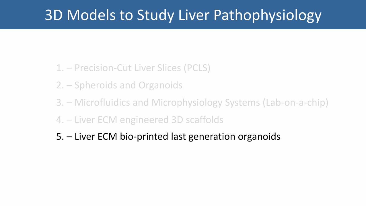
3D Models to Study Liver Pathophysiology
1. – Precision-Cut Liver Slices (PCLS)
2. – Spheroids and Organoids
3. – Microfluidics and Microphysiology Systems (Lab-on-a-chip)
4. – Liver ECM engineered 3D scaffolds
5. – Liver ECM bio-printed last generation organoids
Tissue Engineering

3D Models to Study Liver Pathophysiology
1. – Precision-Cut Liver Slices (PCLS)
2. – Spheroids and Organoids
3. – Microfluidics and Microphysiology Systems (Lab-on-a-chip)
4. – Liver ECM engineered 3D scaffolds
5. – Liver ECM bio-printed last generation organoids

Precision-Cut Liver Slices
Olinga P and Schuppan D

Hepatology. 2019 Apr 4. doi: 10.1002/hep.30651. [Epub ahead of print]

Spheroids, Organoids, Lab-on-a-chip
2D 3D


3D Models to Study Liver Pathophysiology
1. – Precision-Cut Liver Slices (PCLS)
2. – Spheroids and Organoids
3. – Microfluidics and Microphysiology Systems (Lab-on-a-chip)
4. – Liver ECM engineered 3D scaffolds
5. – Liver ECM bio-printed last generation organoids

Reproducing human non alcoholic fatty liver under lipotoxic stress
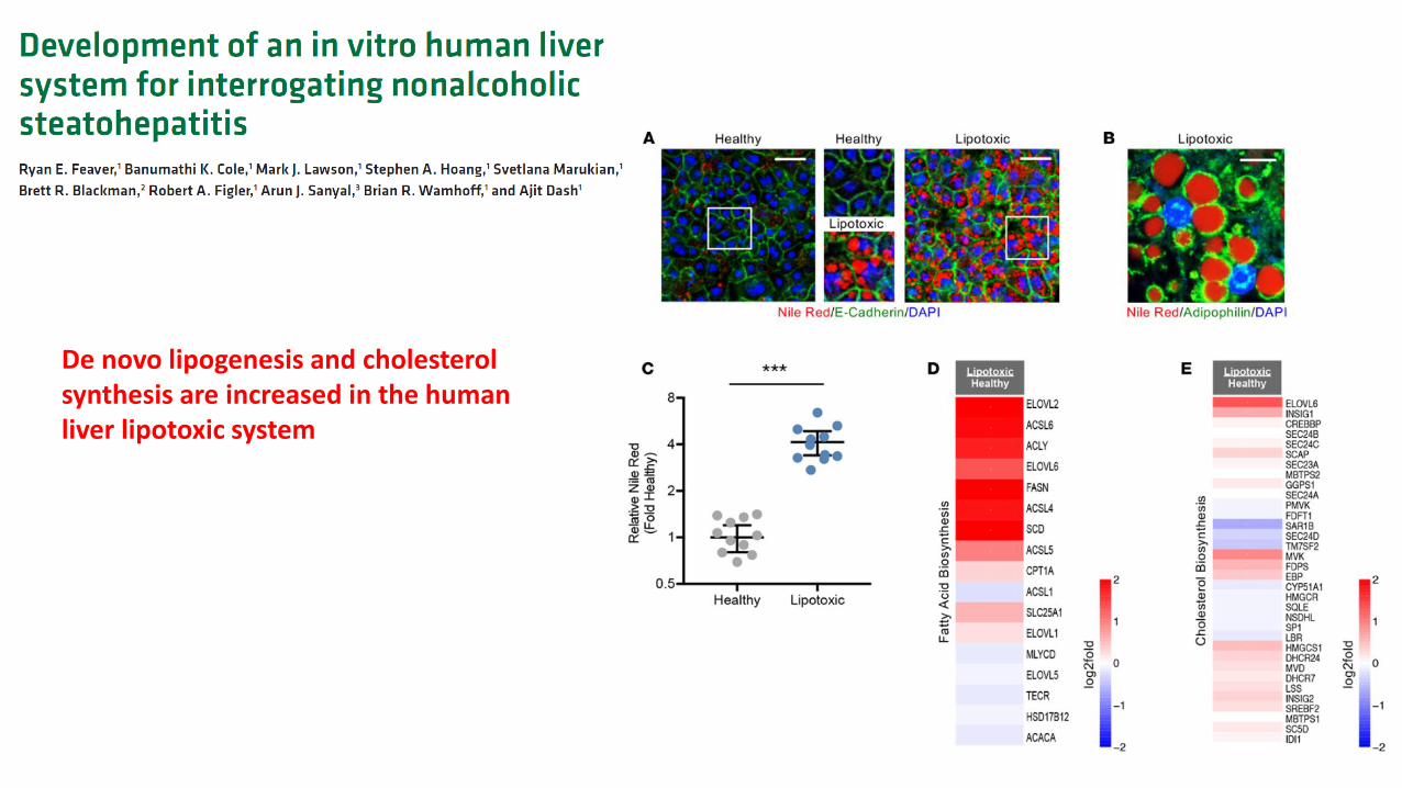
De novo lipogenesis and cholesterol synthesis are increased in the human liver lipotoxic system

3D Models to Study Liver Pathophysiology
1. – Precision-Cut Liver Slices (PCLS)
2. – Spheroids and Organoids
3. – Microfluidics and Microphysiology Systems (Lab-on-a-chip)
4. – Liver ECM engineered 3D scaffolds
5. – Liver ECM bio-printed last generation organoids

3D Human Healthy and Fibrotic Liver ECM
Explanted cirrhotic liver
Donor healthy human liver
Mazza G et al, Nature Scientific Report 2015; Mazza G et al, Nature Scientific Report 2017
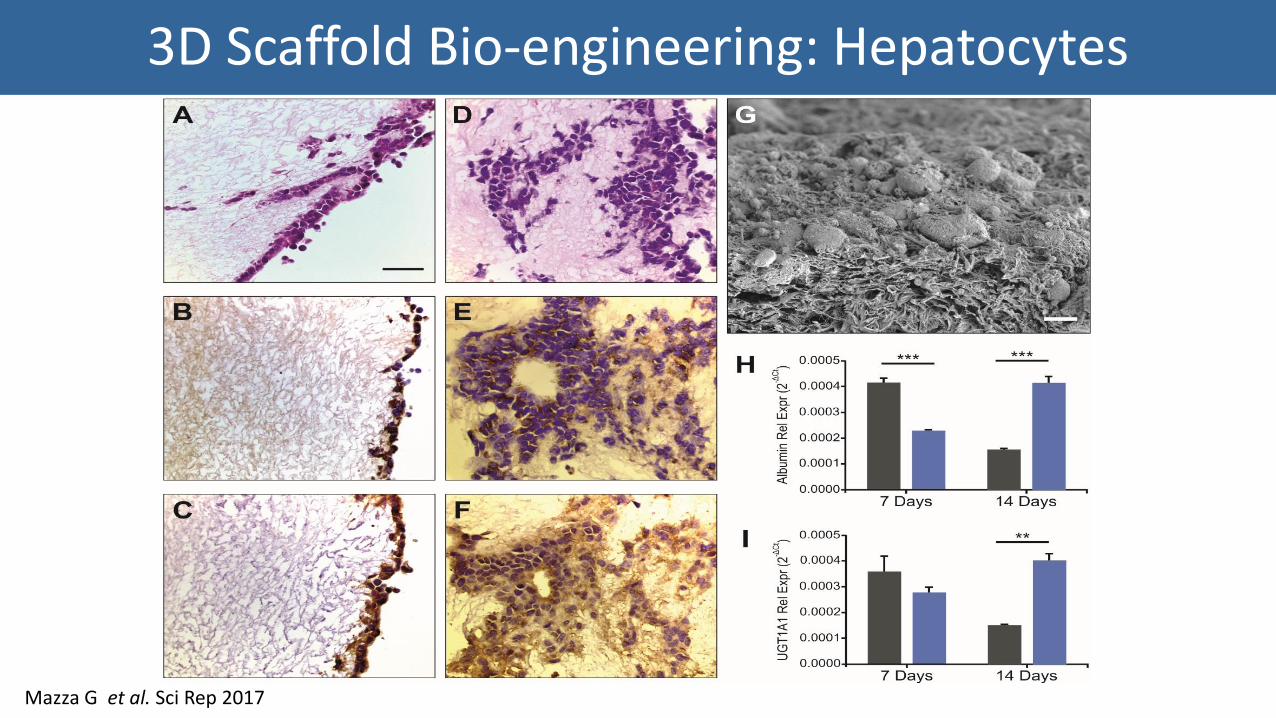
3D Scaffold Bio-engineering: Hepatocytes
Mazza G et al. Sci Rep 2017

3D Scaffold Bio-engineering: Stellate Cells
Mazza G et al. Sci Rep 2017

HSC Gene Expression: 2D vs. 3D
Caon E., et al. Unpublished

LX2 cell line Primary hHSC
Primary hHSC Gene Expression: Normal vs Cirrhotic Scaffolds
Caon E., et al. Unpublished

• LTC repopulated with Hep G2 (hepatocyte-like cell line) and LX2 (hepatic stellate cell line) (first as monoculture, then as co-culture)
• Mimic steatosis by treating with Palmitic Acid (PA) or Oleic Acid (OA) every 24 hours for 7 days
• LTC quadruplicates with 100 µM PA/OA or 1% isopropanol (vehicle control)• LTC culture stopped at Day 14 to evaluate extent and distribution of steatosis
A Tissue-engineered 3D Model of NAFLD Employing Human ECM

Targets for Evaluation Method of Analysis
Ultrastructure of ECM and Cell Distribution Histology
Fat Accumulation, Extent of Steatosis Triglyceride LevelsOil Red O Staining (lipid droplet internalisation)
Insulin Sensitivity levels of insulin-stimulated phosphorylation of AKT and Insulin Receptor Substrate-1 (IRS-1)
Production of pro-inflammatory factors Multiplex ELISA
Cell Viability MTS
Cytotoxicity Western BlotTUNEL assay for in situ studies
Cell Death Pathways Western Blot, Immunohistochemistry
Production of lipotoxic mediatorsModulation of key lipid metabolic pathways
QT-PCR
Hepatic Stellate Cell Activation Markers for fibrogenesis, markers of myofibroblastic activation (e.g. cytoskeletal proteins, collagen type I secretion)
Collagen Deposition Sirius Red Staining
NAFLD 3D Model: Experimental Objectives
3D long-term with chronic treatment Vs. 2D short term models
Optimization:1. FBS concentration2. FFAA concentration3. Insulin concentration

HepG2 Vehicle Control HepG2 100mM Oleic Acid HepG2 100mM Palmitic Acid
A Tissue-engineered 3D Model of NAFLD Employing Human ECM
Longato L., et al. Unpublished
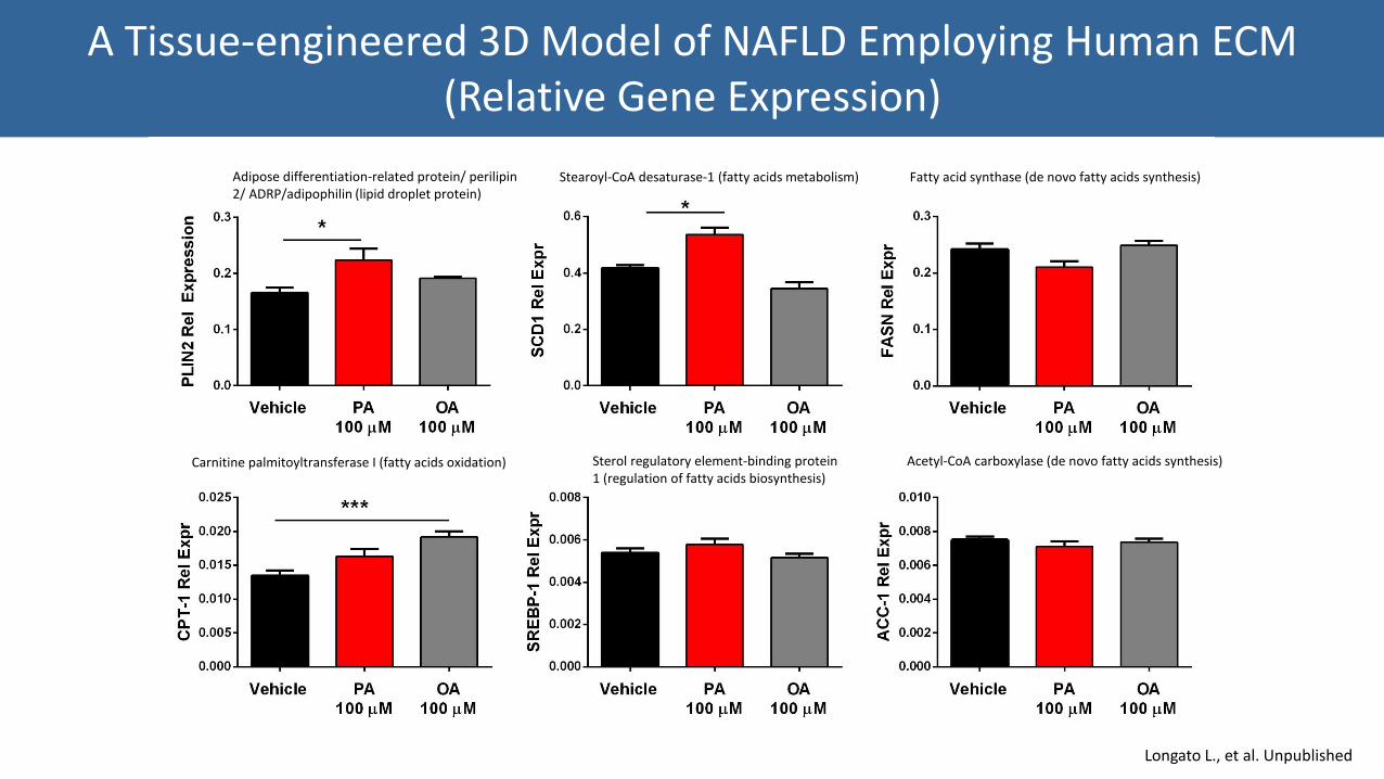
A Tissue-engineered 3D Model of NAFLD Employing Human ECM (Relative Gene Expression)
Adipose differentiation-related protein/ perilipin 2/ ADRP/adipophilin (lipid droplet protein)
Stearoyl-CoA desaturase-1 (fatty acids metabolism) Fatty acid synthase (de novo fatty acids synthesis)
Carnitine palmitoyltransferase I (fatty acids oxidation) Sterol regulatory element-binding protein 1 (regulation of fatty acids biosynthesis)
Acetyl-CoA carboxylase (de novo fatty acids synthesis)
Longato L., et al. Unpublished

Does not satisfy the criteria
Does partially satisfy the criteria
Does satisfy the criteria.
3D Human Scaffold Cultures Vs. Other 3D Systems
3D Printed in ECM Bio-Inks
3D Models to Study Liver Pathophysiology
1. – Precision-Cut Liver Slices (PCLS)
2. – Spheroids and Organoids
3. – Microfluidics and Microphysiology Systems (Lab-on-a-chip)
4. – Liver ECM engineered 3D scaffolds
5. – Liver ECM bio-printed last generation organoids

Worm-like micelles Sphere stage
Synthetic diblock copolymer mixed with human ECM solution:
•Human liver ECM solution•2-hydroxypropylmethacrylate (HPMA)•Poly(glycerolmonomethacrylate) (PGMA) macro-CTA
• Thermo-responsive hydrogels (e.g Matrigel) with human tissue-specific ECM (healthy – possibility to develop disease Hep-Gel)
• Cells are mixed with the soluble form and then dispensed into well plates
• Low volume and low cell concentration (1ul/333.3 cells)
• Currently used in 48 well plates (50ul/well) or 96 well plates (10ul/well)
• Spheroids and HTS assays
Hep-Gel: Liver ECM last generation (LG) organoids for HTS*
*HTS: high throughput screening

Bio-printed Cholangiocytes: Ductal Formation

Day
0
Day
3
Day
7
Day
10
Day
13
0
50000
100000
150000
200000
250000100 cells/mL
300 cells/mL
600 cells/mL
Flu
ore
sc
en
ce
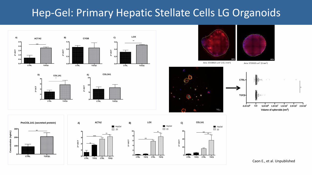
Hep-Gel: Primary Hepatic Stellate Cells LG Organoids
Caon E., et al. Unpublished
CTRL TGFβ10.0
0.5
1.0
1.5
2.0
2.5
ACTA2
2^
-DC
T
***
CTRL TGFβ10
2
4
6
8
COL1A1
2^
-DC
T
*
CTRL TGFβ10.0
0.1
0.2
0.3
CYGB
2^
-DC
T
CTRL TGFβ10
5
10
15
COL3A12
^-D
CT
CTRL TGFβ10.0
0.2
0.4
0.6
LOX
2^
-DC
T
**
A) B) C)
D) E)
CTRL TGFβ10.0
0.5
1.0
1.5
2.0
2.5
ACTA2
2^
-DC
T
***
CTRL TGFβ10
2
4
6
8
COL1A1
2^
-DC
T
*
CTRL TGFβ10.0
0.1
0.2
0.3
CYGB
2^
-DC
T
CTRL TGFβ10
5
10
15
COL3A1
2^
-DC
T
CTRL TGFβ10.0
0.2
0.4
0.6
LOX
2^
-DC
T
**
A) B) C)
D) E)
CTRL TGFβ10
100
200
300
Co
nc
en
tra
tio
n (n
g/m
L)
ProCOL1A1 (secreted protein)
**
CTRL TGFβ CTRL TGFβ0
1
2
3
4
ACTA2
2^
-DC
T
HepGel
2D
**
****
ns
CTRL TGFβ CTRL TGFβ0
2
4
6
8
10
LOX
2^
-DC
T2D
HepGel
***
**
CTRL TGFβ CTRL TGFβ0
20
40
60
COL1A1
2^
-DC
T
2D
HepGel
*****
A) B) C)
-5.0×10⁶ 0.0 5.0×10⁶ 1.0×10⁷ 1.5×10⁷ 2.0×10⁷ 2.5×10⁷
TGFβ
CTRL
*
Volume of spheroids (nm3)
Caon E., et al. Unpublished
Hep-Gel: Primary Hepatic Stellate Cells LG Organoids
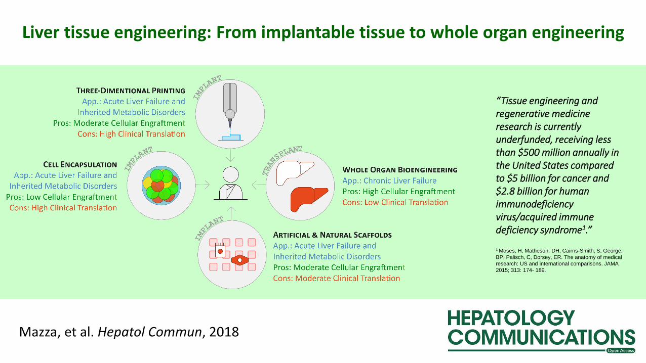
Mazza, et al. Hepatol Commun, 2018
Liver tissue engineering: From implantable tissue to whole organ engineering
“Tissue engineering and regenerative medicine research is currently underfunded, receiving less than $500 million annually in the United States compared to $5 billion for cancer and $2.8 billion for human immunodeficiency virus/acquired immune deficiency syndrome1.”
1 Moses, H, Matheson, DH, Cairns‐Smith, S, George,
BP, Palisch, C, Dorsey, ER. The anatomy of medical
research: US and international comparisons. JAMA
2015; 313: 174‐ 189.

UCL Institute for Liver and Digestive Health: Tissue Engineering Laboratory

UCL ILDHGiuseppe MAZZAKrista ROMBOUTSWalid Al-AkkadLuca FrenguelliLisa LongatoKessarin ThanapiromMaria Giovanna ViliaElisabetta CaonMartina MarraliEric FelliBailin ChenYutaka YasuiZhen Zhen Zhang
Kevin MooreDouglas Thorburn
UCL Div. of SurgeryBrian DavidsonBarry FullerAmir GanderJoerg PollockBettina Hansen
UCL Cellular PathologyAlberto QuagliaTu Vinh LuongAndy Hall
UCL Institute for Child Health, Div. Of SurgeryPaolo De CoppiPanagiotis MaghsoudlouLuca Urbani
Armando Del Rio HernandezBenjamin Robinson
Ludovic VallierFotios Sampaziotis