liver fluke – a growing threat to uk livestock production cp paper... · liver fluke – a...
TRANSCRIPT

CATTLE PRACTICE VOLUME 21 PART 2
2013 138
Liver fluke – A growing threat to UK livestock productionSkuce, P.J., Zadoks, R.N., Moredun Research Institute, Pentlands Science Park, Bush Loan, Penicuik, EH26 0PZ
ABSTRACTThe liver fluke, Fasciola hepatica, is becoming increasingly common in cattle and sheep in the UK. The suitability of different methods for detection of liver fluke infection depends on the type of animal, the purpose of the investigation (individual or herd level) and the stage of the liver fluke life-cycle. Treatment options are also influenced by the type of animal (beef, dairy, sheep) and the fluke’s life-cycle and by regulatory issues that determine availability of effective products. To some extent, forecasting of fluke risk at regional or herd level is possible based on weather conditions and farm management. Official guidelines for evaluation of treatment efficacy do not exist, but several methods have been proposed based on reduction in faecal egg counts or coproantigen secretion. Detection of liver fluke infection and treatment efficacy may be compromised by the presence of rumen fluke. The predominant rumen fluke species in the UK has been shown to be Calicophoron daubneyi. This rumen fluke shares an intermediate snail host, Galba truncatula, with the liver fluke, which contributes to the risk of mixed infections. In this paper, we review the respective flukes’ life-cycles, their impact on production and health, and diagnostic and control options in cattle and sheep.
FASCIOLOSIS AS AN EMERGING DISEASEAn emerging disease is one that has appeared in a population for the first time, or that may have existed previously but is rapidly increasing in incidence or geographic range. Based on this World Health Organisation (WHO) definition, liver fluke disease or fasciolosis would be classed as an emerging disease in the UK. Liver fluke has been present in the UK for decades, but the incidence and geographic range of this trematode parasite has increased rapidly in the last 10 years, with a disastrous liver fluke season in winter 2012/2013. An important factor in the emergence of liver fluke is its life-cycle (Figure 1). Unlike roundworms, which have direct life-cycles (host-pasture-host), most flatworms have life-cycles that involve intermediate snail hosts. In the case of the liver fluke in the UK, the preferred snail host is a tiny mud snail, Galba truncatula
(formerly Lymnaea truncatula), although other snails serve as the main intermediate host in other countries. It is worth understanding the liver fluke’s complicated life-cycle because it has implications for the increase in disease occurrence, for diagnostics, treatment options and herd or flock management.
Adult liver fluke live in the host’s bile duct system where they produce massive numbers of eggs, up to 50,000 each a day, which are excreted via the host’s faeces. In water, the eggs become embryonated and miracidia hatch in 2 to 4 weeks depending on prevailing temperature and rainfall. The miracidia are motile and have a “battery life” of about 3 hours, long enough to allow them to find a host snail in wet environmental conditions. The miracidia penetrate the snail where they go through several life stages, which may lead to shedding of an average of ca. 240 cercariae per snail over a period of approximately 4 months (Dreyfuss and Rondelaud 1994). The cercariae have a tail, which enables them to move through wet environments until they have found an appropriate plant, where they attach, lose their tail, and encyst. The cyst stage is very hardy and can survive in dry and cold conditions. There are anectodal reports of the presence of viable cysts on hay and in silage, but the biggest risk factor by far would be the ingestion of fresh herbage with viable liver fluke cysts. After ingestion, juvenile liver fluke excyst in the duodenum and start their migration through the gut wall and peritoneal cavity towards the liver, where they work their way through the parenchyma to the bile ducts. Here, the liver fluke mature and several months after ingestion of the infective cysts, they start to lay eggs and the life-cycle resumes.
Because a large part of the liver fluke’s life cycle takes place in the outside environment, it is highly dependent on temperature and moisture. This explains why the recent spate of wet summers and relatively mild winters have supported the expansion of the host
KEYWORDS: Liver fluke, rumen fluke, cattle, sheep
Figure 1. The life-cycle of the liver fluke, Fasciola hepatica. ©Moredun Research Institute.
CATTLE PRACTICE VOLUME 21 PART 2
2013 139
snail and liver fluke populations (Kenyon and others 2009, Van Dijk and others 2010). Other factors that may contribute to emergence of fasciolosis are movement of animals, environmental management schemes and the development of drug resistance, which will be discussed in a subsequent section. Before we leave the liver fluke’s life-cycle, however, we should emphasise another major distinction between roundworms and this flatworm. Most roundworm species are host-specific. Not so for liver fluke. Any mammal that ingests plants with liver fluke cysts may become infected, including cattle and sheep, pigs and donkeys (Valero and others 2001), wildlife such as deer, rabbits and hares (Ménard and others 2000, Walker and others 2011), kangaroos and other Australian marsupials (Spratt and Presidente 1981) and people (Laird and Boray 1992, Winkelhagen and others 2012). Liver fluke has even been reported in farmed emus and rhea in Australia and South America, respectively (Vaughan and others 1997, Soares and others 2007). The lack of host specificity of Fasciola hepatica has several important implications: (i) Eradication is unlikely to be feasible in the presence of a significant wildlife reservoir; (ii) F. hepatica is a zoonotic pathogen, i.e. it can affect animals and humans alike (although there is no direct host-to-host transmission); and (iii) Cross-infection between cattle and sheep is possible, which has relevance to treatment and management strategies as described later.
IMPACT IN CATTLE AND SHEEPAlthough liver fluke may affect cattle and sheep, the impact in the two host species can be quite different, which has relevance to diagnosis and treatment options. Acute clinical disease or sudden death due to liver fluke is rare in cattle, probably because the liver tissue is relatively tough in this host species. This limits the damage that is done by migrating juvenile liver fluke. In cattle, liver fluke infection has been associated with reduced milk production (Mezo and others 2011, Charlier and others 2012a), reduced milk fat content and increased calving interval in dairy cattle (Charlier
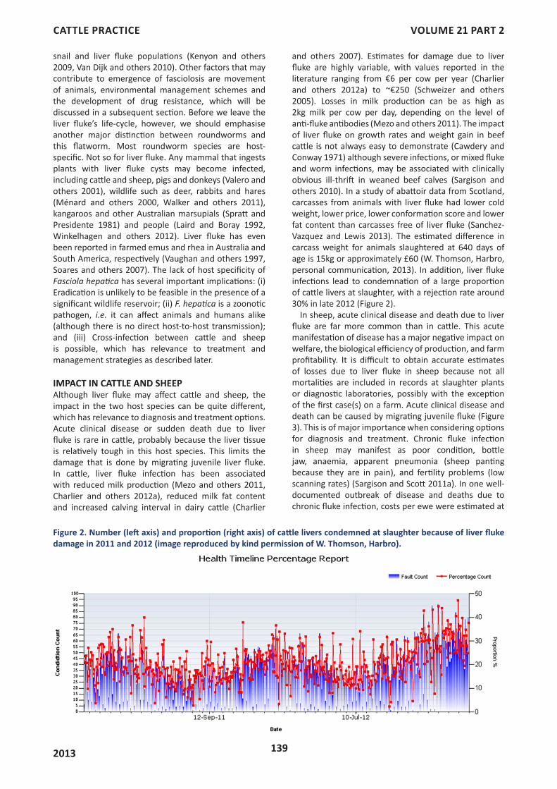
and others 2007). Estimates for damage due to liver fluke are highly variable, with values reported in the literature ranging from €6 per cow per year (Charlier and others 2012a) to ~€250 (Schweizer and others 2005). Losses in milk production can be as high as 2kg milk per cow per day, depending on the level of anti-fluke antibodies (Mezo and others 2011). The impact of liver fluke on growth rates and weight gain in beef cattle is not always easy to demonstrate (Cawdery and Conway 1971) although severe infections, or mixed fluke and worm infections, may be associated with clinically obvious ill-thrift in weaned beef calves (Sargison and others 2010). In a study of abattoir data from Scotland, carcasses from animals with liver fluke had lower cold weight, lower price, lower conformation score and lower fat content than carcasses free of liver fluke (Sanchez-Vazquez and Lewis 2013). The estimated difference in carcass weight for animals slaughtered at 640 days of age is 15kg or approximately £60 (W. Thomson, Harbro, personal communication, 2013). In addition, liver fluke infections lead to condemnation of a large proportion of cattle livers at slaughter, with a rejection rate around 30% in late 2012 (Figure 2).
In sheep, acute clinical disease and death due to liver fluke are far more common than in cattle. This acute manifestation of disease has a major negative impact on welfare, the biological efficiency of production, and farm profitability. It is difficult to obtain accurate estimates of losses due to liver fluke in sheep because not all mortalities are included in records at slaughter plants or diagnostic laboratories, possibly with the exception of the first case(s) on a farm. Acute clinical disease and death can be caused by migrating juvenile fluke (Figure 3). This is of major importance when considering options for diagnosis and treatment. Chronic fluke infection in sheep may manifest as poor condition, bottle jaw, anaemia, apparent pneumonia (sheep panting because they are in pain), and fertility problems (low scanning rates) (Sargison and Scott 2011a). In one well-documented outbreak of disease and deaths due to chronic fluke infection, costs per ewe were estimated at
Figure 2. Number (left axis) and proportion (right axis) of cattle livers condemned at slaughter because of liver fluke damage in 2011 and 2012 (image reproduced by kind permission of W. Thomson, Harbro).

CATTLE PRACTICE VOLUME 21 PART 2
2013 140
£8.73 (Sargison and Scott 2011a) but costs may increase rapidly if many animals succumb to the infection.
Chronic fluke infection was particularly evident in the winter/spring of 2012-2013, with numerous deaths of adult sheep reported, often despite flukicide treatment in the autumn. Out-wintered stock, especially pregnant ewes, were still at risk of infection due to the mild Jan-Feb and many succumbed due to a combination of liver fluke and the exceptionally cold March-April with lack of adequate grass growth around lambing time.
DETECTION OF INFECTION AND TREATMENT OUTCOMEToo often, we hear or read stories from farmers who have administered multiple anthelmintic treatments to sick animals or who have waited for a considerable number of animals to die before they finally sought a diagnosis for their problem or an assessment of treatment efficacy. This does not need to happen. Diagnostic tests for liver fluke are far from perfect, but many useful methods are available.
Detection of liver fluke infection in cattle is easier than in sheep. To begin with, there is no urgent need to detect juvenile fluke, for which diagnostic tests are limited. Secondly, access and sample collection is easier for animals that are housed than for sheep that are on
pasture for most of the year, particularly in Less Favoured Areas. Thirdly, milk samples can be used in dairy herds. Finally, the value of the individual animal may justify collection of blood samples, which is harder to justify economically in sheep. Diagnostic tests are needed to detect liver fluke infection and to assess treatment efficacy. Detection of liver fluke infection can be done at farm level or individual animal level, in live or dead animals, and using invasive or non-invasive methods. In addition, the farm-level risk of liver fluke can be assessed based on biological, meteorological and management data.
Post-mortem inspections happen routinely at slaughter and in diagnostic laboratories. Slaughter data may lack accuracy, both in sensitivity and specificity of detection of liver fluke and in correlation of post mortem findings with slaughtered individuals or batches of animals, but their routine availability should be an incentive to use them and to improve their accuracy. Several abattoirs are quite active in this area because they incur increasing losses due to the need to discard affected livers. Some abattoirs even strive to make a distinction between current fluke infection, as diagnosed based on the presence of live liver fluke and past fluke infection with thickening and hardening of bile ducts in cattle. In diagnostic laboratories such as those at the Animal Health and Veterinary Laboratories Agency (AHVLA) and Scotland’s Rural College (SAC Consulting, Veterinary Services), many cases of liver fluke are identified and a distinction can be made between juvenile and mature liver fluke.
Invasive in vivo assays are those that require collection of a blood sample. Blood samples can be used to assess liver function or damage based on levels of liver and bile duct enzymes. Increased levels of liver enzymes are not specific for liver fluke and levels above threshold values may naturally occur in young animals (Gordon and others 2012a). The usefulness of liver enzymes as an indicator of the ability of animals to metabolise drugs or recover from infection has not been explored in depth. Blood samples are routinely used to detect antibodies to liver fluke by means of enzyme linked immunosorbent assay (ELISA). In lactating dairy cattle, there is no need to collect blood to perform ELISA testing, as antibody presence can be detected in milk. The agreement between ELISA results from blood and milk is very good (Salimi-Bejestani and others 2007, Mezo and others 2010, Duscher and others 2011). For some ELISAs, the titre is indicative of fluke burden (Salimi-Bejestani and others 2008, Kuerpick and others 2013). Antibody titres decrease after successful treatment but this drop is not always significant or persistent (Boulard and others 1995, Charlier and others 2012b) and the change is not routinely used to monitor treatment outcome. For herd level diagnosis, bulk milk samples can be used. Estimates of the sensitivity of bulk milk ELISA vary considerably. Some reports state that a within-herd prevalence of 12 to 20% can be detected in bulk milk whereas other authors put the bar at 60% within-herd prevalence (Reichel and others 2005, Mezo and others 2010, Duscher and others 2011). In winter 2006/2007,
Figure 3. Panel (A) acute fluke damage and associated haemorrhage in a sheep liver, panel (B) chronic fluke damage in a sheep liver, note distended gall bladder and damaged liver lobe. Panel C, classic ‘pipestem fibrosis’ in a chronically infected cattle liver. Images reproduced by kind permission AHVLA (Crown Copyright), ©Moredun Research Institute and by kind permission Prof. Neil Sargison, R(D)SVS, respectively.

CATTLE PRACTICE VOLUME 21 PART 2
2013 141
72% and 84% of dairy herds in England and Wales, respectively, were positive for liver fluke based on bulk milk testing, with almost half of the herds in north-west England showing high levels of antibodies, reflecting high levels of exposure (McCann and others 2010a).
Another ELISA is the coproantigen ELISA (cELISA). This test does not detect antibodies but rather liver fluke antigens in the host’s faeces. Thus, this test does not require invasive sampling. This is an advantage it shares with the traditional faecal egg count (FEC). In experimental challenge studies, the cELISA has the ability to detect liver fluke infections several weeks before the start of the patent period. In addition, it can detect infections with a single adult liver fluke and is more or less quantitative, i.e. cELISA values are indicative of fluke burdens (Mezo and others 2004, Valero and others 2009, Brockwell and others 2013). Under field conditions, however, cELISA does not always perform quite as well. In our work, a commercially available cELISA did not detect liver fluke infections any sooner than FEC in sheep with a relatively low burden of infection (Gordon and others 2012a). On several sheep farms, multiple faecal samples that contained liver fluke eggs were negative by cELISA (Gordon, personal communication, 2012) and this anomaly is thus far unexplained. Similar observations have been published for dairy cattle, with 17.8% of animals testing positive by FEC and only 13.4% by cELISA (Duscher and others 2011). Pooling of faecal samples from sheep leads to further loss of sensitivity (Gordon, personal communication, 2012). By contrast, pooled faecal samples are useful for FEC testing in sheep (Daniel and others 2012). Advantages of the cELISA include the limited “hands-on” time needed in the laboratory and the shorter delay in evaluation of treatment outcome compared to FEC (Gordon and others 2012a). We also need to keep in mind that, in face of a high fluke burden, cELISA may well have the ability to diagnose juvenile liver fluke infections in the field, something that the FEC will never be able to achieve. Although the FEC has been a mainstay of liver fluke diagnostics, it is not without its flaws either. The most critical one is its inability to detect juvenile fluke, i.e. the first 3 months of fluke infection, which may be crucial, particularly in sheep. In addition, egg shedding is highly variable, both between animals and within animals over time (Düwel and Reisenleiter 1990). This can cause false-negative results and makes it difficult to correlate FEC to liver fluke burdens.
Comparison of the sensitivity and specificity of different diagnostic tests for liver fluke is not straightforward. In the study that reported 17.8% prevalence based on FEC, antibody prevalence in individual milk and blood samples was approximately 45% (Duscher and others 2011). This does not necessarily mean that FEC lacks sensitivity, but rather that it detects a different underlying biological mechanism. Antibody-based tests obviously detect antibodies. Those may be present within 2 weeks from infection, potentially several months ahead of the patent stage of infection (Boulard and others 1995). Antibodies may persist after successful treatment, whilst liver fluke may not be present in the liver anymore at that stage. This does not imply that the ELISA is less specific
than FEC or post-mortem inspection, but goes back to different underlying biological mechanisms. In some areas, exposure to liver fluke is so high that veterinarians see limited value in using antibody based testing, as all grazing animals are expected to be positive regardless of current infection status. For animals in their first grazing season, however, ELISA would be the earliest indicator of liver fluke infection. Thus, the choice of diagnostic tests depends on the farm situation, animal type and age.
Treatment with flukicides may kill liver fluke and may reduce egg production by surviving flukes (Hanna and others 2006). In fact, many studies report discrepancies between treatment efficacy based on faecal egg count reduction testing (FECRT) as compared to counts of surviving fluke at post mortem. FECRT tends to overestimate cure rates compared to fluke counting (Bradley and others 1981, Malone and others 1982, Hutchinson and others 2009). In practice, we cannot routinely assess treatment efficacy by killing animals, which means that we rely on FECRT. For roundworms, guidelines have been formulated for conduct and interpretation of FECRT. In analogy with approaches used for roundworms, a 90% reduction in FEC is sometimes used as cut-off. The observed reduction will depend on the number of animals tested, the fluke burden in those animals, the amount of faeces processed per animal and the method used for calculation of the % reduction. No official guidelines exist for conduct and interpretation of FECRT for liver fluke yet if we want to evaluate treatment efficacy in the absence of guidelines, FECRT is one of the few available tests. In addition, we do not need to be too worried about whether the cut-off should be 85%, 90% or 95%, considering that treatment efficacies from 0 to 60% have been observed when resistant fluke were present in experimental and natural infections, respectively (Walker and others 2004, Hutchinson and others 2009, Gordon and others 2012a). Animals may naturally be infected with a mixed population of liver fluke strains (Vilas and others 2012). This could potentially be a mixture of susceptible and resistant strains. Killing of susceptible strains and concomitant survival or resistant strains could theoretically explain observed partial treatment efficacy.
A recent alternative is the use of FECRT is the cELISA reduction test or CRT. Official guidelines for interpretation of the CRT do not exist but studies in sheep and cattle suggest that the CRT is a reliable indicator of treatment outcome (Gordon and others 2012a, Brockwell and others 2013). For FECRT, a 3-week interval between initial sample collection plus treatment and follow-up sampling is recommended so that residual eggs can be flushed out of the gall bladder. For CRT, a 1-week interval seems to suffice (Gordon and others 2012a, Brockwell and others 2013). This has advantages in terms of management (follow-up can be done before other priorities overshadow evaluation of treatment efficacy; information relevant to animal movement becomes available quickly) and in case additional treatment is needed. Interestingly, several studies also show very good results for FECRT at 1-week post treatment (Gordon and others 2012a, Brockwell and others 2013) and the

CATTLE PRACTICE VOLUME 21 PART 2
2013 142
need for a 3-week interval may need to be re-evaluated.In addition to samples obtained from animals, farm
level information may be used to determine the risk of liver fluke infection. Biological data on the intermediate host and the free-living parasite stages, such as cysts and snails, are used to monitor the exposure to liver fluke in some countries, e.g. The Netherlands and Switzerland (Gaasenbeek and others 1992, Knubben-Schweizer and others 2010). This approach is informative but also very labour intensive and it is rarely used anymore in the UK. Instead, meteorological data and farm management information are used. Models based on weather and pasture data explained 70% of the observed variation in liver fluke prevalence on dairy farms in England and Wales (McCann and others 2010b). However, considerable differences could sometimes be observed between farms within the same postcode area and weather patterns. In temperate climate zones without large climatic and environmental variation, management factors affect the spatial distribution of F. hepatica (Bennema and others 2011). In Belgium, 85% of observed variation in bulk milk ELISA results for liver fluke could be explained based on the number of potential habitats, the presence of snails, drainage of pastures, month of turnout of the cows, stocking rate, type of watering place and post code area (Charlier and others 2011). This shows that, at least on dairy farms, pasture management tools can make a significant contribution to liver fluke control.
TREATMENT OPTIONS AND TREATMENT FAILUREIn sheep, clinical disease due to early immature liver fluke is a major problem. Currently, there is only a single drug that is effective against the early immature stages of liver fluke, i.e. triclabendazole. Because of the importance of this drug to sheep farming, and because of increasing concerns about triclabendazole resistance, it is of the utmost importance that this drug is preserved for treatment of early immature liver fluke in sheep. Therefore, this drug should, in our opinion, not be recommended for treatment of cattle where early immature fluke rarely cause clinical disease. The realities
of current product registrations, however, may dictate otherwise in practice. Whilst triclabendazole should be reserved for the treatment of sheep, clorsulon is only commercially available for the treatment of cattle. There is an extensive body of literature regarding the efficacy of different fasciolicides at different stages of infection. A summary of recommended combinations of host species, liver fluke ages and active compounds is provided in Figure 4. Note that many active compounds can be sold under multiple trade names. Farmers may change from one treatment to another, thinking that they alternate treatments, but they may actually use various presentations of the same active compound rather than truly changing treatments.
In dairy cattle, treatment options are limited compared to beef cattle due to concerns about residues in milk. For example, after administration to lactating animals, triclabendazole and nitroxynil may be detected in milk for several weeks and almost 2 months, respectively (Whelan and others 2011, Power and others 2013). The concentration of residues in cheese can be 13-fold higher (Imperiale and others 2011). Constraints also apply to the use of products in sheep that produce milk for human consumption. When cattle are treated in the non-lactating period, residue in milk may not be detectable after calving (Power and others 2013) and milk production in the subsequent lactation may increase (Charlier and others 2012b). Considerable milk withdrawal periods need to be taken into account, which is particularly important when cows come into lactation earlier than anticipated. Residue in beef can be a problem when animals are slaughtered unexpectedly, e.g. after injury. In Ireland, injured (casualty) cattle were shown to be more likely to test positive for anthelmintic residues, including the flukicide Closantel, than retail beef (Cooper and others 2012).
In conversations with veterinarians and farmers, we often hear questions about the combined use of multiple flukicide drugs, particularly when there are concerns about juvenile fluke infections in sheep in combination with suspicions of triclabendazole treatment failure. There is limited scientific evidence to support or reject
Figure 4. Active compounds that can be used for different stages of liver fluke infection in sheep and cattle, respectively. ©Moredun Research Institute.

CATTLE PRACTICE VOLUME 21 PART 2
2013 143
combined drug use. In one experimental study in rats, a potential beneficial effect of combined use of triclabendazole and clorsulon was reported (Meaney and others 2006) but this is currently of limited practical relevance in the UK as the drugs are primarily or only used for sheep or cattle, respectively. It is important to ensure that farmers do not actually combine two products in a single syringe or dosing gun, which is unfortunately how some farmers interpret “combination” of drugs. It is also important to keep in mind that both efficacy and safety may be affected by combined drug use, which is of particular concern for drugs with a narrow safety margin and for animals with compromised liver function such as caused by liver fluke. Finally, it is important to differentiate between drug resistance and treatment failure, as detailed below.
Treatment failure may have many causes, most of which are associated with farm management rather than drug resistance. Factors that contribute to treatment failure include inaccurate dosing of animals, e.g. due to inaccurate estimation or measurement of the animals’ body weight or due to the use of non-calibrated or incorrect dosing equipment. Farmers may use a single dosing gun for multiple products without realising that a single dose of one product may have a different volume than a single dose of another product. Incorrect storage of product may also have a negative impact on efficacy (Sargison and Scott 2011b). When products are stored and administered correctly, treatment efficacy may be affected by the time of year, particularly for pour-on products. Depending on season, the efficacy of certain pour-on products ranged from ca. 75% to 99.6% after administration to cattle (Sargent and others 2009). This was not due to the age of the fluke, as all treatments were given after artificial infection, i.e. using fluke of known and standardised age. The observed differences were thought to be linked to the impact of seasonal differences in hair coat and weather on transdermal uptake of pour-on products (Sargent and others 2009). For treatment of 2-week old fluke, pour-on treatment of triclabendazole lacked efficacy whilst oral administration did reduce fluke burdens (Hutchinson and others 2009). Oral administration may result in higher efficacy than administration of the same active compound using a pour-on whilst using a lower quantity of the compound per animal, as demonstrated for triclabendazole treatment of 4-week old fluke (Martin and others 2009), but this result is not consistent across studies (Hutchinson and others 2009). In sheep, apparent treatment failure may be due to re-infection of animals that are not housed or moved to safe grazing after treatment. In the winter of 2012/2013, exposure levels are thought to have been so high that animals became re-exposed to high numbers of liver fluke cysts immediately after treatment, resulting in severe liver damage and clinical disease within weeks from the initial treatment. Because several products that are used for roundworm treatment have residual effect, i.e. efficacy that lasts beyond the day of administration, farmers may expect this to be true for flukicides also. However, none of the current flukicides have residual
effect and animals are vulnerable to re-infection immediately after treatment. Another complicating factor is that products that are deemed to be effective against immature fluke do not necessarily kill all immature fluke. For example, closantel will lead to a significant reduction in fluke burden compared to control cattle when used to treat 6- or 8-week old fluke, but efficacy has been reported as 68% and 91%, respectively, rather than 100% (Geurden and others 2012). Efficacy of triclabendazole against 4-week old fluke is approximately 90% (Richards and others 1990, Hutchinson and others 2009, Geurden and others 2012). Thus, an expectation of 100% treatment efficacy may be unrealistic, even when the product is appropriate for the age of the fluke. This, combined with the limitations of diagnostic tests to assess liver fluke burdens, makes formal demonstration of flukicide resistance extremely difficult. A recent twist to this story is the increasing detection of rumen fluke in British livestock, which may affect assessment of flukicide treatment efficacy as discussed in a subsequent section.
To the extent that detection of flukicide resistance is possible (for discussion, see Fairweather 2011 and Sargison and Scott 2011b), resistance to triclabendazole has been demonstrated in a number of countries and continents (Gaasenbeek and others 2001, Gordon and others 2012a,b, Olaechea and others 2011). Although triclabendazole is primarily used in sheep, resistance to triclabendazole has been reported for liver fluke from sheep as well as cattle (Olaechea and others 2011) and it is suspected in liver fluke from hares (Walker and others 2011) and people (Winkelhagen and others 2012). To date, all demonstrations of triclabendazole resistance are based on the host response to treatment as evaluated in vivo or at post-mortem. It would be very useful to have in vitro assays to detect triclabendazole resistance. Proof-of-concept for an egg hatch assay to detect resistance has been published (Fairweather and others 2012). Histological examination of mature fluke harvested at post-mortem also allows for differentiation of triclabendazole susceptible and – resistant liver fluke (Hanna and others 2013). The limitation of such assays is that they require mature flukes from dead animals, which means that the assay may come too late for sheep, where tools for detection of triclabendazole resistance are most needed. Efforts are underway to identify genetic markers of triclabendazole resistance in liver fluke (reviewed by Hodgkinson and others 2013). One specific mutation in the P-glycoprotein of F. hepatica has been linked to triclabendazole resistance (Wilkinson and others 2012) but this mutation was not present in a confirmed triclabendazole resistant isolate from Scotland (Gordon and others 2012b) so the absence of this mutation does not guarantee triclabendazole susceptibility.
To end the treatment section on a positive note, the majority of liver fluke treatments are successful, particularly when applied in the right manner at the right time with adequate follow-up to assess treatment efficacy. Treatment can limit the clinical and economic impact of liver fluke infections. Furthermore, strategic

CATTLE PRACTICE VOLUME 21 PART 2
2013 144
dosing schemes can be used to suppress the faecal egg output at critical times of the year e.g. spring/early summer, which limits infection of the intermediate host snail population and reduces the subsequent contamination of the pasture with metacercariae (Parr and Gray 2000). In combination with pasture management, and in the absence of vaccines, treatment is an important and potentially effective liver fluke control tool (Knubben-Schweizer and others 2010).
RUMEN FLUKE IN CATTLE AND SHEEPA relatively recent finding is the emergence of rumen fluke infection in both cattle and sheep in the UK and Ireland. Rumen fluke (or paramphistomes) are also trematode parasites with a very similar life-cycle to liver fluke. Rumen fluke have been described at post mortem in the UK as far back as the 1950s (Willmott 1950) and their eggs have been observed during routine faecal egg counting for liver fluke since the ~mid-1980s (Foster and others 2008, Murphy and others 2008). Rumen fluke diagnoses by faecal egg count are now more common in Ireland (NI and ROI) than liver fluke e.g. ca. 30% sheep; 40% cattle (e.g. Anon 2011) and AHVLA report more diagnoses of rumen fluke in GB in 2012 than the previous 5 years combined (R. Daniel, AHVLA, personal communication 2013). The adult rumen fluke resemble small, pink, fleshy maggots and appear to be well-tolerated on the surface of the rumen (Figure 5). Rumen fluke disease (paramphistomosis) is invariably associated with the presence of large numbers of immature rumen fluke in the intestine, usually in the duodenum. This sets up an inflammatory response, resulting in non-responsive, watery diarrhoea and marked reddening of the proximal intestinal mucosa at post mortem together with mesenteric lymphadenopathy and watery large intestinal content (Millar and others 2012). Immature rumen fluke were also reported as the cause of death in sheep (Mason and others 2012). Whilst juvenile rumen fluke has been shown to be pathogenic, there is little data on production effects of adult rumen fluke infection on livestock or the knock-on economic impact, other than anecdotal reports of animals improving in condition after successful treatment.
For decades, it has been assumed that the predominant rumen fluke species infecting livestock in UK and ROI is Paramphistomum cervi (e.g. Taylor and others 2007). This species is believed to have originated from the wild deer population and to favour aquatic e.g. planorbid or bulinid snails as its intermediate host, rather than the lymnaeid mud snail intermediate host of the liver fluke. Recent DNA sequencing of rumen fluke specimens sourced from home-bred sheep and cattle from England, Ireland, Scotland and Wales, somewhat surprisingly, revealed them all to be Calicophoron daubneyi (Gordon and others 2013 and unpublished data from the authors’ laboratory). This is the predominant rumen fluke species in mainland Europe, most notably France, Italy, Portugal and Spain (Mage and others 2002, Rinaldi and others 2005, Arias and others 2011, González-Warleta and others 2013). The species identity is important here because C. daubneyi in Europe is known to favour the same mud snail intermediate host as liver fluke, i.e. G. truncatula (Mage and others 2002, Sanabria and others 2012), so this finding may have implications for the epidemiology of rumen fluke in the UK and Ireland, including its presence as a co-infection with liver fluke. Wildlife reservoirs for C. daubneyi have not been investigated in depth, but a study from Spain suggests that roe deer are not an important reservoir (González-Warleta and others 2013).
Differential diagnosis of rumen fluke and liver fluke infections is also very important because the treatment options are different. Although not licensed for the treatment of rumen fluke, only one flukicide has been demonstrated to have activity against rumen fluke, both immature and adults and that is oxyclozanide (Rolfe and others 1987). Oxyclozanide treatment will reduce egg output by C. daubneyi in cattle (Rolfe and Boray 1987, Diaz and others 2006). It is also a liver fluke drug (and licensed as such in the UK) but it is not able to kill liver fluke within the first 3 months after infection (Richards and others 1990). Furthermore, the eggs of rumen fluke and liver fluke look very similar and could be confused, leading to erroneous declarations of liver fluke treatment failure. Mixed infections with rumen fluke and liver fluke have been reported in cattle and sheep, including animals from Scotland (Arias and others 2011, Gordon and others 2013), making incorrect diagnosis of disease or treatment outcome of practical concern in the UK. As ever, treatment decisions based on accurate diagnosis are recommended for best practice fluke control. In herds or flocks with mixed liver fluke and rumen fluke infection, the coproantigen ELISA is a useful test for the diagnosis of liver fluke and evaluation of liver fluke treatment outcome, because the test does not cross-react with C. daubneyi and provides a good indication of treatment success within a week post treatment (Gordon and others 2012a, 2013).
CONCLUSIONS AND OUTLOOKThe presence of the snail intermediate host is integral to the life-cycle of the liver fluke (and rumen fluke). As a result, methods aimed at reducing snail numbers or snail habitat e.g. through effective drainage
Figure 5. Adult rumen fluke on the surface of a bovine stomach (Image reproduced by kind permission, W. Thomson, Harbro).

CATTLE PRACTICE VOLUME 21 PART 2
2013 145
and/or reducing access of livestock to potential snail habitats during high risk periods by fencing off ‘fluky’ pasture, have all been advocated as part of integrated fluke control strategies on-farm, especially before the advent of effective flukicidal drugs. In the past, several commercial molluscicides were available to kill snails on pasture but these have all been banned since the ~1970s for environmental reasons. Biological control using predatory snails has been explored in France but is unlikely to gain widespread acceptance in the UK (Rondelaud and others 2011). Considering the increasing prevalence of liver fluke and rumen fluke, the devastating impact of liver fluke on welfare and production, and the “perfect storm” in sheep farming of major damage by juvenile fluke, availability of a single active compound against early immature fluke, and the emergence of resistance to that compound, it is time to dust off pasture management measures. Although there are limits to how much wet pasture (or any pasture, for that matter) can be fenced off, drainage is considered a valuable and underutilised strategy according to several farmers we have spoken to. Environmental schemes that encourage wetland conditions for breeding and migratory birds and invertebrates have been linked with a higher risk of liver fluke in livestock (Pritchard and others 2005). Furthermore, wading birds have been implicated in the dispersal of aquatic organisms, including the liver fluke’s intermediate host snail, G. truncatula (van Leeuwen 2012). Thus, there may also be an imperative to balance policy aimed at maintaining biodiversity with policy aimed at maintaining livestock farming.
A vaccine to protect livestock against liver fluke or reduce the clinical signs or production losses would be highly desirable. This was attempted as far back as the 1960s (Ross 1967). Despite several decades of research since and considerable optimism around the turn of the century (Dalton and Mulcahy 2001), production of a commercial liver fluke vaccine is still some way off. The development of vaccines against parasitic helminths has been difficult for a number of reasons, but liver fluke is particularly challenging. Firstly, there is no evidence of a strong protective natural immunity to build on (Clery and others 1996, Bossaert and others 2000a,b); the amplification of fluke numbers through the snail intermediate host can lead to extremely high parasite challenge on pasture; and, finally, liver fluke is a complicated eukaryotic organism, with a genome comprising >20,000 proteins. That said, several potential candidate vaccine antigens have been identified and tested in a number of laboratory- and natural host species. These include, amongst others, fatty acid binding proteins, haemoglobin, glutathione-S-transferases, cathepsin L proteases and leucine aminopeptidases, which have been shown to reduce fluke burdens by between ~40% and 80% (for a review, see Rojo-Vazquez and others 2010). The fact that a commercial liver fluke vaccine remains elusive several decades from antigen discovery is testament to the biological, immunological and technological challenges associated with this objective.
Many of the current liver fluke drugs were discovered
many years e.g. triclabendazole is 30 years old and oxyclozanide is approaching its 50th birthday (Walley 1966, Boray and others 1983)! As far as we are aware, there are no new liver fluke drugs under development or near market. This is most likely because it is extremely expensive and takes considerable time to bring new drugs to market and, unless there is a strong business case to do so, it is unlikely the multinational drug companies will embark on such a venture any time soon. There has been considerable interest in evaluating the flukicidal potential of new actives either alone or in combination within the academic research sector but little/no evidence of uptake by industry. For example, artemisinin derivatives have broad-spectrum activity against trematodes and have been evaluated therapeutically in rats, showing activity against a triclabendazole-resistant Fasciola isolate (Duthaler and others 2012). ‘Compound alpha’, a derivative of triclabendazole, has shown activity similar to the parent compound against susceptible F. hepatica in sheep, and some activity against triclabendazole resistant fluke (McConville and others 2009)[See review by Rojo-Vazquez and others 2010].
Whilst current diagnostic methods are reasonably adequate for detection of liver fluke infection in cattle, better methods for reliable detection of juvenile liver fluke in sheep are urgently needed. One recent avenue of research that may hold some promise is DNA-based detection of liver fluke. There is recent evidence in the literature that Fasciola DNA can be detected by PCR amplification of host faeces from as early as 2-weeks post-infection in an experimental challenge model (Robles-Perez and others 2013). The biological relevance of this finding e.g. what is the source of this fluke DNA, how does the detection of DNA relate to infection status and how would the DNA signal respond to treatment, successful or otherwise, all remain to be evaluated. However, this does open up the potential of rapid, pen-side testing through technological advances in amplification methods e.g. loop mediated isothermal amplification (LAMP) testing and lateral flow devices (Ai and others 2010).
In summary, liver fluke is becoming increasingly common in cattle and sheep and has a major impact on animal welfare and farm profitability, particularly in sheep farming. Major issues include the decreasing availability and efficacy of flukicides, particularly in dairy cattle and sheep, and the increased infection pressure which may be aggravated by changes in weather patterns and land use policy. With no vaccine or new drugs within reach, it is imperative that we make the best possible use of the liver fluke management tools that are available to us, including diagnostic testing, appropriate anthelmintic treatment and pasture management strategies. Rumen fluke is relevant in the context of liver fluke diagnostics and may have clinical relevance in its own right but we should not let the relative novelty of rumen fluke distract us from the fact that liver fluke is currently the major trematode threat to our livestock industry.
ACKNOWLEDGEMENTSWork reported in this paper has been financially
CATTLE PRACTICE VOLUME 21 PART 2
2013 146
supported by the Scottish Government, the Moredun Foundation and Quality Meat Scotland.
REFERENCESAi, L., Li, C., Elsheikha, H.M., Hong, S.J., Chen, J.X., Chen, S.H., Li, X., Cai, X.Q., Chen, M.X., Zhu, X.Q. (2020) Rapid identification and differentiation of Fasciola hepatica and Fasciola gigantica by a loop-mediated isothermal amplification (LAMP) assay. Veterinary Parasitology. 174(3-4): 228-33Anon. (2011) Northern Ireland Disease Surveillance Report, January to March 2011. Veterinary Record, 168: 529-532 doi:10.1136/vr.d3065Arias, M., Lomba, C., Dacal, V., Vázquez, L., Pedreira, J., Francisco, I., Piñeiro, P., Cazapal-Monteiro, C., Suárez, J.L., Díez-Baños, P., Morrondo, P., Sánchez-Andrade, R., Paz-Silva, A. (2011) Prevalence of mixed trematode infections in an abattoir receiving cattle from northern Portugal and north-west Spain. Veterinary Record. 168(15): 408Bennema, S.C., Ducheyne, E., Vercruysse, J., Claerebout, E., Hendrickx, G., Charlier, J. (2011) Relative importance of management, meteorological and environmental factors in the spatial distribution of Fasciola hepatica in dairy cattle in a temperate climate zone. International Journal of Parasitology, 41(2): 225-33Boray, J.C., Crowfoot, P.D., Strong, M.B., Allison, J.R., Schellenbaum, M., Von Orelli, M., Sarasin, G. (1983) Treatment of immature and mature Fasciola hepatica infections in sheep with triclabendazole. Veterinary Record. 113(14): 315-7 Bossaert, K., Jacquinet, E., Saunders, J., Farnir, F., Losson, B. (2000a) Cell-mediated immune response in calves to single-dose, trickle, and challenge infections with Fasciola hepatica. Veterinary Parasitology. 88(1-2): 17-34Bossaert, K., Farnir, F., Leclipteux, T., Protz, M., Lonneux, J.F., Losson, B. (2000b) Humoral immune response in calves to single-dose, trickle and challenge infections with Fasciola hepatica. Veterinary Parasitology. 87(2-3): 103-23Boulard, C., Carreras, F., Van Gool, F. (1995) Evaluation of nitroxynil and closantel activity using ELISA and egg counts against Fasciola hepatica in experimentally and naturally infected cattle. Veterinary Research. 26(4): 249-55Bradley, R.E., Randell, W.F., Armstrong, D.A. (1981) Anthelmintic efficacy of albendazole in calves with naturally acquired Fasciola hepatica infections. Am J Vet. Res. 42(6): 1062-4Brockwell, Y.M., Spithill, T.W., Anderson, G.R., Grillo, V., Sangster, N.C. (2013) Comparative kinetics of serological and coproantigen ELISA and faecal egg count in cattle experimentally infected with Fasciola hepatica and following treatment with triclabendazole. Veterinary Parasitology. 2013 Apr 16. pii: S0304-4017(13)00223-9.Brockwell, Y.M., Spithill, T.W., Anderson, G.R., Grillo, V., Sangster, N.C. (2013) Comparative kinetics of serological and coproantigen ELISA and faecal egg count in cattle experimentally infected with Fasciola hepatica and following treatment with triclabendazole. Veterinary Parasitology http://dx.doi.org/10.1016/j.vetpar.2013.04.012Cawdery, M.J., Conway, A. (1971) Production effects of the liver fluke, Fasciola hepatica, on beef cattle. Veterinary Record. 89(24): 641-3Charlier, J., Duchateau, L., Claerebout, E., Williams, D., Vercruysse, J. (2007) Associations between anti-Fasciola hepatica antibody levels in bulk-tank milk samples and production parameters in dairy herds. Preventative Veterinary Medicine. 78(1): 57-66Charlier, J., Bennema, S.C., Caron, Y., Counotte, M.,
Ducheyne, E., Hendrickx, G., Vercruysse, J. (2011) Towards assessing fine-scale indicators for the spatial transmission risk of Fasciola hepatica in cattle. Geospatial Health. 5(2): 239-45Charlier, J., Van der Voort, M., Hogeveen, H., Vercruysse, J. (2012a) ParaCalc®- a novel tool to evaluate the economic importance of worm infections on the dairy farm. Veterinary Parasitology. 184(2-4): 204-11Charlier, J., Hostens, M., Jacobs, J., Van Ranst, B., Duchateau, L., Vercruysse, J. (2012b) Integrating fasciolosis control in the dry cow management: the effect of closantel treatment on milk production. PLoS One. 7(8): e43216.Clery, D., Torgerson, P., Mulcahy, G. (1996) Immune responses of chronically infected adult cattle to Fasciola hepatica. Veterinary Parasitology. 62(1-2): 71-82Cooper, K.M., Whyte, M., Danaher, M., Kennedy, D.G. (2012) Emergency slaughter of casualty cattle increases the prevalence of anthelmintic drug residues in muscle. Food Additives and Contaminants, Part A: Chemistry, Analysis, Control, Exposure and Risk Assessment. 29(8): 1263-71Dalton, J.P., Mulcahy, G. (2001) Parasite vaccines-a reality? Veterinary Parasitology. 98(1-3): 149-67Daniel, R., van Dijk, J., Jenkins, T., Akca, A., Mearns, R., Williams, D.J. (2012) Composite faecal egg count reduction test to detect resistance to triclabendazole in Fasciola hepatica. Veterinary Record. 171(6): 153, 1-5Díaz, P., Lomba, C., Pedreira, J., Arias, M., Sánchez-Andrade, R., Suárez, J.L., Díez-Baños, P., Morrondo, P., Paz-Silva, A. (2006) Analysis of the IgG antibody response against Paramphistomidae trematoda in naturally infected cattle. Application to serological surveys. Veterinary Parasitology. 140(3-4): 281-8Dreyfuss, G., Rondelaud, D. (1994) Fasciola hepatica: a study of the shedding of cercariae from Lymnaea truncatula raised under constant conditions of temperature and photoperiod. Parasite. 1(4): 401-4Duscher, R., Duscher, G., Hofer, J., Tichy, A., Prosl, H., Joachim, A. (2011) Fasciola hepatica - monitoring the milky way? The use of tank milk for liver fluke monitoring in dairy herds as base for treatment strategies. Veterinary Parasitology. 178(3-4): 273-8Duthaler, U., Huwyler, J., Rinaldi, L., Cringoli, G., Keiser, J. (2012) Evaluation of the pharmacokinetic profile of artenusate, artemether and their metaobolites in sheep naturally infected with Fasciola hepatica. Veterinary Parasitology 186: 270-280Düwel, D., Reisenleiter, R. (1990) Fasciola hepatica: coprological diagnosis in comparison to the worm burden in sheep and cattle. Angew Parasitology. 31(4): 211-7 Fairweather, I. (2011) Raising the bar on reporting ‘triclabendazole resistance’. Veterinary Record. 168(19): 514-5Fairweather, I., McShane, D.D., Shaw, L., Ellison, S.E., O’Hagan, N.T., York, E.A., Trudgett, A., Brennan, G.P. (2012) Development of an egg hatch assay for the diagnosis of triclabendazole resistance in Fasciola hepatica: proof of concept. Veterinary Parasitology. 183(3-4): 249-59Foster, A.P., Otter, A., O’Sullivan, T., Cranwell, M.P., Twomey, D.F., Millar, M.F., Taylor, M.A. (2008) Rumen fluke (paramphistomosis) in British cattle. Veterinary Record. 162(16): 528Gaasenbeek, C.P., Over, H.J., Noorman, N., de Leeuw, W.A. (1992) An epidemiological study of Fasciola hepatica in The Netherlands. Veterinary Quarterly. 14(4): 140-4Gaasenbeek, C.P., Moll, L., Cornelissen, J.B., Vellema, P., Borgsteede, F.H. (2001) An experimental study on triclabendazole resistance of Fasciola hepatica in sheep. Veterinary Parasitology. 95(1): 37-43Geurden, T., Bartram, D., Van, Brussel, L., Bo, L., Scott-Baird,

CATTLE PRACTICE VOLUME 21 PART 2
2013 147
E., Rugg, D. (2012) Evaluation of the comparative efficacy of a moxidectin plus triclabendazole pour-on solution against adult and immature liver fluke, Fasciola hepatica, in cattle. Veterinary Parasitology. 189(2-4): 227-32González-Warleta, M., Lladosa, S., Castro-Hermida, J.A., Martínez-Ibeas, A.M., Conesa, D., Muñoz, F., López-Quílez, A., Manga-González, Y., Mezo, M. (2013) Bovine paramphistomosis in Galicia (Spain): prevalence, intensity, aetiology and geospatial distribution of the infection. Veterinary Parasitology. 191(3-4): 252-63Gordon, D.K., Zadoks, R.N., Stevenson, H., Sargison, N.D., Skuce, P.J. (2012a) On farm evaluation of the coproantigen ELISA and coproantigen reduction test in Scottish sheep naturally infected with Fasciola hepatica. Veterinary Parasitology. 187(3-4): 436-44Gordon, D., Zadoks, R., Skuce, P., Sargison, N. (2012b) Confirmation of triclabendazole resistance in liver fluke in the UK. Veterinary Record. 171(6): 159-60 Gordon, D.K,, Roberts, L.C., Lean, N., Zadoks, R.N., Sargison, N.D., Skuce, P.J. (2013) Identification of the rumen fluke, Calicophoron daubneyi, in GB livestock: possible implications for liver fluke diagnosis. Veterinary Parasitology. 195(1-2): 65-71González-Warleta, M., Lladosa, S., Castro-Hermida, J.A., Martínez-Ibeas, A.M., Conesa, D., Muñoz, F., López-Quílez, A., Manga-González, Y., Mezo, M. (2013) Bovine paramphistomosis in Galicia (Spain): prevalence, intensity, aetiology and geospatial distribution of the infection. Veterinary Parasitology. 191(3-4): 252-63Hanna, R.E., Cromie, L., Taylor, S.M., Couper, A. (2006)The effect of a parenteral ivermectin/closantel injection on the growth and reproductive development of early immature Fasciola hepatica in cattle. Veterinary Parasitology. 142(1-2): 78-90Hanna, R.E., Forster, F.I., Brennan, G.P., Fairweather, I. (2013) Fasciola hepatica: histological demonstration of apoptosis in the reproductive organs of flukes of triclabendazole-sensitive and triclabendazole-resistant isolates, and in field-derived flukes from triclabendazole-treated hosts, using in situ hybridisation to visualise endonuclease-generated DNA strand breaks. Veterinary Parasitology. 191(3-4): 240-51Hodgkinson, J., Cwiklinski, K., Beesley, N.J., Paterson, S., Williams, D.J. (2013) Identification of putative markers of triclabendazole resistance by a genome-wide analysis of genetically recombinant Fasciola hepatica. Parasitology. 31: 1-11Hutchinson, G.W., Dawson, K., Fitzgibbon, C.C., Martin, P.J. (2009) Efficacy of an injectable combination anthelmintic (nitroxynil+clorsulon+ivermectin) against early immature Fasciola hepatica compared to triclabendazole combination flukicides given orally or topically to cattle. Veterinary Parasitology. 162(3-4): 278-84Imperiale, F., Ortiz, P., Cabrera, M., Farias, C., Sallovitz, J.M., Iezzi, S., Pérez, J., Alvarez, L., Lanusse, C. (2011)Residual concentrations of the flukicidal compound triclabendazole in dairy cows’ milk and cheese. Food Additives and Contaminants, Part A: Chemistry, Analysis, Control, Exposure and Risk Assessment 28(4): 438-45Kenyon, F., Sargison, N.D., Skuce, P.J., Jackson, F. (2009) Sheep helminth parasitic disease in south eastern Scotland arising as a possible consequence of climate change. VeterinaryParasitology. 163(4): 293-7Knubben-Schweizer, G., Rüegg, S., Torgerson, P.R., Rapsch, C., Grimm, F., Hässig, M., Deplazes, P., Braun, U. (2010) Control of bovine fasciolosis in dairy cattle in Switzerland with emphasis on pasture management. Veterinary Journal. 186(2): 188-91
Kuerpick, B., Schnieder, T., Strube, C. (2013) Evaluation of a recombinant cathepsin L1 ELISA and comparison with the Pourquier and ES ELISA for the detection of antibodies against Fasciola hepatica. Veterinary Parasitology. 193(1-3): 206-13Laird, P.P., Boray, J.C. (1992) Human fascioliasis successfully treated with triclabendazole. Australian and New Zealand Journal of Medicine. 22(1): 45-7Mage, C., Bourgne, H., Toullieu, J.M., Rondelaud, D., Dreyfuss, G. (2002) Fasciola hepatica and Paramphistomum daubneyi: changes in prevalences of natural infections in cattle and in Lymnaea truncatula from central France over the past 12 years. Veterinary Research. 33(5): 439-47Malone, J.B., Smith, P.H., Loyacano, A.F., Hembry, F.G., Brock, L.T. (1982) Efficacy of albendazole for treatment of naturally acquired Fasciola hepatica in calves. Am J Vet. Res. 43(5): 879-81Martin, P., Chambers, M., Hennessy, D. (2009) Efficacy against Fasciola hepatica and the pharmacokinetics of triclabendazole administered by oral and topical routes. Australian Veterinary Journal. 87(5): 200-3Mason, C., Stevenson, H., Cox, A., Dick, I., Rodger, C. (2012) Disease associated with immature paramphistome infection in sheep. Veterinary Record. 170(13): 343-4McCann, C.M., Baylis, M., Williams, D.J. (2010a) Seroprevalence and spatial distribution of Fasciola hepatica-infected dairy herds in England and Wales. Veterinary Record. 166(20): 612-7McCann, C.M., Baylis, M., Williams, D.J. (2010b) The development of linear regression models using environmental variables to explain the spatial distribution of Fasciola hepatica infection in dairy herds in England and Wales. International Journal of Parasitology. 40(9): 1021-8McConville, M., Brennan, G.P., Flanagan, A., Edgar, H.W., Hanna, R.E., McCoy, M., Gordon, A.W., Castillo, R., Hernández-Campos, A., Fairweather, I. (2009) An evaluation of the efficacy of compound alpha and triclabendazole against two isolates of Fasciola hepatica. Veterinary Parasitology. 162(1-2): 75-88Meaney, M., Allister, J., McKinstry, B., McLaughlin, K., Brennan, G.P., Forbes, A.B., Fairweather, I. (2006) Fasciola hepatica: morphological effects of a combination of triclabendazole and clorsulon against mature fluke. Parasitology Research. 99(5): 609-21Ménard, A., L’Hostis, M., Leray, G., Marchandeau, S., Pascal, M., Roudot, N., Michel, V., Chauvin, A. (2000) Inventory of wild rodents and lagomorphs as natural hosts of Fasciola hepatica on a farm located in a humid area in Loire Atlantique (France). Parasite. 7(2): 77-82Mezo, M., González-Warleta, M., Carro, C., Ubeira, F.M. (2004) An ultrasensitive capture ELISA for detection of Fasciola hepatica coproantigens in sheep and cattle using a new monoclonal antibody (MM3). Journal of Parasitology. 90(4): 845-52Mezo, M., González-Warleta, M., Castro-Hermida, J.A., Muiño, L., Ubeira, F.M. (2010) Field evaluation of the MM3-SERO ELISA for detection of anti-Fasciola IgG antibodies in milk samples from individual cows and bulk milk tanks. Parasitology International. 59(4): 610-5Mezo, M., González-Warleta, M., Castro-Hermida, J.A., Muiño, L., Ubeira, F.M. (2011) Association between anti-F. hepatica antibody levels in milk and production losses in dairy cows. Veterinary Parasitology. 180(3-4): 237-42Millar, M., Colloff, A., Scholes, S. (2012) Disease associated with immature paramphistome infection. Veterinary Record. 171(20): 509-10Murphy, T.M., Power, E.P., Sanchez-Miguel, C., Casey, M.J., Toolan, D.P., Fagan, J.G. (2008) Paramphistomosis in Irish

CATTLE PRACTICE VOLUME 21 PART 2
2013 148
cattle. Veterinary Record. 162: 831Olaechea, F., Lovera, V., Larroza, M., Raffo, F., Cabrera, R. (2011) Resistance of Fasciola hepatica against triclabendazole in cattle in Patagonia (Argentina). Veterinary Parasitology 178: 364–366Parr, S.L., Gray, J.S. (2000) A strategic dosing scheme for the control of fasciolosis in cattle and sheep in Ireland. Veterinary Parasitology, 88: 187-197Power, C., Whelan, M., Danaher, M., Bloemhoff, Y., Sayers, R., O’Brien, B., Furey, A., Jordan. (2013) Investigation of the persistence of triclabendazole residues in bovine milk following lactating-cow and dry-cow treatments. Food Additives and Contaminants, Part A: Chemistry, Analysis, Control, Exposure and Risk Assessment. 30(6): 1080-6Pritchard, G.C., Forbes, A.B., Williams, D.J., Salimi-Bejestani, M.R., Daniel, R.G. (2005) Emergence of fasciolosis in cattle in East Anglia. Veterinary Record. 157(19): 578-82Reichel, M.P., Vanhoff, K., Baxter, B. (2005) Performance characteristics of an enzyme-linked immunosorbent assay performed in milk for the detection of liver fluke (Fasciola hepatica) infection in cattle. Veterinary Parasitology. 129(1-2): 61-6Richards, R.J., Bowen, F.L., Essenwein, F., Steiger, R.F., Büscher, G. (1990) The efficacy of triclabendazole and other anthelmintics against Fasciola hepatica in controlled studies in cattle. Veterinary Record. 126(9): 213-6Rinaldi, L., Perugini, A.G., Capuano, F., Fenizia, D., Musella, V., Veneziano, V., Cringoli, G. (2005) Characterization of the second internal transcribed spacer of ribosomal DNA of Calicophoron daubneyi from various hosts and locations in southern Italy. Veterinary Parasitology. 131(3-4): 247-53Robles-Pérez, D., Martínez-Pérez, J.M., Rojo-Vázquez, F.A., Martínez-Valladares, M. (2013) The diagnosis of fasciolosis in faeces of sheep by means of a PCR and its application in the detection of anthelmintic resistance in sheep flocks naturally infected. Veterinary Parasitology. doi:10.1016/j.vetpar.2012.06.002Rojo-Vazquez, F.A., Meana, A., Valcarcel, F., Martinez-Valladares, M. (2010) Update on trematode infections in sheep. Veterinary Parasitology 189(1): 15-38 doi:10.1016/j.vetpar.2012.03.029Rolfe, P.F., Boray, J.C. (1987) Chemotherapy of paramphistomosis in cattle. Australian Veterinary Journal, 64(11): 328-332Rondelaud, D., Hourdin, P., Vignoles, P., Dreyfuss, G., Cabaret, J. (2011) The detection of snail host habitats in liver fluke infected farms by use of plant indicators. Veterinary Parasitology. 181(2-4): 166-73Ross J.G. (1967) Studies of immunity to Fasciola hepatica: acquired immunity in cattle, sheep and rabbits following natural infection and vaccine procedures. Journal of Helminthology. 41(4): 393-9Salimi-Bejestani, M.R., Daniel, R., Cripps, P., Felstead, S., Williams, D.J. (2007) Evaluation of an enzyme-linked immunosorbent assay for detection of antibodies to Fasciola hepatica in milk. Veterinary Parasitology. 149(3-4): 290-3Salimi-Bejestani, M.R., Cripps, P., Williams, D.J. (2008) Evaluation of an ELISA to assess the intensity of Fasciola hepatica infection in cattle. Vet. Rec. 162(4): 109-11Sanabria, R., Titi, A., Mekroud, A., Vignoles, P., Dreyfuss, G., Rondelaud, D., Romero, J. (2012) Paramphistomum daubneyi: characteristics of infection in three lymnaeid species. Parasite. 19(4): 445-9Sanchez-Vazquez, M.J., Lewis, F.I. (2013) Investigating the impact of fasciolosis on cattle carcase performance. Veterinary Parasitology. 193(1-3): 307-11Sargent, R.M., Chambers, M., Elliott, T. (2009) Seasonal
differences in the efficacy of pour-on formulations of triclabendazole and ivermectin or abamectin against late immature liver fluke (Fasciola hepatica) in cattle. Veterinary Parasitology. 161(1-2): 133-7Sargison, N.D., Scott, P.R. (2011a) Diagnosis and economic consequences of triclabendazole resistance in Fasciola hepatica in a sheep flock in south-east Scotland. Veterinary Record. 168(6): 159Sargison, N.D., Scott, P.R. (2011b) Anthelmintic resistance: potential benefits of ‘over-diagnosis’. Veterinary Record. 168(24): 646-7Sargison, N.D., Wilson, D.J., Penny, C.D., Bartley, D.J. (2010) Unexpected production loss caused by helminth parasites in weaned beef calves. Veterinary Record. 167(19): 752-4Schweizer, G., Braun, U., Deplazes, P., Torgerson, P.R. (2005) Estimating the financial losses due to bovine fasciolosis in Switzerland. Veterinary Record. 157(7): 188-93Soares, M.P., da Silva, S.S., Nizoli, L.Q., Felix, S.R., Schild, A.L. (2007) Chronic fascioliasis in farmed and wild greater rheas (Rhea americana). Veterinary Parasitology. 145(1-2): 168-71Spence, S.A., Fraser, G.C., Chang, S. (1996) Responses in milk production to control of gastrointestinal nematode and paramphistome parasites in dairy cattle. Australian Veterinary Journal. 74(6): 456-9 Spratt, D.M., Presidente, P.J. (1981) Prevalence of Fasciola hepatica infection in native mammals in southeastern Australia. Australian Journal of Experimental Biology and Medical Science. 59(6): 713-21Taylor, M.A., Coop, R.L., Wall, R.L. (2007) Chapter 2, Parasites of Cattle in ‘Veterinary Parasitology’, Third Edition, 2007, Blackwell Publishing Ltd., Oxford OX4 2DQ, UK, pp. 52-54Valero, M.A., Darce, N.A., Panova, M., Mas-Coma, S. (2001) Relationships between host species and morphometric patterns in Fasciola hepatica adults and eggs from the northern Bolivian Altiplano hyperendemic region. Veterinary Parasitology. 102(1-2): 85-100Valero, M.A., Ubeira, F.M., Khoubbane, M., Artigas, P., Muiño, L., Mezo, M., Pérez-Crespo, I., Periago, M.V., Mas-Coma, S. (2009) MM3-ELISA evaluation of coproantigen release and serum antibody production in sheep experimentally infected with Fasciola hepatica and F. gigantica. Veterinary Parasitology. 159(1): 77-81van Dijk, J., Sargison, N.D., Kenyon, F., Skuce, P.J. (2010)Climate change and infectious disease: helminthological challenges to farmed ruminants in temperate regions. Animal. 4(3): 377-92van Leeuwen, C. (2012) Speeding up the snail’s pace: Bird-mediated dispersal of aquatic organisms. PhD thesis Wageningen University, The Netherlands.Vaughan, J.L., Charles, J.A., Boray, J.C. (1997) Fasciola hepatica infection in farmed emus (Dromaius novaehollandiae). Australian Veterinary Journal. 75(11): 811-3Vilas, R., Vázquez-Prieto, S., Paniagua, E. (2012) Contrasting patterns of population genetic structure of Fasciola hepatica from cattle and sheep: implications for the evolution of anthelmintic resistance. Infection, Genetics and Evolution. 12(1): 45-52Walker, S.M., McKinstry, B., Boray, J.C., Brennan, G.P., Trudgett, A., Hoey, E.M., Fletcher, H., Fairweather, I. (2004) Response of two isolates of Fasciola hepatica to treatment with triclabendazole in vivo and in vitro. Parasitology Research. 94(6): 427-38Walker, S.M., Johnston, C., Hoey, E.M., Fairweather, I., Borgsteede, F.H., Gaasenbeek, C.P., Prodohl, P.A., Trudgett, A. (2011) Potential role of hares in the spread of liver fluke in the Netherlands. Veterinary Parasitology. 177(1-2): 179-81Walley, J.K. (1966) Oxyclozanide (3,3’,5,5’,6-pentachloro-2,2’-

CATTLE PRACTICE VOLUME 21 PART 2
2013 149
dihydroxybenzanilide--6Zanil”) in the treatment of the liver fluke Fasciola hepatica in sheep and cattle. Veterinary Record. 78(8): 267-76Whelan, M., Bloemhoff, Y., Furey, A., Sayers, R., Danaher, M. (2011) Investigation of the persistence of nitroxynil residues in milk from lactating dairy cows by ultra performance liquid chromatography tandem mass spectrometry. Journal of Agricultural Food Chemistry. 59(14): 7793-7Wilkinson, R., Law, C.J., Hoey, E.M., Fairweather, I., Brennan, G.P., Trudgett, A. (2012) An amino acid substitution in Fasciola hepatica P-glycoprotein from triclabendazole-resistant and triclabendazole-susceptible populations. Molecular and Biochemical Parasitology. 186(1): 69-72Willmott, S. (1950) On the species of Paramphistomum Fischoeder, 1901 occurring in Britain and Ireland with notes on some material from the Netherlands and France. Journal of Helminthology. 24: 155–170Winkelhagen, A.J., Mank, T., de Vries, P.J., Soetekouw, R. (2012) Apparent triclabendazole-resistant human Fasciola hepatica infection, the Netherlands. Emerging Infectious Diseases. 18(6): 1028-9