liver cell failure done by : fatima s. bajari ohood al bajri malath al shereef
TRANSCRIPT

LIVER CELL FAILUREDONE BY :
Fatima S. BajariOhood Al Bajri
Malath Al Shereef
OBJECTIVES
• Liver Anatomy • Physiology • Liver Cirrhosis

• liver is the largest gland of the body.
• the liver is divided into right and left lobes by the external marking of the falciform ligament.
Liver Anatomy

The Right Lobe The Left Lobe
VIII
V
IV
IV
III
II
VII
VI
• Each segment receives its own portal pedicle (triad of portal vein, hepatic artery, and bile duct).
Liver Segments
• composed of 4 lobes (left, right, caudate, quadrate) and divided into 8 segments

Portal v. 70%
Hepatic a. 30%

The most common anatomy of the hepatic artery

The anatomy of the portal vein

The anatomy of the Bile duct

Liver Physiology
the liver performs several important functions including:
Protein metabolism (Synthesis and storage) Carbohydrate metabolism Lipid metabolism Formation of bile (Bile secretion and bile acid
metabolism) Hormone and drug inactivation Immunological function

Protein metabolism (Synthesis and storage)
The liver is the principal site of synthesis of all circulating proteins, which are produced in the reticuloendothelial system.
Plasma contains 60–80 g/L of protein, mainly in the form of albumin, globulin and fibrinogen .
Albumin main functions are first to maintain the intravascular oncotic (colloid osmotic) pressure, and second to transport water-insoluble substances such as bilirubin,hormones, fatty acids and drugs.
Transport or carrier proteins such as transferrin and caeruloplasmin. The liver also synthesizes all factors involved in coagulation ( fibrinogen,
prothrombin, factors V, VII, IX, X and XIII, proteins C and S and antithrombin as well as components of the complement system)
The liver stores large amounts of vitamins, particularly A, D and B12, vitamin
K and folate), and also minerals – iron in ferritin and haemosiderin and copper.
• As The liver receives amino acids degraded by transamination and oxidative
deamination ammonia, converted to urea excreted by kidneys.
Liv
er
Ph
ysio
log
y C
on
t…

Carbohydrate metabolism
• Glucose homeostasis and the maintenance of the blood sugar is a major function of the liver.
• In the immediate fasting state, blood glucose is maintained either by glucose released from the breakdown of glycogen (glycogenolysis) or by newly synthesized glucose (gluconeogenesis).
• Sources for gluconeogenesis are lactate, pyruvate, amino acids from muscles (mainly alanine and glutamine) and glycerol from lipolysis of fat stores.
• In prolonged starvation, ketone bodies and fatty acids are used as alternative sources of fuel as the body tissues adapt to a lower glucose requirement
Liv
er
Ph
ysio
log
y C
on
t…

Lipid metabolism
• Fats are insoluble in water and are transported in the plasma as protein–lipid complexes (lipoproteins).
• The liver has a major role in the metabolism of lipoproteins. It synthesizes (VLDLs) and (HDLs).
• Hepatic lipase removes triglyceride from (IDLs) to produce (LDLs) which are degraded by the liver after uptake by specific cell receptors.
• Oxidation of FFA occurs in the liver, depending on the availability of dietary fat .Cholesterol may be of dietary origin but most is synthesized from acetyl-CoA mainly in the liver, adrenal cortex and skin.L
iver
Ph
ysio
log
y C
on
t…

Formation of bile (Bile secretion and bile acid metabolism)
- RBCs >> heme Oxidized into >> bileverdine >> reduction to (indirect bilirubin).
1- Uptake of indirect bilirubin through Y and Z receptors in the liver. 2- Conjugation and Secretion:
- Conjugation occurs in the liver by glucouronyl transferease and the conjugated bilirubin is secreted with the bile into the small intestine in the form of stercobilinogen which passing into three directions: • Part of it enters the enterohepatic circulation. • Another part escapes to the systemic circulation reaching the kidney and secreted in urine (urobilinogen).
• The last part passes to the large intestine to be transformed into stercobilin.
Liv
er
Ph
ysio
log
y C
on
t…

Hormone and drug inactivation
• The liver catabolizes hormones such as insulin, glucagon, estrogens, growth hormone, glucocorticoids and parathyroid hormone.
• It is also the prime target organ for many hormones (e.g. insulin). It is the major site for the metabolism of drugs and alcohol .
• Fat-soluble drugs are converted to water-soluble substances that facilitate their excretion in the bile or urine.
• Cholecalciferol is converted to 25-hydroxycholecalciferol.Liv
er
Ph
ysio
log
y C
on
t…

Immunological function
• The reticuloendothelial system of the liver contains many immunologically active cells.
• The liver acts as a sieve for the bacterial and other antigens carried to it through the portal vein from G.I.T., they are phagocytosed by Kupffer cells, these cells secrete interleukins and tumour necrosis factor (TNF).
• The reticuloendothelial system plays a role in tissue repair, T and B lymphocyte interaction, and cytotoxic activity in disease processes.
Liv
er
Ph
ysio
log
y C
on
t…

Liver Cirrhosis
Definition:
• It is a chronic liver disease results from necrosis of hepatocytes followed by fibrous tissue deposition and formation of regenerating nodules with loss of hepatic architecture.
• This derangement eventually produces portal hypertension and liver cell failure.

Aetiology• Alcohol is now the most common cause in the
West, but viral infection is the most common cause world-wide.
• Young patients with cirrhosis must be investigated carefully as the cause may be treatable (e.g.Wilson’s disease).
Causes of Cirrhosis
Others Common - Biliary cirrhosis:primarysecondary- Autoimmune hepatitis- Hereditary haemochromatosis- Hepatic venous congestion- Budd–Chiari syndrome- Wilson’s disease- Drugs (e.g. methotrexate)- α1-Antitrypsin deficiency- Cystic fibrosis- Non-alcoholic fatty liver disease- Galactosaemia- Glycogen storage disease- Veno-occlusive disease- Idiopathic (cryptogenic)
- Alcohol- Hepatitis B ± D- Hepatitis C

Pathogenesis
• Long standing injury to the liver lead to inflammation, necrosis and eventually fibrosis (initiated by activation of stellate cells).
• These liver injuries e.g. (virus, alcohol, .... ) stimulate kupffer cells release of cytokines stimulate into (stellate) cells excessive release and deposition of collagen fibres loss of hepatic architecture (cirrhosis).
PathologyThe characteristic features of cirrhosis are regenerating nodules separated by fibrous septa and loss of the normal lobular architecture within the nodules .Two types of cirrhosis have been described which give clues to the underlying cause:
■ Micronodular cirrhosis:
Regenerating nodules are usually less than 3 mm in size and the liver is involved uniformly. This type is often caused by ongoing alcohol damage or biliary tract disease.
■ Macronodular cirrhosis.:
The nodules are of variable sizeand normal acini may be seen within the larger nodules. This type is often seen following chronic viral hepatitis.
A mixed picture with small and large nodules is sometimes seen.

Investigations
• These are performed to assess the severity and type of liver disease.1- Severity■ Liver function: Serum albumin and prothrombin time are the best indicators of liver function■ Liver biochemistry: This can be normal, depending onthe severity of cirrhosis. In most cases there is at least a slight elevation in the serum ALP and serum aminotransferases. ■ Serum electrolytes: A low sodium indicates severe liver disease due to a defect in free water clearance or toexcess diuretic therapy.■ Serum creatinine: An elevated concentration > 130 μmol/L is a marker of worse prognosis. In addition, serum α-fetoprotein if > 200 ng/mL is strongly suggestive of the presence of a hepatocellular carcinoma.

Cont. Investigations2- Type This can be determined by:■ viral markers■ serum autoantibodies■ serum immunoglobulins■ iron indices and ferritin (Total iron-binding capacity(TIBC) and ferritin should be measured to exclude hereditaryHaemochromatosis)■ copper, caeruloplasmin (Serum copper and serum α1-antitrypsin should always bemeasured in young cirrhotics. )■ α1-antitrypsin ■ Ultrasound examination: This can demonstrate changes in size and shape of the liver. Fatty changeand fibrosis produce a diffuse increased echogenicity. The patency of the portal and hepatic veins can be evaluated. It is useful in detecting hepatocellular carcinoma■ CT scan ■ Endoscopy is performed for the detection and treatment of varices, and portal hypertensive■ Biopsy:This is usually necessary to confirm the severity and type of liver disease.

Management
• Patients should have 6-monthly ultrasound to detect the early development of a hepatocellular carcinoma.
• Treatment of the underlying cause may arrest or occasionally reverse the cirrhotic changes .
• Patients with compensated cirrhosis should lead a normal life.
• The only dietary restriction is to reduce salt intake.
• Aspirin and NSAIDs should be avoided.
• Alcohol should be avoided ??
LIVER TRANSPLANTATIONWhat are the Indications?
- Acute liver disease: Patients with fulminant hepatic failure of any cause, including acute viral hepatitis may be considered.
- Chronic liver disease: The indications for transplantation are usually for complications of cirrhosis, no longer responsive to therapy.
- All patients with end-stage (Child’s grade C) cirrhosis.
. In addition specific extrahepatic complications of cirrhosis, even with preserved liver function, such as hepatopulmonary syndrome (shunting in the lung leading to hypoxia) and porto-pulmonary hypertension,can be reversed by liver transplantation.
- Primary biliary cirrhosis: Patients with this disease should be transplanted when their serum bilirubin is persistently > 100 μmol/L or symptoms such as itching are intolerable.
- Chronic hepatitis B if HBV DNA negative or levels falling under therapy. Following transplantation, recurrence of hepatitis is prevented by hepatitis B immunoglobulin

- Chronic hepatitis C is the most common indication.
- Autoimmune hepatitis. In patients who have failed to respond to medical treatment or have major side-effects of corticosteroid therapy.
- Alcoholic liver disease !!
- Primary metabolic disorders. Examples are Wilson’s disease, hereditary haemochromatosis and α1- antitrypsin deficiency.
- Other conditions, such as sclerosing cholangitis
LIVER TRANSPLANTATION Cont..

Contraindications:
Absolute contraindications:- active sepsis outside the hepatobiliary tree- malignancy outside the liver, - liver metastases- If the patient is not psychologically committed.Relative contraindications:- mainly anatomical considerations that would make surgery more difficult,
such as extensive splanchnic venous thrombosis.
• With exceptions, patients aged 65 years or over are not usually transplanted.• In hepatocellular carcinoma the recurrence rate is high unless there are
fewer than three small (< 3 cm) lesions, or a solitary nodule of < 5 cm.
LIVER TRANSPLANTATION Cont..

Prognosis

COMPLICATIONS AND EFFECTS OF CIRRHOSIS

Portal hypertension
Variceal haemorrhage
Ascites
Portosystemic encephalopathy (PSE)
Renal failure (hepatorenal syndrome)
Hepatopulmonary syndrome
Porto-pulmonary hypertension
Primary hepatocellular carcinoma
COMPLICATIONS AND EFFECTS OF CIRRHOSIS

Co
nt.
co
mp
lic
ati
on
s a
nd
eff
ec
ts o
f c
irrh
os
is:
1-P
ort
al
hy
pe
rte
ns
ion
• The portal vein is formed by the union of the superior mesenteric and splenic veins.
• The pressure within it is normally 5–8 mmHg with only a small gradient across the liver to the hepatic vein in which blood is returned to the heart via the inferior vena cava.
• Portal hypertension can be classified according to the site of obstruction:
■ prehepatic – due to blockage of the portal vein before the liver
■ intrahepatic – due to distortion of the liver architecture, which can be presinusoidal (e.g. in schistosomiasis) or postsinusoidal (e.g. in cirrhosis)
■ posthepatic – due to venous blockage outside the liver(rare).
Portal hypertension

• As portal pressure rises above 10–12 mmHg the compliant venous system dilates and collaterals occur within the systemic venous system.
• The main sites of the collaterals are at the gastro-oesophageal junction, the rectum, the left renal vein, the diaphragm, the retroperitoneum and the anterior
abdominal wall via the umbilical vein.
• The collaterals at the gastro-oesophageal junction (varices) • Rectal varices are found frequently (30%)
• gut becomes congested giving rise to portal hypertensive gastropathy and colopathy, in which there is punctate erythema sometimes erosions, which can bleed.
Portal hypertensionC
on
t. c
om
pli
ca
tio
ns
an
d e
ffe
cts
of
cir
rho
sis
: 1
-Po
rta
l h
yp
ert
en
sio
n

Pathophysiology• Portal vascular resistance is increased in chronic liver disease.
• During liver injury: stellate cells are activated and transform into myofibroblasts.
expression of the specific smooth muscle protein α-actin under the influence of endothelin, nitricoxide or prostaglandins.
• the contraction of these activated cells contributes to abnormal blood flow patterns and increased resistance to blood flow.
• the balance of fibrogenic and fibrolytic factors is shifted towards fibrogenesis.leads to portal hypertension and opening of portosystemic anastomoses in both precirrhotic and cirrhotic livers. Neoangiogenesis also occurs.
• Patients with cirrhosis have a hyperdynamic circulation due to the release of nitric oxide and glucagon, which leads to peripheral and splanchnic vasodilatation.
• This effect is followed by plasma volume expansion due to sodium retention and this has a significant effect in maintaining portal hypertension.
Co
nt.
co
mp
lic
ati
on
s a
nd
eff
ec
ts o
f c
irrh
os
is:
1-P
ort
al
hy
pe
rte
ns
ion

Causes
Intrahepatic causes
Posthepatic causes
Prehepatic causes
Co
nt.
co
mp
lic
ati
on
s a
nd
eff
ec
ts o
f c
irrh
os
is:
1-P
ort
al
hy
pe
rte
ns
ion


• Extrahepatic blockage is due to portal vein thrombosis.
• The cause is often unidentified, but some cases are due to portal vein occlusion secondary to congenital portal venous abnormalities or neonatal sepsis of the umbilical vein.
• Many are due to inherited defects causing prothrombotic conditions, e.g. factor V Leiden.
• Patients usually present with bleeding, often at a young age.
• They have normal liver function and, prognosis excellent.
• The portal vein blockage can be identified by ultrasound with Doppler imaging; CT and MR angiography.
• Treatment is repeated endoscopic therapy or nonselective beta-blockade.
• Splenectomy is only performed if there is isolated splenic vein thrombosis.
• Anticoagulation prevents further thrombosis and does not increase the riskof bleeding; used when there is a high risk of recurrent thrombosis.
Prehepatic causesC
on
t. c
om
pli
ca
tio
ns
an
d e
ffe
cts
of
cir
rho
sis
: 1
-Po
rta
l h
yp
ert
en
sio
n

cirrhosis is the most common intrahepatic cause of portal hypertension
other causes:■ Non-cirrhotic portal hypertension. portal hypertension and variceal bleeding without cirrhosis. Histologically, the liver shows mild portal tract fibrosis. The aetiology is unknown, but arsenic, vinyl chloride, antiretroviral therapy and other toxic agents have been implicated.A similar disease is found frequently in India. liver lesion does not progress and the prognosis is therefore good.
■ Schistosomiasis with extensive fibrosis is the commonest cause, endemic areas such as Egypt and Brazil. often there may be concomitant liver disease such as HCV infection.■ Other causes include congenital hepatic fibrosis, nodular regenerative hyperplasia and partial nodular transformation(rare). The common features of hyperplastic liver cell growth in the form of nodules but in contrast to cirrhosis, fibrosis is typically absent. A wedge liver biopsy is usually required to establish the diagnosis. hormones are none implicated in aetiology or progression.
Intrahepatic causesC
on
t. c
om
pli
ca
tio
ns
an
d e
ffe
cts
of
cir
rho
sis
: 1
-Po
rta
l h
yp
ert
en
sio
n

Posthepatic causes
• Prolonged severe heart failure with tricuspid incompetence
• constrictive pericarditis
both lead to portal hypertension.
Co
nt.
co
mp
lic
ati
on
s a
nd
eff
ec
ts o
f c
irrh
os
is:
1-P
ort
al
hy
pe
rte
ns
ion

Clinical features
Patients with portal hypertension are often asymptomatic and the only clinical evidence of portal hypertension is splenomegaly.
Clinical features of chronic liver disease are usually present.
Presenting features may include:
■ haematemesis or melaena from rupture of gastro-oesophageal varices or portal hypertensive gastropathy
■ ascites
■ encephalopathy
■ breathlessness due to porto-pulmonary hypertension orhepatopulmonary syndrome (rare).C
on
t. c
om
pli
ca
tio
ns
an
d e
ffe
cts
of
cir
rho
sis
: 1
-Po
rta
l h
yp
ert
en
sio
n

Variceal haemorrhage
• Approximately 90% of patients with cirrhosis will develop gastro-oesophageal varices, over 10 years, only onethird of these will bleed from them.
• Bleeding is likely to occur with large varices, red signs on varices (diagnosed at endoscopy) and in severe liver disease.
Co
nt.
co
mp
lic
ati
on
s a
nd
eff
ec
ts o
f c
irrh
os
is:
2-V
ari
ce
al
ha
em
orr
ha
ge

Management
• Management can be divided into the active bleeding episode,the prevention of rebleeding, and prophylactic measures to prevent the first haemorrhage.
• the prognosis depends on the severity of the underlying liver disease, with an overall mortality from variceal haemorrhage of 25%, reaching 50% in Child’s grade C.
Co
nt.
co
mp
lic
ati
on
s a
nd
eff
ec
ts o
f c
irrh
os
is:
2-V
ari
ce
al
ha
em
orr
ha
ge

Initial management of acute variceal bleedingC
on
t. c
om
pli
ca
tio
ns
an
d e
ffe
cts
of
cir
rho
sis
: 2
-Va
ric
ea
l h
ae
mo
rrh
ag
e

Resuscitation
■ Assess the general condition of the patient – pulse and blood pressure.
■ Insert an intravenous line and obtain blood for grouping and crossmatching, haemoglobin, PT/INR, urea, electrolytes, creatinine, liver biochemistry and bloodcultures.
■ Restore blood volume with plasma expanders or blood transfusion. Prompt correction of hypovolaemia is necessary in patients with cirrhosis as their baroreceptor reflexes are diminished.
■ Ascitic tap.
■ Monitor for alcohol withdrawal. Give thiamine i.v.
■ Start prophylactic antibiotics – third generation cephalosporins, e.g. cefotaxime. These treat and prevent infection and early rebleeding and reduce mortality.
Co
nt.
co
mp
lic
ati
on
s a
nd
eff
ec
ts o
f c
irrh
os
is:
2-V
ari
ce
al
ha
em
orr
ha
ge

Urgent endoscopy• to confirm the diagnosis of varices.
• excludes bleeding from other sites (e.g. gastric ulceration) or portal hypertensive (or congestive) gastropathy.
• injected with a sclerosing agent that may arrest bleeding by producing vessel thrombosis
Co
nt.
co
mp
lic
ati
on
s a
nd
eff
ec
ts o
f c
irrh
os
is:
2-V
ari
ce
al
ha
em
orr
ha
ge
Balloon tamponade is used mainly to control bleeding if endoscopic therapy or vasoconstrictor therapy has failed
Transjugular intrahepatic portocaval shunt (TIPS)is used when bleeding cannot be stopped after twosessions of endoscopic therapy within 5 days.
Emergency surgeryThis is used when other measures fail or if TIPS is not availableand, particularly, if the rebleeding is from gastric fundalvarices.

Long-term measuresOral propranolol in a dose sufficient to reduce resting pulse rate by 25% has been shown to decrease portal pressure.
Portal inflow is reduced by two mechanisms:1. decrease in cardiac output (β1), 2. blockade of β2 vasodilator receptors on the splanchnic arteries, leaving an
unopposed vasoconstrictor effect.
Co
nt.
co
mp
lic
ati
on
s a
nd
eff
ec
ts o
f c
irrh
os
is:
2-V
ari
ce
al
ha
em
orr
ha
ge

Ascites
The pathogenesis of ascites in liver disease is secondary to renal sodium and water retention.
Several factors are involved:■ Sodium and water retention results from peripheral arterial vasodilatation and consequent reduction in the effective blood volume.
■ Portal hypertension exerts a local hydrostatic pressure and leads to increased hepatic and splanchnic production of lymph and transudation of fluid into the peritoneal cavity.
■ Low serum albumin (a consequence of poor synthetic liver function) may further contribute by a reduction in plasma oncotic pressure.
Co
nt.
co
mp
lic
ati
on
s a
nd
eff
ec
ts o
f c
irrh
os
is:3
- A
sc
ite
s

Clinical features
The abdominal swelling associated with ascites develops over many weeks or as rapidly as a few days.
Precipitating factors include a high sodium diet or the development of a hepatocellular carcinoma or splanchnic vein thrombosis.
Mild generalized abdominal pain and discomfort are common.
Respiratory distress accompanies tense ascites, and also causes difficulty in eating.
confirmed by the demonstration of shifting dullness also have peripheral oedema.
A pleural effusion (usually on the right side) may infrequently be found and arises from the passage of ascitic fluid through congenital diaphragmatic defects.
Co
nt.
co
mp
lic
ati
on
s a
nd
eff
ec
ts o
f c
irrh
os
is:3
- A
sc
ite
s

InvestigationsA diagnostic aspiration of 10–20 mL of fluid should beobtained and the following performed:
■ Cell count. A neutrophil count above 250 cells/mm3 is indicative of an underlying (usually spontaneous) bacterial peritonitis.
■ Gram stain and culture – for bacteria and acid-fast bacilli.
■ Protein. A high serum–ascites albumin gradient of > 11 g/L suggests portal hypertension, and a low gradient < 11 g/L is associated with abnormalities ofthe peritoneum, e.g. inflammation, infections, neoplasia
■ Cytology – for malignant cells.
■ Amylase – to exclude pancreatic ascites
Co
nt.
co
mp
lic
ati
on
s a
nd
eff
ec
ts o
f c
irrh
os
is:3
- A
sc
ite
s

Co
nt.
co
mp
lic
ati
on
s a
nd
eff
ec
ts o
f c
irrh
os
is:3
- A
sc
ite
s

Management
The aim is to reduce sodium intake and increase renal excretion of sodium, ■ Check serum electrolytes and creatinine at the start and every other day; weigh patient and measure urinary output daily.
■ Bed rest alone will lead to a diuresis in a small proportion of people by improving renal perfusion, but in practice is not helpful.
■ By dietary sodium restriction it is possible to reduce sodium intake to 40 mmol in 24 hours and still maintain an adequate protein and calorie intake with a palatable diet.
■ Drugs: many contain significant amounts of sodium (up to 50 mmol daily). include antacids, antibiotics (particularly the penicillins and cephalosporins) and effervescent tablets. Sodium-retaining drugs (nonsteroidals, corticosteroids) should be avoided.
Co
nt.
co
mp
lic
ati
on
s a
nd
eff
ec
ts o
f c
irrh
os
is:3
- A
sc
ite
s

Cont. Management
■ Fluid restriction is probably not necessary unless the serum sodium is under 128 mmol/L.
■ The diuretic of first choice is the aldosterone antagonist spironolactone
■ Paracentesis This is used to relieve symptomatic tense ascites.
■ Shunts a transjugular intrahepatic portosystemic shunt (TIPS) is used for resistant ascites providing there is no spontaneous portosystemic encephalopathy and minimal disturbance of renal function. C
on
t. c
om
pli
ca
tio
ns
an
d e
ffe
cts
of
cir
rho
sis
:3-
As
cit
es

Spontaneous bacterial peritonitis (SBP)• A serious complication of ascites with cirrhosis.
• occurs in approximately 8%. The infecting.
• Organisms access to the peritoneum by haematogenous spread
• most are Escherichia coli, Klebsiella or enterococci.
• The condition should be suspected in any patient with ascites who clinically deteriorates.
• A raised neutrophil count in ascites is alone sufficient evidence to start treatmentimmediately.
• A third-generation cephalosporin (cefotaxime or ceftazidime) is used and is modified on the basis of culture results.
• Mortality is 10–15%.
• Recurrence is common (70% within a year) and an oral quinolone
• SBP is an indication to refer to a liver transplant centre.
Co
nt.
co
mp
lic
ati
on
s a
nd
eff
ec
ts o
f c
irrh
os
is:3
- A
sc
ite
s
Portosystemic encephalopathy (PSE)
• This is a chronic neuropsychiatric syndrome secondary to cirrhosis.
• Acute encephalopathy can occur in acute hepatic Failure.
• can occur in portal hypertensive patients due to spontaneous shunting or in those with surgical or TIPS shunts.
• Encephalopathy is potentially reversible.
Co
nt.
co
mp
lic
ati
on
s a
nd
eff
ec
ts o
f c
irrh
os
is:3
- A
sc
ite
s

Pathogenesis
The mechanism is unknown but several factors are involved.
In cirrhosis portal blood bypasses the liver via the collaterals and the ‘toxic’ metabolites pass directly to the brain to produce the encephalopathy.
Many toxic substances may be causative factors including ammonia, free fatty acids, mercaptans and accumulation of false neurotransmitters (octopamine) or activation of the γ-aminobutyric acid (GABA) inhibitory neurotransmittersystem.
Increased blood levels of aromatic amino acids (tyrosine and phenylalanine) and reduced branched-chain amino acids (valine, leucine and isoleucine) also occur.
Ammonia has a major role ammonia-induced alteration of brain neurotransmitter balance – especially at the astrocyte– neurone interface is the leading pathophysiological mechanism.
Ammonia is produced by intestinal bacteria breaking down protein.
Co
nt.
co
mp
lic
ati
on
s a
nd
eff
ec
ts o
f c
irrh
os
is:3
- A
sc
ite
s

Clinical features
drowsy and comatose
Chronically: disorder of personality, mood and intellect, reversal of normal sleep rhythm.
irritable, confused, disorientated and has slow slurred speech.
General features include nausea, vomiting and weakness.
Coma occurs as the encephalopathy becomes more marked, there is always hyperreflexia and increased tone.
Signs include:■ fetor hepaticus (a sweet smell to the breath)■ a coarse flapping tremor ■ decreased mental function
Co
nt.
co
mp
lic
ati
on
s a
nd
eff
ec
ts o
f c
irrh
os
is:3
- A
sc
ite
s
Additional investigations
■ Electroencephalography (EEG) shows a decrease in the frequency of the normal α-waves
■ Visual evoked responses also detect subclinical encephalopathy.
Co
nt.
co
mp
lic
ati
on
s a
nd
eff
ec
ts o
f c
irrh
os
is:3
- A
sc
ite
s
Management■ Identify and remove the possible precipitating cause.
■ Give purgation and enemas to empty the bowels of nitrogenous substances.
■ Maintain nutrition with adequate calories if necessary via a fine-bore nasogastric tube, do not restrict protein for more than 48 hours.
■ Give antibiotics. Rifaximin is mainly unabsorbed and well tolerated long term. Metronidazole (200 mg four times daily) is also effective in the acute situation.Neomycin should be avoided.Stop or reduce diuretic therapy.
■ Give intravenous fluids as necessary (beware of too much sodium).
■ Treat any infection.
■ Increase protein in the diet to the limit of tolerance as the encephalopathy improves.
Co
nt.
co
mp
lic
ati
on
s a
nd
eff
ec
ts o
f c
irrh
os
is:3
- A
sc
ite
s

Course and prognosis
• Acute encephalopathy in acute liver failure has a very poor prognosis with high mortality.
• In cirrhosis chronic PSE is very variable and adversely affects prognosis.
• Very rarely with chronic portosystemic shunting an organic syndrome with cerebellar signs or choreoathetosis can develop
• myelopathy leading to a spastic paraparesis due to demyelination.
• Patients should be referred to a liver transplant centre.Co
nt.
co
mp
lic
ati
on
s a
nd
eff
ec
ts o
f c
irrh
os
is:3
- A
sc
ite
s

Renal failure (hepatorenal syndrome)• occurs in a patient with advanced cirrhosis, portal hypertension with jaundice
and ascites.
• The urine output is low with a low urinary sodium concentration
• The renal failure is described as functional sometimes precipitated by diuretic, NSAIDs, diarrhoea or paracentesis, and infection, particularly spontaneous bacterial peritonitis.
• The initiating factor is thought to be extreme peripheral vasodilatation possibly due to nitric oxide leading to an extreme decrease in the effective blood volume and hypotension.
• This activates the homeostatic mechanisms, causing a rise in plasma renin, aldosterone, norepinephrine and vasopressin leading to vasoconstriction of the renal vasculature.
• There is an increased preglomerular vascular resistance causing the blood flow to be directed away from the renal cortex.
• This leads to a reduced glomerular filtration rate and plasma renin remains high. Salt and water retention occur with reabsorption of sodium from the
renal tubules.Co
nt.
co
mp
lic
ati
on
s a
nd
eff
ec
ts o
f c
irrh
os
is:4
- R
en
al
fail
ure
(h
ep
ato
ren
al
sy
nd
rom
e)

• Other mediators have been incriminated in the pathogenesis of the hepatorenal syndrome, in particular the eicosanoids.
• This has been supported by the precipitation of the syndrome by inhibitors of prostaglandin synthase such as non-steroidal anti-inflammatory drugs (NSAIDs).
• Diuretic therapy should be stopped and intravascular hypovolaemia corrected, preferably with albumin.
• Terlipressin or noradrenaline with intravenous albumin improves renalfunction in one-third of patients.
• Liver transplantation is the best option.
Cont. Renal failure (hepatorenal syndrome)C
on
t. c
om
pli
ca
tio
ns
an
d e
ffe
cts
of
cir
rho
sis
:4-
Re
na
l fa
ilu
re (
he
pa
tore
na
l s
yn
dro
me
)
Hepatopulmonary syndrome
• This is defined as a hypoxaemia occurring in patients with advanced liver disease.
• It is due to intrapulmonary vascular dilatation with no evidence of primary pulmonary disease.
• The patients have features of cirrhosis with spider naevi and clubbing as well as cyanosis.
• Most patients have no respiratory symptoms, but with more severe disease, patients are breathless on standing.
• Transthoracic ECHO shows intrapulmonary shunting, and arterial blood gases confirm the arterial oxygen desaturation.
• These changes are improved with liver transplantation.
Co
nt.
co
mp
lic
ati
on
s a
nd
eff
ec
ts o
f c
irrh
os
is:5
-He
pa
top
ulm
on
ary
sy
nd
rom
e

Porto-pulmonary hypertension
• there is pulmonary hypertension.
• It occurs in 1–2% of patients with cirrhosis related to portal hypertension.
• It may respond to medical therapy.
• Severe pulmonary hypertension is a contraindication for liver transplantation.
Co
nt.
co
mp
lic
ati
on
s a
nd
eff
ec
ts o
f 6
-Po
rto
-pu
lmo
na
ry h
yp
ert
en
sio
n

Primary hepatocellular carcinomaHepatocellular carcinoma (HCC) is the fifth most common cancer worldwide.
Aetiology• Carriers of HBV and HCV have an extremely high risk of developing HCC.
• In areas where HBV is prevalent 90% of patients with this cancer are positive for the hepatitis B virus.
• Cirrhosis is present in approximately 80% of these patients.
• The development of HCC is related to the integration of viral HBV DNA into the genome of the host hepatocyte and the degree of viral replication (> 10 000 copies/mL).
• The risk of HCC in HCV is higher than in HBV (even higher with both HBV and HCV) despite no viral integration.
• Unlike HBV infection, cirrhosis is always present.
• Primary liver cancer is also associated with other forms of cirrhosis, such as alcoholic cirrhosis and haemochromatosis.
Co
nt.
co
mp
lic
ati
on
s a
nd
eff
ec
ts o
f 7
-Pri
ma
ry h
ep
ato
ce
llu
lar
ca
rcin
om
a

• Non-alcoholic fatty liver disease is associated with HCC probably secondary to the presence of cirrhosis.
• Males are affected more than females.
• Other aetiological factors are aflatoxin (a metabolite of a fungus found in groundnuts) and androgenic steroids, and there is a weak association with the contraceptive pill.
Cont. Primary hepatocellular carcinomaC
on
t. c
om
pli
ca
tio
ns
an
d e
ffe
cts
of
7-P
rim
ary
he
pa
toc
ell
ula
r c
arc
ino
ma

Pathology• The tumour is either single or occurs as multiple nodules throughout the liver.
• Histologically it consists of cells resembling hepatocytes.
• It can metastasize via the hepatic or portal veins to the lymph nodes, bones and lungs.
Clinical features• The clinical features include weight loss, anorexia, fever, an ache in the right
hypochondrium and ascites.
• The rapid development of these features in a cirrhotic patient is suggestiveof HCC.
• On examination an enlarged irregular tender liver may be felt.
• Increasingly due to surveillance HCC is found without symptoms in cirrhotics.
Co
nt.
co
mp
lic
ati
on
s a
nd
eff
ec
ts o
f 7
-Pri
ma
ry h
ep
ato
ce
llu
lar
ca
rcin
om
a
Investigations
• Serum α-fetoprotein may be raised.
• Ultrasound scans show filling defects.
• Enhanced CT scans identify HCC but it is difficult to confirm the diagnosis in lesions < 2 cm.
• MRI can further help to delineate lesions.
• Tumour biopsy, particularly under ultrasonic guidance.
Co
nt.
co
mp
lic
ati
on
s a
nd
eff
ec
ts o
f 7
-Pri
ma
ry h
ep
ato
ce
llu
lar
ca
rcin
om
a

Treatment and prognosis
• Surgical resection is occasionally possible.
• Conventional chemotherapy and radiotherapy.
• Radiofrequency ablation is also effective.
• Antiangiogenic compounds are being evaluated: sorafenib prolongs survivalin patients with non-resectable tumours.
• Liver transplantation is curative in patients with smaller tumours (< 3 nodules< 3 cm diameter or a single tumour < 5 cm).
• In advanced cases survival is seldom more than 6 months.Co
nt.
co
mp
lic
ati
on
s a
nd
eff
ec
ts o
f 7
-Pri
ma
ry h
ep
ato
ce
llu
lar
ca
rcin
om
a

Types of cirrhosis

Type of cirrhosis
Common
Alcohol
Less common
Metabolic
Wilsons disease
Alpha -1 antitrypsin deficiency
hemochromatosis
Biliary obstruction
biliary cirrhosis
Hepatic congestion
Budd- chiari syndrome

Alcoholic cirrhosis
acetaldehyde
responsible for liver cell damaged
Alcohol( ethanol )
metabolized in the liver
Oxidation
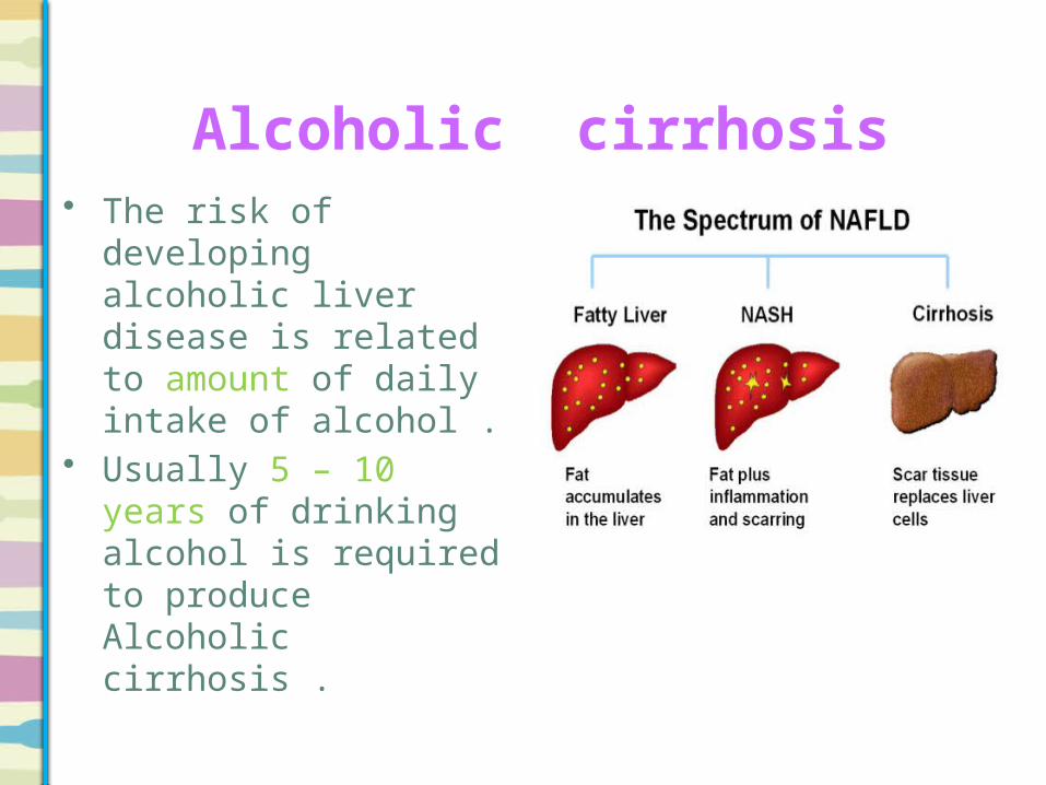
Alcoholic cirrhosis • The risk of developing
alcoholic liver disease is related to amount of daily intake of alcohol .
• Usually 5 – 10 years of drinking alcohol is required to produce Alcoholic cirrhosis .

Alcoholic cirrhosis • Signs of chronic liver
diseases :1. Drowsiness2. Hyperventilation 3. Flapping tremor 4. Jaundice5. Ascites6. Leukonychia 7. Peripheral Oedema 8. Bruising9. Dupuytren's contracture

Alcoholic cirrhosis
Usually the patient present with one of the Complications of cirrhosis :
1. Portal Hypertension • Ascites • Hypersplenism (with or without splenomegaly) • Varices (lower oesophageal and rectal)
2. Synthetic Dysfunction • Hypoalbuminaemia • Coagulopathy
3. Hepatopulmonary Syndrome 4. Hepatorenal Syndrome 5. Encephalopathy 6. Hepatocellular Carcinoma

Investigations
• CBC :1. Macrocytosis in the absence of
anemia .2. Leukocytosis or leukopenia.3. Thrombocytopenia.• LFTs :All LFTs are high.• Liver biopsy :It is diagnostic and shows mallory
bodies ( alcoholic hyaline )
Eosinophilic Mallory bodies are seen in hepatocytes, which are surrounded by fibrous tissue
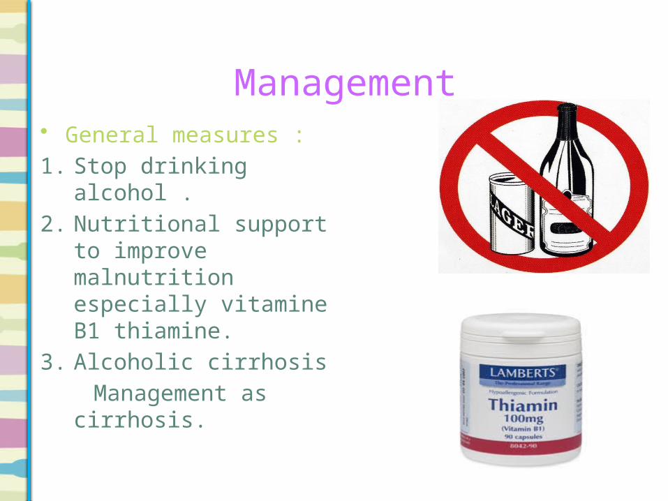
Management • General measures :1. Stop drinking alcohol .2. Nutritional support to
improve malnutrition especially vitamine B1 thiamine.
3. Alcoholic cirrhosis Management as
cirrhosis.

Management
• Be carful if patient need glucose
administration increase thiamine requirement and precipitate wernicke – korsakoff syndrome therefore thiamine should be given IV before glucose infusion .

Biliary cirrhosis • Biliary cirrhosis results from
prolonged biliary obstruction anywhere between the small interlobular bile duct and papilla of vater .
• Types of biliary cirrhosis :1. Primary 2. Secondary

Primary biliary cirrhosis • Primary biliary cirrhosis is a chronic
disease of liver in which small interlobular bile ducts of liver become progressively damaged and leading to cirrhosis and cholestasis.
• Etiology is unknown antimitochondrial
antibodies are found in almost all patients.

Clinical features Signs Symptoms
Hepatomegaly Asymptomatic
xanthelasma Itching (earlist)
Jaundice Yellow discoloration eyes
and skin (late)
Sign of portal HTN Diarrhea (steatorhea)
Fractures

Investigations
1) Liver function test : ALK ( very high)2) Atimitochondrial antibodies (AMA) >
95 % of cases.3) Serum cholesterol is high .4) Serum IgM may be very high.5) Ultrasound : diffuse alteration in
liver architecture.6) Liver biopsy show chareacteristic
features such as :A. Infiltration of portal tract by lymphocyte
and plasma cells .B. Loss of small bile ducts
C. Portal tract fibrosis
D. Periductal epithelioid granulomata about 40% of cases.

Management 1. Ursodeoxycholic acid (Ursodiol) is the most frequently
used treatment. This helps reduce the cholestasis and improves liver function tests
2. Pruritus :A. Cholestyramine.
B. Rifampicin .C. Opioid antaonists .
3. Malabsorption:D. Monthly inject vit K.
E. Vit D and calcium supplents F. Reducing fat intake to 40 g/d.
G. Bisphosphonate for osteoporosis.4. Statins for hypercholesterolemia.

Secondary biliary cirrhosis• This develops after
prolonged large bile duct obstruction due to :
1) Gall stones2) Bile duct stricture.Sing and symptoms are
same as that of PBC.Diagnosis is based on
ultrasound which common bile duct dilated .

Hemochromatosis • Hemochromatosis is a condition in which the
amount of total body iron is increase that damaged the organs . It may be primary or secondary.
• primary Hemochromatosis :is inherited autosomal recessive disease
characterized by excess iron deposition in various organs in form of hemosiderin leading to fibrosis and functional organs failure.

primary Hemochromatosis
Mostly organs affected are :1)Liver 2) pancreas 3)heart
4) adrenals 5) testes 6)kidneys 7) Pituitary
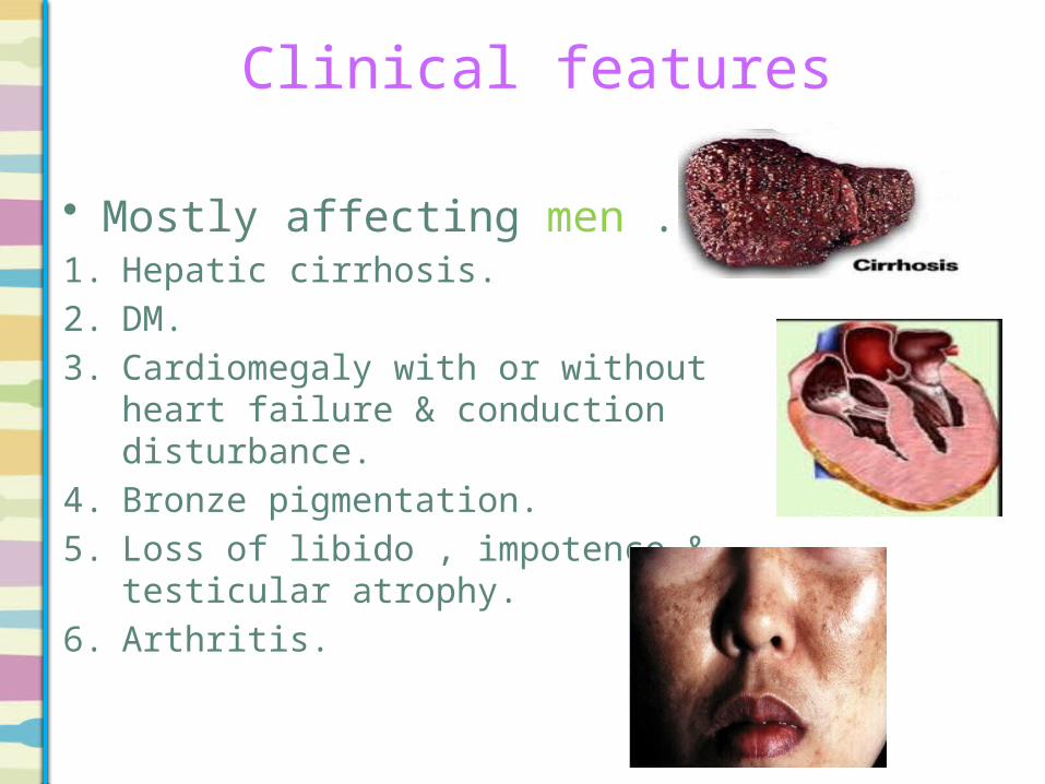
Clinical features
• Mostly affecting men .1. Hepatic cirrhosis.2. DM.3. Cardiomegaly with or without heart
failure & conduction disturbance.4. Bronze pigmentation.5. Loss of libido , impotence & testicular
atrophy.6. Arthritis.
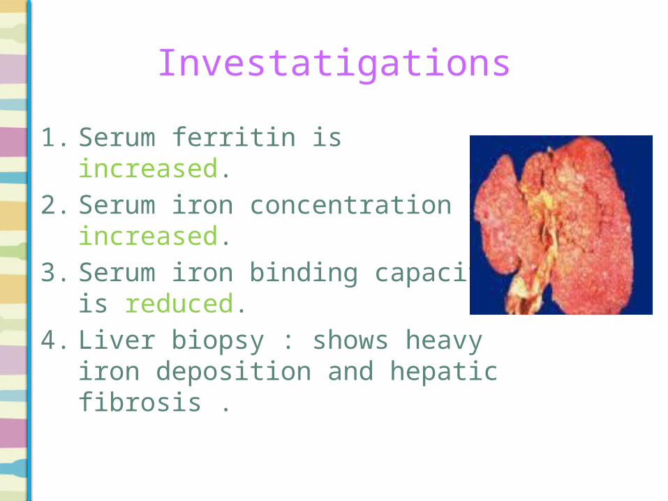
Investatigations
1. Serum ferritin is increased.2. Serum iron concentration
increased.3. Serum iron binding capacity is
reduced.4. Liver biopsy : shows heavy iron
deposition and hepatic fibrosis .

Treatment
1. Avoid food rich by in iron such as red meat , vitamin C , alcohol.
2. venesection of 500ml (250 mg iron ) weekly until the serum iron is normal .
3. Chelating agent deferoxamine subcutaneous injection. If the dose not tolerate venesection .

Secondary Hemochromatosis
• Secondry causes of iron overload are hemolytic anemia such as thalassemia in which multiple transfusions are required .
• Treatment: chelating therapy with deferoxamine is usually required.

Wilson's disease
• autosomal recessive genetic disorder in which copper accumulates in tissues.
• Increase copper content is due to increase absorption from small intestine and decrease excretion in bile .
• The most affected organs are:1)Liver 2)basal ganglia ( brain) 3)eyes
4) Kidneys 5) Skeleton

Clinical features Hormones
Hypoparathyroidism
Heart and kidney
Eyes Neuropsychiatric symptoms
Liver disease
Pain in the face, legs, and feet
cardiac arrhythmias
Kayser–Fleischer
rings
behavioral changes,
depression, anxiety and psychosis
fatigue
Abdominal pain cardiomyopathy
Resting tremor confusion
Convulsions (seizures)
renal tubular acidosis
Dystopia of bulbar muscle
such as dysarthria and
dysphagia
Bruising
Muscle cramps Tetanic
contractions
portal hypertension:1. Upper GI
bleeding2. Abdominal
distension
investigations
1. serum copper:Is reduce but it may be normal.2. Serum Ceruloplasmin :abnormally low (<0.2 g/L) .3. 24 – hours urine copper:Urinary copper excretion is increase (>100-1000
μg/24h .4. liver function tests :Are high .5. Liver biopsy :May show acute or chronic hepatitis or cirrhosis.

Treatment
1. Pencillamine: is the drug of choice that chelates and excretes copper through kidney in urine. Pyrodoxine 50mg / week should be added, since pencillamine is an antimetabolite of this vitamin and causes deficiency. The dose can be reduced once the disease is controlled, but treatment must continue for life. Side effects are skin rashes, leukopenia, and renal damage.
2. Trientine dihydrochoride: if patient develops side effects of pencillamine

Continue Treatment
3. Zinc acetate: Oral zinc acetate daily promotes fecal excretion of copper and used as maintenance therapy after chelation with pencillamine or as a first-line therapy in presymptomatic or pregnant patients.
4. Ammonium tetrathiomolybdate: is an initial therapy for neurologic Wilson’s disease.
5. Liver transplantation: indicated for fulminant hepatic failure, end stage liver disease and selected cases of severe neurological disease.

Prognosis
• Prognosis is good in patients effectively treated before liver or brain damage has occurred.
• Neurological damage is permanent.

α1-Antitrypsin Deficienacy
• A deficiency of α1-Antitrypsin (α1-AT) is sometimes associated with liver disease and pulmonary emphysema (particularly in smokers).
• Autosomal dominant.• Is synthesized in the liver. • Main role of alpha1-antitrypsin is to protect the
tissues, and especially the elastic tissueof the lungs, against the enzyme neutrophil elastase .
• The gene is located on chromosome 14.
α1-Antitrypsin Deficiency• The genetic variants of α1-AT are characterized
by their electrophoretic mobilities as medium (M), slow (S) or very slow (Z).
• The normal geneotype is protease inhibitor MM the homozygote for Z is PiZZ, and the heterozygotes are PiMZ and PiSZ.
• This results in decreased synthesis and secretion of the protein by the liver.
• Causes liver disease is uncertain.• The failure of secretion of the abnormal protein
leads to an accumulation in the liver, causing liver damage.
Clinical features
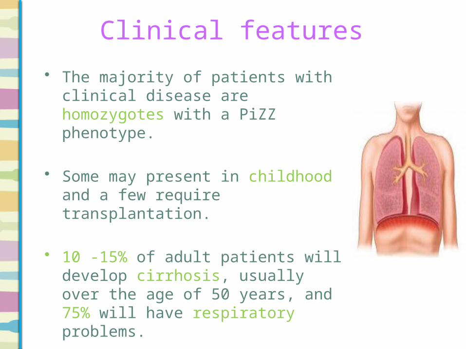
• The majority of patients with clinical disease are homozygotes with a PiZZ phenotype.
• Some may present in childhood and a few require transplantation.
• 10 -15% of adult patients will develop cirrhosis, usually over the age of 50 years, and 75% will have respiratory problems.

Clinical features
• Approximately 5% of patients die of their liver disease.
• Heterozygotes may develop liver disease, but the risk is small.

• Serum α1- antrypsin: low
• Liver biopsy: Periodic Acid – Schiff (PAS)- positive globules are present seen in periportal hepatocytes.
Investatigations
The periportal red hyaline globules seen here with periodic acid-Schiff (PAS) stain are characteristic for alpha-1-antitrypsin (AAT) deficiency
Treatment
• No specific treatment is available other than management of complications of liver disease. Liver transplantation may be advised for advanced liver disease.

On physical examination
Sign of Chronic Liver
Disease+
Dupuytren’s Contracture
+Peripheral
Neuropathy+
Cerebellar Signs
Alcohol
Sign of Chronic Liver
Disease+
Young+
Kayser-Fleisher Rings
+Cerebellar Signs
Wilson's
Sign of Chronic Liver
Disease
+
Increased pigmentation of
the skin
Haemochromatosis
Sign of Chronic Liver
Disease
+
Tattoos
Hepatitis C

Reference

Thank You