liver abscess secondary to foreign body clinical case presentation and subject · pdf...
TRANSCRIPT

ESCRIBA EL TÍTULO DEL DOCUMENTO 1
L IVER ABSCESS SECONDARY TO FOREIGN BODY CLINICAL CASE PRESENTATION AND SUBJECT
REVISION
Dr. Verónica Gigirey (1), Dr. Martín Rodríguez Parodi (2), Prof. Dr. Nelson Di Trapani (3).
(1)Ex. Assistant of Clinical Department of Imaging, University of the Oriental Republic of Uruguay, (2) Resident., (3)Ex. Professor of Clinical Department of Imaging, University of Oriental Republic of Uruguay.
ABSTRACT Liver abscess due to gastrointestinal perforation by foreign body is not frequent. Clinical symptoms are non-specific and variable. This article describes and illustrates a case of liver abscess secondary to fish bone that pierced the wall of the gastric antrum, diagnosed by computed tomography (CT). We performed percutaneous drainage of the abscess. Preoperative diagnosis of liver abscess due to penetration of foreign bodies in the gastrointestinal tract is rare using conventional imaging methods. The value of CT in preoperative diagnosis in such cases becomes very relevant, so that in patients with known risk factors of foreign body ingestion it should be studied using this method in order to establish an early treatment.
Keywords: liver abscess, foreign bodies, penetration, perforation, computed tomography. ABBREVIATIONS CT: computed tomography, CXR: chest radiography; FGC: fibrogastroscopy; MIP: maximum intensity projections
2 Revista Uruguaya de Imagenología - Epoca II - Vol. XVI, N°1 / Octubre 2012
INTRODUCTION
Intra-abdominal abscesses secondary to perforation of gastrointestinal tract due to ingestion of foreign bodies are not uncommon in clinical practice. However, liver abscesses secondary to a foreign body, which penetrates the gastrointestinal tract, are very rare. Very few cases have been reported in the literature.
Furthermore, preoperative diagnosis of penetration of foreign bodies of the digestive tract associated to liver abscess is not frequent using conventional imaging methods. These methods only show foreign bodies with high density: metal or bone.
We present a case of penetration of fish bone through the gastric wall with migration towards the lower edge of left liver lobe and presence of liver abscess, stressing the role of CT in the preoperative diagnosis.
CASE REPORT
62 years old man seeks emergency consultation due to clinical profile of fever witch started 15 days ago, adding during the process slight abdominal pain in right hypochondrium.
Physical examination reveals soft, depressible abdomen, no visceromegaly, slight pain in right hypochondrium when palpated, no signs of peritoneal irritation.
Requested paraclinical reveals presence of leukocytosis (13000 x 10`3uL),
Gamma GT 90000, C- reactive protein of 52 and speed of erythrosedimentation of 79 mm/hr.
For imaging evaluation, an XR is requested, which did not show any alterations.
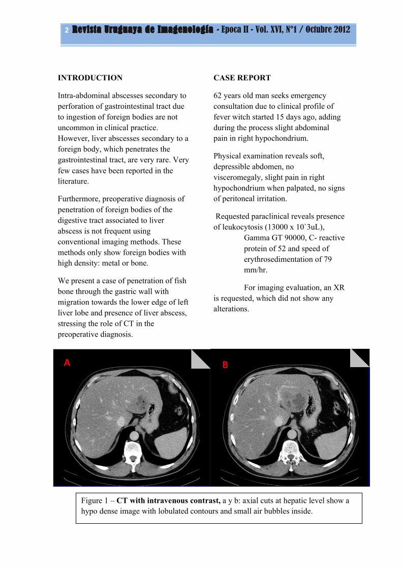
Figure 1 – CT with intravenous contrast, a y b: axial cuts at hepatic level show a hypo dense image with lobulated contours and small air bubbles inside.
A B
ESCRIBA EL TÍTULO DEL DOCUMENTO 3
A CT was carried out with 16-raw multi-detector equipment, with oral and intravenous contrast, which shows:
- Liver reduced in density with steatosic aspect
- In segments II-III a hypo-dense image can be observed, grossly rounded, of somewhat irregular contours, lobulated, with badly defined limits, of approx. 60 mm of diameter, with small air bubbles in its interior, compatible with abscess (fig. 1 A and B).
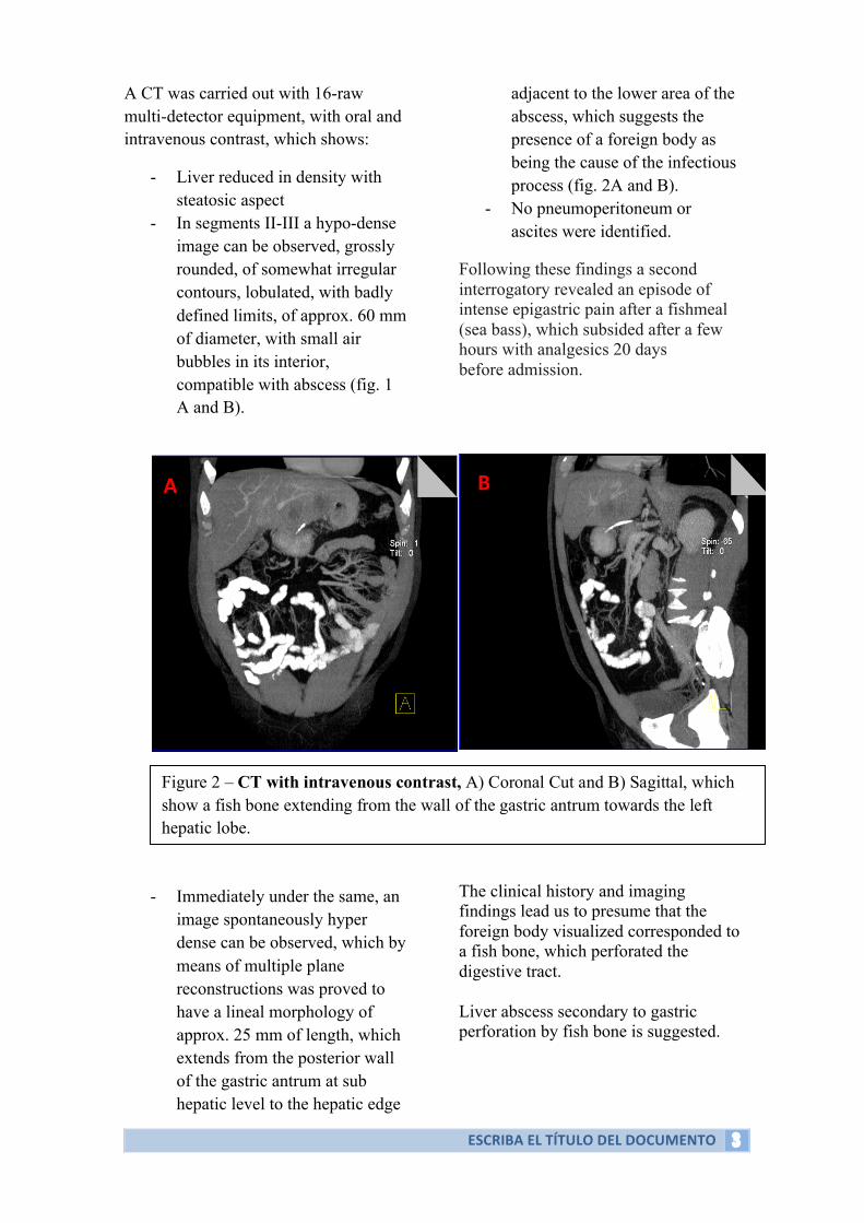
- Immediately under the same, an image spontaneously hyper dense can be observed, which by means of multiple plane reconstructions was proved to have a lineal morphology of approx. 25 mm of length, which extends from the posterior wall of the gastric antrum at sub hepatic level to the hepatic edge
adjacent to the lower area of the abscess, which suggests the presence of a foreign body as being the cause of the infectious process (fig. 2A and B).
- No pneumoperitoneum or ascites were identified.
Following these findings a second interrogatory revealed an episode of intense epigastric pain after a fishmeal (sea bass), which subsided after a few hours with analgesics 20 days before admission. The clinical history and imaging findings lead us to presume that the foreign body visualized corresponded to a fish bone, which perforated the digestive tract. Liver abscess secondary to gastric perforation by fish bone is suggested.
Figure 2 – CT with intravenous contrast, A) Coronal Cut and B) Sagittal, which show a fish bone extending from the wall of the gastric antrum towards the left hepatic lobe.
A B

4 Revista Uruguaya de Imagenología - Epoca II - Vol. XVI, N°1 / Octubre 2012
FGC is requested which shows erosive antrum gastritis in healing process. Percutaneous drainage of liver abscess was performed using the Seldinger method guided by echography and CT, with no complications, leaving in the drainage tube (fig.3). CT control of abscess is requested, which shows a positive progress, due to which the drainage tube is withdrawn. The foreign body persists without any changes in its topography.
A month after the drainage was performed, a new control is requested, which shows:
- Liver of normal shape and size, of reduced density as seen in the steatosis, without evidence of focal lesions, proving the complete resolution of the abscess.
- The foreign body inside the gastric cavity is identified (fig. 4).
- Expectant management is adopted and during progressive process elimination of the foreign body together with stools is confirmed.
DISCUSSION Most foreign bodies ingested (80-90 %) pass through the digestive tract with no complications (3, 4), but occasionally objects may become stuck in areas of anatomical narrowing ( 5 ). Gastrointestinal perforation by ingested foreign bodies is rare, their incidence is less than 1%. Those that perforate it are usually sharp, pointed. However, a blunt object can also perforate the intestinal wall through necrosis by pressure (4).
Figure 3 - Control CT, without intravenous contrast, post percutaneous drainage.
Figure 4 - Control CT, without intravenous contrast, A) Coronal Cut, B) Oblique and C) Sagittal, show the fish bone inside the gastric chamber.

ESCRIBA EL TÍTULO DEL DOCUMENTO 5
Clinical manifestations of perforation include peritonitis, formation of abscess, inflammatory masses, fistulous, obstruction and hemorrhage (8, 9). Formation of liver abscess due to perforation by foreign body in the digestive tract is not a frequent complication, they represent between 0 and 5 % of the same, there are very few cases reported in literature; the first case was described in 1898; since then another 47 cases have been published, and fish bones are usually the most frequent isolated causal agents, as occurred in our case, followed by toothpicks and in third place, chicken bones. Other etiologies such as needles, piercings etc. are less frequent (9). In these cases the foreign body usually perforates the digestive tract in the area of the stomach or duodenum with the abscess most commonly developing in the left hepatic lobe, because of its proximity. Typical cases of liver abscess present fever and abdominal pain, jaundice is present in a small percentage of patients and can make its debut as an episode of cholangitis. Most patients present non-specific symptoms. The foreign body´s migration is usually silent for a long time and is discovered only if there is infection or abscess (1, 2,10). Suspecting the diagnosis through clinical history is difficult, especially in the absence of specific symptoms and because most patients usually do not remember ingesting a foreign body, although it may help to ask if food consumed during the previous days included or not chicken or fish or if they used toothpicks or ate anything that might carry them inside. Although the area of penetration cannot be proved in most cases, localization of perforation in cases of liver abscess secondary to the same is generally observed in the stomach or duodenum and the foreign body is frequently lodged in the left lobe of the liver. Diagnosis by means of conventional
radiology is not common due to non-specific signs and performing an abdominal tomography is of great help, as happened with our patient (7.10). In this case, clinical progress was non-specific. However, CT allowed us to diagnose liver abscess and identify the cause of the same by detecting a fish bone, which perforated the posterior wall of the gastric antrum and migrated towards the left hepatic lobe. Nowadays it is relatively easy to detect the foreign body thanks to instruments such as MIP or multiple plane reconstructions. CT simplifies preoperative diagnosis in this kind of cases, leading to early treatment. Mortality due to liver abscess is significant, although it has decreased substantially in recent studies that report between 11 and 31 %. A foreign body migrating from the digestive tract is an unusual cause of liver abscess. The most common etiologies (6,7, 8) are: 1) Complication of cholangitis; 2) bloodstream dissemination through portal vein coming from gastrointestinal tract or from hepatic artery by systemic sepsis; 4) local spreading due to suppuration which affects contiguous tissue; and 5) by traumatic injuries, closed or penetrating. Among germs identified in this kind of abscess, the Streptococcus genre is the one most frequently isolated. However, in close to half the cases, the causal germ is not identified. The characteristic aspect of liver abscess in CT is a rounded mass of irregular shape, hypo dense with a peripheral capsule, which enhances after contrast administration. Usually there is a narrow area of transition, of slightly inferior density, between the central hypo dense portion of the mass and the hyper dense ring. In some cases, a dynamic tomography shows a double target image which consists of a central

6 Revista Uruguaya de Imagenología - Epoca II - Vol. XVI, N°1 / Octubre 2012
hypo dense area surrounded by a hyper dense ring which in itself is also surrounded by a hypo dense area, believed to be related to localized edema. Liver abscesses can be unilocular or multilocular. Central gas, forming multiple bubbles or a gas-fluid level, highly suggests an abscess but is only found in the minority of cases. An early diagnosis is a challenge and one must be highly suspicious, since the most frequent clinical sign is fever (3), although findings are typically non-specific.
Conventional treatment consists in surgery, draining of the abscess and withdrawing the foreign body, although some cases of percutaneous or endoscopic extraction have been described (2, 9, 11). It is advisable to remove the foreign body to avoid the presence of recurring abscesses. In this case, as an anecdote, this was not necessary since the fish bone migrated again within gastric lumen to be naturally eliminated by stools.
CONCLUSIONS
This report presents a case of liver abscess caused by fish bone, which perforated the gastric antrum wall with migration towards left hepatic lobe diagnosed by tomography. A tomography can simplify diagnosing this kind of case, allowing us to perform an early and adequate treatment. BIBLIOGRAPHY 1- Berk, R.N. Intraabdominal chicken-bone abscess. Diagnos. Radiol.101:311-313; 1971. 2- Abel, R.M.; Fischer, J.E.; He&en, W.H. Penetration of the alimentary tract
by a foreign body with migration to the liver. Arch. Surg. 102:227-228; 1971. 3- Strauss, J.E.; Balthazar, E.J.; Naidich, D.P. Jejunal perforation by a toothpick: CT demonstration. J. Comput. Assist. Tomogr. 9:812-814; 1985. 4- McCanse, D..E.; Kurchin, A.; Hinshaw, J.R. Gastrointestinal foreign bodies. Am. J. Surg. 142:335-337; 1981. 5- Spiz, L. Management of ingested foreign bodies in childhood. Br. Med. J. 4:469472; 1971. 6- Shin-ichiro M., Mitsuyuki A., Toshio I., Motokazu A., Shunsuke M., Ichiro F. Hepatic abscess secondary to a fishbone penetrating the gastric wall: CT demonstration. Computerized Medical Imaging and Graphics, Volume 15, Issue 2, March-April 1991, Pages 113-116 7- Maleki, M., Evans W.E. Foreign-body perforation of the intestinal tract. Report of 12 cases and review of the literature. Arch. Surg. 101:475477; 1970. 8- Schwartz J.T., Graham D.Y. Toothpick perforation of the intestines. Ann. Surg., 1977, 18:564-66;. 9- Kimbrell, FT., Tepas, J. J., Mullen, J.T. Chicken bone perforation of the sigmoid colon: a report of three cases. Am. Surg. 1975, 41:814-817. 10- Drnouvsek V, Fontanez-Garcia D, Wakabayashi M, Plavsic B. Gastrointestinal case of the day. RadioGraphics 1999; 19;820-822. 11- Adán Merino L, Gómez Senent S, Martin Arranz, MD, Martín Arranz E, Segura Cabral JM. Cuerpo extraño: una causa infrecuente de absceso hepático. Gastroenterología y hepatología. 2009; 32:179-80.