lipoprotein lipase activity is required for cardiac lipid ... · even the substrate used for heart...
TRANSCRIPT

This article is available online at http://www.jlr.org Journal of Lipid Research Volume 55, 2014 645
Copyright © 2014 by the American Society for Biochemistry and Molecular Biology, Inc.
and did not reduce heart function ( 3 ). Overexpression of DGAT1 in skeletal muscle also increased TG storage in mice with diet-induced obesity and mimicked the “athlete’s par-adox” observed in endurance-trained humans; skeletal muscle DGAT1 transgenic mice had increased FA oxidation and improved insulin sensitivity ( 4 ). In contrast, increased TG accumulation in the human heart correlates with reduced heart function ( 5, 6 ). Moreover, greater TG stores are often associated with greater FA oxidation and greater injury during ischemia/reperfusion in isolated perfused hearts ( 7 ). Thus, the role of TG stores in the heart is unclear.
Even the substrate used for heart TG production has not been established. One physiologic stimulus that causes lipid accumulation in mouse hearts is prolonged fasting ( 8 ). Because starvation is a threat to survival, lipid accumu-lation in the heart may be an adaptation to accommodate future energetic demands, to protect the heart from lipo-toxicity, or to do both. Understanding the features of this adaptation may provide insight into mechanisms that drive lipid accumulation under physiologic and pathological conditions. During fasting, animals rely exclusively on stored energy. While the liver produces and releases glu-cose under fasting conditions, this is insuffi cient to meet the energetic demands of the body ( 9 ). Adipose tissue is the major storage depot for energy in the form of TGs. During the fed state, dietary glucose stimulates insulin se-cretion, which simultaneously promotes glucose utiliza-tion and lipid storage. During fasting, circulating insulin levels fall while glucagon and catecholamines increase. This shift in the hormonal milieu leads to an activation of
Abstract The rodent heart accumulates TGs and lipid droplets during fasting. The sources of heart lipids could be either FFAs liberated from adipose tissue or FAs from lipo-protein-associated TGs via the action of lipoprotein lipase (LpL ). Because circulating levels of FFAs increase during fasting, it has been assumed that albumin transported FFAs are the source of lipids within heart lipid droplets. We stud-ied mice with three genetic mutations: peroxisomal prolifer-ator-activated receptor � defi ciency, cluster of differentiation 36 (CD36) defi ciency, and heart-specifi c LpL deletion. All three genetically altered groups of mice had defective ac-cumulation of lipid droplet TGs. Moreover, hearts from mice treated with poloxamer 407, an inhibitor of lipopro-tein TG lipolysis, also failed to accumulate TGs, despite in-creased uptake of FFAs. TG storage did not impair maximal cardiac function as measured by stress echocardiography. Thus, LpL hydrolysis of circulating lipoproteins is required for the accumulation of lipids in the heart of fasting mice. —Trent, C. M., S. Yu, Y. Hu, N. Skoller, L. A. Huggins, S. Homma, and I. J. Goldberg. Lipoprotein lipase activity is required for cardiac lipid droplet production. J. Lipid Res. 2014. 55: 645–658.
Supplementary key words PPAR • Cd36 • triglyceride
The human heart will accumulate TGs in lipid droplets in disease states such as obesity and diabetes. Whether TG storage directly leads to reduced heart function, i.e., lipo-toxicity ( 1, 2 ), or is a marker for accumulation of other toxic lipids is unclear. Evidence suggesting that stored TGs in cardiomyocytes are not always toxic has come from ex-periments in genetically modifi ed mice. For instance, overexpression of the fi nal enzyme in TG synthesis, diacyl-glycerol acyltransferase (DGAT)1, in cardiomyocytes in-creased TG stores but reduced accumulation of toxic lipids
This work was supported by National Heart, Lung, and Blood Institute Grants HL73029 and HL45095. C.M.T. is supported by American Heart Association pre-doctoral fellowship 12PRE11770023. The authors have no confl icts of inter-est to declare.
�� Author’s Choice —Final version full access Manuscript received 5 September 2013 and in revised form 27 January 2014.
Published, JLR Papers in Press, February 3, 2014 DOI 10.1194/jlr.M043471
Lipoprotein lipase activity is required for cardiac lipid droplet production
Chad M. Trent , * ,† Shuiqing Yu , * Yunying Hu , * Nathan Skoller , † Lesley A. Huggins , * Shunichi Homma , § and Ira J. Goldberg 1, *
Division of Preventive Medicine and Nutrition,* Institute of Human Nutrition, † and Division of Cardiology, § Columbia University College of Physicians and Surgeons , New York, NY 10032
Abbreviations: Acox, acyl CoA oxidase; Atgl, adipose TG lipase; CD36, cluster of differentiation 36; Cpt, carnitine palmitoyl transferase; DGAT, diacylglycerol acyltransferase; Fabp, FA binding protein; Fatp, FA transport protein; FS, fractional shortening; Glut, glucose transporter; HSL, hormone-sensitive lipase; LVEDd, left ventricular end-diastolic dimension; LVEDs, left ventricular end-systolic dimension; MHC, alpha-myosin heavy chain; P407, poloxamer 407; PAS, periodic acid-Schiff; Pdk, pyruvate dehydrogenase kinase; PLIN, perilipin .
1 To whom correspondence should be addressed. e-mail: [email protected]
The online version of this article (available at http://www.jlr.org) contains supplementary data in the form of text, four fi gures, and one table.
�� Author’s Choice
by guest, on Septem
ber 16, 2018w
ww
.jlr.orgD
ownloaded from
.html http://www.jlr.org/content/suppl/2014/02/03/jlr.M043471.DC1Supplemental Material can be found at:

646 Journal of Lipid Research Volume 55, 2014
protein analysis and 3 ml of 2:1 chloroform:methanol was added to the rest and vortexed. Samples were then centrifuged for 10 min at 3,000 rpm at 4°C. The lower organic phase was then collected and dried under nitrogen gas. The dried lipid was then dissolved in 500 � l of 1% Triton X-100 in chloroform, further dried, and then dissolved in 100 � l of double distilled water.
Lipid and protein measurements of tissues The sample of tissue lysate retained from the lipid extraction
protocol was assayed for protein content using Bradford reagent (Bio-Rad) following the instructions of the manufacturer. Using the tissue lipid extract, assays for TGs and FFAs were performed as previously described for plasma lipids. Lipid measurements were normalized to protein content or tissue weight.
Microscopy for cardiac lipid visualization Heart pieces were embedded in Tissue-Tek OCT compound
(Sakura) and then air dried and fi xed with formalin. Sections were washed with distilled water and isopropanol. Lipids were then stained with Oil Red O for 18 min, washed with isopropanol and distilled water, and then counterstained with hematoxylin. Slides were once again washed with distilled water and covered with clear nail polish. Images were taken using a Leica DMLB microscope and digital camera. Representative images obtained from fi ve animals of each genotype are shown.
Glycogen staining and quantifi cation Periodic acid-Schiff (PAS) reagent staining was used to dem-
onstrate heart glycogen. Sections of OCT embedded hearts were placed in 10% neutral buffered formalin. Ventricular tissue sec-tions were fi xed in methanol for 10 min and stained with PAS reagent (Poly Scientifi c), hematoxylin, and eosin. Images were taken using a Leica DMLB microscope and digital camera. Four to fi ve mouse hearts were used for each genotype and for each feeding condition, and several representative images were cap-tured for each mouse.
Glycogen was also measured by extracting total insoluble carbo-hydrates and digesting with amyloglucosidase; free glucose was then measured and reported as ratio to tissue weight used for mea-surement as previously described ( 16 ). Ventricular tissue was hy-drolyzed in 300 � l of 5.4 M KOH in a 100°C water bath for 30 min. Then, 100 � l 1 M Na 2 SO 4 and 800 � l of 100% ethanol were added to each sample. Samples were boiled for 5 min and then centri-fuged at 10,000 g for 5 min. The glycogen pellet was dissolved in 200 � l water and ethanol precipitation was performed twice with addition of 800 � l of 100% ethanol. Finally, the glycogen pellet was dissolved in 200 � l of 60 U/ml amyloglucosidase (Sigma) in 0.2 M sodium acetate (pH 4.8) and incubated for 3 h at 40°C. Each sam-ple was diluted fi ve times and glucose concentration was mea-sured using the Wako Autokit Glucose kit (Wako Life Sciences).
Cardiac gene expression Total RNA was purifi ed from a 30–50 mg piece of heart using
TRIzol reagent (Invitrogen) according to the instructions of the manufacturer. cDNA was synthesized using the SuperScript III First-Strand Synthesis SuperMix (Invitrogen) and quantitative real-time PCR was performed with SYBR Green PCR Core re-agents (Agilent Technologies) using an Mx3000 sequence detec-tion system (Stratagene, La Jolla, CA). Genes of interest were normalized against 18s rRNA. Primer sequences are listed in sup-plementary Table I.
Western blotting Hearts were excised as previously described. Approximately
20 mg of tissue was homogenized in RIPA buffer containing
glycogenolysis in the skeletal muscle and liver and lipolysis in the adipose tissue. However, prolonged fasting will de-plete glycogen stores and thus the energy demands of pe-ripheral tissues must rely on both the liver, to secrete glucose and ketone bodies, and TGs and adipose tissue, to secrete FFAs and glycerol. Adipose tissue secreted glycerol, as well as lactate secreted from both adipose tissue and muscle, are taken up by the liver and used as substrates for gluconeogenesis.
We tested to determine whether reduced FA oxidation increased fasting-induced TG accumulation in the heart. To do this we studied PPAR � knockout mice. Surprisingly, we found that fasted Ppara � / � mice had no lipid droplet ac-cumulation in hearts and had a marked reduction in mRNA levels of the FA transporter cluster of differentiation 36 (CD36) as well as lipoprotein lipase (LpL) ( 10, 11 ); LpL is required for heart uptake of FFAs from lipoprotein TGs. We then assessed the specifi c roles of CD36 and LpL in heart TG accumulation. Our data show that LpL activity is required for the accumulation of heart lipid droplets. In addition, we demonstrated that lipid droplet accumulation does not affect maximal systolic function of the heart.
MATERIALS AND METHODS
Animals and fasting We used 3–4-month-old male C57BL/6 mice, Ppara � / � mice
( 12 ), Cd36 � / � mice ( 13 ), fl oxed LpL mice (LpL fl ox/fl ox ), and heart-specifi c LpL knockout (hLpL0) mice ( 14 ). Mice were raised on a normal chow diet. C57BL/6 mice were used as con-trols for both Ppara � / � and Cd36 � / � mice and LpL fl ox/fl ox litter-mates served as controls for the hLpL0 studies. Mice of each genotype were divided into two groups. One group was subjected to a 16 h overnight fast and the other group was fed ad libitum over the same time period. These mice were then euthanized with a lethal injection of 100 mg/kg ketamine and 10 mg/kg xy-lazine. All procedures were approved by the Columbia University Institutional Animal Care and Use Committee.
Tissue collection A ventral incision was made after administration of ketamine-
xylazine. The left ventricle of the heart was perfused with 10 ml of PBS or until the liver appeared blanched. Tissues were rapidly removed and frozen in liquid nitrogen. Heart pieces were embedded into Tissue-Tek OCT compound (Sakura) for histology.
Measurement of plasma lipids and glucose Two hundred microliters of blood were drawn from each ani-
mal and then centrifuged at 2,000 rpm for 10 min to obtain plasma. Plasma was utilized for measurement of TGs, FFAs, and glucose by colorimetric assays. TG measurements were made using the Thermo Scientifi c Infi nity assay (Thermo Scientific), FFAs were measured using the Wako NEFA kit, and plasma glu-cose was measured using the Wako Autokit Glucose kit (Wako Life Sciences).
Lipid extraction from tissues The lipid extraction protocol was adapted from the Folch
method ( 15 ). Approximately 100 mg of tissue in 1 ml of PBS were homogenized using stainless steel beads for 1 min in a bead beater homogenizer. From each sample, 50 � l were removed for
by guest, on Septem
ber 16, 2018w
ww
.jlr.orgD
ownloaded from
.html http://www.jlr.org/content/suppl/2014/02/03/jlr.M043471.DC1Supplemental Material can be found at:

LpL activity is required for cardiac lipid droplet production 647
for 16 h. Fasting increased plasma FFAs 2-fold in Ppara +/+ mice and 3-fold in Ppara � / � mice, but had no signifi cant effect on plasma TGs ( Fig. 1A ). Plasma glucose decreased approximately 30% in fasted Ppara +/+ mice and approxi-mately 60% in fasted Ppara � / � mice. Fasting increased heart TGs 5-fold in Ppara +/+ mice, but there was no signifi -cant TG accumulation in Ppara � / � mice ( Fig. 1B ). Heart FA levels increased approximately 30% in Ppara +/+ mice, but were not increased in Ppara � / � mice ( Fig. 1B ). Fasted Ppara +/+ mice had increased Oil Red O staining, but Ppara � / � mice had minimal staining ( Fig. 1C ).
We then assessed heart glycogen storage in Ppara � / � mice to determine whether these hearts depleted their stored carbohydrate. There was no difference in PAS reagent stain-ing of glycogen ( Fig. 1D ) or extracted glycogen content ( Fig. 1E ) after fasting. There tended to be increased glyco-gen in Ppara � / � hearts both before and after fasting.
Changes in genes required for lipid and glucose metab-olism were determined in hearts of fed and fasted mice. Adipose TG lipase ( Atgl ), the rate limiting enzyme for intra-cellular TG lipolysis ( 18 ), was decreased by 50% in both fed and fasted Ppara � / � mice compared with Ppara +/+ mice ( Fig. 1F ). Expression of carnitine palmitoyl transferase ( Cpt ) 1b , the rate limiting enzyme for mitochondrial lipid oxidation, was minimal in both fed and fasted Ppara � / � hearts. Surprisingly, mRNA of acyl CoA oxidase ( Acox ) 1 , the fi rst enzyme in the lipid oxidation pathway, was increased in fasted Ppara � / � hearts. However, decreased FA oxidation has been previously observed in these hearts ( 19, 20 ).
Therefore, absence of TG stores was not likely due to increased FA oxidation. As expected, mRNA levels of lipid droplet protein genes Plin2 and Plin5 were minimal in both fed and fasted Ppara � / � mice compared with Ppara +/+ mice ( 21 ).
Fasting dramatically increased expression of pyruvate dehydrogenase kinase ( Pdk ) 4 , the negative regulator of glucose oxidation, in hearts from Ppara +/+ mice. Fasted Ppara � / � mice also had increased Pdk4 mRNA levels com-pared with fed Ppara � / � mice, but these levels were still reduced compared with the Ppara +/+ counterparts. Fasted Ppara � / � mice had increased mRNA expression of glucose transporter ( Glut ) 1 , the insulin insensitive glucose trans-porter, compared with fasted Ppara +/+ hearts, but there was no difference in expression of Glut4 , the insulin sensitive glucose transporter . Most remarkable was that fed and fasted Ppara � / � hearts had an � 80% reduction in lipid up-take genes Cd36 and Lpl ( Fig. 1F ).
We assessed heart mRNA levels of several genes involved in both de novo lipogenesis and TG formation. Expression of acetyl-CoA carboxylase ( Acc ) 2 , which is rate-limiting for de novo lipogenesis, was increased in the hearts of fed Ppara � / � mice (supplementary Fig. IIIA). mRNA expres-sion of Fasn , the second rate-limiting enzyme for de novo lipogenesis, was increased in the hearts of both fed and fasted Ppara � / � mice. mRNA levels of Dgat1 , the rate limit-ing enzyme for TG synthesis, were decreased in hearts from both fed and fasted Ppara � / � mice. We also measured gene expression of FA transporters other than Cd36 . Hearts from both fed and fasted Ppara � / � mice had decreased
protease inhibitor cocktail (Sigma-Aldrich). Twenty-fi ve micro-grams of protein extract was applied to SDS-PAGE and trans-ferred onto polyvinylidene fl uoride membranes . Antibodies for perilipin (PLIN)2 and PLIN5 were obtained from Santa Cruz Bio-technology (PLIN2, � -actin) and American Research Products (PLIN5). Band density measurements were made using ImageJ software. PLIN2 and PLIN5 band densities were normalized to � -actin band density.
Stress echocardiography Echocardiography was performed on 3–4-month-old male Cd36 +/+
(wild-type), Cd36 � / � , LpL fl ox/fl ox , and hLpL0 mice fasted for 16 h. Two-dimensional echocardiography was performed using a high-resolution imaging system with a 30 MHz imaging transducer (Vevo 770; VisualSonics) in unconscious mice. The mice were anesthetized with 1.5–2% isofl urane and thereafter maintained on 0.5% isofl urane throughout the procedure. Care was taken to minimize sedation by monitoring the heart rate of the mice. Two-dimensional echocardiographic images were obtained using short-axis views at the level of papillary muscles, and each parameter was measured using M-mode view. Images were analyzed offl ine by a researcher blinded to the murine genotype. Left ventricular end-diastolic dimension (LVEDd) and left ventricular end-systolic dimension (LVEDs) were measured. Percentage fractional short-ening (FS), which quantifi es contraction of the ventricular wall and is an indication of muscle function, was calculated as FS = ([LVEDd � LVEDs]/LVEDd) × 100. To assess stress response, 0.3 mg/kg isoproterenol (Sigma-Aldrich) was administered in-traperitoneally. Successful administration of drug was confi rmed by observation of increased heart rate.
Pharmacologic inhibition of LpL Poloxamer 407 (P407) was prepared in PBS as previously de-
scribed ( 17 ). Mice were injected intraperitoneally with 1 mg/g body weight of P407 and then fasted for 16 h. Control mice were injected with an equivalent volume of PBS. Mice were euthanized and analyzed as previously described.
In vivo assessment of cardiac glucose and FFA uptake FFA and glucose uptake were assessed in mice that were in-
jected with PBS or P407 and then fasted for 16 h. [9,10- 3 H(N)]oleate (PerkinElmer Life Sciences) was complexed to 6% FA-free BSA (Sigma). Mice were injected intravenously with 1 � Ci [9,10- 3 H(N)]oleate-BSA and blood was collected at 0.5, 1, 3, and 5 min after injection. Five minutes after injection, the body cavity was per-fused with 10 ml of PBS by cardiac puncture and tissues were ex-cised. Tissue was homogenized in PBS and radioactive counts were measured. Basal glucose uptake was measured in hearts following an intravenous administration of 2.5 � Ci of 2-deoxy-D-[1- 14 C]glucose (PerkinElmer Life Sciences). Blood was collected 2, 30, and 60 min following injection. At 60 min, hearts were perfused with PBS, tissues were excised, and radioactive counts were measured. For all turnover studies, radioactivity per gram of tissue was normalized to the respective 30 s or 2 min plasma counts (injected dose).
Statistical analysis Data are expressed as mean ± SE. Data were analyzed by the
use of unpaired Student’s t -test or two-way ANOVA.
RESULTS
Fasted Ppara � / � mice do not store lipids in the heart We fi rst assessed heart lipid storage in Ppara � / � mice.
We fasted Ppara +/+ (wild-type) and Ppara � / � mice overnight
by guest, on Septem
ber 16, 2018w
ww
.jlr.orgD
ownloaded from
.html http://www.jlr.org/content/suppl/2014/02/03/jlr.M043471.DC1Supplemental Material can be found at:

648 Journal of Lipid Research Volume 55, 2014
signifi cant TG accumulation in Cd36 � / � hearts ( Fig. 2B, C ). Heart FA content also did not increase in hearts from Cd36 � / � mice ( Fig. 2B ). Glycogen content was similar in all hearts ( Fig. 2D, E ). Finally, Cd36 � / � mouse hearts tended to have decreased expression of lipid metabolism genes ( Atgl , Cpt1b , Acox1 , Atgl , Plin2 , and Plin5 ) in the fed state, but fasted Cd36 � / � mouse hearts had similar gene expression to hearts of Cd36 +/+ mice ( Fig. 2F ). LpL mRNA levels were comparable to control mice in both fasting and fed hearts. Glucose oxidation and uptake genes ( Pdk4 , Glut1 , and Glut4 ) were comparable between genotypes and feeding conditions. Fed and fasted Cd36 � / � mice had decreased ex-pression of Dgat2 (supplementary Fig. IIIB). Hearts of fasted Cd36 � / � mice had increased expression of Slc27a1 , and both fed and fasted hearts had decreased expression of FA
expression of FA transport protein 1 ( Slc27a1 , or Fatp1 ), but increased expression of FA binding protein-plasma mem-brane ( Got2 , or FABPpm ). Finally, we measured TG lipase activity in hearts of fed and fasted Ppara +/+ and Ppara � / � mice. Both fed and fasted Ppara � / � mice had 80–90% in-creased heart TG lipase activity compared with Ppara +/+ mice of the same nutritional status (supplementary Fig. IVA).
Fasted Cd36 � / � mice do not store lipids in the heart Next, we determined whether CD36 defi ciency would be
suffi cient to prevent heart TG accumulation during the fasted state. Fasting increased plasma FFAs 3-fold in Cd36 � / � mice ( Fig. 2A ). Plasma TGs tended to be higher in fasted Cd36 � / � mice compared with Cd36 +/+ mice. Fasted Cd36 � / � mice had an � 50% decrease in plasma glucose. There was no
Fig. 1. Overnight fasting of Ppara +/+ and Ppara � / � mice. A: Plasma FFAs, TGs, and glucose were measured in 3–4-month-old male Ppara +/+ (n = 9) and Ppara � / � (n = 5) mice that were fed or fasted for 16 h. Data were compared by Student’s t -test. * P < 0.05. B: Total lipids were extracted from hearts, and TGs and FFAs were measured. Lipid content was normalized to protein content. Data were compared by Stu-dent’s t -test. * P < 0.05. C: Heart sections of fed and fasted Ppara +/+ and Ppara � / � mice (n = 5) were stained with Oil Red O, indicating neutral lipid content (1,000× magnifi cation). D: Heart sections of fed and fasted Ppara +/+ and Ppara � / � mice (n = 5) were stained with PAS reagent, indicating glycogen content (400× magnifi cation). E: Total glycogen was extracted from hearts of fed and fasted Ppara +/+ and Ppara � / � mice and digested with amyloglucosidase, and free glucose was measured. Glucose content was normalized to tissue weight. F: Gene expression of Atgl , Cpt1b , Acox1 , Plin2 , Plin5 , Cd36 , Lpl , Pdk4 , Glut1 , and Glut4 was assessed using quantitative real-time PCR. Gene expression is ex-pressed relative to fed Ppara +/+ mice. Data were compared by two-way ANOVA. * P < 0.05 compared within genotype; # P < 0.05 compared with Ppara +/+ mice of same feeding status.
by guest, on Septem
ber 16, 2018w
ww
.jlr.orgD
ownloaded from
.html http://www.jlr.org/content/suppl/2014/02/03/jlr.M043471.DC1Supplemental Material can be found at:

LpL activity is required for cardiac lipid droplet production 649
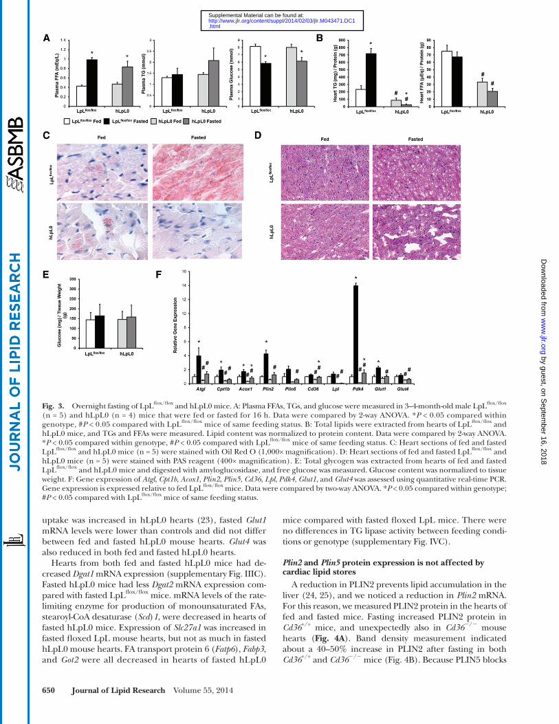
LpL fl ox/fl ox and hLpL0 mice. Surprisingly, hLpL0 mice did not accumulate cardiomyocyte TGs during fasting ( Fig. 3B, C ). Glycogen content was similar in all hearts ( Fig. 3D, E ).
We predicted that changes in gene expression in the fasting hLpL0 hearts would explain the lack of TG stores. mRNA levels of Atgl , Cpt1b , and Acox1 were reduced in both fed and fasted hLpL0 mouse hearts ( Fig. 3F ), consis-tent with the reduction in FA oxidation in these hearts ( 23 ). hLpL0 mice also had decreased expression of Plin2 and Plin5 compared with fasted LpL fl ox/fl ox mice. Although these hearts do not have reduced FFA uptake, Cd36 mRNA was reduced in both fed and fasted hLpL0 hearts. Pdk4 expression was reduced in both fed and fasted hLpL0 hearts compared with LpL fl ox/fl ox hearts. Although glucose
binding protein ( Fabp ) 3 . There were no differences in TG lipase activity between feeding conditions or genotype (sup-plementary Fig. IVB).
Fasted hLpL0 mice do not store lipids in the heart If circulating FFAs are the source of heart TG stores dur-
ing fasting, we would expect that loss of lipoprotein TG hydrolysis in the heart would not affect lipid droplet accu-mulation during fasting. To test this, we fasted hLpL0 mice and compared them to LpL fl ox/fl ox littermates. After fasting hLpL0 mice had normal increases in plasma FFA levels, an approximately 2-fold increase ( Fig. 3A ). hLpL0 mice tended to have slightly elevated TGs in the fasted state, as has been reported ( 22 ). Plasma glucose fell 20% in both
Fig. 2. Overnight fasting of Cd36 +/+ and Cd36 � / � mice. A: Plasma FFAs, TGs, and glucose were measured in 3–4-month-old male Cd36 +/+ (n = 9) and Cd36 � / � (n = 9) mice that were fed or fasted for 16 h. Data were compared by 2-way ANOVA. * P < 0.05 compared within genotype, # P < 0.05 compared to Cd36 +/+ mice of same feeding status. B: Total lipids were extracted from hearts of Cd36 +/+ and Cd36 � / � mice, and TGs and FFAs were measured. Lipid content was normalized to protein content. Data were compared by 2-way ANOVA. * P < 0.05 compared within genotype, # P < 0.05 compared to Cd36 +/+ mice of same feeding status. C: Heart sections of fed and fasted Cd36 +/+ and Cd36 � / � mice (n = 5) were stained with Oil Red O (1,000× magnifi cation). D: Heart sections of fed and fasted Cd36 +/+ and Cd36 � / � mice (n = 5) were stained with PAS reagent (400× magnifi cation). E: Total glycogen was extracted from hearts of fed and fasted Cd36 +/+ and Cd36 � / � mice and digested with amy-loglucosidase, and free glucose was measured. Glucose content was normalized to tissue weight. F: Gene expression of Atgl , Cpt1b , Acox1 , Plin2 , Plin5 , Cd36 , Lpl , Pdk4 , Glut1 , and Glut4 was assessed using quantitative real-time PCR. Gene expression is expressed relative to fed Cd36 +/+ mice. Data were compared by two-way ANOVA. * P < 0.05 compared within genotype; # P < 0.05 compared with Cd36 +/+ mice of same feeding status.
by guest, on Septem
ber 16, 2018w
ww
.jlr.orgD
ownloaded from
.html http://www.jlr.org/content/suppl/2014/02/03/jlr.M043471.DC1Supplemental Material can be found at:
650 Journal of Lipid Research Volume 55, 2014
mice compared with fasted fl oxed LpL mice. There were no differences in TG lipase activity between feeding condi-tions or genotype (supplementary Fig. IVC).
Plin2 and Plin5 protein expression is not affected by cardiac lipid stores
A reduction in PLIN2 prevents lipid accumulation in the liver ( 24, 25 ), and we noticed a reduction in Plin2 mRNA. For this reason, we measured PLIN2 protein in the hearts of fed and fasted mice. Fasting increased PLIN2 protein in Cd36 +/+ mice, and unexpectedly also in Cd36 � / � mouse hearts ( Fig. 4A ). Band density measurement indicated about a 40–50% increase in PLIN2 after fasting in both Cd36 +/+ and Cd36 � / � mice ( Fig. 4B ). Because PLIN5 blocks
uptake was increased in hLpL0 hearts ( 23 ), fasted Glut1 mRNA levels were lower than controls and did not differ between fed and fasted hLpL0 mouse hearts. Glut4 was also reduced in both fed and fasted hLpL0 hearts.
Hearts from both fed and fasted hLpL0 mice had de-creased Dgat1 mRNA expression (supplementary Fig. IIIC). Fasted hLpL0 mice had less Dgat2 mRNA expression com-pared with fasted LpL fl ox/fl ox mice. mRNA levels of the rate-limiting enzyme for production of monounsaturated FAs, stearoyl-CoA desaturase ( Scd ) 1 , were decreased in hearts of fasted hLpL0 mice. Expression of Slc27a1 was increased in fasted fl oxed LpL mouse hearts, but not as much in fasted hLpL0 mouse hearts. FA transport protein 6 ( Fatp6 ), Fabp3 , and Got2 were all decreased in hearts of fasted hLpL0
Fig. 3. Overnight fasting of LpL fl ox/fl ox and hLpL0 mice. A: Plasma FFAs, TGs, and glucose were measured in 3–4-month-old male LpL fl ox/fl ox (n = 5) and hLpL0 (n = 4) mice that were fed or fasted for 16 h. Data were compared by 2-way ANOVA. * P < 0.05 compared within genotype, # P < 0.05 compared with LpL fl ox/fl ox mice of same feeding status. B: Total lipids were extracted from hearts of LpL fl ox/fl ox and hLpL0 mice, and TGs and FFAs were measured. Lipid content was normalized to protein content. Data were compared by 2-way ANOVA. * P < 0.05 compared within genotype, # P < 0.05 compared with LpL fl ox/fl ox mice of same feeding status. C: Heart sections of fed and fasted LpL fl ox/fl ox and hLpL0 mice (n = 5) were stained with Oil Red O (1,000× magnifi cation). D: Heart sections of fed and fasted LpL fl ox/fl ox and hLpL0 mice (n = 5) were stained with PAS reagent (400× magnifi cation). E: Total glycogen was extracted from hearts of fed and fasted LpL fl ox/fl ox and hLpL0 mice and digested with amyloglucosidase, and free glucose was measured. Glucose content was normalized to tissue weight. F: Gene expression of Atgl , Cpt1b , Acox1 , Plin2 , Plin5 , Cd36 , Lpl , Pdk4 , Glut1 , and Glut4 was assessed using quantitative real-time PCR. Gene expression is expressed relative to fed LpL fl ox/fl ox mice. Data were compared by two-way ANOVA. * P < 0.05 compared within genotype; # P < 0.05 compared with LpL fl ox/fl ox mice of same feeding status.
by guest, on Septem
ber 16, 2018w
ww
.jlr.orgD
ownloaded from
.html http://www.jlr.org/content/suppl/2014/02/03/jlr.M043471.DC1Supplemental Material can be found at:

LpL activity is required for cardiac lipid droplet production 651
in P407-treated mouse hearts compared with PBS-treated mouse hearts ( Fig. 5C ).
We then measured uptake of circulating FFAs in control and P407-treated mice. Plasma turnover of the label was identical in the control and treated mice ( Fig. 5D ). Heart uptake of FFAs was greater in the lipolysis-inhibited mice ( Fig. 5E ), consistent with a greater reliance of the heart on FFAs than TGs. Liver uptake of the label did not differ between groups. Next, we measured uptake of circulating glucose in control and P407-treated mice. Plasma turnover of the labeled glucose in plasma was the same in both groups of mice ( Fig. 5F ). Uptake of plasma glucose tended to be increased in P407-treated mouse hearts ( P = 0.08) and livers ( Fig. 5G ). Total glycogen content was not changed between groups ( Fig. 5H, I ).
Lipid metabolism genes tended to be increased with P407, but not all increases reached statistical signifi cance. Cpt1b , Acox1 , and Lpl were increased in P407-treated mice while increases in Atgl , Plin2 , Plin5 , and Cd36 were less ro-bust ( Fig. 5J ). Pdk4 expression was not affected by P407-treatment, but Glut1 and Glut4 were both increased in hearts of P407-treated mice. P407-treated fasted mice had increased heart expression of Fabp3 and tended to have
TG lipolysis ( 26–28 ), a lack of change in mRNA but reduced protein could allow more rapid degradation of stored TGs. Therefore, we measured Plin5 protein in the fed and fasted state. PLIN5 protein levels were highly variable ( Fig. 4A ), but not signifi cantly different between the fed and fasted states ( Fig. 4B ). Similar changes in both PLIN2 and PLIN5 protein were observed in hLpL0 mice ( Fig. 4C, D ).
Blocking circulating TG degradation prevents cardiac lipid accumulation
Because of the surprising observation that hLpL0 mouse hearts did not accumulate TGs during fasting, a result sug-gesting that circulating TGs are the primary substrate for cardiac lipid accumulation, we used a drug that blocks li-polysis of circulating TGs. Mice were administered P407 and then fasted for 16 h. Plasma TGs increased 10-fold in the P407-treated mice, indicating a complete block of TG uptake ( Fig. 5A ). Plasma FFA and glucose levels were also higher in the P407-treated mice. Next, we looked at the heart lipids. As we found in the hLpL0 hearts, cardiac FFAs were not affected by P407 treatment, but P407-treated mice had 60% less TGs than PBS-treated mice ( Fig. 5B ). There was decreased Oil Red O staining of neutral lipids
Fig. 4. Western blot of PLIN2 and PLIN5 in hearts. A: PLIN2 and PLIN5 content was assessed with Western blotting in hearts of Cd36 +/+ and Cd36 � / � mice. B: PLIN2 and PLIN5 protein content in hearts of Cd36 +/+ and Cd36 � / � mice. Protein was quantifi ed by band density measurements of the Western blot. Band densities were normalized to � -actin content within each sample. Data are expressed as relative amount compared with fed Cd36 +/+ mice. Data were compared by Student’s t -test. * P < 0.05. C: PLIN2 and PLIN5 content was assessed with Western blotting in hearts of LpL fl ox/fl ox and hLpL0 mice. D: PLIN2 and PLIN5 protein content in hearts of LpL fl ox/fl ox and hLpL0 mice was quantifi ed by band density measurements of the Western blot. Data were normalized to � -actin content within each sample and expressed as relative amount compared with fed LpL fl ox/fl ox mice. Data were compared by Student’s t -test. * P < 0.05.
by guest, on Septem
ber 16, 2018w
ww
.jlr.orgD
ownloaded from
.html http://www.jlr.org/content/suppl/2014/02/03/jlr.M043471.DC1Supplemental Material can be found at:

652 Journal of Lipid Research Volume 55, 2014
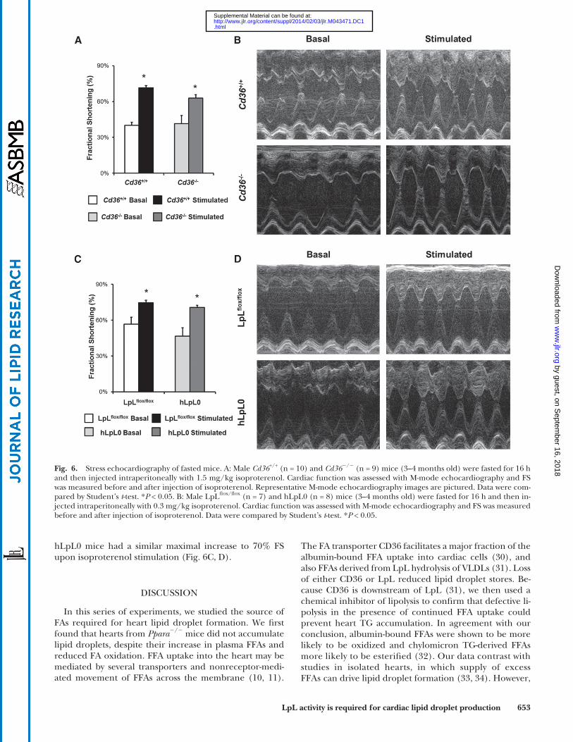
function, we fasted mice overnight and measured FS before and after administration of isoproterenol. We used young mice, 3–4 months old, prior to the development of severe heart dysfunction that occurs in the hLpL0 mice ( 23 ). Basal FS was similar ( � 40%) in fasted Cd36 +/+ and Cd36 � / � mice. Isoproterenol injection resulted in a maximum FS of 70% in fasted Cd36 +/+ hearts and 60% in Cd36 � / � hearts ( Fig. 6A, B ). Basal FS tended to be greater in LpL fl ox/fl ox mice (56%) compared with hLpL0 mice (47%), but LpL fl ox/fl ox and
increased expression of Fatp6 (supplementary Fig. IIID). There was a 30% increase in TG lipase activity in fasted mice treated with P407 (supplementary Fig. IVD).
Cardiac TG accumulation during fasting does not impair heart function
Cardiac TG accumulation has been postulated to cause toxicity and to reduce heart function ( 2, 29 ). To determine whether TG accumulation during fasting affects cardiac
Fig. 5. Overnight-fasted mice treated with P407. A: Plasma FFAs, TGs, and glucose in 3–4-month-old C57/BL6 male mice (n = 5) that were injected intraperitoneally with 1 mg/g P407 or an equivalent volume of PBS. Data were compared by Student’s t -test. * P < 0.05. B: Total lipids were extracted from hearts of fasted PBS- and P407-treated mice, and TGs and FFAs were measured. Lipid content was normalized to heart weight . Data were compared by Student’s t -test. * P < 0.05. C: Heart sections of fasted PBS- and P407-treated mice (n = 5) were stained with Oil Red O (1,000× magnifi cation). D: Plasma disappearance of the [ 3 H]oleate was measured at 30 s, 1 min, 3 min, and 5 min after injection. E: Cardiac and hepatic FFA uptake were assessed using [ 3 H]oleate. Data were compared by Student’s t -test. * P < 0.05. F: Plasma disappearance of the [ 14 C]2-deoxyglucose was measured at 2 min, 30 min, and 60 min after injection. G: Cardiac and hepatic glucose uptake were assessed using [ 14 C]2-deoxyglucose. H: Total glycogen was extracted from hearts of fasted PBS- and P407-treated mice and digested with amyloglucosi-dase, and free glucose was measured. Glucose content was normalized to tissue weight. I: Heart sections of fasted PBS- and P407-treated mice (n = 5) were stained with PAS reagent, indicating glycogen content (400× magnifi cation). J: Gene expression of Atgl , Cpt1b , Acox1 , Plin2 , Plin5 , Cd36 , Lpl , Pdk4 , Glut1 , and Glut4 was assessed using quantitative real-time PCR. Data were compared by Student’s t -test. * P < 0.05.
by guest, on Septem
ber 16, 2018w
ww
.jlr.orgD
ownloaded from
.html http://www.jlr.org/content/suppl/2014/02/03/jlr.M043471.DC1Supplemental Material can be found at:
LpL activity is required for cardiac lipid droplet production 653
The FA transporter CD36 facilitates a major fraction of the albumin-bound FFA uptake into cardiac cells ( 30 ), and also FFAs derived from LpL hydrolysis of VLDLs ( 31 ). Loss of either CD36 or LpL reduced lipid droplet stores. Be-cause CD36 is downstream of LpL ( 31 ), we then used a chemical inhibitor of lipolysis to confi rm that defective li-polysis in the presence of continued FFA uptake could prevent heart TG accumulation. In agreement with our conclusion, albumin-bound FFAs were shown to be more likely to be oxidized and chylomicron TG-derived FFAs more likely to be esterifi ed ( 32 ). Our data contrast with studies in isolated hearts, in which supply of excess FFAs can drive lipid droplet formation ( 33, 34 ). However,
hLpL0 mice had a similar maximal increase to 70% FS upon isoproterenol stimulation ( Fig. 6C, D ).
DISCUSSION
In this series of experiments, we studied the source of FAs required for heart lipid droplet formation. We fi rst found that hearts from Ppara � / � mice did not accumulate lipid droplets, despite their increase in plasma FFAs and reduced FA oxidation. FFA uptake into the heart may be mediated by several transporters and nonreceptor-medi-ated movement of FFAs across the membrane ( 10, 11 ).
Fig. 6. Stress echocardiography of fasted mice. A: Male Cd36 +/+ (n = 10) and Cd36 � / � (n = 9) mice (3–4 months old) were fasted for 16 h and then injected intraperitoneally with 1.5 mg/kg isoproterenol. Cardiac function was assessed with M-mode echocardiography and FS was measured before and after injection of isoproterenol. Representative M-mode echocardiography images are pictured. Data were com-pared by Student’s t -test. * P < 0.05. B: Male LpL fl ox/fl ox (n = 7) and hLpL0 (n = 8) mice (3–4 months old) were fasted for 16 h and then in-jected intraperitoneally with 0.3 mg/kg isoproterenol. Cardiac function was assessed with M-mode echocardiography and FS was measured before and after injection of isoproterenol. Data were compared by Student’s t -test. * P < 0.05.
by guest, on Septem
ber 16, 2018w
ww
.jlr.orgD
ownloaded from
.html http://www.jlr.org/content/suppl/2014/02/03/jlr.M043471.DC1Supplemental Material can be found at:

654 Journal of Lipid Research Volume 55, 2014
fasting-induced increase of TG lipase activity in the hearts of wild-type animals ( 41 ); we did not fi nd this to be the case.
mRNA levels for Cd36 and Lpl were both markedly re-duced in the Ppara � / � mice. Therefore, we fasted mice that were defi cient in CD36 or heart LpL. Cd36 � / � mice have decreased cardiac FFA uptake and decreased VLDL-TG uptake, whereas hLpL0 mice have normal to increased cardiac FFA uptake and decreased VLDL-TG uptake ( 31, 42 ). Because the concentration of circulating FFAs in-creases during fasting, and mice that do not mobilize adi-pose lipid stores during fasting do not accumulate cardiac lipids ( 38 ), we hypothesized that FFAs are the primary sub-strate for lipid accumulation in the hearts of fasted mice. We expected that Cd36 � / � mice would not form lipid droplets after an overnight fast, but hLpL0 mice would form lipid droplets. As expected, fasted Cd36 � / � mice failed to accumulate lipids in the heart. Surprisingly, the hLpL0 mice also failed to accumulate cardiac lipids.
We still believed that FFAs were driving this process, so we hypothesized that lipid droplets were not stabilized and were turned over more rapidly in hLpL0 mice. hLpL0 mice have decreased PPAR � target gene expression ( 22, 43 ), so we suspected that lipid droplet proteins PLIN2 and PLIN5 were also reduced. We found that Plin2 mRNA lev-els were reduced in both the fed and fasted state. How-ever, Plin5 mRNA was normal. As there is often a mismatch between Plin2 and Plin5 message and protein level, we as-sayed protein levels with Western blotting. Protein levels in the heart were comparable for each genotype, and fast-ing increased heart PLIN2 modestly in all genotypes. PLIN5 was comparable between control and hLpL0 mice with both feeding conditions. Finally, we measured heart intracellular TG lipase activity, and there was no differ-ence between LpL fl ox/fl ox mice and hLpL0 mice. Thus, we concluded that despite decreased PPAR � target gene ex-pression, it was unlikely that a defect in storage was pre-venting lipid accumulation in hLpL0 mice.
We then posited that VLDL-TGs, and not FFAs, were the source of the cardiac lipids for droplet accumulation ob-served in fasting. Indeed, cardiac LpL activity is increased after an overnight fast, suggesting an important role for circulating TGs in supplying the heart with lipids ( 44, 45 ). To test this hypothesis, we treated mice with P407, a deter-gent which interferes with lipolysis of circulating lipopro-teins. P407 treatment dramatically reduced cardiac lipid accumulation, but did not reduce PPAR � target gene ex-pression. Furthermore, P407-treated mice had increased FFA uptake, yet still failed to form lipid droplets. However, the decrease in lipid droplet formation could be the result of increased intracellular TG lipase activity; it is unclear why intracellular TG lipase activity was increased in the P407-treated mice. We observed that P407 treatment and fasting together dramatically increased plasma FFAs; plasma FFA concentrations in P407-treated mice were 5-fold greater than those in plasma of nontreated fasting mice. This increase may be partially due to the partition-ing of plasma FFAs onto VLDL particles (supplementary Fig. I). We concluded that TGs, and not FFAs, are the pri-mary substrate for cardiac lipid accumulation observed in
in vivo the majority of circulating FAs are esterifi ed as TGs and phospholipids (85–90%). Thus it would not be sur-prising that they also are a major supplier of heart FFAs for TG storage. Finally, we showed that TG accumulation dur-ing overnight fasting did not impair or improve stimulated cardiac function.
Lipid uptake, oxidation, and storage are controlled by the PPAR family of transcription factors. The family mem-bers, PPAR � , PPAR � / � , and PPAR � , have overlapping tran-scriptional control of lipid metabolism gene expression. Two mouse models of heart-specifi c PPAR overexpression, the alpha-myosin heavy chain (MHC)-PPAR � and MHC-PPAR � transgenic mice, have increased lipid uptake, in-creased neutral lipid storage, and increased lipid oxidation associated with cardiac dysfunction ( 35, 36 ). Although Ppara � / � mice have increased fasting levels of plasma FFAs ( 37 ), the accumulation of heart lipids during fasting had not been assessed. We hypothesized that Ppara � / � mice, which have reduced FA oxidation in the heart ( 12 ), would accumulate more lipids during a prolonged fast. We found just the opposite; TG accumulation was drastically re-duced. Was this due to a defect in lipid droplet produc-tion, greater lipolysis of the stored TGs, or reduced lipid uptake into these hearts? Because lipid uptake is upstream of lipid storage and turnover, it seems most likely that a defi ciency of lipid uptake would precede any intracellular metabolic abnormalities.
There are several mouse models that do not accumulate cardiac lipids during fasting. Mice defi cient in hormone-sensitive lipase (HSL), an intracellular TG and diacylglyc-erol lipase, do not accumulate cardiac lipids during fasting, presumably due to the reduced ability of the adipose tissue to lipolyze stored TGs and release FFAs ( 38 ). HSL knock-out mice have lower fasting plasma FFA and TG levels and decreased heart FFA uptake ( 38 ). Conversely, cardiomyo-cyte-specifi c HSL-overexpressing mice also do not accu-mulate lipids after prolonged fasting because of rapid lipid droplet turnover ( 39 ). Ppara � / � mice did not have an in-crease in ATGL expression; in fact, mRNA levels of ATGL were dramatically reduced.
Another modulator of intracellular TG lipolysis is the lipid droplet protein PLIN5. Recently, Plin5 � / � mice were described to have defective storage of lipids in the heart ( 28 ); PLIN5 is responsible for regulating ATGL activity, thus with PLIN5 defi ciency intracellular lipase activity is constantly turned on. Plin4 deletion resulted in a similar phenotype due to a consequent decrease in Plin5 expres-sion ( 40 ). Overexpression of PLIN5 increased heart TG content ( 26, 27 ). Plin2 � / � mice had decreased lipid accu-mulation in the liver, suggesting that Plin2 promotes lipid accumulation ( 24 ). As others have reported ( 21 ), we ob-served that Plin2 and Plin5 mRNA were dramatically reduced in Ppara � / � mouse hearts. Therefore, defects in lipid drop-let formation in Ppara � / � mice could be due to lack of PLIN2 or PLIN5 or, more likely, the reductions in PLIN2 and PLIN5 are secondary to reduced PPAR � activation. Surprisingly, intracellular TG lipase activities were dramati-cally increased in Ppara � / � mice, perhaps due to decreased Plin2 and Plin5 expression. Others have demonstrated a
by guest, on Septem
ber 16, 2018w
ww
.jlr.orgD
ownloaded from
.html http://www.jlr.org/content/suppl/2014/02/03/jlr.M043471.DC1Supplemental Material can be found at:

LpL activity is required for cardiac lipid droplet production 655
TG lipase activity in Ppara � / � mice, despite the reduction in Atgl mRNA. The defi ciency of Plin2 and Plin5 that we and others have observed ( 21 ) in these mice could also indicate greater TG turnover. We also observed an in-crease in heart TG lipase activity in fasted mice treated with P407. However, there were no differences in heart TG lipase activity between Cd36 � / � or hLpL0 mice and wild-type or fl oxed controls.
Although our data and others implicate LpL and CD36 as the primary players in cardiac lipid accumulation, it is possible that mice defi cient in these proteins might have a reduction in other lipid transporters or de novo lipogen-esis. We assessed mRNA expression of a number of other genes and found that some of these genes had increased expression, but this was not suffi cient to restore TG lipid droplet accumulation.
Lipid droplet accumulation is thought to occur due to an imbalance between lipid uptake and oxidation. This in-volves PPAR � driven expression of LpL and CD36, allowing increased uptake of circulating lipoprotein TGs ( Fig. 7 ). Whether TG accumulation is toxic is unresolved ( 3 ). Lipid droplets are found in hearts of patients with diabetes and metabolic syndrome ( 5, 6, 53 ) and in hearts of high-fat diet-fed rodents and genetically altered mice ( 2 ). Lipotoxicity can occur when ceramides, diacylglycerol, or other lipid species can alter cardiac cell signaling, disrupt membrane function, or cause apoptosis ( 54 ). We measured ceramides in hearts of fasted wild-type mice and did not observe an increase in ceramides coinciding with increased TG content (supplementary Fig. II).
Excessive FFA oxidation has been proposed to lead to mitochondrial dysfunction, apoptosis, and heart failure ( 7 ). For this reason, we tested to determine whether increased stored lipid would reduce stimulated heart function. It did
fasting, and that PPAR � is necessary, but not suffi cient, for cardiac lipid accumulation.
In these studies, we focused on lipid uptake pathways that modulate TG accumulation in the hearts of fasted ani-mals. As the heart is a dynamic organ that possesses the ability to use multiple substrates, we asked whether hearts in fasted animals that have defective lipid uptake may have changes in glucose utilization. Mice that have decreased lipid uptake in cardiomyocytes tend to increase glucose uptake and catabolism ( 23, 46 ). We suspected that this might be happening in mice that failed to store lipid drop-lets in the cardiomyocytes. Heart gene expression of Pdk4 , a negative regulator of glucose oxidation, was increased in all fasted mice, but to a lesser degree in mice that had de-fi ciencies in lipid uptake. This likely indicates an increased reliance on glucose oxidation, i.e., pyruvate conversion to acetyl-CoA. Although gene expression of the glucose trans-porters GLUT1 and GLUT4 was variable, it has been previ-ously demonstrated that Ppara � / � mice ( 47 ), Cd36 � / � mice ( 48 ), and hLpL0 mice ( 23 ) all have increased cardiac glucose uptake. P407-treated fasting mice, which did not accumulate TGs in cardiomyocytes, tended to have in-creased glucose uptake. However, glycogen content of hearts was similar for all genotypes, regardless of feeding status. We should note that others have reported increased heart glycogen with fasting ( 49 ).
We would have liked to assess where the albumin-bound FFAs from adipose lipolysis and the FFAs from LpL’s ac-tion on circulating TGs were going once they entered the cardiomyocyte. Different pools of circulating lipids, either FFAs or TGs, may enter the cardiomyocyte and be immedi-ately oxidized, or they may be esterifi ed as TGs and later oxidized, or some combination of the two. FA turnover in the heart is very rapid with hydrolysis of much the pool of FAs within 10 min ( 23 ). Thus, assessing the residual FAs from labeled FFAs or VLDL-TGs is challenging. There are a number of elegant studies that have used isolated per-fused hearts to track uptake, oxidation, and esterifi cation of labeled TGs in chylomicrons and VLDLs ( 29, 50–52 ). However, methods to study tracer uptake and oxidation in vivo and not in perfused hearts are not available. It is likely that LpL-mediated accumulation of heart TGs during fast-ing is refl ective of the much greater amount of circulating lipids that reside in TG particles. The total amount of albu-min-bound FFAs might be insuffi cient to alone promote heart lipid accumulation during fasting. In support of the importance of TGs as a source of heart lipids, lipotoxic mice that have excessive cardiac lipid accumulation are cured with cardiac-specifi c LpL knockout ( 43 ). Further-more, it might also be that FAs from different sources di-verge in their intracellular fate. In support of this, one study of isolated perfused hearts found that FFAs were pri-marily oxidized, while FAs from chylomicrons divided equally into oxidation and cellular storage ( 32 ).
We assessed intracellular TG lipase activity to determine whether increased turnover of lipid droplets might explain why we did not see fasting-induced cardiac lipid accumula-tion in Ppara � / � , Cd36 � / � , hLpL0, and P407-treated mice. Surprisingly, we found there was dramatically increased
Fig. 7. Pathways of lipid uptake leading to lipid droplet formation. PPAR � drives lipid accumulation by regulating transcription of lipid uptake proteins LpL and CD36. Although both LpL and CD36 are required for cardiac lipid accumulation, LpL-mediated lipolysis of lipoprotein TGs is required for lipid droplet accumulation.
by guest, on Septem
ber 16, 2018w
ww
.jlr.orgD
ownloaded from
.html http://www.jlr.org/content/suppl/2014/02/03/jlr.M043471.DC1Supplemental Material can be found at:

656 Journal of Lipid Research Volume 55, 2014
very high level of cardiac LpL expression is involved not only in acquisition of circulating FAs for energy, but also for allowing storage.
REFERENCES
1 . Abel , E. D. , K. M. O’Shea , and R. Ramasamy . 2012 . Insulin resis-tance: metabolic mechanisms and consequences in the heart. Arterioscler. Thromb. Vasc. Biol. 32 : 2068 – 2076 .
2 . Goldberg , I. J. , C. M. Trent , and P. C. Schulze . 2012 . Lipid metabo-lism and toxicity in the heart. Cell Metab. 15 : 805 – 812 .
3 . Liu , L. , X. Shi , K. G. Bharadwaj , S. Ikeda , H. Yamashita , H. Yagyu , J. E. Schaffer , Y. H. Yu , and I. J. Goldberg . 2009 . DGAT1 expression increases heart triglyceride content but ameliorates lipotoxicity. J. Biol. Chem. 284 : 36312 – 36323 .
4 . Liu , L. , X. Shi , C. S. Choi , G. I. Shulman , K. Klaus , K. S. Nair , G. J. Schwartz , Y. Zhang , I. J. Goldberg , and Y. H. Yu . 2009 . Paradoxical coupling of triglyceride synthesis and fatty acid oxidation in skel-etal muscle overexpressing DGAT1. Diabetes . 58 : 2516 – 2524 .
5 . Marfella , R. , C. Di Filippo , M. Portoghese , M. Barbieri , F. Ferraraccio , M. Siniscalchi , F. Cacciapuoti , F. Rossi , M. D’Amico , and G. Paolisso . 2009 . Myocardial lipid accumulation in patients with pressure-overloaded heart and metabolic syndrome. J. Lipid Res. 50 : 2314 – 2323 .
6 . Sharma , S. , J. V. Adrogue , L. Golfman , I. Uray , J. Lemm , K. Youker , G. P. Noon , O. H. Frazier , and H. Taegtmeyer . 2004 . Intramyocardial lipid accumulation in the failing human heart re-sembles the lipotoxic rat heart. FASEB J. 18 : 1692 – 1700 .
7 . Lopaschuk , G. D. , J. R. Ussher , C. D. Folmes , J. S. Jaswal , and W. C. Stanley . 2010 . Myocardial fatty acid metabolism in health and disease. Physiol. Rev. 90 : 207 – 258 .
8 . Suzuki , J. , W. J. Shen , B. D. Nelson , S. P. Selwood , G. M. Murphy , Jr ., H. Kanehara , S. Takahashi , K. Oida , I. Miyamori , and F. B. Kraemer . 2002 . Cardiac gene expression profi le and lipid accumulation in response to starvation. Am. J. Physiol. Endocrinol. Metab. 283 : E94 – E102 . [Erratum. 2002. Am. J. Physiol. Endocrinol. Metab. 283: follow-ing table of contents.]
9 . Cahill , G. F. , Jr . 2006 . Fuel metabolism in starvation. Annu. Rev. Nutr. 26 : 1 – 22 .
10 . Kazantzis , M. , and A. Stahl . 2012 . Fatty acid transport proteins, im-plications in physiology and disease. Biochim. Biophys. Acta . 1821 : 852 – 857 .
11 . Glatz , J. F. , J. J. Luiken , and A. Bonen . 2010 . Membrane fatty acid transporters as regulators of lipid metabolism: implications for metabolic disease. Physiol. Rev. 90 : 367 – 417 .
12 . Leone , T. C. , C. J. Weinheimer , and D. P. Kelly . 1999 . A critical role for the peroxisome proliferator-activated receptor alpha (PPARalpha) in the cellular fasting response: the PPARalpha-null mouse as a model of fatty acid oxidation disorders. Proc. Natl. Acad. Sci. USA . 96 : 7473 – 7478 .
13 . Febbraio , M. , N. A. Abumrad , D. P. Hajjar , K. Sharma , W. Cheng , S. F. Pearce , and R. L. Silverstein . 1999 . A null mutation in murine CD36 reveals an important role in fatty acid and lipoprotein me-tabolism. J. Biol. Chem. 274 : 19055 – 19062 .
14 . Noh , H. L. , K. Okajima , J. D. Molkentin , S. Homma , and I. J. Goldberg . 2006 . Acute lipoprotein lipase deletion in adult mice leads to dyslipidemia and cardiac dysfunction. Am. J. Physiol. Endocrinol. Metab. 291 : E755 – E760 . [Erratum. 2006. Am. J. Physiol. Endocrinol. Metab. 292: E367.]
15 . Folch , J. , M. Lees , and G. H. Sloane Stanley . 1957 . A simple method for the isolation and purifi cation of total lipides from animal tis-sues. J. Biol. Chem. 226 : 497 – 509 .
16 . Suzuki , Y. , C. Lanner , J. H. Kim , P. G. Vilardo , H. Zhang , J. Yang , L. D. Cooper , M. Steele , A. Kennedy , C. B. Bock , et al . 2001 . Insulin control of glycogen metabolism in knockout mice lacking the muscle-specifi c protein phosphatase PP1G/RGL. Mol. Cell. Biol. 21 : 2683 – 2694 .
17 . Millar , J. S. , D. A. Cromley , M. G. McCoy , D. J. Rader , and J. T. Billheimer . 2005 . Determining hepatic triglyceride production in mice: comparison of poloxamer 407 with Triton WR-1339. J. Lipid Res. 46 : 2023 – 2028 .
18 . Haemmerle , G. , A. Lass , R. Zimmermann , G. Gorkiewicz , C. Meyer , J. Rozman , G. Heldmaier , R. Maier , C. Theussl , S. Eder , et al . 2006 . Defective lipolysis and altered energy metabolism in mice lacking adipose triglyceride lipase. Science . 312 : 734 – 737 .
not. We found that short-term starvation and consequent TG accumulation did not decrease cardiac output.
Although several models of lipid-induced heart dysfunc-tion have massively increased amounts of heart TGs, others have increases similar to those we found with fasting. ATGL knockout mice have 20 times the amount of cardiac lipids at 12 weeks of age and dramatically decreased heart function leading to 50% mortality by 18 weeks of age ( 18 ). However, two groups recently reported that PLIN5 overexpression and a 3- to 10-fold increase in cardiac TGs led to only mild heart dysfunction ( 26, 27 ). Less dramatic increases in TG levels are occasionally associated with heart dysfunction, however, in those hearts diacylgycerols and ceramides are also increased ( 55 ). We have previously reported a model of 50% increased cardiac TGs in the fed state that is not associ-ated with decreased cardiac function, the MHC-DGAT1 mouse model ( 3 ). In fact, this transgene reduces toxicity in other models without changing TG levels, but reducing heart ceramide 20% ( 3 ). In another model, transgenic MHC-PPAR � mice, a similar 2- to 3-fold increase in myocar-dial TGs but coupled to increased diacylglyceride and cer-amide was associated with more than a 50% reduction in FS ( 8, 35, 56 ). The effects of diets and diabetes on heart TGs and other lipids and cardiac function are less clear. Three weeks of high-fat diet feeding increased heart TGs by 2- to 3-fold, but a 20% decrease of FS was only observed after 20 weeks of high-fat diet feeding ( 57, 58 ). Streptozotocin-diabetic mice had a 50% increase in cardiac TGs after 12 weeks associated with a 20% reduction of FS ( 59 ). Ob / ob and db / db mice all have varying degrees of increased cardiac TGs (from 2-fold to 4-fold) associated with heart dysfunc-tion depending on the duration of the study ( 60, 61 ). Levels of ceramides and diacylglycerides were often not mea-sured in these models. Increased heart TG content is some-times, but not always, a hallmark of increased accumulation of other lipids, many of which are harmful to the heart.
Storage of TGs in the heart may be an adaptive response to decreased energy intake during starvation. However, it also occurs in the setting of pathological conditions in-cluding obesity, diabetes, and nonischemic heart failure ( 62, 63 ). Moreover, one report has suggested that heart lipid droplet accumulation occurs postischemia and is as-sociated with more tissue damage ( 64 ). As we noted above, lipid droplet accumulation is sometimes associated with heart dysfunction and also with increased concentrations of potentially toxic lipids such as ceramides and diacylglyc-erols, but in other situations TG storage alone does not lead to heart dysfunction ( 2 ). Whether the association be-tween heart TGs and function in humans is due to TGs or other accumulated lipids is not obvious. It should be noted that severe heart failure in humans leads to reduced heart TGs, but an increase in potentially toxic ceramides and diacylglycerols ( 65 ). Our data show that lipid droplets, at least those that occur during fasting, do not affect heart function.
In summary, our studies show that LpL lipolysis of TGs is required to store lipids in the hearts of fasting mice. This study further confi rms previous work showing that hearts are active organs in TG metabolism. We now show that the
by guest, on Septem
ber 16, 2018w
ww
.jlr.orgD
ownloaded from
.html http://www.jlr.org/content/suppl/2014/02/03/jlr.M043471.DC1Supplemental Material can be found at:

LpL activity is required for cardiac lipid droplet production 657
2002 . The cardiac phenotype induced by PPARalpha overexpres-sion mimics that caused by diabetes mellitus. J. Clin. Invest. 109 : 121 – 130 .
37 . Muoio , D. M. , P. S. MacLean , D. B. Lang , S. Li , J. A. Houmard , J. M. Way , D. A. Winegar , J. C. Corton , G. L. Dohm , and W. E. Kraus . 2002 . Fatty acid homeostasis and induction of lipid regula-tory genes in skeletal muscles of peroxisome proliferator-activated receptor (PPAR) alpha knock-out mice. Evidence for compensa-tory regulation by PPAR delta. J. Biol. Chem. 277 : 26089 – 26097 .
38 . Suzuki , J. , M. Ueno , M. Uno , Y. Hirose , Y. Zenimaru , S. Takahashi , J. Osuga , S. Ishibashi , M. Takahashi , M. Hirose , et al . 2009 . Effects of hormone-sensitive lipase disruption on cardiac energy metabo-lism in response to fasting and refeeding. Am. J. Physiol. Endocrinol. Metab. 297 : E1115 – E1124 .
39 . Suzuki , J. , W. J. Shen , B. D. Nelson , S. Patel , J. H. Veerkamp , S. P. Selwood , G. M. Murphy , Jr ., E. Reaven , and F. B. Kraemer . 2001 . Absence of cardiac lipid accumulation in transgenic mice with heart-specifi c HSL overexpression. Am. J. Physiol. Endocrinol. Metab. 281 : E857 – E866 .
40 . Chen , W. , B. Chang , X. Wu , L. Li , M. Sleeman , and L. Chan . 2013 . Inactivation of Plin4 downregulates Plin5 and reduces cardiac lipid accumulation in mice. Am. J. Physiol. Endocrinol. Metab. 304 : E770 – E779 .
41 . Zierler , K. A. , D. Jaeger , N. M. Pollak , S. Eder , G. N. Rechberger , F. P. Radner , G. Woelkart , D. Kolb , A. Schmidt , M. Kumari , et al . 2013 . Functional cardiac lipolysis in mice critically depends on comparative gene identifi cation-58. J. Biol. Chem. 288 : 9892 - 9904 .
42 . Coburn , C. T. , T. Hajri , A. Ibrahimi , and N. A. Abumrad . 2001 . Role of CD36 in membrane transport and utilization of long-chain fatty acids by different tissues. J. Mol. Neurosci. 16 : 117 – 121 ; discus-sion 151–157.
43 . Duncan , J. G. , K. G. Bharadwaj , J. L. Fong , R. Mitra , N. Sambandam , M. R. Courtois , K. J. Lavine , I. J. Goldberg , and D. P. Kelly . 2010 . Rescue of cardiomyopathy in peroxisome proliferator-activated receptor-alpha transgenic mice by deletion of lipoprotein lipase identifi es sources of cardiac lipids and peroxisome proliferator-activated receptor-alpha activators. Circulation . 121 : 426 – 435 .
44 . Haemmerle , G. , R. Zimmermann , J. G. Strauss , D. Kratky , M. Riederer , G. Knipping , and R. Zechner . 2002 . Hormone-sensitive lipase defi ciency in mice changes the plasma lipid profi le by affect-ing the tissue-specifi c expression pattern of lipoprotein lipase in adipose tissue and muscle. J. Biol. Chem. 277 : 12946 – 12952 .
45 . Goldberg , I. J. , R. H. Eckel , and N. A. Abumrad . 2009 . Regulation of fatty acid uptake into tissues: lipoprotein lipase- and CD36-mediated pathways. J. Lipid Res. 50 (Suppl) : S86 – S90 .
46 . Angin , Y. , L. K. Steinbusch , P. J. Simons , S. Greulich , N. T. Hoebers , K. Douma , M. A. van Zandvoort , W. A. Coumans , W. Wijnen , M. Diamant , et al . 2012 . CD36 inhibition prevents lipid accumulation and contractile dysfunction in rat cardiomyocytes. Biochem. J. 448 : 43 – 53 .
47 . Nöhammer , C. , F. Brunner , G. Wölkart , P. B. Staber , E. Steyrer , F. J. Gonzalez , R. Zechner , and G. Hoefl er . 2003 . Myocardial dys-function and male mortality in peroxisome proliferator-activated receptor alpha knockout mice overexpressing lipoprotein lipase in muscle. Lab. Invest. 83 : 259 – 269 .
48 . Hajri , T. , X. X. Han , A. Bonen , and N. A. Abumrad . 2002 . Defective fatty acid uptake modulates insulin responsiveness and metabolic re-sponses to diet in CD36-null mice. J. Clin. Invest. 109 : 1381 – 1389 .
49 . Schneider , C. A. , V. T. Nguyen , and H. Taegtmeyer . 1991 . Feeding and fasting determine postischemic glucose utilization in isolated working rat hearts. Am. J. Physiol. 260 : H542 – H548 .
50 . Stanley , W. C. , F. A. Recchia , and G. D. Lopaschuk . 2005 . Myocardial substrate metabolism in the normal and failing heart. Physiol. Rev. 85 : 1093 – 1129 .
51 . Lopaschuk , G. D. , D. D. Belke , J. Gamble , T. Itoi , and B. O. Schonekess . 1994 . Regulation of fatty acid oxidation in the mam-malian heart in health and disease. Biochim. Biophys. Acta . 1213 : 263 – 276 .
52 . Niu , Y. G. , D. Hauton , and R. D. Evans . 2004 . Utilization of triacylg-lycerol-rich lipoproteins by the working rat heart: routes of uptake and metabolic fates. J. Physiol. 558 : 225 – 237 .
53 . McGavock , J. M. , I. Lingvay , I. Zib , T. Tillery , N. Salas , R. Unger , B. D. Levine , P. Raskin , R. G. Victor , and L. S. Szczepaniak . 2007 . Cardiac steatosis in diabetes mellitus: a 1H-magnetic resonance spectroscopy study. Circulation . 116 : 1170 – 1175 .
54 . Drosatos , K. , K. G. Bharadwaj , A. Lymperopoulos , S. Ikeda , R. Khan , Y. Hu , R. Agarwal , S. Yu , H. Jiang , S. F. Steinberg , et al. 2011 .
19 . Watanabe , K. , H. Fujii , T. Takahashi , M. Kodama , Y. Aizawa , Y. Ohta , T. Ono , G. Hasegawa , M. Naito , T. Nakajima , et al . 2000 . Constitutive regulation of cardiac fatty acid metabolism through peroxisome proliferator-activated receptor alpha associated with age-dependent cardiac toxicity. J. Biol. Chem. 275 : 22293 – 22299
20 . Campbell , F. M. , R. Kozak , A. Wagner , J. Y. Altarejos , J. R. Dyck , D. D. Belke , D. L. Severson , D. P. Kelly , and G. D. Lopaschuk . 2002 . A role for peroxisome proliferator-activated receptor alpha (PPARalpha) in the control of cardiac malonyl-CoA levels: reduced fatty acid oxidation rates and increased glucose oxidation rates in the hearts of mice lacking PPARalpha are associated with higher concentrations of malonyl-CoA and reduced expression of malo-nyl-CoA decarboxylase. J. Biol. Chem. 277 : 4098 – 4103 .
21 . Yamaguchi , T. , S. Matsushita , K. Motojima , F. Hirose , and T. Osumi . 2006 . MLDP, a novel PAT family protein localized to lipid droplets and enriched in the heart, is regulated by peroxisome proliferator-activated receptor alpha. J. Biol. Chem. 281 : 14232 – 14240 .
22 . Augustus , A. , H. Yagyu , G. Haemmerle , A. Bensadoun , R. K. Vikramadithyan , S. Y. Park , J. K. Kim , R. Zechner , and I. J. Goldberg . 2004 . Cardiac-specifi c knock-out of lipoprotein lipase alters plasma lipoprotein triglyceride metabolism and cardiac gene expression. J. Biol. Chem. 279 : 25050 – 25057 .
23 . Augustus , A. S. , J. Buchanan , T. S. Park , K. Hirata , H. L. Noh , J. Sun , S. Homma , J. D’Armiento , E. D. Abel , and I. J. Goldberg . 2006 . Loss of lipoprotein lipase-derived fatty acids leads to increased cardiac glucose metabolism and heart dysfunction. J. Biol. Chem.
24 . Chang , B. H. , L. Li , A. Paul , S. Taniguchi , V. Nannegari , W. C. Heird , and L. Chan . 2006 . Protection against fatty liver but normal adipogenesis in mice lacking adipose differentiation-related pro-tein. Mol. Cell. Biol. 26 : 1063 – 1076 .
25 . McManaman , J. L. , E. S. Bales , D. J. Orlicky , M. Jackman , P. S. MacLean , S. Cain , A. E. Crunk , A. Mansur , C. E. Graham , T. A. Bowman , et al . 2013 . Perilipin-2-null mice are protected against diet-induced obesity, adipose infl ammation, and fatty liver disease. J. Lipid Res. 54 : 1346 – 1359 .
26 . Pollak , N. M. , M. Schweiger , D. Jaeger , D. Kolb , M. Kumari , R. Schreiber , S. Kolleritsch , P. Markolin , G. F. Grabner , C. Heier , et al . 2013 . Cardiac-specifi c overexpression of perilipin 5 provokes severe cardiac steatosis via the formation of a lipolytic barrier. J. Lipid Res. 54 : 1092 – 1102 .
27 . Wang , H. , U. Sreenivasan , D. W. Gong , K. A. O’Connell , E. R. Dabkowski , P. A. Hecker , N. Ionica , M. Konig , A. Mahurkar , Y. Sun , et al . 2013 . Cardiomyocyte-specifi c perilipin 5 overexpression leads to myocardial steatosis and modest cardiac dysfunction. J. Lipid Res. 54 : 953 – 965 .
28 . Kuramoto , K. , T. Okamura , T. Yamaguchi , T. Y. Nakamura , S. Wakabayashi , H. Morinaga , M. Nomura , T. Yanase , K. Otsu , N. Usuda , et al . 2012 . Perilipin 5, a lipid droplet-binding protein, pro-tects heart from oxidative burden by sequestering fatty acid from excessive oxidation. J. Biol. Chem. 287 : 23852 – 23863 .
29 . Zhang , L. , W. Keung , V. Samokhvalov , W. Wang , and G. D. Lopaschuk . 2010 . Role of fatty acid uptake and fatty acid beta-oxi-dation in mediating insulin resistance in heart and skeletal muscle. Biochim. Biophys. Acta . 1801 : 1 – 22 .
30 . Su , X. , and N. A. Abumrad . 2009 . Cellular fatty acid uptake: a path-way under construction. Trends Endocrinol. Metab. 20 : 72 – 77 .
31 . Bharadwaj , K. G. , Y. Hiyama , Y. Hu , L. A. Huggins , R. Ramakrishnan , N. A. Abumrad , G. I. Shulman , W. S. Blaner , and I. J. Goldberg . 2010 . Chylomicron- and VLDL-derived lipids enter the heart through dif-ferent pathways: in vivo evidence for receptor- and non-receptor-mediated fatty acid uptake. J. Biol. Chem. 285 : 37976 – 37986 .
32 . Mardy , K. , D. D. Belke , and D. L. Severson . 2001 . Chylomicron metabolism by the isolated perfused mouse heart. Am. J. Physiol. Endocrinol. Metab. 281 : E357 – E364 .
33 . Saddik , M. , and G. D. Lopaschuk . 1992 . Myocardial triglycer-ide turnover during reperfusion of isolated rat hearts subjected to a transient period of global ischemia. J. Biol. Chem. 267 : 3825 – 3831 .
34 . Saddik , M. , and G. D. Lopaschuk . 1991 . Myocardial triglyceride turnover and contribution to energy substrate utilization in iso-lated working rat hearts. J. Biol. Chem. 266 : 8162 – 8170 .
35 . Son , N. H. , T. S. Park , H. Yamashita , M. Yokoyama , L. A. Huggins , K. Okajima , S. Homma , M. J. Szabolcs , L. S. Huang , and I. J. Goldberg . 2007 . Cardiomyocyte expression of PPARgamma leads to cardiac dysfunction in mice. J. Clin. Invest. 117 : 2791 - 2801 .
36 . Finck , B. N. , J. J. Lehman , T. C. Leone , M. J. Welch , M. J. Bennett , A. Kovacs , X. Han , R. W. Gross , R. Kozak , G. D. Lopaschuk , et al .
by guest, on Septem
ber 16, 2018w
ww
.jlr.orgD
ownloaded from
.html http://www.jlr.org/content/suppl/2014/02/03/jlr.M043471.DC1Supplemental Material can be found at:

658 Journal of Lipid Research Volume 55, 2014
Cardiomyocyte lipids impair beta-adrenergic receptor function via PKC activation. Am. J. Physiol. Endocrinol. Metab. 300 : E489 – E499 .
55 . Park , T. S. , Y. Hu , H. L. Noh , K. Drosatos , K. Okajima , J. Buchanan , J. Tuinei , S. Homma , X. C. Jiang , E. D. Abel , et al . 2008 . Ceramide is a cardiotoxin in lipotoxic cardiomyopathy. J. Lipid Res. 49 : 2101 – 2112 .
56 . Park , S. Y. , Y. R. Cho , B. N. Finck , H. J. Kim , T. Higashimori , E. G. Hong , M. K. Lee , C. Danton , S. Deshmukh , G. W. Cline , et al . 2005 . Cardiac-specifi c overexpression of peroxisome proliferator-activated receptor-alpha causes insulin resistance in heart and liver. Diabetes . 54 : 2514 – 2524 .
57 . Park , S. Y. , H. J. Kim , S. Wang , T. Higashimori , J. Dong , Y. J. Kim , G. Cline , H. Li , M. Prentki , G. I. Shulman , et al . 2005 . Hormone-sensitive lipase knockout mice have increased hepatic insulin sen-sitivity and are protected from short-term diet-induced insulin resistance in skeletal muscle and heart. Am. J. Physiol. Endocrinol. Metab. 289 : E30 – E39 .
58 . Park , S. Y. , Y. R. Cho , H. J. Kim , T. Higashimori , C. Danton , M. K. Lee , A. Dey , B. Rothermel , Y. B. Kim , A. Kalinowski , et al . 2005 . Unraveling the temporal pattern of diet-induced insulin resistance in individual organs and cardiac dysfunction in C57BL/6 mice. Diabetes . 54 : 3530 – 3540 .
59 . Nielsen , L. B. , E. D. Bartels , and E. Bollano . 2002 . Overexpres-sion of apolipoprotein B in the heart impedes cardiac triglyceride
accumulation and development of cardiac dysfunction in diabetic mice. J. Biol. Chem. 277 : 27014 – 27020 .
60 . Bugger , H. , and E. D. Abel . 2009 . Rodent models of diabetic car-diomyopathy. Dis. Model. Mech. 2 : 454 – 466 .
61 . Christoffersen , C. , E. Bollano , M. L. Lindegaard , E. D. Bartels , J. P. Goetze , C. B. Andersen , and L. B. Nielsen . 2003 . Cardiac lipid accumulation associated with diastolic dysfunction in obese mice. Endocrinology . 144 : 3483 – 3490 .
62 . Harmancey , R. , C. R. Wilson , and H. Taegtmeyer . 2008 . Adapta-tion and maladaptation of the heart in obesity. Hypertension . 52 : 181 – 187 .
63 . Schaffer , J. E. 2003 . Lipotoxicity: when tissues overeat. Curr. Opin. Lipidol. 14 : 281 – 287 .
64 . Perman , J. C. , P. Boström , M. Lindbom , U. Lidberg , M. StÅhlman , D. Hägg , H. Lindskog , M. Scharin Täng , E. Omerovic , L. Mattsson Hultén , et al . 2011 . The VLDL receptor promotes lipotoxicity and increases mortality in mice following an acute myocardial infarc-tion. J. Clin. Invest. 121 : 2625 – 2640 .
65 . Chokshi , A. , K. Drosatos , F. H. Cheema , R. Ji , T. Khawaja , S. Yu , T. Kato , R. Khan , H. Takayama , R. Knoll , et al. 2012 . Ventricular assist device implantation corrects myocardial lipotoxicity, reverses insulin resistance, and normalizes cardiac metabolism in patients with advanced heart failure. Circulation . 125 : 2844 – 2853 .
by guest, on Septem
ber 16, 2018w
ww
.jlr.orgD
ownloaded from
.html http://www.jlr.org/content/suppl/2014/02/03/jlr.M043471.DC1Supplemental Material can be found at: