lipoprotein biosynthesis by prolipoprotein diacylglyceryl transferase is required for efficient...
TRANSCRIPT

Lipoprotein biosynthesis by prolipoprotein diacylglyceryltransferase is required for efficient spore germination andfull virulence of Bacillus anthracismmi_7915 96..109
Shu Okugawa, Mahtab Moayeri,Andrei P. Pomerantsev, Inka Sastalla,Devorah Crown, Pradeep K. Gupta andStephen H. Leppla*Laboratory of Bacterial Diseases, National Institute ofAllergy and Infectious Diseases, National Institutes ofHealth, Bethesda, MD 20892, USA.
Summary
Bacterial lipoproteins play a crucial role in virulence insome Gram-positive bacteria. However, the role oflipoprotein biosynthesis in Bacillus anthracis isunknown. We created a B. anthracis mutant strainaltered in lipoproteins by deleting the lgt gene encod-ing the enzyme prolipoprotein diacylglyceryl trans-ferase, which attaches the lipid anchor to pro-lipoproteins. 14C-palmitate labelling confirmed that themutant strain lacked lipoproteins, and hydrocarbonpartitioning showed it to have decreased surfacehydrophobicity. The anthrax toxin proteins weresecreted from the mutant strain at nearly the samelevels as from the wild-type strain. The TLR2-dependent TNF-a response of macrophages to heat-killed lgt mutant bacteria was reduced. Spores of thelgt mutant germinated inefficiently in vitro and inmouse skin. As a result, in a murine subcutaneousinfection model, lgt mutant spores had markedlyattenuated virulence. In contrast, vegetative cells ofthe lgt mutant were as virulent as those of the wild-typestrain. Thus, lipoprotein biosynthesis in B. anthracisis required for full virulence in a murine infectionmodel.
Introduction
Bacillus anthracis is a Gram-positive, rod-shaped bacte-rium and the causative agent of anthrax. B. anthracisundergoes a developmental life cycle, alternating between
two distinct forms, spores and vegetative cells. Spores aremetabolically dormant and can survive for long periodsunder harsh conditions. However, once they gain accessto an animal host, the spores germinate and grow outas vegetative bacteria (Setlow, 2003). Vegetative cellssecrete several virulence factors, most prominently theanthrax toxins, which are composed of the three proteins,protective antigen (PA), lethal factor (LF) and oedemafactor (EF). These proteins combine to form lethal toxin (LT,the combination of PA and LF) and oedema toxin (ET, thecombination of PA and EF) (Leppla, 2006; Young andCollier, 2007). The toxins individually and cooperativelyattenuate the host innate immune system, allowingmassive bacteraemia and a resulting toxaemia that rapidlykills the host [for reviews see (Moayeri and Leppla, 2009;2011)].
Microbial lipoproteins are expressed on the bacterial cellsurface and have important functions in the growth andsurvival of bacteria. These include substrate binding forABC transport systems, processing of exported proteins,sporulation and germination (Kontinen and Sarvas, 1993;Dartois et al., 1997; Igarashi et al., 2004; Deka et al., 2006;Khandavilli et al., 2008; Hutchings et al., 2009), as recentlyreviewed (Kovacs-Simon et al., 2011). Microbial lipopro-teins are synthesized as precursors carrying a conservedsequence termed a ‘lipobox’ at the C-terminus of the signalpeptide. A diacylglyceryl moiety is transferred to the cys-teine residue within the lipobox by lipoprotein diacylglyc-eryl transferase (Lgt), and the signal peptide of theprolipoprotein is then cleaved by the signal peptidase(Tokunaga et al., 1982; Hayashi et al., 1985). In Gram-negative bacteria, lipoproteins are further modified byN-acyltransferase (Lnt), which transfers an N-acyl group tothe diacylglyceryl cysteine, yielding mature triacylated lipo-proteins, which are then often transferred to the outermembrane (Robichon et al., 2005).Although an equivalentenzyme has not been found in Gram-positive bacteria,some N-acylation of lipoproteins was reported to occur inBacillus subtilis and Staphylococcus aureus (Hayashiet al., 1985; Navarre et al., 1996). The lipoprotein biosyn-thetic pathway is essential for growth of Gram-negativebacteria, but is dispensable for growth of Gram-positivebacteria such as B. subtilis, S. aureus and Streptococcus
Accepted 29 October, 2011. *For correspondence. E-mail [email protected]; Tel. (+1) 301 594 2865; Fax (+1) 301 480 3633.
Molecular Microbiology (2012) 83(1), 96–109 � doi:10.1111/j.1365-2958.2011.07915.xFirst published online 22 November 2011
Published 2011. This article is a U.S. Government work and is in the public domain in the USA.

pneumoniae (Leskela et al., 1999; Petit et al., 2001; Stollet al., 2005).
In an infected animal host, microbial lipoproteins arerecognized by Toll-like receptors (TLRs), which play acentral role in the innate immune system by sensingpathogen-associated molecular patterns (Akira et al.,2006). MyD88 is a crucial adaptor protein in TLR signaltransduction except that occurring through TLR3. MyD88-deficient mice have increased susceptibility to B. anthracis(Okugawa et al., 2011). TLR2, one of the TLR family pro-teins, is the primary receptor recognizing diacylated andtriacylated lipoproteins and leading to induction of cytokineand chemokine synthesis (Takeuchi et al., 2001; 2002).Inactivation of lgt eliminates lipoproteins and allows bacte-ria to escape from TLR recognition. As a result, Lgt defi-ciency in bacteria such as S. aureus and Streptococcusagalactiae produces a hypervirulent phenotype in mouseinfection models (Bubeck Wardenburg et al., 2006;Henneke et al., 2008). In contrast, Lgt-deficient mutants ofListeria monocytogenes and S. pneumoniae are attenu-ated in virulence (Petit et al., 2001; Baumgartner et al.,2007). Thus, microbial lipoproteins appear to havespecies-specific roles in virulence.
In this study, we investigated the role of lipoproteins inB. anthracis. We constructed a B. anthracis lgt mutantthat was unable to carry out lipid modification of prelipo-proteins and used this to investigate the role of lipoproteinbiosynthesis in B. anthracis.
Results
Identification of candidate lipoproteins in B. anthracis
The programs ScanProsite and G + LPPv2 identified 145candidate lipoproteins in the genome of the B. anthracisAmes 35 strain. Because the Ames strain sequence doesnot include the B. anthracis pXO1 and pXO2 plasmidsequences, these sequences were analysed from theB. anthracis Ames Ancestor strain. Three proteinsencoded on each of the two plasmids were identified asputative lipoproteins, making a total of 151 candidates. Theprogram LipoP scored 138 of the 151 candidates as lipo-proteins (Table S1). Forty-one proteins could confidentlybe assigned a name and function, with most of the restidentified only as putative lipoproteins orABC transporters.
The construction and complementation of lgt-deficientB. anthracis
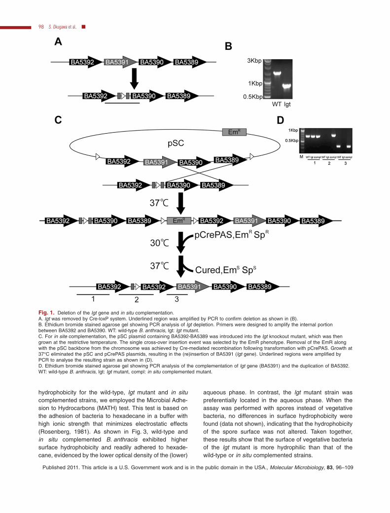
To determine the role of lipoproteins in B. anthracis, weconstructed an lgt-deficient mutant. The lgt gene (BA5391in the Ames strain) was disrupted without polar effectsusing the Cre-loxP system (Pomerantsev et al., 2006) asshown in Fig. 1A. We confirmed by PCR and sequencing
that the lgt gene was deleted (Fig. 1B). We complementedthe lgt gene in 2 ways. The first (‘in situ’) method placedan intact gene back into the chromosome. This wasaccomplished with the single cross-over plasmid pSC,into which the DNA region encompassing BA5389,BA5390, BA5391 and BA5392 was cloned at the multi-cloning site between the loxP sites, allowing the insertionof lgt (and neighbouring genes) into the chromosome(Fig. 1C). Because the region containing BA5389 toBA5392 was reported to be operonic (Passalacqua et al.,2009), we inserted the lgt gene along with the putativeco-operonic genes to avoid any disruption of transcriptionof the adjacent genes. After the single cross-over, Crerecombinase treatment was performed to remove theerythromycin resistance gene along with the pSC back-bone and duplicated genes. Although we anticipated thata single cross-over would spontaneously remove thesecond copy of BA5392, it was still duplicated afterseveral passages as was shown by PCR and sequencing(Fig. 1C and D). To exclude the possibility that the dupli-cated BA5392 affected the phenotype in these in situcomplemented strains, we also complemented the lgtmutation with the plasmid pSW4-lgt in which lgt transcrip-tion is driven by the pagA promoter of B. anthracis. Foreach phenotype examined below, the in situ comple-mented strain displayed properties like those of the origi-nal wild-type strain.
Lgt is required for lipid modification of prelipoproteins
To determine whether lgt performs the expected proteinlipidation in B. anthracis, we carried out metabolic label-ling experiments with [14C]-palmitic acid. Autoradiographyof lipoprotein extracts separated by SDS-PAGE revealedseveral [14C]-labelled protein bands in the samples of thewild-type strain and the lgt mutant complemented witheither the pSW4-lgt plasmid or by in situ gene replace-ment (Fig. 2A and B). In contrast, no labelled proteinswere detected in the lgt mutant (Fig. 2A and B). Thestrains having the mutation complemented in either of thetwo ways showed the same [14C]-labelled proteins as thewild-type strain, suggesting that the duplicated BA5392gene in the in situ complemented mutant did not affectlipoprotein processing. These results demonstrated thatlgt is required for the lipid modification of prolipoproteins inB. anthracis.
Deletion of lgt alters bacterial surface hydrophobicity
Bacterial surfaces are composed of hydrophobic andhydrophilic components, and many of them are proteins(Doyle, 2000). We considered whether the loss of surface-associated lipoproteins in the lgt mutant would alter bac-terial surface hydrophobicity. Thus, to determine surface
Role of lipoprotein biosynthesis in B. anthracis 97
Published 2011. This article is a U.S. Government work and is in the public domain in the USA., Molecular Microbiology, 83, 96–109
hydrophobicity for the wild-type, lgt mutant and in situcomplemented strains, we employed the Microbial Adhe-sion to Hydrocarbons (MATH) test. This test is based onthe adhesion of bacteria to hexadecane in a buffer withhigh ionic strength that minimizes electrostatic effects(Rosenberg, 1981). As shown in Fig. 3, wild-type andin situ complemented B. anthracis exhibited highersurface hydrophobicity and readily adhered to hexade-cane, evidenced by the lower optical density of the (lower)
aqueous phase. In contrast, the lgt mutant strain waspreferentially located in the aqueous phase. When theassay was performed with spores instead of vegetativebacteria, no differences in surface hydrophobicity werefound (data not shown), indicating that the hydrophobicityof the spore surface was not altered. Taken together,these results show that the surface of vegetative bacteriaof the lgt mutant is more hydrophilic than that of thewild-type or in situ complemented strains.
Fig. 1. Deletion of the lgt gene and in situ complementation.A. lgt was removed by Cre-loxP system. Underlined region was amplified by PCR to confirm deletion as shown in (B).B. Ethidium bromide stained agarose gel showing PCR analysis of lgt depletion. Primers were designed to amplify the internal portionbetween BA5392 and BA5390. WT: wild-type B. anthracis, lgt: lgt mutant.C. For in site complementation, the pSC plasmid containing BA5392-BA5389 was introduced into the lgt knockout mutant, which was thengrown at the restrictive temperature. The single cross-over insertion event was selected by the EmR phenotype. Removal of the EmR alongwith the pSC backbone from the chromosome was achieved by Cre-mediated recombination following transformation with pCrePAS. Growth at37°C eliminated the pSC and pCrePAS plasmids, resulting in the (re)insertion of BA5391 (lgt gene). Underlined regions were amplified byPCR to analyse the resulting strain as shown in (D).D. Ethidium bromide stained agarose gel showing PCR analysis of the complementation of lgt gene (BA5391) and the duplication of BA5392.WT: wild-type B. anthracis, lgt: lgt mutant, compl: in situ complemented mutant.
98 S. Okugawa et al. �
Published 2011. This article is a U.S. Government work and is in the public domain in the USA., Molecular Microbiology, 83, 96–109

Deletion of lgt impairs germination
It was previously reported that inactivation of lgt impairsgermination in B. subtilis (Igarashi et al., 2004). To testwhether the same is true for B. anthracis, we assessedgermination by measuring optical density at 600 nm andthe loss of heat resistance. The lgt mutant showed signifi-cantly decreased germination efficiency in both assays(Fig. 4A and B), when compared with the fully germination-proficient in situ complemented mutant and wild-typestrains. These data show that lipoproteins are needed forefficient spore germination in B. anthracis. Once germina-tion occurred, both the lgt mutant and the complementedstrain grew as well as the wild-type strain in BHI mediumand modified G medium (data not shown).
Anthrax toxin secretion is not prevented inthe lgt mutant
The anthrax toxins play a crucial role in virulence towardsanimal hosts (Moayeri and Leppla, 2011). Because lgtmutants of B. subtilis were reported to be impaired inprotein secretion due to decreased levels of the PrsAlipoprotein chaperone/foldase (Kontinen and Sarvas,1993; Leskela et al., 1999), we considered the possibilitythat the lack of lgt in B. anthracis would prevent anthraxtoxin secretion. We found that PA, LF and EF weresecreted from the wild-type, lgt -, and in situ complementedstrains grown in BHI medium in air (data not shown) or inNBY medium supplemented with 0.9% Na2HCO3 in 9%CO2 (Fig. 5). [NBY medium was used because it appears tomimic the induction of toxin secretion by Na2HCO3/CO2
that occurs in a host that is infected with B. anthracis(Bartkus and Leppla, 1989; Chitlaru et al., 2006)]. Quanti-fication of the band intensities showed no significant differ-ences in the amounts of PA, EF and LF in supernatantsderived from the lgt mutant compared with the parentaland complemented strains (Fig. 5). Thus, lgt deficiencyappears not to effect anthrax toxin secretion or stability inB. anthracis in vitro.
B. anthracis lacking lgt is a less potent activator ofthe macrophage inflammatory response
The lack of lgt in several Gram-positive pathogens wasreported to attenuate host immune responses (BubeckWardenburg et al., 2006; Henneke et al., 2008). To deter-mine whether lgt deficiency in B. anthracis affectsimmune responses, we measured TNF-a production inmacrophages exposed to heat-killed bacteria of the wild-
Fig. 2. Lack of lipidation in lgt mutant. Cells were cultivated in BHImedium with shaking for 4 h at 37°C. Lipoproteins were labelledwith [14C]-palmitic acid and detected by autoradiography.A. Wild-type strain with empty vector pSW4, lgt mutant with emptyvector pSW4, and the complemented mutant with pSW4-lgt.B. Wild-type strain, lgt mutant, and the in situ complementedmutant.
Fig. 3. Lipoproteins of B. anthracis influence bacterial surfacehydrophobicity. Bacteria were cultured in BHI medium to an A600
of 4.0 and the washed bacterial suspensions were tested foradherence to hexadecane, which results in reduced optical densityin the lower aqueous phase. Significant differences (P < 0.0001)between the lgt mutant, wild-type strain, and in situ complementedstrain were determined by ANOVA with a Tukey’s multiplecomparison post-test and are indicated by asterisks.
Role of lipoprotein biosynthesis in B. anthracis 99
Published 2011. This article is a U.S. Government work and is in the public domain in the USA., Molecular Microbiology, 83, 96–109

type, lgt mutant and in situ complemented strains. Wecompared the cytokine responses of macrophages fromwild-type C57BL/6J, TLR2 and MyD88-deficient mice(Fig. 6A). The Lgt-deficient bacteria caused significantlyless TNF-a induction in the macrophages from C57BL/6Jstrains than did wild-type or complemented bacteria. Asignificant portion of the TNF-a response induced by wild-type and in situ complemented bacteria was mediated byTLR2. Other signalling pathways also contributed to theTNF-a response, as the lower cytokine response inducedby the lgt mutant was not TLR2 dependent (Fig. 6A). Asexpected, none of the bacterial strains induced a cytokineresponse in macrophages from MyD88-deficient mice(Fig. 6A).
Next, we asked whether lipoproteins of B. anthracisinduce a TNF-a response through TLR2-mediated immuneactivation, as occurs with lipoproteins of other Gram-positive bacteria such as S. aureus and S. pneumoniae.We isolated lipoprotein-enriched fractions from the wild-type strain using Triton X-114 (TX-114) detergent extrac-tion and partition procedures, as described in Experimentalprocedures. Coomassie blue-stained gels showed severalbands in the TX-114 fraction (Fig. 6B). The lipoproteinsamples strongly activated TNF-a production fromC57BL/6J macrophages, but had little effect on macroph-ages from TLR2-deficient mice. To address the concernthat the TX-114 fraction may contain other bacterial com-ponents such as DNA and peptidoglycan that may activatemacrophages, the TX-114 fraction was treated with lipo-protein lipase to inactivate the lipoproteins. Lipoproteinlipase was reported to eliminate the ability of lipoproteins
samples to activate via TLR2 (Shimizu et al., 2005). Thistreatment significantly reduced the TNF-a response of thewild-type macrophages but did not affect the response ofthe TLR2-deficient macrophages (Fig. 6C). We confirmedthat the decrease in cytokine production was not due tomacrophage cell death by measuring viability (data notshown). These results indicated that lipoproteins of B. an-thracis play a role in inducing inflammatory cytokines,acting at least in part through TLR2.
Lipoprotein biogenesis is required for efficientgermination and virulence in vivo
To investigate the contribution of lipoproteins toB. anthracis virulence, we challenged two strains of mice.Complement-deficient A/J mice are highly susceptible tonon-encapsulated toxigenic B. anthracis (Loving et al.,2007), whereas complement-sufficient C57BL/6J micerequire much higher doses of bacteria or spores for infec-tion (Moayeri et al., 2010). Mice were infected with wild-type, lgt- and in situ complemented strain spores orvegetative cells at 1 ¥ LD100 for the particular mousestrain. Both A/J and C57BL/6J mice infected with the lgtmutant spores had significantly decreased mortality com-pared to mice infected with the wild-type strain (Fig. 7Aand C). In contrast, no differences between strains wereseen when vegetative bacteria were used (Fig. 7B and D).Complementation with the lgt gene restored virulence(Fig. 7A and C). These results suggested that the reducedvirulence of the lgt mutant strains results from theirimpaired germination ability.
Fig. 4. Lgt deficiency attenuates germination.A. Germination of wild-type strain, lgt mutant and in situ complemented mutant as monitored by the decrease in A600 of spore suspensionsduring incubation in BHI medium. Results are mean values of three independent experiments. Standard deviations are < 10% of the mean.The A600 curve of lgt mutant spores differed with statistical significance from the curves of the wild-type and in situ complemented strainspores.B. Spores of wild-type strain, lgt mutant and complemented mutant were incubated at 37°C for 30 min. At 30 min, samples were heat-treatedat 65°C for 30 min. Before and after heat treatment, samples were plated on BHI agar. Plates were incubated overnight and colonies werecounted. The fractions of total cfu that were heat-sensitive (i.e. germinated) was calculated and are plotted as the mean values � standarddeviation of three independent experiments. Significant differences using one-way ANOVA with Tukey’s post-test (P < 0.001) are indicated bythree asterisks (***).
100 S. Okugawa et al. �
Published 2011. This article is a U.S. Government work and is in the public domain in the USA., Molecular Microbiology, 83, 96–109

To address in another animal model the question ofwhether the lgt mutant is impaired for germination in vivo,we measured oedema at early times after spore injection,which we previously showed was representative of theoutgrowth of vegetative bacteria (Moayeri et al., 2010).A/J mice were infected with equal numbers of lgt mutant
or in situ complemented spores in the foreleg in a smallvolume (50 ml). ET-mediated local oedema was quantifiedby measuring anterior to posterior and sagittal dorsal/ventral dimensions at various times post infection. Even atthe earliest (3 h) measurement, the lgt mutant inducedsignificantly less oedema than the complemented strain(Fig. 8). This difference persisted through the 6 and 24 hmeasurements (Fig. 8).
For a more direct quantification of the lgt mutant’s ger-mination ability, we assessed bacterial growth and survival[as colony forming units (cfu)] in mouse skin and internalorgans. Skin was homogenized in a germination-prohibitive buffer 6 h after subcutaneous (SC) injection ofspores. Plating of the homogenates before and after heattreatment (to kill vegetative bacteria) allowed enumerationof ungerminated spores and vegetative bacteria. In paral-lel, intravenous (IV) injection of spores provided an alter-native in vivo environment for germination and rapiddelivery to organ sites. Spleens and livers from mice werepooled and processed together to quantify vegetative bac-teria vs. total bacteria (ungerminated spores + vegetativebacteria). Results from these studies are presented inFig. 9A (skin) and Fig. 9B (liver + spleen). While the wild-type and complemented strain bacteria were present asvegetative bacteria and spores in both skin and the internalorgans, the lgt mutant appeared to be present only asspores, reflecting little to no germination. Interestingly, thetotal number of bacteria isolated from the skin of miceinjected with the lgt mutant was consistently lower thanfrom those injected with the wild-type and the comple-mented mutant by 6 h (Fig. 9A). This could be due in part toa more efficient ingestion and killing of the lgt-vegetativebacteria, present in smaller numbers due to slower sporegermination, and their subsequent inability to producesufficient anthrax toxins to inactivate immune cells. Thisexplanation is consistent with our recent demonstrationthat germination and the resultant production of anthraxtoxin is required for effective evasion of myeloid cell-mediated killing of B. anthracis (Liu et al., 2010). Alterna-tively, the lower total number of bacteria may be due toinefficient germination of the lgt mutant spores retrievedfrom the animals. However it is clear that almost all thebacteria retrieved from mice injected with the mutant were
Fig. 5. Anthrax toxin protein secretion from wild-type strain, lgtmutant, and in situ complemented mutant. Wild-type strain (W), lgtmutant (G) and in situ complemented mutant (C) were cultivated inNBY medium in air, or NBY supplemented with 0.9% sodiumbicarbonate and 9% CO2 until A600 reached 2.0. Supernatantswere separated by SDS-PAGE on 4–20% polyacrylamide gels,transferred to nitrocellulose, and blotted with antibody to PA, LF orEF. Intensities of bands were measured with the Odyssey software.Results shown in graph represent the mean � SD of threeindependent experiments, and statistical analyses using one-wayANOVA with a Tukey’s post-test for pairwise comparisons revealedno significant differences.
Role of lipoprotein biosynthesis in B. anthracis 101
Published 2011. This article is a U.S. Government work and is in the public domain in the USA., Molecular Microbiology, 83, 96–109

spores, and analyses of skin sections from mice injectedwith spores also showed a higher number of ungerminatedlgt mutant spores compared with the complemented strainspores (Fig. 9C). Altogether, these data verify that lgt defi-ciency leads to an attenuation of virulence primarilythrough impaired in vivo germination, with a possible con-
sequence of higher efficiency of killing by the innateimmune response.
Discussion
Bacillus anthracis, like other Gram-positive bacteria, ispredicted from genomic sequence analysis to have a largenumber of cell surface-associated lipoproteins (Hutchingset al., 2009), but these have not been studied as a group.In fact, no putative B. anthracis lipoproteins appear to haveexperimentally been localized to the cell or spore mem-branes through a lipid anchor. Several proteins that areexpected to be anchored in the cell wall by lipid modifica-tions are implicated as playing important roles in normalphysiology and/or pathogenesis. Examples include themanganese ABC transporter MntA (BA3189) (Gat et al.,2005), the polyglutamic capsule anchoring protein CapD(pXO2 BA0063) (Richter et al., 2009), the InhA protease(BA0672) (Chung et al., 2009) and the three PrsAchaperone/foldases (BA1041, 1169 and 2336) (Kontinenand Sarvas, 1993) (See also Table S1 and commentsbelow.)
To further understand the role of lipoproteins in B. an-thracis, we began by screening databases using bioinfor-matic tools. We used ScanProsite with the improvedG + Lppv2 motif, followed by LipoP. G + LPPv2 wasreported to have 100% sensitivity to 90 experimentallyverified Gram-positive bacterial lipoproteins whereasLipoP has a higher specificity and ability to discriminateagainst false positives than G + LPPv2 (Rahman et al.,2010). This analysis identified 138 proteins as putativelipoproteins (Table S1), indicating that lipoproteins repre-sent about 2.5% of the proteome. This percentage issimilar to that in the proteomes of other Gram-positivebacteria (Sutcliffe and Harrington, 2002; Babu et al.,2006). The largest group of the candidate lipoproteins inB. anthracis consists of substrate-binding proteins of ABCtransporter uptake systems. The next largest group com-prises proteins involved in sensing germinants and trigger-ing germination. This analysis helped to suggest possiblephenotypic changes that might be observed in bacteriadeficient in lipoprotein biosynthesis.
We disrupted the lgt gene using the previously reportedCre-loxP system (Pomerantsev et al., 2009). To verify thatthe phenotypes described here are due to the lgt muta-tion, it was essential to complement the mutation. Thein situ complementation method used here took advan-tage of the residual loxP site generated during gene dele-tion (Fig. 1A). This method also allowed complementationto be done without having to maintain a plasmid underselective pressure. The strain complemented in this wayretained two copies of the gene adjacent to lgt, BA5392(annotated as a bifunctional histidine-containing proteinkinase/phosphatase), but this had no detectable effect on
Fig. 6. Induction of TNF-a secretion from macrophages byheat-killed B. anthracis and lipoprotein extracts.A. Bone marrow-derived macrophages from the three types of miceindicated were exposed to heat-killed wild-type strain, lgt mutant,or complemented mutant at 100 mg ml-1. Macrophages wereincubated for 18 h and supernatants were analysed for TNF-a byenzyme-linked immunosorbent assay. Data are plotted as the meanvalues � standard deviation of three independent experiments.Significant differences where P < 0.001 are indicated by threeasterisks (***) and where P < 0.0001 are indicated by four asterisks(****).B. Coomassie blue staining of proteins in TX-114 fraction ofB. anthracis (second lane). First lane contains molecular weightmarkers.C. C57BL/6J bone marrow-derived macrophages from the twotypes of mice indicated were incubated with medium, TX-114fraction, or lipoprotein lipase digested TX-114 fraction for 18 hand supernatants were analysed for TNF-a by enzyme-linkedimmunosorbent assay. Data are plotted as the mean values �standard deviation of three independent experiments. Significantdifference (P < 0.05) between lipoprotein lipase treatment and notreatment is indicated by an asterisk (*). All statistical tests used theunpaired two-tailed t-test.
102 S. Okugawa et al. �
Published 2011. This article is a U.S. Government work and is in the public domain in the USA., Molecular Microbiology, 83, 96–109

any of the phenotypes of B. anthracis studied here. Thestrain complemented with a plasmid expressing lgt alsobehaved in every regard like the wild-type strain.
While lgt is essential for the survival of Gram-negativebacteria, it is dispensable in Gram-positive bacteria, asshown for S. aureus and S. pneumoniae (Petit et al., 2001;Stoll et al., 2005). In B. anthracis, the lgt mutant grows aswell as the wild-type strain in either nutrient-rich medium(BHI medium) or poor-nutrient medium (e.g. modified Gmedium) (data not shown). This is somewhat surprisinggiven the large number of ABC transporters that are lipo-proteins. ABC transporters mediate the uptake of manynutrients, such as peptides, amino acids, metals and vita-mins (Davidson et al., 2008), and the uptake of thesematerials could promote growth in nutrient-poor media.However, B. anthracis can apparently satisfy its essentialrequirements through de novo synthesis, or by using otheruptake mechanisms that do not involve lipoproteins.
Although B. subtilis lacking lgt shows impairment ofprotein secretion (Kontinen and Sarvas, 1993), there wereno significant changes in anthrax toxin secretion in theB. anthracis lgt mutant under the conditions used here.A secretion defect might have been expected becausethe three similar putative B. anthracis PrsA secretion
Fig. 7. Survival curves of mice challenged subcutaneously (SC) with spores or vegetative bacteria. A/J mice (A, B) or C57BL/6J (C, D) micewere injected SC with 2 ¥ 103 spores (A, n = 10), 20 vegetative cells (B, n = 8), 2 ¥ 107 spores (C, n = 5) or 1 ¥ 105 vegetative cells (D, n = 5).The survival curves following lgt mutant spore challenge differed with statistical significance (by the log-rank test) from curves of the wild-typeand complemented strains for both A/J challenges (P < 0.0001), as well as C57BL/6J challenges (P < 0.05), while no significant differenceswere found in vegetative challenges.
Fig. 8. Oedema in mouse forelegs following infection with lgtmutant or in situ complemented strain. Groups of three A/J micewere injected with lgt mutant or complemented mutant (5 ¥ 107
spores, 50 ml, SC). Percent increases in foot oedema dorsal/ventralmeasurements relative to pre-infection measurements are shown at3, 6 and 24 h following infection. Each symbol representsmeasurements of an individual mouse. P-values comparing lgtmutant to in situ complemented strain at 6 and 24 h are < 0.02 byunpaired two-tailed t-test.
Role of lipoprotein biosynthesis in B. anthracis 103
Published 2011. This article is a U.S. Government work and is in the public domain in the USA., Molecular Microbiology, 83, 96–109

chaperone/foldases are predicted to be involved in proteinsecretion and all are lipoproteins (Table S1). In fact, thesethree B. anthracis genes supported modest increases inproduction of recombinant PA when expressed in B. sub-tilis (Williams et al., 2003).
The largest phenotypic changes we found in the lgtmutant involved spore germination. Spore germinationplays a crucial role in anthrax disease initiation (Carret al., 2009) and the ability to germinate efficiently may beviewed as a key virulence factor. In our infection model wefound that the lgt mutant has reduced virulence, likely
caused by its impaired germination. Genomic and geneticanalyses have identified five distinct germination operonsin B. anthracis, GerH, GerK, GerL, GerS and GerX (Fisherand Hanna, 2005; Carr et al., 2009). Each operon con-tains three genes, denoted A, B and C (e.g. gerHA,gerHB, gerHC, etc.), of which the C gene in each operonencodes a lipoprotein (Table S1). The three proteinsencoded by each operon associate to form a receptor thatrecognizes the specific small molecule germinants suchas amino acids and purine nucleosides that initiategermination. In addition, two less well-characterizedgenes that affect germination, gerD (Pelczar et al., 2007)and gerM (Rigden and Galperin, 2008), are also predictedto encode lipoproteins (Table S1). The GerD protein isbelieved to colocalize with the germinant receptor pro-teins and accelerate their signalling to downstreameffectors. Mutation of the cysteine in the lipobox of GerDleads to spores lacking the protein and having a severegermination defect like that of a complete gerD deletion(Pelczar et al., 2007; Mongkolthanaruk et al., 2009). Bothtypes of gerD mutations therefore accurately phenocopythe lgt mutation described here. Although we did notdirectly demonstrate that specific germinant proteins andpathways are impaired in the lgt mutant strain, the keyroles that lipoproteins play in the germinant sensing path-ways appear sufficient to account for the impairment ofgermination. It is interesting to note that the first identifi-cation of lgt in B. subtilis came from a search for germi-nation mutants, and the gene was first designated gerF(Igarashi et al., 2004), before its biochemical role in lipo-protein biosynthesis being recognized.
While lipoproteins were found to be essential in aspore-initiated infection, they were dispensable whenvegetative bacteria were inoculated into mice. This reten-tion of virulence was surprising given that up to 150 pro-teins probably depend on a lipid anchor for correctlocalization and function, and that lgt is an essential genein Gram-negative bacteria. A possible decrease in proteinsecretion (affecting proteinaceous virulence factors)
Fig. 9. Spore germination following SC and IV infection.A. A/J mice (n = 3/group) were injected SC in the foreleg with5 ¥ 107 spores in 50 ml, and (B) C57BL/6J mice (n = 2/group) wereinjected IV with 1 ¥ 106 spores in 200 ml. Skin and underlyingtissues were obtained at 6 h from the A/J mice and spleen and liverwere obtained at 90 min from the C57BL/6J mice. Tissues wereprocessed as described in Experimental procedures and total cfudetermined before and after heat treatment. Values plotted are thenumber of spores (solid bars, ‘Heat’) and the sum ofspores + germinated bacteria (open bars, ‘No Heat’). Asterisksindicate significant differences (P < 0.05) by two-way ANOVA test.C. Representative histology images showing staining of vegetativebacteria (left panels) and spores (right panels) in foreleg skinsections harvested 6 h post-infection from A/J mice injected SCwith 1 ¥ 108 spores. Upper panels show lgt mutant and lowerpanels show complemented strain. The upper and lower pairs ofimages were prepared from proximal but independent sections.
104 S. Okugawa et al. �
Published 2011. This article is a U.S. Government work and is in the public domain in the USA., Molecular Microbiology, 83, 96–109

might have been expected to decrease virulence (Leskelaet al., 1999), but appears to be of little significance in theinfection model used here. Furthermore, the apparentability to dispense with a large number of ABC transport-ers during growth in animals suggests that this is a richenvironment where nutrient acquisition is not limiting.
It should be noted, in considering explanations for therestricted impact of the lgt mutation, that the precise effectof Lgt deficiency on the anchoring of specific lipoproteinsmay vary. The prolipoproteins will be secreted onto thesurface of the mutant bacteria, or into the matrix of thepeptidoglycan cell wall, and will initially be anchored thereby their signal peptides. In the absence of the Lgt enzyme,the prolipoproteins may remain cell surface-associatedand retain some function. For certain lipoproteins, thisresidual function may approach that of the normal, fullylipidated protein that would be produced in a wild-typebacterium (Hutchings et al., 2009). This may reconcile ourfinding that the lgt mutant retains virulence (Fig. 7) withthe report that the deletion of the manganese ABC trans-porter and lipoprotein MntA (BA3189) severely impairsvirulence (Gat et al., 2005). Some insights into this ques-tion could come from a comparison of the ‘surfaceomes’(Desvaux et al., 2006) of the lgt- and wild-type strains, asmight be achieved by mass spectrometry of protease-released peptides.
The creation of the lgt mutant strain also allowed us toexamine the role of lipoproteins in host innate immuneresponses. It was reported that heat-killed B. anthracis arerecognized by TLR2 (Hughes et al., 2005), but it was notdetermined which bacterial components induce TLR2 acti-vation. Here we showed that lipoproteins of B. anthracisinduce TNF-a production in macrophages primarilythrough the TLR2 pathway. We considered that TLR2may play a protective role against B. anthracis infectionbecause of its recognition of B. anthracis. However wefound no difference in susceptibility of TLR2-deficient andwild-type mice to subcutaneous B. anthracis infection(data not shown). This finding was similar to previousfindings in an inhalational anthrax mouse model (Hugheset al., 2005). Thus, while TLR2 contributes to cytokineresponses induced by B. anthracis, a protective or detri-mental role for the TLR2-dependent innate immuneresponse is not observed in mice.
In conclusion, lipoproteins synthesized and anchored tomembranes through the action of the Lgt enzyme areessential for full virulence of B. anthracis in spore-inducedinfections, apparently due to the role of lipoproteins insensing germinants present in host tissues. Lipoproteinsin B. anthracis are recognized by TLR2; however, TLR2activation does not play a crucial role in a mouse infectionmodel. These results expand our understanding thepathogenesis of B. anthracis and could aid in developingnew therapeutic approaches.
Experimental procedures
Bioinformatic analyses
We used two tools, ScanProsite with G + LPPv2 pattern andLipoP (Rahman et al., 2008). First, protein sequence data-bases (UniprotKB; Taxonomy identifier 1392 B. anthracis)were screened using ScanProsite and the G + LPPv2pattern ([MV]-X(0,13)-[RK]-{DERK}(6,20)-[LIVMFESTAGPC]-[LVIAMFTG]-[IVMSTAGCP]-[AGS]-C). Second, proteins iden-tified by G + LPPv2 were validated by the Hidden-Markov-Model-based tool LipoP that screens for type II signalpeptidase sequences. LipoP can eliminate potential false-positives. Sequences selected by LipoP were considered asputative lipoproteins. Finally, the genes of the Ames strain thatwe used in this study were selected because both Ames andSterne strain data exist in the protein sequence database. Forthe screening of the pXO1 and pXO2 plasmids, B. anthracisAmes Ancestor strain protein sequence databases were usedbecause B. anthracis Ames strain databases does not includeplasmid sequences.
Bacterial strains, growth condition andspore preparation
The bacterial strains and plasmids used in this study arelisted in Table S2. E. coli was grown at 37°C with 225 r.p.m.shaking in Luria–Burtani broth. B. anthracis was grown at37°C with shaking at 225 r.p.m. in Brain Heart Infusion (BHI)broth (Difco). SOC medium (Quality Biologicals) was used foroutgrowth of transformation mixtures before plating on selec-tive medium to isolate transformants. When required, mediawere supplemented with antibiotics as follows: 100 mg ml-1
ampicillin, 20 mg ml-1 erythromycin, 20 mg ml-1 kanamycinand 150 mg ml-1 spectinomycin.
Spores for germination assays and murine challengeswere prepared by a previously reported method with somemodifications (Hu et al., 2006). Briefly, bacteria were grown at30°C for 1 day and at room temperature for 2 days on Schaef-fer’s sporulation agar, and spores were collected with ice-colddistilled water. Residual vegetative cells were killed by heat at65°C for 30 min. To remove vegetative cell debris, the sporeswere washed multiple times with ice-cold distilled water.Spore purity was assessed under a phase contrast micro-scope and was greater than 99%. Spore quantification wasperformed using a Petroff-Hausser counting chamber. Veg-etative cells for murine challenge were grown at 37°C over-night in BHI broth, diluted 100-fold in fresh broth, andincubated with shaking at 37°C until A600 of 0.4–0.6 (early logphase). Cells were washed and diluted with phosphate buff-ered saline (PBS). Cell quantification was performed using aPetroff-Hausser counting chamber and confirmed by colonyforming count on BHI agar.
DNA isolation and manipulation
Preparation of plasmid DNA from E. coli, transformation ofE. coli, and recombinant DNA techniques were carried out bystandard procedures (Sambrook and Russell, 2001). Recom-binant plasmid construction was carried out in E. coli TOP10(Invitrogen). Chromosomal DNA from B. anthracis was iso-
Role of lipoprotein biosynthesis in B. anthracis 105
Published 2011. This article is a U.S. Government work and is in the public domain in the USA., Molecular Microbiology, 83, 96–109

lated with the Wizard genomic purification kit (Promega) inaccordance with the protocol for isolation of genomic DNAfrom Gram-positive bacteria. B. anthracis was electroporatedwith unmethylated plasmid DNA isolated from E. coli SCS110(dam - dcm -, Stratagene). Electroporation-competent B. an-thracis cells were prepared and transformed as previouslydescribed (Park and Leppla, 2000).
Deletion of the lgt gene
The primers used in this study are listed in Table S2. TheCre-loxP-generated deletion method was previouslydescribed (Pomerantsev et al., 2009). Briefly, to delete the lgtgene, we amplified an upstream fragment of the lgt gene usingthe forward primer LLF containing an EcoRI site and thereverse primer LLR containing a SpeI site. The downstreamfragment was amplified with the forward primer LRF contain-ing an EcoRI site and the reverse primer LRR containing aSpeI site. These fragments were separately cloned into thetemperature-sensitive pSC plasmid between loxP sites. pSCcontaining the upstream fragment was transformed into B. an-thracis Ames 35, which was then grown at the restrictivetemperature. Cre-mediated recombination was achieved bythe transforming the strain with pCrePAS. The same processwas repeated for the downstream fragment. After Cre-loxP-generated deletion, 774 bp of the 813 bp lgt gene wereremoved. Deletion of lgt was confirmed by PCR using forwardprimer BA5391genoF and reverse primer BA5391genoR, andalso by sequence analysis.
Complementation of the lgt gene
For plasmid-based complementation of the lgt mutant, the lgtgene was cloned into plasmid pSW4 behind the pagApromoter. The lgt gene was amplified from B. anthracis Ames35 strain genomic DNA by PCR using the forward primersBA5391F containing an NdeI site and the reverse primerBA5391R containing a BamHI site. Plasmid pSW4-lgt wasidentified by restriction analysis and sequencing and wastransformed into the lgt mutant by electroporation, with selec-tion for kanamycin resistance. For in situ complementation ofthe lgt gene in the genome of the lgt-deficient mutant, a3413 bp fragment including BA5389 to BA5392 was amplifiedby PCR using the forward primer BA5389-5392F containing aPstI site and the reverse primer BA5389-5392R containing aBamHI site. The PCR product was cut with PstI and BamHI(New England Biolabs) and the fragment was ligated intopSC with T4 DNA ligase (Invitrogen). Erythromycin-resistantcolonies obtained by transformation with this pSC plasmidand grown at 37°C were presumed to have undergone asingle cross-over. The erythromycin resistance gene alongwith the pSC backbone and duplicated genes between theloxP sites were removed by Cre-mediated recombinationafter transforming the strain with pCrePAS at 30°C. BothpCrePAS and pSC were eliminated by growth at 37°C.Complementation of genes was confirmed by PCR using theforward primer BA5392-BA5392F and the reverse primerBA5392-BA5392R for the junction between BA5392 andBA5392, the forward primer BA5393-BA5392F and thereverse primer BA5392-BA5392R for the junction betweenBA5393 and BA5392, and the forward primer BA5392-
BA5391F and the reverse primer BA5392-BA5391R for thejunction between BA5392 and BA5391.
Metabolic labelling of lipoproteins with [14C] palmitic acid
[14C] palmitic acid (MP Biomedicals) was dried under a nitro-gen stream and redissolved in 0.2% Tween 80. To an expo-nential phase culture in BHI medium, 4 mCi [14C] palmitic acidml-1 was added. The bacteria were cultivated with shaking for4 h at 37°C. The cells were disrupted by 0.1 mm diameterglass beads (Next Advance, Averill Park, NY, USA). Lysedsuspensions were centrifuged and were separated by sodiumdodecyl sulphate-polyacrylamide gel electrophoresis on a4–20% Tris-glycine gel (Invitrogen) under denaturing condi-tions. Gels were dried and autoradiographed for 2 weeks.
Measurement of cell surface hydrophobicity
Bacteria were grown in BHI broth at 37°C and 225 r.p.m. to latelogarithmic growth phase (A600 nm of 4.0). Bacteria werecentrifuged and washed twice in PUM buffer (17 g l-1 K2HPO4,7.3 g l-1 KH2PO4, 1.8 g l-1 urea, 0.2 g l-1 MgSO4 ¥ 7H2O, pH7.1) and resuspended in PUM to 2 ¥ the original volume.Bacteria (2 ml) were transferred to glass tubes and overlaidwith 1 ml of hexadecane (Sigma-Aldrich), incubated for 10 minat 30°C, and vortexed rigorously for 2 min. The aqueous andhydrocarbon phases were allowed to separate at room tem-perature for 15 min before the aqueous (lower) phase wascarefully removed, and its optical density determined at400 nm.
Toxin production
Bacillus anthracis was cultured in NBY medium with or without0.9% Na2HCO3 in either air or air supplemented with 9% CO2
(Chitlaru et al., 2007; Sastalla et al., 2009). Cultures weregrown at 37°C with shaking at 225 r.p.m. until A600 of 2.0(mid-log phase) and then centrifuged for 10 min at 15 000 gand filtered using 0.22 mm polyvinylidene fluoride (PVDF) filterunits (Millipore). The supernatants were denatured by theaddition of 1/5 volume of 6 ¥ sodium dodecyl sulphate (SDS)sample buffer (10% SDS, 500 mM Tris, 0.01% bromophenolblue, 0.6 M dithiothreitol, 30% glycerol, pH 7.5). The sampleswere boiled for 5 min and equal volumes were resolved usingSDS-polyacrylamide gel electrophoresis (4–20% Tris-glycinegel, Invitrogen) and blotted to nitrocellulose membranes (Invit-rogen). The membranes were probed for the presence of PA,EF and LF using anti-PA serum (#5308 at a dilution of 1:5000),anti-EF serum (#5900 at a dilution of 1:1000), and anti-LF goatpolyclonal antibodies (List Biological Laboratories, Campbell,CA, at 0.5 mg ml-1) respectively. Primary antibodies weredetected using IR-dye-conjugated secondary antibodies(Licor Biosciences, 1:5000) and signals were imaged andquantified with the Odyssey Infrared Imaging System (LicorBiosciences, Lincoln, NE, USA).
In vitro germination assay
Spores were activated by incubation at 70°C for 20 min indistilled water. Subsequently, spores were washed with dis-
106 S. Okugawa et al. �
Published 2011. This article is a U.S. Government work and is in the public domain in the USA., Molecular Microbiology, 83, 96–109

tilled water and resuspended in BHI medium to an A600 ofª 0.3 (ª 2.0 ¥ 107 spores ml-1) and the germination processwas monitored by A600. The A600 of the samples were mea-sured every 5 min for 120 min. Spores were routinelyobserved for germination by phase contrast microscopy. Toevaluate the loss of heat resistance, spores incubated in BHImedium for 30 min were heated at 65°C for 30 min to killgerminated cells. Samples taken before and after heat treat-ment were plated on BHI agar. One hundred percent sensi-tive to a heat treatment was considered 100% germinated.
Preparation of heat-killed bacteria for TNF-a assays
To generate heat-killed bacteria, vegetative cells were grownat 37°C overnight in BHI broth, diluted 100-fold in fresh broth,and incubated with shaking at 37°C until A600 was 0.65 (early-log phase). The bacterial suspension was washed threetimes with PBS, resuspended in PBS, and A600 for differentstrains normalized to each other before heat inactivation(70°C for 70 min). After a final spin and removal of PBS, thebacterial cell pellet was lyophilized. Lyophilized heat-killedbacteria were weighed and resuspended in PBS before addi-tion to bone marrow-derived macrophages. To confirm thatbacteria were killed, heat-killed bacteria were plated on BHIagar and incubated overnight at 37°C.
Extraction of lipoproteins
To extract a fraction containing lipoproteins, exponentialphase B. anthracis cells at A600 of 1.0 were subjected to TritonX-114 (TX-114) phase partitioning (Thakran et al., 2008).Briefly, the cell pellets were resuspended in PBS supple-mented with 350 mM NaCl, 2% v/v TX-114 (Sigma-Aldrich),and protease inhibitor cocktail (Complete mini, Roche Diag-nostics) and incubated at 4°C for 1 h. Samples were centri-fuged at 10 000 g at 4°C for 30 min and the supernatant wasincubated at 37°C for 10 min to induce detergent phaseseparation. The upper aqueous phase was discarded andreplaced with a similar volume of PBS supplemented with350 mM NaCl. The procedure of phase separation wasrepeated twice and the final detergent phase was retained. Toremove the detergent, the crude lipoprotein fraction was pre-cipitated at -20°C overnight by addition of 2.5 volumes ofmethanol. After centrifugation at 10 000 g and 4°C for 30 min,the pellet was washed with methanol, air-dried, and resus-pended in PBS. Protein concentration of the suspension wasmeasured with BCA protein assay (Thermo Scientific Pierce).To digest lipoproteins, 10 mg ml-1 lipoprotein lipase fromPseudomonas sp. (Sigma-Aldrich) was added to crude lipo-proteins and then incubated for 18 h at 37°C. Lipoproteinlipase was inactivated by heating at 72°C for 20 min.
TNF-a assays
Bone marrow-derived macrophages were isolated fromC57BL/6J mice (Jackson Laboratories, Bar Harbor, ME,USA), TLR2-deficient mice (knockouts on C57BL/6J back-ground, kindly provided by Alan Sher, National Institutes ofHealth, Bethesda, MD, USA) or MyD88-deficient mice(gift from Shizuko Akira, Osaka University, Osaka, Japan;received via Tod J. Merkel, Food and Drug Administration,
Bethesda, MD, USA). Cells were cultured in differentiationmedium consisting of 30% L929 cell-conditioned medium inDulbecco-modified Eagle medium (DMEM) supplementedwith 10% fetal bovine serum, 10 mM HEPES, 50 mg ml-1 gen-tamicin (all obtained from Invitrogen) as previously described(Newman et al., 2009). On day 7, cells were plated in 96-wellplates (1 ¥ 105 cells well-1). On day 8 cells were treated with100 mg ml-1 heat-killed bacteria for 20 min and expressionlevels of TNF-a were measured with a TNF-a ELISA kit (R&DSystems, Minneapolis, MN, USA).
Mouse infection studies
A/J or C57BL/6J mice (8–12 weeks, female) were purchasedfrom Jackson Laboratories. In survival studies, mice wereinjected subcutaneously (SC, 200 ml) with spores or vegeta-tive cells (see figure legends for doses), and observed for up to10 days. For indirect assessment of germination by measure-ment of ET activity, 5 ¥ 107 spores in 50 ml PBS were injectedSC into the foreleg of A/J mice and oedema quantified aspreviously described (Moayeri et al., 2010). Direct measure-ments of in vivo germination were made by completely remov-ing skin and underlying foreleg tissue 6 h after injection of A/Jmice (5 ¥ 107 spores, SC). Tissues were immediately frozen inliquid nitrogen and homogenized in 7.5 ml of a germinationinhibitory buffer (PBS + 150 mM D-alanine, 200 mM 6-thioguanosine, both from Sigma, St Louis, MO, USA). Dilutionplating of homogenates to assess differential vegetative andspore-based cfu was performed both before and after heattreatment (70°C, 70 min). In separate experiments, C57BL/6Jmice were injected with spores (1 ¥ 106) by the IV route andspleen and liver from individual mice were harvested, pooledand processed for vegetative and spore-based cfu quantifica-tion as described above. For histology, A/J mouse forelegswere injected (1 ¥ 108 spores, 50 ml, SC) and skin and under-lying tissue sections were harvested 90 min and 6 h afterinfection, fixed overnight in 10% formalin followed by repeated70% ethanol washes. Section preparation, haematoxylin andeosin (H&E) staining, Brown and Hopps tissue Gram stain (forvegetative bacteria), and a modified carboxyl fuchsin stain/malachite green counterstain (for spores) were performed onadjacent serial sections by Histoserv (Gaithersburg, MD,USA). For the spore stain, the Accustain Acid Fast Kit (Sigma)was used according to manufacturer’s protocol, with a 1 hstaining period in heated (steaming) carbol fuchsin solution(0.85% pararosaniline dye, 2.5% phenol, 5% glycerol, 5%DMSO, in deionized water) before counterstaining with mala-chite green (1.5% malachite green oxalate, 10% acetic acid,17% glycerol in deionized water) for 2 min at room tempera-ture. All protocols using mice were approved by the NationalInstitute of Allergy and Infectious Diseases Animal Care andUse Committee.
Statistical analysis
All analyses were performed using Graph Pad Prism 5(Graph Pad System, San Diego, CA, USA). Statistical testsused for each experiment are listed in the figure legends.
Acknowledgements
We thank Alan Sher for kindly providing TLR2-deficient miceand Drs. Akira and Merkel for MyD88 knockout mice. We also
Role of lipoprotein biosynthesis in B. anthracis 107
Published 2011. This article is a U.S. Government work and is in the public domain in the USA., Molecular Microbiology, 83, 96–109

thank Mini Varughese for editing the manuscript. This researchwas supported by the Intramural Research Program of theNIH, National Institute of Allergy and Infectious Diseases.
References
Akira, S., Uematsu, S., and Takeuchi, O. (2006) Pathogenrecognition and innate immunity. Cell 124: 783–801.
Babu, M.M., Priya, M.L., Selvan, A.T., Madera, M., Gough, J.,Aravind, L., and Sankaran, K. (2006) A database of bacte-rial lipoproteins (DOLOP) with functional assignments topredicted lipoproteins. J Bacteriol 188: 2761–2773.
Bartkus, J.M., and Leppla, S.H. (1989) Transcriptional regu-lation of the protective antigen gene of Bacillus anthracis.Infect Immun 57: 2295–2300.
Baumgartner, M., Karst, U., Gerstel, B., Loessner, M.,Wehland, J., and Jansch, L. (2007) Inactivation of Lgtallows systematic characterization of lipoproteins from List-eria monocytogenes. J Bacteriol 189: 313–324.
Bubeck Wardenburg, J., Williams, W.A., and Missiakas, D.(2006) Host defenses against Staphylococcus aureusinfection require recognition of bacterial lipoproteins. ProcNatl Acad Sci USA 103: 13831–13836.
Carr, K.A., Lybarger, S.R., Anderson, E.C., Janes, B.K., andHanna, P.C. (2009) The role of Bacillus anthracis germi-nant receptors in germination and virulence. Mol Microbiol75: 365–375.
Chitlaru, T., Gat, O., Gozlan, Y., Ariel, N., and Shafferman, A.(2006) Differential proteomic analysis of the Bacillusanthracis secretome: distinct plasmid and chromosomeCO2-dependent cross talk mechanisms modulate extracel-lular proteolytic activities. J Bacteriol 188: 3551–3571.
Chitlaru, T., Gat, O., Grosfeld, H., Inbar, I., Gozlan, Y., andShafferman, A. (2007) Identification of in-vivo expressedIimunogenic proteins by serological proteome analysis ofBacillus anthracis secretome. Infect Immun 75: 2841–2852.
Chung, M.C., Jorgensen, S.C., Popova, T.G., Tonry, J.H.,Bailey, C.L., and Popov, S.G. (2009) Activation of plasmi-nogen activator inhibitor implicates protease InhA in theacute-phase response to Bacillus anthracis infection.J Med Microbiol 58: 737–744.
Dartois, V., Djavakhishvili, T., and Hoch, J.A. (1997) KapB isa lipoprotein required for KinB signal transduction and acti-vation of the phosphorelay to sporulation in Bacillussubtilis. Mol Microbiol 26: 1097–1108.
Davidson, A.L., Dassa, E., Orelle, C., and Chen, J. (2008)Structure, function, and evolution of bacterial ATP-bindingcassette systems. Microbiol Mol Biol Rev 72: 317–364.
Deka, R.K., Brautigam, C.A., Yang, X.F., Blevins, J.S.,Machius, M., Tomchick, D.R., and Norgard, M.V. (2006)The PnrA (Tp0319; TmpC) lipoprotein represents a newfamily of bacterial purine nucleoside receptor encodedwithin an ATP-binding cassette (ABC)-like operon in Tre-ponema pallidum. J Biol Chem 281: 8072–8081.
Desvaux, M., Dumas, E., Chafsey, I., and Hebraud, M. (2006)Protein cell surface display in Gram-positive bacteria: fromsingle protein to macromolecular protein structure. FEMSMicrobiol Lett 256: 1–15.
Doyle, R.J. (2000) Contribution of the hydrophobic effect tomicrobial infection. Microbes Infect 2: 391–400.
Fisher, N., and Hanna, P. (2005) Characterization of Bacillus
anthracis germinant receptors in vitro. J Bacteriol 187:8055–8062.
Gat, O., Mendelson, I., Chitlaru, T., Ariel, N., Altboum, Z.,Levy, H., et al. (2005) The solute-binding component of aputative Mn(II) ABC transporter (MntA) is a novel Bacillusanthracis virulence determinant. Mol Microbiol 58: 533–551.
Hayashi, S., Chang, S.Y., Chang, S., Giam, C.Z., and Wu,H.C. (1985) Modification and processing of internalizedsignal sequences of prolipoprotein in Escherichia coli andin Bacillus subtilis. J Biol Chem 260: 5753–5759.
Henneke, P., Dramsi, S., Mancuso, G., Chraibi, K., Pellegrini,E., Theilacker, C., et al. (2008) Lipoproteins are criticalTLR2 activating toxins in group B streptococcal sepsis.J Immunol 180: 6149–6158.
Hu, H., Sa, Q., Koehler, T.M., Aronson, A.I., and Zhou, D.(2006) Inactivation of Bacillus anthracis spores in murineprimary macrophages. Cell Microbiol 8: 1634–1642.
Hughes, M.A., Green, C.S., Lowchyj, L., Lee, G.M., Grippe,V.K., Smith, M.F., Jr, et al. (2005) MyD88-dependent sig-naling contributes to protection following Bacillus anthracisspore challenge of mice: implications for Toll-Like receptorsignaling. Infect Immun 73: 7535–7540.
Hutchings, M.I., Palmer, T., Harrington, D.J., and Sutcliffe,I.C. (2009) Lipoprotein biogenesis in Gram-positive bacte-ria: knowing when to hold ‘em, knowing when to fold ‘em.Trends Microbiol 17: 13–21.
Igarashi, T., Setlow, B., Paidhungat, M., and Setlow, P. (2004)Effects of a gerF (lgt) mutation on the germination ofspores of Bacillus subtilis. J Bacteriol 186: 2984–2991.
Khandavilli, S., Homer, K.A., Yuste, J., Basavanna, S., Mitch-ell, T., and Brown, J.S. (2008) Maturation of Streptococcuspneumoniae lipoproteins by a type II signal peptidase isrequired for ABC transporter function and full virulence. MolMicrobiol 67: 541–557.
Kontinen, V.P., and Sarvas, M. (1993) The PrsA lipoprotein isessential for protein secretion in Bacillus subtilis and sets alimit for high-level secretion. Mol Microbiol 8: 727–737.
Kovacs-Simon, A., Titball, R.W., and Michell, S.L. (2011)Lipoproteins of bacterial pathogens. Infect Immun 79: 548–561.
Leppla, S.H. (2006) Bacillus anthracis toxins. In The Com-prehensive Sourcebook of Bacterial Protein Toxins. Alouf,J.E., and Popoff, M.R. (eds). Burlington, MA: AcademicPress, pp. 323–347.
Leskela, S., Wahlstrom, E., Kontinen, V.P., and Sarvas, M.(1999) Lipid modification of prelipoproteins is dispensablefor growth but essential for efficient protein secretion inBacillus subtilis: characterization of the Lgt gene. MolMicrobiol 31: 1075–1085.
Liu, S., Miller-Randolph, S., Crown, D., Moayeri, M., Sastalla,I., Okugawa, S., and Leppla, S.H. (2010) Anthrax toxintargeting of myeloid cells through the CMG2 receptor Isessential for establishment of Bacillus anthracis infectionsin mice. Cell Host Microbe 8: 455–462.
Loving, C.L., Kennett, M., Lee, G.M., Grippe, V.K., andMerkel, T.J. (2007) A murine aerosol challenge model ofanthrax. Infect Immun 75: 2689–2698.
Moayeri, M., and Leppla, S.H. (2009) Cellular and systemiceffects of anthrax lethal toxin and edema toxin. MolAspects Med 30: 439–455.
108 S. Okugawa et al. �
Published 2011. This article is a U.S. Government work and is in the public domain in the USA., Molecular Microbiology, 83, 96–109

Moayeri, M., and Leppla, S.H. (2011) Anthrax toxins. In Bacil-lus Anthracis and Anthrax. Bergman, N.H. (ed.). Hoboken,NJ: John Wiley & Sons, pp. 121–156.
Moayeri, M., Crown, D., Newman, Z.L., Okugawa, S.,Eckhaus, M., Cataisson, C., et al. (2010) Inflammasomesensor Nlrp1b-dependent resistance to anthrax Is mediatedby caspase-1, IL-1 signaling and neutrophil recruitment.PLoS Pathog 6: e1001222.
Mongkolthanaruk, W., Robinson, C., and Moir, A. (2009)Localization of the GerD spore germination protein inthe Bacillus subtilis spore. Microbiology 155: 1146–1151.
Navarre, W.W., Daefler, S., and Schneewind, O. (1996) Cellwall sorting of lipoproteins in Staphylococcus aureus.J Bacteriol 178: 441–446.
Newman, Z.L., Leppla, S.H., and Moayeri, M. (2009)CA-074Me protection against anthrax lethal toxin. InfectImmun 77: 4327–4336.
Okugawa, S., Moayeri, M., Eckhaus, M.A., Crown, D., Miller-Randolph, S., Liu, S., et al. (2011) MyD88-dependent sig-naling protects against anthrax lethal toxin-inducedimpairment of intestinal barrier function. Infect Immun 79:118–124.
Park, S., and Leppla, S.H. (2000) Optimized production andpurification of Bacillus anthracis lethal factor. Protein ExprPurif 18: 293–302.
Passalacqua, K.D., Varadarajan, A., Ondov, B.D., Okou, D.T.,Zwick, M.E., and Bergman, N.H. (2009) The structure andcomplexity of a bacterial transcriptome. J Bacteriol 191:3203–3211.
Pelczar, P.L., Igarashi, T., Setlow, B., and Setlow, P. (2007)Role of GerD in germination of Bacillus subtilis spores.J Bacteriol 189: 1090–1098.
Petit, C.M., Brown, J.R., Ingraham, K., Bryant, A.P., andHolmes, D.J. (2001) Lipid modification of prelipoproteins isdispensable for growth in vitro but essential for virulence inStreptococcus pneumoniae. FEMS Microbiol Lett 200:229–233.
Pomerantsev, A.P., Sitaraman, R., Galloway, C.R., Kivovich,V., and Leppla, S.H. (2006) Genome engineering in Bacil-lus anthracis using Cre recombinase. Infect Immun 74:682–693.
Pomerantsev, A.P., Camp, A., and Leppla, S.H. (2009) A newminimal replicon of Bacillus anthracis plasmid pXO1.J Bacteriol 191: 5134–5146.
Rahman, O., Cummings, S.P., Harrington, D.J., and Sutcliffe,I.C. (2008) Methods for the bioinformatic identification ofbacterial lipoproteins encoded in the genomes of Gram-positive bacteria. World J Microbiol Biotechnol 24: 2377–2382.
Rahman, O., Cummings, S.P., Harrington, D.J., and Sutcliffe,I.C. (2010) Methods for the bioinformatic identification ofbacterial lipoproteins encoded in the genomes of Gram-positive bacteria. World J Microbiol Biotechnol 24: 2377–2382.
Richter, S., Anderson, V.J., Garufi, G., Lu, L., Budzik, J.M.,Joachimiak, A., et al. (2009) Capsule anchoring in Bacillusanthracis occurs by a transpeptidation reaction that isinhibited by capsidin. Mol Microbiol 71: 404–420.
Rigden, D.J., and Galperin, M.Y. (2008) Sequence analysis ofGerM and SpoVS, uncharacterized bacterial ‘sporulation’
proteins with widespread phylogenetic distribution. Bio-informatics 24: 1793–1797.
Robichon, C., Vidal-Ingigliardi, D., and Pugsley, A.P. (2005)Depletion of apolipoprotein N-acyltransferase causes mis-localization of outer membrane lipoproteins in Escherichiacoli. J Biol Chem 280: 974–983.
Rosenberg, M. (1981) Bacterial adherence to polystyrene: areplica method of screening for bacterial hydrophobicity.Appl Environ Microbiol 42: 375–377.
Sambrook, J., and Russell, D.W. (2001) Molecular Cloning. ALaboratory Manual. Cold Spring Harbor, New York: ColdSpring Harbor Laboratory Press.
Sastalla, I., Chim, K., Cheung, G.Y., Pomerantsev, A.P., andLeppla, S.H. (2009) Codon-optimized fluorescent proteinsdesigned for expression in low GC Gram-positive bacteria.Appl Environ Microbiol 75: 2099–2110.
Setlow, P. (2003) Spore germination. Curr Opin Microbiol 6:550–556.
Shimizu, T., Kida, Y., and Kuwano, K. (2005) A dipalmitoy-lated lipoprotein from Mycoplasma pneumoniae activatesNF-kappa B through TLR1, TLR2, and TLR6. J Immunol175: 4641–4646.
Stoll, H., Dengjel, J., Nerz, C., and Gotz, F. (2005) Staphylo-coccus aureus deficient in lipidation of prelipoproteins isattenuated in growth and immune activation. Infect Immun73: 2411–2423.
Sutcliffe, I.C., and Harrington, D.J. (2002) Pattern searchesfor the identification of putative lipoprotein genes in Gram-positive bacterial genomes. Microbiology 148: 2065–2077.
Takeuchi, O., Kawai, T., Muhlradt, P.F., Morr, M., Radolf, J.D.,Zychlinsky, A., et al. (2001) Discrimination of bacterial lipo-proteins by Toll-like receptor 6. Int Immunol 13: 933–940.
Takeuchi, O., Sato, S., Horiuchi, T., Hoshino, K., Takeda, K.,Dong, Z., et al. (2002) Cutting edge: role of Toll-like recep-tor 1 in mediating immune response to microbiallipoproteins. J Immunol 169: 10–14.
Thakran, S., Li, H., Lavine, C.L., Miller, M.A., Bina, J.E., Bina,X.R., and Re, F. (2008) Identification of Francisella tularen-sis lipoproteins that stimulate the toll-like receptor (TLR)2/TLR1 heterodimer. J Biol Chem 283: 3751–3760.
Tokunaga, M., Tokunaga, H., and Wu, H.C. (1982) Post-translational modification and processing of Escherichiacoli prolipoprotein in vitro. Proc Natl Acad Sci USA 79:2255–2259.
Williams, R.C., Rees, M.L., Jacobs, M.F., Pragai, Z., Thwaite,J.E., Baillie, L.W., et al. (2003) Production of Bacillusanthracis protective antigen is dependent on the extracel-lular chaperone, PrsA. J Biol Chem 278: 18056–18062.
Young, J.A., and Collier, R.J. (2007) Anthrax toxin: receptor-binding, internalization, pore formation, and translocation.Annu Rev Biochem 76: 243–265.
Supporting information
Additional supporting information may be found in the onlineversion of this article.
Please note: Wiley-Blackwell are not responsible for thecontent or functionality of any supporting materials suppliedby the authors. Any queries (other than missing material)should be directed to the corresponding author for the article.
Role of lipoprotein biosynthesis in B. anthracis 109
Published 2011. This article is a U.S. Government work and is in the public domain in the USA., Molecular Microbiology, 83, 96–109