lipidomics analysis reveals efficient storage of hepatic ... · 2010 lipidomics analysis reveals...
TRANSCRIPT

Washington University School of MedicineDigital Commons@Becker
Open Access Publications
2010
Lipidomics analysis reveals efficient storage ofhepatic triacylglycerides enriched in unsaturatedfatty acids after one bout of exercise in miceChunxiu HuDalian Institute of Chemical Physics, Chinese Academy of Sciences, Dalian, China
Miriam HoeneUniversity Hospital of Tuebingen, Tuebingen, Germany
Xinjie ZhaoDalian Institute of Chemical Physics, Chinese Academy of Sciences, Dalian, China
Hans U. HaringUniversity Hospital of Tuebingen, Tuebingen, Germany
Erwin SchleicherUniversity Hospital of Tuebingen, Tuebingen, Germany
See next page for additional authors
Follow this and additional works at: http://digitalcommons.wustl.edu/open_access_pubs
Part of the Medicine and Health Sciences Commons
This Open Access Publication is brought to you for free and open access by Digital Commons@Becker. It has been accepted for inclusion in OpenAccess Publications by an authorized administrator of Digital Commons@Becker. For more information, please contact [email protected].
Recommended CitationHu, Chunxiu; Hoene, Miriam; Zhao, Xinjie; Haring, Hans U.; Schleicher, Erwin; Lehmann, Rainer; Han, Xianlin; Xu, Guowang; andWeigert, Cora, ,"Lipidomics analysis reveals efficient storage of hepatic triacylglycerides enriched in unsaturated fatty acids after onebout of exercise in mice." PLoS One.5,10. e13318. (2010).http://digitalcommons.wustl.edu/open_access_pubs/733

AuthorsChunxiu Hu, Miriam Hoene, Xinjie Zhao, Hans U. Haring, Erwin Schleicher, Rainer Lehmann, Xianlin Han,Guowang Xu, and Cora Weigert
This open access publication is available at Digital Commons@Becker: http://digitalcommons.wustl.edu/open_access_pubs/733

Lipidomics Analysis Reveals Efficient Storage of HepaticTriacylglycerides Enriched in Unsaturated Fatty Acidsafter One Bout of Exercise in MiceChunxiu Hu2., Miriam Hoene1,4., Xinjie Zhao2, Hans U. Haring1,4, Erwin Schleicher1,4, Rainer
Lehmann1,4, Xianlin Han3, Guowang Xu2*, Cora Weigert1,4*
1 Division of Endocrinology, Diabetology, Angiology, Nephrology, Pathobiochemistry and Clinical Chemistry, Department of Internal Medicine, University Hospital of
Tuebingen, Tuebingen, Germany, 2 CAS Key Laboratory of Separation Science for Analytical Chemistry, Dalian Institute of Chemical Physics, Chinese Academy of Sciences,
Dalian, China, 3 Division of Bioorganic Chemistry and Molecular Pharmacology, Department of Medicine, Washington University School of Medicine, St. Louis, Missouri,
United States of America, 4 Paul Langerhans Institute Tuebingen, Member of the German Center for Diabetes Research (DZD), Tuebingen, Germany
Abstract
Background: Endurance exercise induces lipolysis, increases circulating concentrations of free fatty acids (FFA) and theuptake and oxidation of fatty acids in the working muscle. Less is known about the regulation of lipid metabolism in theliver during and post-exercise.
Methodology/Principal Findings: We performed an ultra fast liquid chromatography-mass spectrometry (UFLC-MS) basedlipidomics analysis of liver tissue samples obtained from C57Bl/6J mice immediately after a 60 min treadmill run of moderateintensity, and after 3 h of recovery. The PLS-DA scores plot for 115 quantified lipid molecular species revealed a clearseparation of the hepatic lipid profile of sedentary from recovering mice, but not from mice immediately after running. 21 lipidspecies were considered to be most responsible for the difference in the hepatic lipid profiles, including 17 triacylglycerides(TG), one lysophosphatidylcholine (LPC) and three phosphatidylcholines (PC). TG species were found to be more abundant inthe recovery phase, while PC species were decreased. The degree of accumulation of individual TG species correlated well withthe amount of theoretical energy stored whereas no increase was found for TG species containing only saturated or onemonounsaturated fatty acid. Total liver TG content as assayed by an enzymatic method was increased to 163% in the recoveryphase, while it was significantly decreased in skeletal muscle by the exercise bout and remained less in the recovery phase.Results from fasted and refed mice indicate that fasting-induced lipolysis was associated with a pronounced accumulation ofhepatic TG, which is reversed by refeeding for 5 h. Thus food intake per se did not elevate hepatic TG.
Conclusion: These data indicate that high availability of FFA induced by endurance exercise or fasting resulted in a transienthepatic TG accumulation, while muscle TG content was decreased during exercise presumably due to increased muscle fattyacid oxidation.
Citation: Hu C, Hoene M, Zhao X, Haring HU, Schleicher E, et al. (2010) Lipidomics Analysis Reveals Efficient Storage of Hepatic Triacylglycerides Enriched inUnsaturated Fatty Acids after One Bout of Exercise in Mice. PLoS ONE 5(10): e13318. doi:10.1371/journal.pone.0013318
Editor: Lorraine Brennan, University College Dublin, Ireland
Received July 14, 2010; Accepted September 16, 2010; Published October 13, 2010
Copyright: � 2010 Hu et al. This is an open-access article distributed under the terms of the Creative Commons Attribution License, which permits unrestricteduse, distribution, and reproduction in any medium, provided the original author and source are credited.
Funding: This study was supported by grants from the Deutsche Forschungsgemeinschaft to C.W. (WE 4176) and to E.S. (GRK 1302/1), by the KompetenznetzDiabetes mellitus (Competence Network for Diabetes mellitus) funded by the Federal Ministry of Education and Research to R.L. and H.U.H. (FKZ 01GI0803-04), bya grant from the German Federal Ministry of Education and Research (BMBF) to the German Center for Diabetes Research (DZD e.V.), the Sino-German Center forResearch Promotion to R.L. and G.X. (DFG and NSFC, GZ 364), the 973 Project of the State Ministry of Science and Technology of China to G.X.(No. 2006CB503902), the National Natural Science Foundation of China to G.X. (No. 20835006), and the Knowledge Innovation Program of the Chinese Academyof Sciences to G.X. (KSCX1-YW-02). The funders had no role in study design, data collection and analysis, decision to publish, or preparation of the manuscript.
Competing Interests: The authors have declared that no competing interests exist.
* E-mail: [email protected] (GW); [email protected] (CW)
. These authors contributed equally to this work.
Introduction
Fatty acids are the major fuel during prolonged moderate intensity
exercise. The availability of plasma free fatty acids (FFA) is markedly
increased by the action of hormone-sensitive lipase and adipose
tissue triacylglyceride (TG) lipase on the fat depots in adipose tissue
and skeletal muscle [1,2]. During exercise the relative proportion of
FFA uptake in the working muscle is substantially increased [3,4]
and FFA are oxidized to provide energy or reesterified and
incorporated into intramyocellular TG [5]. The liver has a central
function in lipid metabolism by repartitioning FFA derived from
body fat stores and integrating dietary FFA into whole body fuel
oxidation and energy storage via secretion of lipoproteins. However
its role in the process of FFA supply and tissue redistribution during
and after exercise is not well characterized.
Studies both in rodents and humans provide evidence that the
hepatic tissue concentration of TG is influenced by regular
exercise. Training of rodents shows that enhanced physical activity
prevents high-fat diet-induced hepatic steatosis when performed
concurrently with the diet [6,7] or thereafter [7]. Voluntary wheel
PLoS ONE | www.plosone.org 1 October 2010 | Volume 5 | Issue 10 | e13318

running reduces the hepatic TG content in Otsuka Long-Evans
Tokushima Fatty rats, a rodent model for obesity and type 2
diabetes [8]. These lipid-lowering effects of training could be
absent in young, non-obese rodents [9]. Application of the
noninvasive proton magnetic resonance spectroscopy (1H-MRS)
to quantify liver fat in humans reveals that regular exercise
training leads to the reduction of hepatic lipid content even in the
absence of weight loss [10–12]. Of note, liver fat shows the highest
percentage of reduction in response to exercise interventions
compared with visceral and subcutaneous lipid stores [13,14].
Further training-related effects on hepatic lipid metabolism are
the regulation of lipoprotein secretion from the liver leading to a
reduction of the postprandial concentrations of plasma TG found
both after one single bout of acute exercise [15–18] and after
regular exercise training [6,19–22]. The contribution of distinct
mechanisms leading to this hypotriglyceridemic effect of exercise
are not completely understood, but it is predominantly a decrease
in the very low density lipoprotein (VLDL)-TG fraction
[18,23,24]. This decrease has been attributed to an increased
plasma clearance rate of VLDL-TG [25–28] and a reduced
VLDL-TG secretion, which has been described in humans [29],
and studies in rats have provided further evidence for that [30,31].
Thus, beneficial effects of regular physical activity on total liver
fat and plasma lipids in humans or rodent obesity models are well
documented, but the response of hepatic lipids to acute exercise
are less clear. We hypothesized that the investigation of the hepatic
lipid profile after one single exercise bout could not only give
insights in the exercise-induced adaptive mechanisms of hepatic
TG storage but also reveal further impact of exercise on other lipid
species found in the liver.
We applied here a recently established reversed-phase ultra fast
liquid chromatography2mass spectrometric (UFLC2MS) method
[32] for the lipidomics analyses of hepatic lipids of mice after one
single bout of treadmill exercise. We combined the hepatic lipid
profiling with conventional determination of plasma and tissue TG
content. The results indicate decreased choline phospholipid and
increased TG levels in the liver after acute exercise, with a more
efficient storage of TG molecular species containing unsaturated
acyl chains.
Results
Plasma parametersThe running protocol applied to the mice was designed to
provide a strong, but non-exhaustive metabolic stimulus, resem-
bling intensive endurance exercise. Immediately after the exercise
bout, plasma FFA levels were significantly increased, and plasma
glucose levels as well as plasma insulin concentrations were
significantly decreased (Fig. 1A–C). After 3 h of recovery with free
access to food in the first 2 h, these metabolic parameters were
similar to those in sedentary mice. Plasma TG levels were not
different in either condition (Fig. 1D).
Hepatic lipidomics reveals differences betweenrecovering mice and sedentary mice
A total of 115 lipid molecular species were identified and
quantified by the current UFLC2MS-based lipidomics platform
with phosphatidylcholines (PC) and TG as most abundant classes
(summarized in Table 1; the complete data are available as
Supporting Information in Table S1).
Figure 1. Plasma parameters. Plasma concentrations are shown for glucose (A), insulin (B), FFA (C), and TG (D) of sedentary (sed), immediatelyafter exercise (run), and recovering (3 h post) mice (n = 12, mean 6 SEM; * p,0.05; ** p,0.01 vs. sed).doi:10.1371/journal.pone.0013318.g001
Hepatic Lipids after Exercise
PLoS ONE | www.plosone.org 2 October 2010 | Volume 5 | Issue 10 | e13318

Based on the hepatic lipid metabolite pattern the PLS-DA
scores plot revealed no separation of sedentary mice and mice
immediately after the run (i.e. the number of components in the
autofit model was zero), but a clear separation between recovering
mice and sedentary mice was achieved (Fig. 2A). To ensure that
the calculated model of the recovering mice vs. sedentary mice is
reliable and the observed clustering has not been obtained by
chance, we performed an internal validation using 7-fold cross-
validation [33]. The calculated goodness of fit (R2Y) was 0.991
and the goodness of prediction (Q2Y) was 0.886 which underlines
the robustness of the model. In addition, a response permutation
test was carried out. The R2Y-intercept and Q2 -intercept were
0.385 and -0.151, respectively, showing that the model is not over-
fitted [33]. The S-plot, visualizing both the covariance and
correlation between the variables and the modeled class
designation, highlighted the lipid species which contributed most
to separation of recovering mice from sedentary mice (Fig. 2B). In
combination with PLS-DA scores plot and the variable impor-
tance in the projection (VIP) values of all lipid ions included in the
dataset for multivariate statistical analysis, 21 hepatic lipid
metabolites (numbered in Fig. 2B) were considered to be most
responsible for the differences between recovering mice and
sedentary mice. Among these 21 affected lipids, 4 choline
phospholipids were decreased whereas 17 TGs were increased in
the liver of recovering mice (Table 2). TG (50:3), TG (52:6) and
TG (54:5) showed the highest relative increase.
In order to further investigate quantitative changes of lipids in
the recovery phase as compared to the sedentary controls,
evaluation of statistical significance for all identified 115 lipid
species were carried out across the groups (Table 3). It was
observed that the lipid species TG (50:3), TG (54:5), TG (54:6),
TG (54:7), TG (56:4), TG (58:6) and TG (58:10) were significantly
increased whereas lipids of PC (36:1), PC (38:3), PC (40:4) and DG
(34:1) were significantly decreased in livers of mice in the recovery
phase versus sedentary controls. Only PC (36:1) and PC (36:3)
detected by VIP analysis were among the eight most abundant PC
species which together comprise approximately 70% of all
detected PCs. The majority of the abundant PCs was unchanged
in the recovery phase (Table 4). The eight most abundant TG
species were all identified as being responsible for the discrimina-
tion of the hepatic lipid profiles of sedentary and recovering mice
in Table 2 and comprised approximately 50% of all detected TGs
(Table 4).
The increase in total hepatic TG in the recovery phase was
verified in liver tissue lysates using a clinical routine enzymatic
method (Fig. 2C). In contrast, in the exercising quadriceps muscle,
TG content was decreased and remained lower in the recovery
phase (p = 0.05 vs. sedentary mice; Fig. 2D).
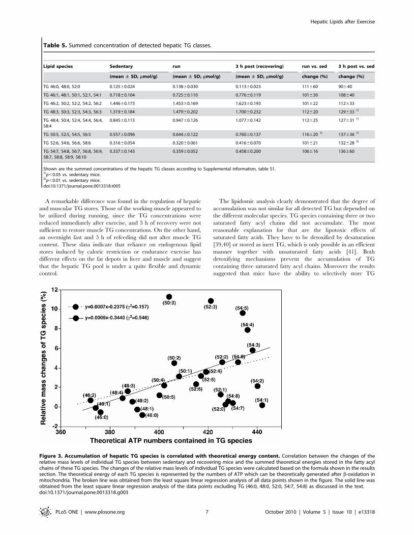
Accumulation of hepatic TG species is correlated withtheoretical energy content
The detected hepatic TG species showed differences in their
response to acute exercise in the recovery phase that could be
related to their content of double bonds (Table 5). TG species
containing only saturated acyl chains (TG 46:0; TG 48:0; and TG
52:0) did not accumulate (mean relative change of 90%640 vs.
sedentary mice), and no significant increase was found for TG
species containing only one monounsaturated acyl chain (TG 46:1,
TG 48:1, TG 50:1, TG 52:1, TG 54:1; mean relative change
108%640 vs. sedentary mice) or two double bonds (TG 46:2, TG
50:2, TG 52:2, TG 54:2, TG 56:2; mean relative change
112%633 vs. sedentary mice). TG species containing more than
two and less than seven double bonds showed a significant
accumulation in the recovery phase (Table 5). The increase in TG
species containing five double bonds was even significant
immediately after exercise. Since the data in Table 5 did not
consider the contribution of individual TG species to the increase
in total TG mass, we calculated the changed relative mass levels of
individual TG species by a formula as follows:
RelativeMassChange~TG 3hpostð Þ{TG sedð Þ
totalTG 3hpostð Þ{totalTG sedð Þ
Moreover, to understand the differences in the accumulation of
TG species we analyzed the relationship between the changed
relative mass levels of individual TG species and the summed
theoretical energies stored in the fatty acyl chains of these TG
species. The theoretical energy of each TG species is represented
by the numbers of ATP which can be theoretically generated after
b-oxidation in mitochondria. We found that the degree of
accumulation of individual TG species correlated well with the
amount of energy stored in it (Fig. 3). The correlation coefficient
(c2) using the least square linear regression analysis is 0.155 for all
determined species containing up to 54 carbons and is 0.546 if
excluding a few TG species which showed large deviations. These
are the TG species containing three saturated fatty acyl chains and
those which contain polyunsaturated fatty acyl chain(s) (e.g., TG
54:7 and TG 54:8). We should point out that TG species
comprising more than 54 carbons were not included in the
correlative analysis. This is due to the fact that at least one fatty
acyl chain containing 20 or more carbons is needed to build up
Table 1. Summary of detected lipid classes.
Lipid class Number of detected lipid species Sum of all detected species
sedentary 0 h (run) 3 h post
(mean ± SD, mmol/g) (mean ± SD, mmol/g) (mean ± SD, mmol/g)
LPC 6 0.46260.063 0.40860.113 0.36560.091
PC 24 4.54261.024 5.34461.115 4.50161.281
PE 14 0.63460.154 0.73960.261 0.62660.184
SM 7 0.35260.039 0.36760.116 0.30760.048
DG 5 0.12960.029 0.13860.021 0.12660.037
TG 59 5.65661.497 6.06361.187 6.90061.850
doi:10.1371/journal.pone.0013318.t001
Hepatic Lipids after Exercise
PLoS ONE | www.plosone.org 3 October 2010 | Volume 5 | Issue 10 | e13318

Hepatic Lipids after Exercise
PLoS ONE | www.plosone.org 4 October 2010 | Volume 5 | Issue 10 | e13318

these species and fatty acids containing 20 or more carbons are
always oxidized to 16 or 18 carbons by peroxisomes prior to being
oxidized in mitochondria. Therefore, these TG species are no
candidates for efficient energy storage.
Refeeding of fasted mice decreases their hepatic lipidcontent
The effects of endurance exercise share some similarities with
fasting, particularly on liver metabolism [34]. Acute exercise
increases circulating FFA levels, activates hepatic glucose produc-
tion, and decreases the expression of genes involved in fatty acid
synthesis. Since the mice had free access to food in the first hours
of the recovery phase, the increased hepatic TG levels might be
due to a refeeding phenomenon. We tested this hypothesis by
analyzing liver tissue lysates of overnight fasted mice and after 5 h
of refeeding. Refed mice had lower circulating FFA levels than
fasted mice and similar plasma TG concentrations (Fig. 4A, B).
Hepatic TG content of refed mice was similar to that of sedentary
mice (14.961.7 vs. 16.261.7 mmol/g, respectively), while fasted
mice had even higher hepatic TG levels than mice after 3 h of
recovery period (43.769.5 vs. 26.462.9 mmol/g, respectively)
(Fig. 2C, 4C). TG concentrations measured in gastrocnemius
muscles were not different in fasted and refed mice (Fig. 4D).
These data indicate a rapid, tissue-specific regulation of the
hepatic TG content with no correlation of TG accumulation in the
liver and food intake.
Discussion
A single bout of endurance exercise followed by 3 h recovery led
to a significant accumulation of TG in the livers of mice, while the
TG content decreased in skeletal muscle. This could be
demonstrated by measurement of total TG in liver and muscle
tissue using a clinical routine laboratory method as well as by a
hepatic lipidomics approach. The analysis of hepatic lipidomics
based on UFLC-MS revealed that a large number of lipid species
are relevant for the differentiation of the hepatic lipid profile of
sedentary and recovering mice. Among the detected LPC, PC, PE,
SM, DG, and TG lipid species, TG clearly dominated the pattern
of exercise-influenced hepatic lipids under the applied analytical
conditions with 17 TG species identified as discriminating lipids in
the total lipid pattern analyzed by multivariate data evaluation.
Seven of all identified TG species (Table 3) showed significant
increases in the recovery phase after one-way ANOVA followed
by post hoc with Dunnett.
Although we did not directly address the mechanism leading to
the hepatic TG accumulation after exercise, several hints point to
an important role of the high availability of plasma FFA during
exercise. Prolonged physical activity leads to increased lipolysis of
body fat stores and elevated levels of plasma FFA. This high
concentration of fatty acids in the circulation might lead not only
to increased uptake and oxidation in the working muscle, but also
to increased influx into the liver, which might be supported by
increased hepatic blood flow in the early recovery phase [35].
Here, fatty acids are reesterified into TG and secreted into
circulation in the form of VLDL or stored in the hepatocytes.
The accumulation of TG in the liver as a result of high plasma
FFA concentrations has been observed during fasting in rodents
when the delivery of fatty acids from peripheral fat stores exceeds
the oxidative requirements [36,37]. Similarly we found high
hepatic TG levels in overnight fasted mice, which returned to
levels of our sedentary control mice after 5 h of refeeding. During
prolonged endurance exercise a similar phenomenon might exist,
with the secreted FFAs exceeding the oxidative capacity and
demands of the working muscle. Therefore it could be speculated
that the liver might serve as a buffer reservoir for high FFA
plasma levels, which are temporarily stored in the liver as TG and
released into circulation when the storage of TG in the adipose
tissue is possible and not blocked by hormonal counterregulation.
Following that consideration, increases in hepatic TG content
should also be detected after long duration of endurance exercise,
Figure 2. Hepatic lipidomics reveals differences between recovering mice and sedentary mice. Partial least squares discriminant analysis(PLS-DA) of hepatic lipidomics data was applied to differentiate the recovering (3h post) mice (n = 8) and the sedentary mice (n = 8) with 115identified lipid species included in analysis as variables. (A) Scores plot of T [2] vs. T [1]. Each data point on the plot represents the individual hepaticlipid profile of one animal analyzed by UFLC-MS; (B) S-plot of the loading of PLS-DA component 1 (p 1; modeled variance) plotted against themodeled correlation [p (corr)], used to identify lipid metabolites most discriminatory for the specified classification. Each data point on the plotrepresents one out of 115 lipid molecular species. Lipids marked by numbers (see Table 2 for the corresponding lipids) were defined as potentialdiscriminators with variable importance in the projection values greater than 1 in at least one of the two groups. (C,D) TG concentrations asdetermined by an enzymatic analysis as described in methodsper g of tissue are shown for liver (C) and quadriceps muscle (D) of sedentary,immediately after exercise (run) and recovering (3 h post) mice (n = 12 (liver), n = 4 (muscle), mean 6 SEM; * p,0.05; *** p,0.001 vs. sed).doi:10.1371/journal.pone.0013318.g002
Table 2. Lipid species responsible for the discrimination ofthe hepatic lipid profile of recovering mice from sedentarymice screened by S-plot from PLS-DA.
No. m/z tR (min)Lipidspecies
Ionadduct
3 h postvs. sed
Up/Down (%)
1 544.342 2.1 LPC (20:4) H+ 227
2 788.619 11.4 PC (36:1) H+ 223
3 784.587 9.9 PC (36:3) H+ 211
4 812.618 10.9 PC (38:3) H+ 223
5 818.726 17.0 TG (48:3) NH4+ 30
6 850.789 19.2 TG (50:1) NH4+ 13
7 848.773 18.5 TG (50:2) NH4+ 15
8 846.759 17.8 TG (50:3) NH4+ 86
9 876.805 19.3 TG (52:2) NH4+ 11
10 874.789 18.7 TG (52:3) NH4+ 23
11 872.775 18.2 TG (52:4) NH4+ 25
12 870.760 17.5 TG (52:5) NH4+ 30
13 868.744 16.8 TG (52:6) NH4+ 50
14 904.837 20.2 TG (54:2) NH4+ 12
15 902.820 19.5 TG (54:3) NH4+ 24
16 900.805 18.8 TG (54:4) NH4+ 27
17 898.789 18.1 TG (54:5) NH4+ 43
18 896.774 17.6 TG (54:6) NH4+ 40
19 932.868 21.0 TG (56:2) NH4+ 21
20 930.851 20.3 TG (56:3) NH4+ 30
21 928.836 19.6 TG (56:4) NH4+ 31
doi:10.1371/journal.pone.0013318.t002
Hepatic Lipids after Exercise
PLoS ONE | www.plosone.org 5 October 2010 | Volume 5 | Issue 10 | e13318

not only in the early recovery phase. This is confirmed by a
previous study that reported on rats performing prolonged
exercise and could show a gradual accumulation of TG in the
liver depending on the duration of the exercise bout with a
doubling at the time of exhaustion [38]. Our exercise protocol of
60 min of treadmill run might be too short to observe such an
effect, but also in our study the concentration of almost all TG
species was higher immediately after exercise, even though this
did not reach statistical significance except the summed increase
of TG species containing five double bonds. Therefore we
conclude that acute exercise is a stimulus for the accumulation of
TG species in the liver similarly to fasting.
Table 3. Lipid species with significant alterations in recovering mice found after one-way ANOVA followed by 2-sided Dunnettpost hoc.
m/ztR
(min)Lipidspecies
Ionadduct sedentary run
3 h post(recovering)
runvs. sed
3 h postvs. sed
One-way ANOVA(p value)
(mean ± SD,
mmol/g)
(mean ± SD,
mmol/g)
(mean ± SD,
mmol/g)change(%)
change(%)
runvs. sed
3 hpostvs. sed
788.619 11.4 PC (36:1) H+ 0.20860.022 0.20660.044 0.16060.021 99 77 0.985 0.009**
812.618 10.9 PC (38:3) H+ 0.19560.020 0.19360.044 0.15060.037 99 77 0.987 0.033*
838.634 11.4 PC (40:4) H+ 0.01760.004 0.01860.007 0.00960.008 108 52 0.898 0.042*
612.559 12.6 DG (34:1) NH4+ 0.01160.004 0.01060.002 0.00660.006 89 49 0.818 0.036*
846.759 17.8 TG (50:3) NH4+ 0.15960.046 0.22960.081 0.29660.103 143 186 0.173 0.005**
898.789 18.1 TG (54:5) NH4+ 0.27060.047 0.34160.076 0.38760.110 126 143 0.170 0.017*
896.774 17.6 TG (54:6) NH4+ 0.13960.021 0.16160.023 0.19560.046 115 140 0.333 0.004**
901.730 17.6 TG (54:6) Na+ 0.02160.002 0.02460.003 0.02860.006 116 136 0.214 0.003**
899.715 16.9 TG (54:7) Na+ 0.01560.003 0.01760.004 0.02360.008 116 155 0.598 0.013*
928.836 19.6 TG (56:4) NH4+ 0.08860.032 0.09860.029 0.11660.036 111 131 0.596 0.031*
952.834 19.1 TG (58:6) NH4+ 0.00860.010 0.00160.003 0.02260.014 15 288 0.330 0.015*
944.773 17.0 TG (58:10) NH4+ 0.01760.006 0.02160.004 0.02660.009 120 150 0.475 0.028*
*p,0.05;**p,0.01 vs. sedentary mice.doi:10.1371/journal.pone.0013318.t003
Table 4. Summary of most abundant lipids from PC and TG lipid classes.
Lipid species Sedentary 0 h (run) 3 h postrunvs. sed
3 h postvs. sed
One-way ANOVA(p value)
(mean ± SD,
mmol/g)
(mean ± SD,
mmol/g)
(mean ± SD,
mmol/g)change(%)
change(%)
runvs. sed
3 h postvs. sed
PC (34:1) 0.56460.244 0.64960.185 0.58760.246 115 104 0.6784 0.9695
PC (34:2) 0.56860.131 0.78360.314 0.63460.263 138 112 0.1692 0.8171
PC (36:1) 0.20860.022 0.20660.044 0.16060.021 99 77 0.9851 0.0090**
PC (36:2) 0.50160.148 0.61760.148 0.50460.142 123 101 0.2136 0.9988
PC (36:3) 0.47560.079 0.52960.127 0.42260.128 111 89 0.5448 0.5588
PC (38:4) 0.34560.143 0.41660.141 0.37160.121 120 107 0.4877 0.9017
PC (38:6) 0.36560.081 0.41860.079 0.36060.094 115 99 0.3689 0.9897
PC (40:6) 0.26260.048 0.27460.054 0.25060.057 104 95 0.8765 0.8502
TG (50:1) 0.30560.093 0.31960.079 0.34360.111 105 113 0.9373 0.6396
TG (50:2) 0.36260.092 0.39660.091 0.41660.132 110 115 0.7410 0.4960
TG (52:2) 0.52660.093 0.49160.137 0.58260.182 93 111 0.8444 0.6520
TG (52:3) 0.56660.083 0.62060.085 0.69860.215 109 123 0.6733 0.1356
TG (54:2) 0.22260.077 0.21160.055 0.24860.090 95 112 0.9389 0.7063
TG (54:3) 0.30060.075 0.3260.056 0.37160.103 107 124 0.8365 0.1590
TG (54:4) 0.35260.077 0.39560.078 0.44760.119 112 127 0.5622 0.0945
TG (54:5) 0.27060.047 0.34160.076 0.38760.110 126 143 0.1700 0.0173*
*p,0.05,**p,0.01 vs. sedentary mice.doi:10.1371/journal.pone.0013318.t004
Hepatic Lipids after Exercise
PLoS ONE | www.plosone.org 6 October 2010 | Volume 5 | Issue 10 | e13318
A remarkable difference was found in the regulation of hepatic
and muscular TG stores. Those of the working muscle appeared to
be utilized during running, since the TG concentrations were
reduced immediately after exercise, and 3 h of recovery were not
sufficient to restore muscle TG concentrations. On the other hand,
an overnight fast and 5 h of refeeding did not alter muscle TG
content. These data indicate that reliance on endogenous lipid
stores induced by caloric restriction or endurance exercise has
different effects on the fat depots in liver and muscle and suggest
that the hepatic TG pool is under a quite flexible and dynamic
control.
The lipidomic analysis clearly demonstrated that the degree of
accumulation was not similar for all detected TG but depended on
the different molecular species. TG species containing three or two
saturated fatty acyl chains did not accumulate. The most
reasonable explanation for that are the lipotoxic effects of
saturated fatty acids. They have to be detoxified by desaturation
[39,40] or stored as inert TG, which is only possible in an efficient
manner together with unsaturated fatty acids [41]. Both
detoxifying mechanisms prevent the accumulation of TG
containing three saturated fatty acyl chains. Moreover the results
suggested that mice have the ability to selectively store TG
Table 5. Summed concentration of detected hepatic TG classes.
Lipid species Sedentary run 3 h post (recovering) run vs. sed 3 h post vs. sed
(mean ± SD, mmol/g) (mean ± SD, mmol/g) (mean ± SD, mmol/g) change (%) change (%)
TG 46:0, 48:0, 52:0 0.12560.024 0.13860.030 0.11360.023 111660 90640
TG 46:1, 48:1, 50:1, 52:1, 54:1 0.71860.104 0.72560.110 0.77660.119 101630 108640
TG 46:2, 50:2, 52:2, 54:2, 56:2 1.44660.173 1.45360.169 1.62360.193 101622 112633
TG 48:3, 50:3, 52:3, 54:3, 56:3 1.31960.184 1.47960.202 1.70060.232 112620 129633 1)
TG 48:4, 50:4, 52:4, 54:4, 56:4,58:4
0.84560.113 0.94760.126 1.07760.142 112625 127631 1)
TG 50:5, 52:5, 54:5, 56:5 0.55760.096 0.64460.122 0.76060.137 116620 1) 137636 1)
TG 52:6, 54:6, 56:6, 58:6 0.31660.054 0.32060.061 0.41660.070 101621 132628 2)
TG 54:7, 54:8, 56:7, 56:8, 56:9,58:7, 58:8, 58:9, 58:10
0.33760.143 0.35960.052 0.45860.200 106616 136660
Shown are the summed concentrations of the hepatic TG classes according to Supplemental information, table S1.1)p,0.05 vs. sedentary mice.2)p,0.01 vs. sedentary mice.doi:10.1371/journal.pone.0013318.t005
Figure 3. Accumulation of hepatic TG species is correlated with theoretical energy content. Correlation between the changes of therelative mass levels of individual TG species between sedentary and recovering mice and the summed theoretical energies stored in the fatty acylchains of these TG species. The changes of the relative mass levels of individual TG species were calculated based on the formula shown in the resultssection. The theoretical energy of each TG species is represented by the numbers of ATP which can be theoretically generated after b-oxidation inmitochondria. The broken line was obtained from the least square linear regression analysis of all data points shown in the figure. The solid line wasobtained from the least square linear regression analysis of the data points excluding TG (46:0, 48:0, 52:0, 54:7, 54:8) as discussed in the text.doi:10.1371/journal.pone.0013318.g003
Hepatic Lipids after Exercise
PLoS ONE | www.plosone.org 7 October 2010 | Volume 5 | Issue 10 | e13318

molecular species that contain high theoretical energy content in
the liver after exercise, since a positive correlation was found
between TG species with a high relative mass change and their
respective theoretical energy content. TG species that must
contain at least one polyunsaturated fatty acyl chain (e.g. TG
54:7) deviate from that correlation. This could be due to the fact
that the energy stored in the polyunsaturated fatty acyl chains
cannot be efficiently harvested through mitochondrial b-oxidation;
instead, they are predominantly oxidized through peroxisomal b-
oxidation resulting in the production of thermal energy instead of
ATP [42]. A previous report describes a similar selective
accumulation of TG species in the liver after a 24 h-fast in rats
[43]. TG species containing at least one linoleyl moiety (C 18:2)
showed a net increase in the liver, while TG species containing
only saturated and monounsaturated fatty acyl chains decrease.
These authors hypothesize that the selective changes in the hepatic
TG composition result from a preferential oxidation of saturated
and monounsaturated fatty acids in the fasted state [43].
It is also possible that the fatty acid composition of the diet fed
to the mice plays a role. The chow has a high content of oleate and
linoleate (6.2 and 18.0 g/kg chow, respectively; see Table S2 for
details) and this could enhance the storage of TG enriched in these
acylmoieties. Our data did not support the hypothesis that food
intake per se increases hepatic TG levels, but we could not
distinguish whether the source of the accumulated hepatic TG
were the dietary lipids or endogenous fatty acids derived from
lipolysis. Further experiments using isotope labeled fatty acids
would be needed to clarify this point.
The effects of the 60 min treadmill run on other lipid molecular
species were less prominent. In general, we found a decrease of
some LPC and PC species in the recovery phase. Phospholipids
have been discussed as energy source, and short periods of fasting
have been shown to lead to substantial decreases in PC and PE
species in the heart, but not in muscle [44]. It needs to be
evaluated whether some phospholipid species were used as
alternate energy source instead of TG in the liver in the recovery
phase after endurance exercise. Since exercise acutely reduces
VLDL-ApoB secretion in humans [26] and hepatic VLDL
secretion has been shown to depend on PC synthesis [45], it
would be tempting to speculate that the detected decrease in PC
contributes to exercise-induced alterations in lipid metabolism.
However, the decreased PC species only contribute to a small
percentage of the total hepatic PC pool, therefore an effect on
VLDL secretion appears rather speculative. To conclude, the
lipidomics analysis did not reveal a pronounced rearrangement in
the fatty acid composition of hepatic phospholipids. In contrast,
exercise training has been shown to alter the phospholipid profile
in rodent or human muscle [46,47], and to a minor extent of
hepatic phospholipids [9].
The current LC-MS lipidomics technology holds great promises
in the discovery of potential lipid biomarkers in relation to, for
example, hepatic steatosis, disease prevention and health promo-
tion. Its feasibility has been demonstrated in revealing the changes
of lipid metabolism in mouse liver after one single bout of exercise.
The most prominent effect of acute exercise on hepatic lipids
shown here is the induction of hepatic TG storage. Whether this
presumably transient hepatic steatosis after exercise is important
for the regulatory mechanisms leading to the reduction of hepatic
TG levels observed after prolonged training periods remains to be
elucidated. But it is possible that exercise might be a physiological
Figure 4. Refeeding of fasted mice decreases their hepatic lipid content. Plasma concentrations of FFA (A) and TG (B) and tissueconcentration of TG in liver (C) and gastrocnemius muscle (D) in fasted and refed mice (n = 10 (plasma, liver), n = 6 (muscle), mean 6 SEM; * p,0.05;** p,0.01 vs. fast).doi:10.1371/journal.pone.0013318.g004
Hepatic Lipids after Exercise
PLoS ONE | www.plosone.org 8 October 2010 | Volume 5 | Issue 10 | e13318

challenge for hepatic lipid metabolism similar to fasting that is
important for the maintenance of the metabolic flexibility of the
liver. A large number of lipid species were responsible for the
discrimination of the hepatic lipid pattern of sedentary and
recovering mice. These lipids provide a starting point for further
understanding of metabolic pathways and investigating integrative
biochemical networks.
Materials and Methods
Ethics statementAll animal experiments were conducted in accordance with the
national guidelines of laboratory animal care and were approved by
the local governmental commission for animal research (M2/05,
Regierungspraesidium Tubingen, Baden-Wurttemberg, Germany).
ChemicalsLiquid chromatography grade chloroform and methanol were
purchased from Merck (Darmstadt, Germany). High pressure liquid
chromatography grade isopropanol and acetonitrile were from
Tedia (Fairfield, OH, USA). Ammonium formate of analytical
grade was obtained from Sigma-Aldrich (St.Louis, MO, USA),
0.9% NaCl was from B.Braun (Melsungen, Germany). Triton X-
100 was from Sigma (Munich, Germany). Synthetic lipid standards
were purchased from Avanti Polar Lipids, Inc. (Alabaster, Alabama,
USA) and Sigma-Aldrich (Munich, Germany).
Animals and exerciseMale C57Bl/6J mice were purchased from The Jackson
Laboratory (Bar Harbor, ME, USA) and kept under an inverted
light-dark cycle (dark period 9:30–21:30, light period 21:30–9:30)
with free access to standard chow (Ssniff, Soest, Germany) and tap
water. Fatty acid content of the diet is available as Table S2 in
Supporting Information. Exercise experiments were performed
between 10:00 and 14:00. Mice were habituated to treadmill
running (Mouse Accupacer treadmill with motorized grade adjust,
Hugo Sachs Elektronik, March-Hugstetten, Germany) for 10 min
at 5 m/min and 5u inclination twice, one and two weeks prior to
the experiment.
In the experiments, 12-week-old mice ran 60 min at 14 m/min
and 14u uphill slope after 5 min warm-up (5 m/min and 5uinclination) and were either killed immediately after the run or
placed back in their cages for 3 h recovery. Sedentary mice
remained in their cages. Mice attempting to rest were encouraged
to continue running by gently tapping on their back. Mice of all
groups had no access to food 60 min before they were killed, either
because of running or because of food withdrawal. In fasting/
refeeding experiments mice were either fasted overnight or fasted
followed by 5 h of free access to food. All animals were
anesthetized with an intraperitoneal injection of ketamine
(150 mg/kg body weight) and xylazine (10 mg/kg body weight)
and killed by decapitation. Livers and muscle tissues were
immediately removed and frozen in liquid nitrogen for later
processing.
Plasma biochemical analysesGlucose was quantified in capillary blood samples taken from
the tail vein using an Accu-Chek Aviva glucometer (Roche,
Mannheim, Germany). Insulin levels were quantified by radio-
immunoassay (Linco Research, St. Charles, MO, USA). FFA and
TG were measured in the EDTA-plasma collected after
decapitation by fully automatic enzymatic methods on the ADVIA
1650 multi analyzer (Siemens Health Care Diagnostics, Fernwald,
Germany). Of note based on the principle of the enzymatic clinical
routine assay applied not only TG, but also diacylglyceride (DG),
monoacylglyceride and free glycerol are captured.
Total tissue TG analysesFrozen liver (100–150 mg) was weighed and homogenized in
1.5 ml of 0.9% NaCl containing 1% Triton X-100 using a
TissueLyser (Qiagen, Hilden, Germany). Lysates were clarified by
centrifugation at 13,000 g for 10 min and total liver TG content
was measured on the ADVIA 1650 multi analyzer. Frozen
quadriceps muscle tissue (approximately 100 mg) was weighed,
mixed with 0.6 ml cold methanol, 0.13 ml cold water and 50 ml
internal standard mixture, and homogenized in a TissueLyser with
stainless steel-beads (Qiagen, Hilden, Germany). After adding
0.6 ml chloroform and 0.3 ml water, the lysates were vortexed for
1 min and incubated 10 min on ice. The samples were then
centrifuged for 10 min at 13,000 g, 4uC for phase separation. A
defined volume of the lower nonpolar layer containing lipids was
collected and dried in a vacuum centrifuge. The lipid residue was
resuspended in 0.2 ml of 0.9% NaCl containing 1% Triton X-100
and total TG content was measured on the ADVIA 1650 multi
analyzer. Determination of hepatic TG content gave similar results
with each method.
Hepatic lipidomics analysisPreparation of lipid standards. Stock solutions of lipid
standards were separately dissolved in organic solvent. Briefly,
LPC (17:0), PE (34:0) and PC (34:0) were separately dissolved in a
chloroform/methanol mixture (2:1, v/v) and TG (51:0) was
dissolved in chloroform by weighing an exact amount of each lipid
standard in new glass vials. The stock solutions were stored at
220uC until further use. To prepare the working solution, a
certain volume of each stock solution was allowed to reach room
temperature, transferred into a new glass vial resulting in a
mixture 1:1:1:1 (v/v/v/v) and vortexed. The working solution was
then diluted 1:10 in a chloroform/methanol mixture (2:1, v/v) for
same-day use to the final concentration used for internal standard
addition. See Table S3 in Supporting Information for details of the
four internal lipid standards.
Liver lipid extraction. Frozen liver pieces (approximately
100 mg) of eight individual mice per group were homogenized and
the lipids were extracted as described above for quadriceps muscle
tissue. The lipid residue was resuspended in 0.3 ml of chloroform/
methanol (2:1, v/v) followed by 20 times dilution with acetonitrile/
isopropanol/water (65:30:5, v/v/v) for lipidomics analysis.
Ultra fast liquid chromatography coupled to ion-trap time-
of-flight mass spectrometry (UFLC2IT-TOF2MS). The
diluted lipid extracts were analyzed on a hybrid ion-trap time-of-
flight mass spectrometer (Shimadzu, Kyoto, Japan) coupled with an
ultra fast liquid chromatography system (Shimadzu, Kyoto, Japan).
The column was an AscentisH Express C8 2.16150 mm packed with
fused-core 2.7 mm diameter particles (Sigma-Aldrich, Munich,
Germany). Each sample was analyzed twice. The UFLC separation
of hepatic lipids was achieved according to the previously published
method [32]. The liver lipid profiling was carried out on Shimadzu
IT-TOF-MS equipped with an electrospray ion source in the positive
ion mode. The voltages of the interface and the detector of the TOF
analyzer were 4.5 kV and 1.6 kV, respectively. The temperatures of
the curved desorption line and heat block were both set at 200uC.
The flow rate of the nebulizing gas was 1.5 L/min. The dry gas
pressure was 0.2 MPa. The flight tube temperature was stable at
40uC and the ion trap pressure was maintained 1.661022 Pa. Ultra-
high purity argon was used for collision and ion cooling. The data
were collected at mass range of m/z 40021500 with an ion
Hepatic Lipids after Exercise
PLoS ONE | www.plosone.org 9 October 2010 | Volume 5 | Issue 10 | e13318

accumulation of 20 ms using LCMS solution software (Shimadzu,
Kyoto, Japan).
Data processing. LC2MS raw data were pre-processed by
the Phenomenome Profiler M Series version 2.5 (AM+, Canada).
Briefly, raw data were first converted to CDF format and then
subjected to peak picking. The parameters for peak picking were
smoothing points, 3; peak noise calculation, cutoff of 0.1% base
peak. The picked peaks were then aligned using Profiler’s
clustering algorithm based on the retention time and m/z of the
peaks. After that, a matrix for multivariate data analysis was
generated by the software with a mass clustering window of 0.005
Da and the retention time clustering window of 0.2 min. The
matrix covered the information of the retention time, m/z and the
ion intensity for each picked peak. Specifically, if certain ion
intensity was less than peak noise value in one sample, zero would
be assigned to that ion intensity in the matrix. The obtained data
set was processed by the ‘‘80% rule’’[48], that is, only the ions with
intensities above zero present in at least 80% of either group were
included for further data analyses. Subsequently internal standard
correction according to the strategy described previously [32],
normalization to mouse liver weight, and averaging the duplicate
injections was performed. Partial least squares discriminant
analysis (PLS-DA) was subsequently applied as a supervised
modeling method to the pareto-scaled data to visualize possible
relations between the samples (scores plot) and possible relations
between lipids (loading plot) related to the samples and the study
groups using SIMCA-P 11.0 (Umetrics AB, Umea, Sweden). The
discriminating individual lipid molecular species of the PLS-DA
model were selected by the S-plot [49].
Statistical analysisData are expressed as means 6 SEM. Unless indicated
otherwise one-way ANOVA followed by the Dunnett post hoc
test was applied to assess the statistical significance between groups
using SPSS statistics (version 17, SPSS Inc., USA). Statistical
significance was set at p,0.05.
Supporting Information
Table S1 Detailed information of the detected 115 lipid species.
Found at: doi:10.1371/journal.pone.0013318.s001 (0.35 MB
DOC)
Table S2 Relative fatty acid content of the standard chow.
Found at: doi:10.1371/journal.pone.0013318.s002 (0.03 MB
DOC)
Table S3 Detailed information of four lipid internal standards.
Found at: doi:10.1371/journal.pone.0013318.s003 (0.03 MB
DOC)
Author Contributions
Conceived and designed the experiments: CH MH RL CW. Performed the
experiments: CH MH XZ. Analyzed the data: CH MH XZ XH GX CW.
Contributed reagents/materials/analysis tools: HUH ES GX. Wrote the
paper: CH RL XH GX CW.
References
1. Schoiswohl G, Schweiger M, Schreiber R, Gorkiewicz G, Preiss-Landl K, et al.(2010) Adipose triglyceride lipase plays a key role in the supply of the working
muscle with fatty acids. J Lipid Res 51: 490–499.
2. Fernandez C, Hansson O, Nevsten P, Holm C, Klint C (2008) Hormone-sensitive lipase is necessary for normal mobilization of lipids during submaximal
exercise. Am J Physiol Endocrinol Metab 295: E179–E186.
3. Ahlborg G, Felig P, Hagenfeldt L, Hendler R, Wahren J (1974) Substrateturnover during prolonged exercise in man. Splanchnic and leg metabolism of
glucose, free fatty acids, and amino acids. J Clin Invest 53: 1080–1090.
4. Burguera B, Proctor D, Dietz N, Guo Z, Joyner M, et al. (2000) Leg free fattyacid kinetics during exercise in men and women. Am J Physiol Endocrinol
Metab 278: E113–E117.
5. Jensen MD (2003) Fate of fatty acids at rest and during exercise: regulatory
mechanisms. Acta Physiol Scand 178: 385–390.
6. Gauthier MS, Couturier K, Charbonneau A, Lavoie JM (2004) Effects ofintroducing physical training in the course of a 16-week high-fat diet regimen on
hepatic steatosis, adipose tissue fat accumulation, and plasma lipid profile.Int J Obes Relat Metab Disord 28: 1064–1071.
7. Vieira VJ, Valentine RJ, Wilund KR, Antao N, Baynard T, et al. (2009) Effects of
exercise and low-fat diet on adipose tissue inflammation and metaboliccomplications in obese mice. Am J Physiol Endocrinol Metab 296: E1164–E1171.
8. Rector RS, Thyfault JP, Morris RT, Laye MJ, Borengasser SJ, et al. (2008) Daily
exercise increases hepatic fatty acid oxidation and prevents steatosis in Otsuka
Long-Evans Tokushima Fatty rats. Am J Physiol Gastrointest Liver Physiol 294:G619–G626.
9. Petridou A, Nikolaidis MG, Matsakas A, Schulz T, Michna H, et al. (2005)
Effect of exercise training on the fatty acid composition of lipid classes in ratliver, skeletal muscle, and adipose tissue. Eur J Appl Physiol 94: 84–92.
10. Johnson NA, Sachinwalla T, Walton DW, Smith K, Armstrong A, et al. (2009)
Aerobic exercise training reduces hepatic and visceral lipids in obese individualswithout weight loss. Hepatology 50: 1105–1112.
11. Larson-Meyer DE, Heilbronn LK, Redman LM, Newcomer BR, Frisard MI, et
al. (2006) Effect of calorie restriction with or without exercise on insulin
sensitivity, beta-cell function, fat cell size, and ectopic lipid in overweightsubjects. Diabetes Care 29: 1337–1344.
12. Tamura Y, Tanaka Y, Sato F, Choi JB, Watada H, et al. (2005) Effects of diet
and exercise on muscle and liver intracellular lipid contents and insulinsensitivity in type 2 diabetic patients. J Clin Endocrinol Metab 90: 3191–3196.
13. Schafer S, Kantartzis K, Machann J, Venter C, Niess A, et al. (2007) Lifestyle
intervention in individuals with normal versus impaired glucose tolerance.Eur J Clin Invest 37: 535–543.
14. Thamer C, Machann J, Stefan N, Haap M, Schafer S, et al. (2007) High visceral
fat mass and high liver fat are associated with resistance to lifestyle intervention.Obesity (Silver Spring) 15: 531–538.
15. Aldred HE, Perry IC, Hardman AE (1994) The effect of a single bout of briskwalking on postprandial lipemia in normolipidemic young adults. Metabolism
43: 836–841.
16. Magkos F, Patterson BW, Mohammed BS, Mittendorfer B (2007) A single 1-hbout of evening exercise increases basal FFA flux without affecting VLDL-
triglyceride and VLDL-apolipoprotein B-100 kinetics in untrained lean men.Am J Physiol Endocrinol Metab 292: E1568–E1574.
17. Cullinane E, Siconolfi S, Saritelli A, Thompson PD (1982) Acute decrease in
serum triglycerides with exercise: is there a threshold for an exercise effect?
Metabolism 31: 844–847.
18. Gill JM, Mees GP, Frayn KN, Hardman AE (2001) Moderate exercise,
postprandial lipaemia and triacylglycerol clearance. Eur J Clin Invest 31:
201–207.
19. Ziogas GG, Thomas TR, Harris WS (1997) Exercise training, postprandialhypertriglyceridemia, and LDL subfraction distribution. Med Sci Sports Exerc
29: 986–991.
20. Leon AS, Sanchez OA (2001) Response of blood lipids to exercise training aloneor combined with dietary intervention. Med Sci Sports Exerc 33: S502–S515.
21. Herd SL, Hardman AE, Boobis LH, Cairns CJ (1998) The effect of 13 weeks of
running training followed by 9 d of detraining on postprandial lipaemia. Br J Nutr80: 57–66.
22. Holloszy JO, Skinner JS, Toro G, Cureton TK (1964) Effects of a six month
program of endurance exercise on the serum lipids of middle-aged man.
Am J Cardiol 14: 753–760.
23. Magkos F, Tsekouras YE, Prentzas KI, Basioukas KN, Matsama SG, et al.(2008) Acute exercise-induced changes in basal VLDL-triglyceride kinetics
leading to hypotriglyceridemia manifest more readily after resistance thanendurance exercise. J Appl Physiol 105: 1228–1236.
24. Borsheim E, Knardahl S, Hostmark AT (1999) Short-term effects of exercise on
plasma very low density lipoproteins (VLDL) and fatty acids. Med Sci SportsExerc 31: 522–530.
25. Tsekouras YE, Magkos F, Prentzas KI, Basioukas KN, Matsama SG, et al.
(2009) A single bout of whole-body resistance exercise augments basal VLDL-
triacylglycerol removal from plasma in healthy untrained men. Clin Sci (Lond)116: 147–156.
26. Magkos F, Wright DC, Patterson BW, Mohammed BS, Mittendorfer B (2006)
Lipid metabolism response to a single, prolonged bout of endurance exercise inhealthy young men. Am J Physiol Endocrinol Metab 290: E355–E362.
27. Sady SP, Cullinane EM, Saritelli A, Bernier D, Thompson PD (1988) Elevated
high-density lipoprotein cholesterol in endurance athletes is related to enhancedplasma triglyceride clearance. Metabolism 37: 568–572.
28. Annuzzi G, Jansson E, Kaijser L, Holmquist L, Carlson LA (1987) Increased
removal rate of exogenous triglycerides after prolonged exercise in man: timecourse and effect of exercise duration. Metabolism 36: 438–443.
Hepatic Lipids after Exercise
PLoS ONE | www.plosone.org 10 October 2010 | Volume 5 | Issue 10 | e13318

29. Tsekouras YE, Magkos F, Kellas Y, Basioukas KN, Kavouras SA, et al. (2008)
High-intensity interval aerobic training reduces hepatic very low-densitylipoprotein-triglyceride secretion rate in men. Am J Physiol Endocrinol Metab
295: E851–E858.
30. Mondon CE, Dolkas CB, Tobey T, Reaven GM (1984) Causes of thetriglyceride-lowering effect of exercise training in rats. J Appl Physiol 57:
1466–1471.31. Simonelli C, Eaton RP (1978) Reduced triglyceride secretion: a metabolic
consequence of chronic exercise. Am J Physiol 234: E221–E227.
32. Hu C, van Dommelen J, van der HR, Spijksma G, Reijmers TH, et al. (2008)RPLC-ion-trap-FTMS method for lipid profiling of plasma: method validation
and application to p53 mutant mouse model. J Proteome Res 7: 4982–4991.33. Eriksson L, Johansson E, Kettaneh-Wold E, Wold S (2001) Multi- and
Megavariate Data Analysis Principles and Applications. Umea, Sweden,Umetrics.
34. Hoene M, Lehmann R, Hennige AM, Pohl AK, Haring HU, et al. (2009) Acute
regulation of metabolic genes and insulin receptor substrates in the liver of miceby one single bout of treadmill exercise. J Physiol 587: 241–252.
35. Nielsen HB, Febbraio MA, Ott P, Krustrup P, Secher NH (2007) Hepatic lactateuptake versus leg lactate output during exercise in humans. J Appl Physiol 103:
1227–1233.
36. van G, V, Verhey E, Poelmann R, Ramakers R, van Dijk KW, et al. (2007)Metabolomics (liver and blood profiling) in a mouse model in response to fasting:
a study of hepatic steatosis. Biochim Biophys Acta 1771: 1263–1270.37. Triscari J, Bryce GF, Sullivan AC (1980) Metabolic consequences of fasting in
old lean and obese Zucker rats. Metabolism 29: 377–385.38. Gorski J, Nowacka M, Namiot Z, Puch U (1988) Effect of prolonged exercise on
the level of triglycerides in the rat liver. Eur J Appl Physiol Occup Physiol 57:
554–557.39. Peter A, Weigert C, Staiger H, Cegan A, Rittig K, et al. (2008) Induction of
stearoyl-CoA desaturase (SCD-1) protects human arterial endothelial cellsagainst lipotoxicity. Am J Physiol Endocrinol Metab 295: E339–E349.
40. Li ZZ, Berk M, McIntyre TM, Feldstein AE (2009) Hepatic lipid partitioning
and liver damage in nonalcoholic fatty liver disease: role of stearoyl-CoA
desaturase. J Biol Chem 284: 5637–5644.
41. Listenberger LL, Han X, Lewis SE, Cases S, Farese RV, Jr., et al. (2003)
Triglyceride accumulation protects against fatty acid-induced lipotoxicity. Proc
Natl Acad Sci U S A 100: 3077–3082.
42. Reddy JK, Hashimoto T (2001) Peroxisomal beta-oxidation and peroxisome
proliferator-activated receptor alpha: an adaptive metabolic system. Annu Rev
Nutr 21: 193–230.
43. Chen ZY, Cunnane SC (1992) Preferential retention of linoleic acid-enriched
triacylglycerols in liver and serum during fasting. Am J Physiol 263: R233–R239.
44. Han X, Cheng H, Mancuso DJ, Gross RW (2004) Caloric restriction results in
phospholipid depletion, membrane remodeling, and triacylglycerol accumula-
tion in murine myocardium. Biochemistry 43: 15584–15594.
45. Vance DE (2008) Role of phosphatidylcholine biosynthesis in the regulation of
lipoprotein homeostasis. Curr Opin Lipidol 19: 229–234.
46. Mitchell TW, Turner N, Hulbert AJ, Else PL, Hawley JA, et al. (2004) Exercise
alters the profile of phospholipid molecular species in rat skeletal muscle. J Appl
Physiol 97: 1823–1829.
47. Andersson A, Sjodin A, Olsson R, Vessby B (1998) Effects of physical exercise on
phospholipid fatty acid composition in skeletal muscle. Am J Physiol 274:
E432–E438.
48. Bijlsma S, Bobeldijk I, Verheij ER, Ramaker R, Kochhar S, et al. (2006) Large-
scale human metabolomics studies: a strategy for data (pre-) processing and
validation. Anal Chem 78: 567–574.
49. Wiklund S, Johansson E, Sjostrom L, Mellerowicz EJ, Edlund U, et al. (2008)
Visualization of GC/TOF-MS-based metabolomics data for identification of
biochemically interesting compounds using OPLS class models. Anal Chem 80:
115–122.
Hepatic Lipids after Exercise
PLoS ONE | www.plosone.org 11 October 2010 | Volume 5 | Issue 10 | e13318