lipid-conjugated fluorescent ph sensors for monitoring ph
TRANSCRIPT

Analyst
PAPER
Cite this: Analyst, 2015, 140, 6313
Received 12th June 2015,Accepted 10th August 2015
DOI: 10.1039/c5an01180a
www.rsc.org/analyst
Lipid-conjugated fluorescent pH sensors formonitoring pH changes in reconstitutedmembrane systems†
Gerdi Christine Kemmer,a Sidsel Ammitzbøll Bogh,b Michael Urban,c
Michael G. Palmgren,a Tom Vosch,b Jürgen Schillerd andThomas Günther Pomorski*a
Accurate real-time measurements of the dynamics of proton concentration gradients are crucial for
detailed molecular studies of proton translocation by membrane-bound enzymes. To reduce complexity,
these measurements are often carried out with purified, reconstituted enzyme systems. Yet the most
paramount problem to detect pH changes in reconstituted systems is that soluble pH reporters leak out
of the vesicle system during the reconstitution procedure. This requires loading of substantial amounts of
pH-sensors into the lumen of unilamellar liposomes during reconstitution. Here, we report the synthesis
and detailed characterisation of two lipid-linked pH sensors employing amine-reactive forms of semi-
naphthorhodafluors (SNARF®-1 dye) and rhodamine probes (pHrodo™ Red dye). Lipid-conjugation of
both dyes allowed for efficient detergent-based reconstitution of these pH indicators into liposomes.
Vesicle-embedded pHrodo™ displayed excellent photostability and an optimal pH-response between 4
and 7. The suitability of the lipid-linked pHrodo™ probe as a pH reporter was demonstrated by assaying
the activity of a plant plasma membrane H+-ATPase (proton pump) reconstituted in proteoliposomes.
Introduction
Cells and organelles depend on electrochemical gradientsacross their membranes. Membrane-embedded biologicalpumps are key players in maintaining these gradients throughprimary active transport of ions, but the precise mechanismsunderlying electrogenic transport are not yet completely under-stood. Among these pumps, plasma membrane H+-ATPasescouple ATP hydrolysis to H+ transport out of the cell. In fungiand plants, they are essential for establishing and maintaining
the crucial transmembrane electrochemical gradient of H+,which provides the driving force for the uptake of nutrientsand other cell constituents through an array of secondarytransport systems.1–4
The study of H+-translocating proteins at the molecularlevel is challenging due to the complexity of the membrane inwhich they are embedded. Thus, as an important step towardsa detailed understanding of their action and molecular func-tioning, current studies focus on isolated organelles and onhomogeneous preparations of membrane proteins reconsti-tuted into an artificial lipid environment. The latter offers awell-defined experimental system where no unknown orunwanted proteins are present. Moreover, reconstitutedsystems allow for a more flexible choice of biochemical, bio-physical and microscopy techniques for characterising therelated proteins.5 H+ fluxes in these systems are typically fol-lowed indirectly by use of pH-sensitive dyes that change absor-bance or fluorescence properties in response to the formationof pH gradients across the membrane.
Dyes such as pyranine (8-hydroxypyrene-1,3,6-trisulphonicacid) have been used to monitor the intravesicular pH valuesin mechanistic studies of H+-translocating membrane-boundproteins.6,7 However, the modest brightness of pyranine canbe a drawback in fluorescence measurements and high con-centrations (up to several millimoles) are required during
†Electronic supplementary information (ESI) available: Scheme of SNARF conju-gation to DOPE is shown (Fig. S1), head group lipid composition of lecithin isdemonstrated (Fig. S2), fluorescence emission spectra of soluble SNARF inbuffer solutions of different pH are presented (Fig. S3), size distribution of lipo-somes containing pH-sensors (Fig. S4), comparison of relative fluorescencechange Pyranine and pHrodo-DOPE (Fig. S5), and conversion of fluorescent inten-sity changes to pH changes is described (Fig. S6). See DOI: 10.1039/c5an01180a
aCentre for Membrane Pumps in Cells and Disease – PUMPKIN, Department of Plant
Biology and Biotechnology, University of Copenhagen, Thorvaldsensvej 40,
1871 Frederiksberg C, Denmark. E-mail: [email protected] Center, Department of Chemistry, University of Copenhagen,
Universitetsparken 5, 2100 Copenhagen, DenmarkcInstitute of Biochemistry, Biocenter, Goethe-University, Max-von-Laue-Str. 9,
D-60438 Frankfurt, GermanydInstitute of Medical Physics and Biophysics, Faculty of Medicine, University of
Leipzig, 04107 Leipzig, Germany
This journal is © The Royal Society of Chemistry 2015 Analyst, 2015, 140, 6313–6320 | 6313
Ope
n A
cces
s A
rtic
le. P
ublis
hed
on 1
0 A
ugus
t 201
5. D
ownl
oade
d on
11/
5/20
21 2
:04:
31 A
M.
Thi
s ar
ticle
is li
cens
ed u
nder
a C
reat
ive
Com
mon
s A
ttrib
utio
n 3.
0 U
npor
ted
Lic
ence
.
View Article OnlineView Journal | View Issue
reconstitution. A number of alternative pH-sensitive dyes withimproved brightness and photostability are available, such asseminaphthorhodafluors (SNARFs) and rhodamine-basedpHrodo dyes, allowing their application in fluorescencemicroscopy and flow cytometry from the single vesicle down tothe single molecule level.8–12 Yet the most paramount problemof reconstituted systems is the loading of sufficient amountsof these fluorophores into unilamellar liposomes (diameter40–400 nm). Most protein reconstitution methods include adetergent removal step usually performed either by dilution,dialysis, or the use of detergent absorbing hydrophobic poly-styrene beads.5,13 This procedure typically results in a signifi-cant loss of the dye from the liposomal lumen.14
In this study, we circumvented this problem by covalentlylinking the pH-sensing fluorophore to a membrane phospho-lipid via an amide bond. Two different pH-sensitivefluorophores, SNARF (SNARF®-1 carboxylic acid, acetate, succi-nimidyl ester) and pHrodo (pHrodo™ Red, succinimidylester) were linked to dioleoylphosphatidylethanolamine(DOPE) by conjugation to the ethanolamine head group. Thecorresponding lipid-conjugated derivatives were characterisedregarding pH dependence of their fluorescence emission, theirreconstitution efficiency and applicability for monitoring pHchanges in reconstituted liposomal systems.
ExperimentalMaterials
DOPE and dioleoylphosphatidylcholine (DOPC) were pur-chased from Avanti Polar Lipids (Alabaster, AL, USA). Soybeanlecithin (Type II-S, 14–23% choline basis) was purchased fromSigma-Aldrich (Brøndby, Denmark). Reactive pH-sensor estersof pHrodo (pHrodo™ Red, succinimidyl ester) and SNARF(SNARF®-1 carboxylic acid, acetate, succinimidyl ester), as wellas water-soluble pH sensors 5-(and-6)-carboxy SNARF®-1 andpHrodo® Red Dextran (10 000 MW) were purchased fromInvitrogen (Eugene, OR, USA). The detergent n-dodecyl-β-D-maltoside (DDM) was obtained from Glycon Biochemicals(Luckenwalde, Germany). Bio-beads SM-2 (Bio-Rad Labora-tories, Hercules, CA) were washed in methanol and rinsed withdouble-distilled water before use. Ultrapure water was obtainedfrom an in-house Millipore water purification system (MerckKGaA, Darmstadt, Germany). Unless indicated otherwise, allother chemicals and reagents were obtained from Sigma-Aldrich (Brøndby, Denmark).
Synthesis and purification of the lipid-conjugated pH-sensors
DOPE (1 mg; 1.35 µmol) in chloroform was evaporated todryness under a stream of nitrogen and re-suspended in 60 µLanhydrous methanol containing trimethylamine (1.14 µmol).After addition of the pH-sensor ester reagent (1 mg; 1.6 µmol)in anhydrous methanol (60 µL), the reaction mixture wasstirred for 1 h at room temperature. Any remaining pH-sensorester reagent was hydrolysed over 30 min by the addition ofwater (1 mL). Afterwards, lipids were extracted from the reac-
tion solution according to Bligh and Dyer.15 The lipid extractswere applied onto normal phase silica gel thin layer chromato-graphy plates (TLC, silica gel 60 F254, Merck) and developedwith a mixture of 90/54/7 (v/v/v) of chloroform, methanol, and25% aqueous ammonium hydroxide. The band containing theproduct was scraped off and extracted using a mixture ofchloroform, methanol, and water (1/2.2/1, by volume). Thisextract was further purified on a second TLC plate which wasdeveloped with a mixture of 50/20/10/10/2.5 (v/v/v/v/v) chloro-form, acetone, methanol, glacial acetic acid, and water. Theband containing the product was scraped off and extracted asdescribed above. The successful labelling of DOPE was con-firmed by matrix-assisted laser desorption ionisation time-of-flight mass spectrometry (MALDI-TOF MS). Before photograph-ing under ambient light or long-wave UV light TLC plates weredried completely to remove any remaining component fromthe mobile phase. To visualise DOPE, TLC plates were sub-sequently stained with primuline (0.005% in acetone/water,8/2; v/v)16 and photographed under long-wave UV light.
MALDI-TOF mass spectrometry
For the acquisition of positive or negative ion mass spectra,0.5 M DHB in methanol or 10 mg mL−1 9-aminoacridine(9-AA) in isopropanol/acetonitrile (60/40, v/v) were used asmatrices, respectively.17,18 As the quality of the spectrarecorded in the presence of 9-AA depends significantly on theapplied solvent system, the applied lipid extracts were dilutedwith isopropanol/acetonitrile (60/40, v/v).19 All samples werepre-mixed with the matrix prior to deposition onto the MALDItarget. All MALDI-TOF mass spectra were acquired on a BrukerAutoflex mass spectrometer (Bruker Daltonics, Bremen,Germany). The system utilises a pulsed nitrogen laser, emit-ting at 337 nm. The extraction voltage was 20 kV and gatedmatrix suppression was applied to prevent the saturation ofthe detector by matrix ions.20 For each mass spectrum, 128single laser shots were averaged. The laser fluence was keptabout 10% above threshold (i.e. the minimum laser fluencerequired to achieve detectable signals) to obtain optimumsignal-to-noise (S/N) ratios. In order to enhance the spectralresolution all spectra were acquired in the reflector modeusing delayed extraction conditions. More detailed methodo-logical descriptions of MALDI-TOF-MS are available inFuchs.21
Liposome preparation
Liposomes were prepared by manual extrusion. Briefly, chloro-form stocks of soybean lecithin (10 mg, ∼11.5 µmol) and lipid-linked pH-sensor (60 nmol for SNARF-DOPE; 20 nmol forpHrodo-DOPE) were dispensed into a round bottom flask,mixed and dried by rotary evaporation, followed by incubationfor 1 h under vacuum (30 mbar) at room temperature. Thelipid film was rehydrated in 667 µL of reconstitution buffer(10 mM Mes-KOH, pH 6.5, 50 mM K2SO4, 20% glycerol) by vor-texing in the presence of a glass pearl, and passed 41 timesthrough 0.2 µm size nucleopore polycarbonate membranes(Nuclepore™, Whatman GmbH, Germany) mounted in a mini-
Paper Analyst
6314 | Analyst, 2015, 140, 6313–6320 This journal is © The Royal Society of Chemistry 2015
Ope
n A
cces
s A
rtic
le. P
ublis
hed
on 1
0 A
ugus
t 201
5. D
ownl
oade
d on
11/
5/20
21 2
:04:
31 A
M.
Thi
s ar
ticle
is li
cens
ed u
nder
a C
reat
ive
Com
mon
s A
ttrib
utio
n 3.
0 U
npor
ted
Lic
ence
.View Article Online
extruder (Avanti Polar Lipids, Alabaster, AL). The resulting lipo-somes were kept at 4 °C and used within 1 week.
pH titration of lipid-conjugated pH-sensors
For assessment of the pH-dependence of DOPE-conjugatedpH-sensors, a range of different buffers between pH 2.5 and10.7 were prepared. Each buffer component was prepared as20 mM stock supplemented with 52.5 mM K2SO4 and the indi-vidual buffer components were mixed to obtain the requiredpH values. Citrate/phosphate buffer was used for pH 2.5–7,phosphate buffer for pH 7–8, and carbonate/bicarbonatebuffer for pH 9–10.7. Water-soluble SNARF and pHrododextran were used at 0.5 µM, and 3–12 µg mL−1, respectively.Prior to fluorescence measurements, liposomes containing theDOPE-conjugated pH-sensors were diluted 1 : 200 in therespective pH-buffers supplemented with the K+ ionophorevalinomycin (62.5 nM) and the protonophore carbonyl cyanidem-chlorophenylhydrazone (CCCP, 5 µM) for at least 15 min toallow equilibration of pH values between the inside and theoutside of the liposomes. The presence of the K+ ionophorevalinomycin and high concentrations of K+ inside and outsidethe vesicles prevent the build-up of a membrane voltage differ-ence. For each buffer condition a fluorescence emission spec-trum was recorded with a Fluoromax-4 spectrofluorometer(Horiba, Edison, NJ, USA) equipped with a LFI-3751 tempera-ture controller (Wavelength Electronics, Bozeman, MT, USA) at25 °C. Spectra were corrected using fluorophore-free liposomesat equal concentration and pH. SNARF was excited at 543 nm,and fluorescence recorded between 560 and 750 nm. For cali-bration curves, the ratio of intensities of low-wavelengthmaximum (at 580 nm) and the high-wavelength maximum (at630 nm for soluble SNARF, and 650 nm for SNARF-DOPE) wascalculated. The pHrodo dye was excited at 532 nm and emis-sion recorded between 550 and 700 nm; the maximum foreach pH was extracted and normalised to the maximum at pH3. For all calibration curves, data (ratio of intensities and nor-malised maximum intensity for SNARF and pHrodo, respecti-vely) was fitted using the built-in Boltzmann function ofOrigin (OriginLab, Northampton, MA).
Photophysical characterisation of DOPE-conjugated pHrodo
Liposomes (30 µL; 125 nmol of lipid) with pHrodo-DOPE werediluted in 2.7 mL transport buffer (20 mM MOPS-KOH, pH 7.0,52.5 mM K2SO4) with the pH value adjusted by addition ofHCl. Absorption spectra were measured on a Lambda 1050(PerkinElmer) with 2 nm slits and were corrected for scatteredlight by subtracting an absorption spectrum of a dye freesample. For the emission measurements pHrodo-DOPE andthe reference dye were excited with a 509 nm pulsed laser witha laser power of 190 μW (model number P-C-510 PicoQuantGmbH) on a Fluotime 300 instrument (PicoQuant GmbH). Allemission spectra were measured with a pulse repetition rate of40 MHz and were corrected for the spectral response of thedetector. Slit widths of 5 nm were used for all emissionmeasurements. Time-correlated single photon counting experi-ments were conducted with the same 509 nm pulsed laser on
a Fluotime 300 instrument. The pulse rate was 16 MHz andthe emission was measured at 590 nm. Measurements wereperformed at 25 °C in a water thermostated cuvette holder.The fluorescence decays were fitted to a bi-exponential func-tion. The full width at half-maximum (FWHM) of the instru-ment response function was typically in the order of 42 ps.
Preparation of plasma membrane H+-ATPase isoform 2 (AHA2)
A 73 amino acid C-terminal truncated version of the protein(aha2Δ73), containing a hexahistidine (6 × His) tag at theN-terminal end of the protein, was overexpressed in Saccharo-myces cerevisiae strain RS-72.22 The cells were lysed by vortex-ing with glass beads, and the protein was solubilised andpurified with DDM at a ratio of 3/1 detergent/protein (w/w)using batch-binding to a Ni2+-NTA resin. The purified proteinwas finally concentrated to 5–10 mg mL−1 using centrifugalconcentrators with a cut-off at 30 kDa (Vivaspin 20, GE Health-care), frozen in liquid nitrogen and stored at −80 °C in storagebuffer containing 50 mM Mes-KOH (pH 7), 50 mM KCl, and20% (v/v) glycerol until further use.
Vesicle reconstitution and analysis
Purified H+-ATPase was reconstituted into preformed lipo-somes as described22 using 75 µg purified protein per reconsti-tution. In short, preformed liposomes (156 µL of ∼11 µmolphospholipid per mL), purified protein (typically 10 μL of7.5 mg mL−1), and detergent (octyl β-D-glucopyranoside,50 mM final concentration) were mixed in a final volume of220 µL reconstitution buffer. Pyranine (8-hydroxypyrene-1,3,6-trisulfonic acid) was added at this step to 25 mM final concen-tration when indicated. The solubilised protein/lipid/detergentmixture was applied to a gel-filtration column (Sephadex G-50Fine, 2 mL packed in 2 mL disposable syringes) and incubatedfor 5 min before centrifugation (180g, 7 min). The eluate wasincubated for 30 min at room temperature with 100 mg of wetbio-beads under overhead rotation to eliminate traces of deter-gent. Protein-free liposomes were prepared similarly by repla-cing purified protein with reconstitution buffer.
Phospholipid vesicle recovery after detergent removal wasdetermined by measuring the amount of phospholipid phos-phorus23 before and after reconstitution. The concentrationsof the DOPE-conjugated pH-sensors and pyranine before andafter reconstitution into liposomes were quantified fluori-metrically (vide supra); comparison between these two numbersyielded the sensor recovery. The size of the vesicles was charac-terised via nanoparticle tracking (NanoSight LM14, NanosightLtd.); typical diameters for extruded liposomes and reconsti-tuted liposomes were 134 ± 45 and 109 ± 31 nm, respectively.
H+ transport measurement
To determine H+ pumping activity, proteoliposomes (5 µL)were added to 1 mL transport buffer (20 mM MOPS-KOH, pH7.0, 52.5 mM K2SO4) containing 62.5 nM valinomycin, andequilibrated for 15–30 min. During measurements, 2 mM ATP/ATP-analogue (buffered at pH 7) was added and H+ pumpingwas initiated by the addition of MgSO4 (3 mM final concen-
Analyst Paper
This journal is © The Royal Society of Chemistry 2015 Analyst, 2015, 140, 6313–6320 | 6315
Ope
n A
cces
s A
rtic
le. P
ublis
hed
on 1
0 A
ugus
t 201
5. D
ownl
oade
d on
11/
5/20
21 2
:04:
31 A
M.
Thi
s ar
ticle
is li
cens
ed u
nder
a C
reat
ive
Com
mon
s A
ttrib
utio
n 3.
0 U
npor
ted
Lic
ence
.View Article Online

tration), and when indicated dissipated by the addition of5 µM CCCP. The non-hydrolysable ATP-analogue adenyl-5′-ylimidodiphosphate (AMP-PNP) was used as a control in thesame concentration as ATP. Fluorescence traces were recordedfor 600 s at 25 °C by a Fluoromax-4 spectrofluorometer at thefollowing settings: 532 nm excitation, 585 nm emission, 3 nmslit widths, 0.1 s resolution (pHrodo) and 460 nm excitation,1 nm slit width, 515 nm emission, 2 nm slit width, 0.1 sresolution (pyranine).
Results and discussionSynthesis and purification of lipid-conjugated fluorescent pHindicators
DOPE was labelled with amine-reactive succinimidyl (NHS)esters of SNARF and pHrodo dyes under mild basic conditionsusing trimethylamine in methanol. The products wereextracted and purified by two subsequent TLC runs. TLC analy-sis of the SNARF labelling reaction revealed multiple fluo-rescent bands (Fig. 1A and B). Under the reaction conditionsused, the SNARF-NHS ester reacts readily with the aminogroup of DOPE yielding SNARF-DOPE; however, the aryl acetylgroup was removed due to the known susceptibility of arylacetates toward bases (Fig. 1C; ESI, Fig. S1†). DOPE-conjugated
SNARF appeared as a pink band under ambient light with Rfvalues of 0.9 and 0.5 using alkaline and acidic TLC conditions,respectively. The positive ion MALDI-TOF mass spectrum ofthe purified product showed three signals in the high massrange (Fig. 1C). These peaks at m/z 1179.6 (+H+), m/z 1201.5(+Na+), and m/z 1223.5 (−H+ + 2Na+) correspond exactly to theexpected molecular weight of SNARF-DOPE. The peak at m/z727.0 is characteristic for the DHB matrix cluster ion generatedin the gas phase,25 while the small peaks at m/z 774.1 and795.1 could not be assigned. Free, unmodified DOPE (expectedat m/z 744.5, 766.5 and 788.5) and free SNARF derivatives werenot detectable, demonstrating efficient purification. Therewere furthermore indications that radical cations are generatedin addition to commonly generated ion adducts: this isobvious from the unusual peak patterns (cf. the insert inFig. 1C) and applies particularly to the peaks corresponding tothe sodiated ions. A more detailed elucidation of theseaspects, however, is beyond the scope of this publication.
TLC analysis of the pHrodo labelling reaction revealed amain band (intense pink under day light) with Rf values of0.99 and 0.3 using alkaline and acidic TLC conditions,respectively (Fig. 1D and E). There were only minor by-productsas assessed by some additional bands with low intensity,detectable under ambient and UV light. The positive ion massspectrum of the purified product gave two peaks at m/z 1270.8
Fig. 1 Purification of lipid-conjugated pH-sensors. SNARF®-1 carboxylic acid, acetate, NHS ester (upper panel, A–C) or pHrodo Red NHS ester(lower panel, D–F) was reacted with DOPE and the resulting derivatives were extracted and resolved by alkaline TLC (A and D, respectively). Afterextraction of the indicated region (filled arrowhead in A and D), the samples were resolved by acidic TLC (B and E, respectively), and the indicatedregion (filled arrowhead in B, resp. E) was extracted as final product. See Experimental for the composition of the used solvent systems. Chromato-grams shown were dried completely before photographing under ambient light (AL) or UV light before (UV) and after staining with primuline (UV +P). The position of non-reacted DOPE is indicated by an open arrowhead. O, origin; F, solvent front, of the chromatograms. The final products wereanalysed by MALDI MS (C and F, positive ion mode). For lipid-conjugated SNARF (C), the annotated peaks correspond to the proton (m/z 1179.6), thesodium (m/z 1201.5), and H+ + 2 Na+ (m/z 1223.5) adducts (insets: chemical structure of SNARF-DOPE and expanded section of the chromatogram).The characteristic matrix peak is indicated by an asterisk. For lipid-conjugated pHrodo (F), the annotated peaks correspond to the proton and thesodium adducts (m/z 1270.8 and m/z 1292.8, respectively) (inset: chemical structure of pHrodo-DOPE; for pHrodo the chemical structure proposedby Ogawa24 was used).
Paper Analyst
6316 | Analyst, 2015, 140, 6313–6320 This journal is © The Royal Society of Chemistry 2015
Ope
n A
cces
s A
rtic
le. P
ublis
hed
on 1
0 A
ugus
t 201
5. D
ownl
oade
d on
11/
5/20
21 2
:04:
31 A
M.
Thi
s ar
ticle
is li
cens
ed u
nder
a C
reat
ive
Com
mon
s A
ttrib
utio
n 3.
0 U
npor
ted
Lic
ence
.View Article Online
and 1292.8 (Fig. 1F) corresponding to the proton and sodiumadducts of pHrodo-DOPE. No unreacted DOPE (expected at m/z744.5, 766.5, and 788.5) or other products were detectable.Thus, the purity of the product is close to 100%.
pH-Dependence of lipid-conjugated fluorescent pH indicators
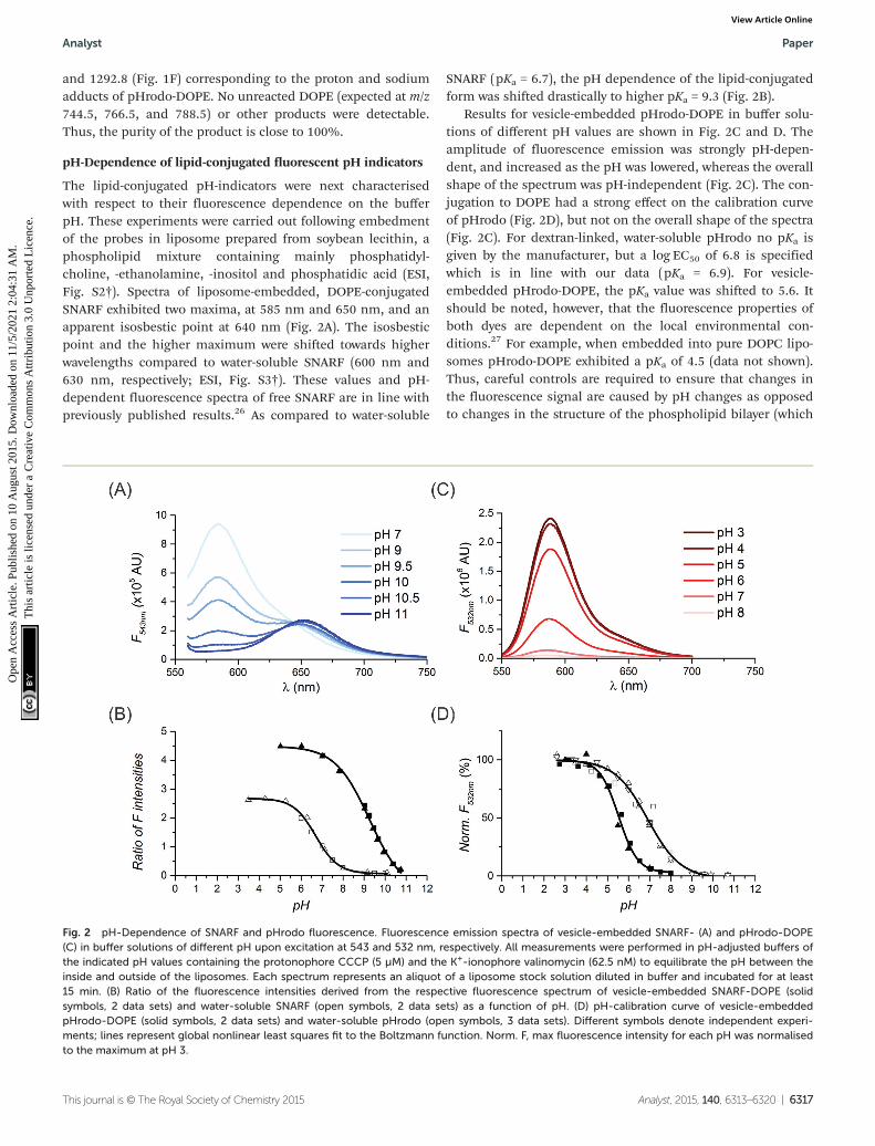
The lipid-conjugated pH-indicators were next characterisedwith respect to their fluorescence dependence on the bufferpH. These experiments were carried out following embedmentof the probes in liposome prepared from soybean lecithin, aphospholipid mixture containing mainly phosphatidyl-choline, -ethanolamine, -inositol and phosphatidic acid (ESI,Fig. S2†). Spectra of liposome-embedded, DOPE-conjugatedSNARF exhibited two maxima, at 585 nm and 650 nm, and anapparent isosbestic point at 640 nm (Fig. 2A). The isosbesticpoint and the higher maximum were shifted towards higherwavelengths compared to water-soluble SNARF (600 nm and630 nm, respectively; ESI, Fig. S3†). These values and pH-dependent fluorescence spectra of free SNARF are in line withpreviously published results.26 As compared to water-soluble
SNARF (pKa = 6.7), the pH dependence of the lipid-conjugatedform was shifted drastically to higher pKa = 9.3 (Fig. 2B).
Results for vesicle-embedded pHrodo-DOPE in buffer solu-tions of different pH values are shown in Fig. 2C and D. Theamplitude of fluorescence emission was strongly pH-depen-dent, and increased as the pH was lowered, whereas the overallshape of the spectrum was pH-independent (Fig. 2C). The con-jugation to DOPE had a strong effect on the calibration curveof pHrodo (Fig. 2D), but not on the overall shape of the spectra(Fig. 2C). For dextran-linked, water-soluble pHrodo no pKa isgiven by the manufacturer, but a log EC50 of 6.8 is specifiedwhich is in line with our data (pKa = 6.9). For vesicle-embedded pHrodo-DOPE, the pKa value was shifted to 5.6. Itshould be noted, however, that the fluorescence properties ofboth dyes are dependent on the local environmental con-ditions.27 For example, when embedded into pure DOPC lipo-somes pHrodo-DOPE exhibited a pKa of 4.5 (data not shown).Thus, careful controls are required to ensure that changes inthe fluorescence signal are caused by pH changes as opposedto changes in the structure of the phospholipid bilayer (which
Fig. 2 pH-Dependence of SNARF and pHrodo fluorescence. Fluorescence emission spectra of vesicle-embedded SNARF- (A) and pHrodo-DOPE(C) in buffer solutions of different pH upon excitation at 543 and 532 nm, respectively. All measurements were performed in pH-adjusted buffers ofthe indicated pH values containing the protonophore CCCP (5 µM) and the K+-ionophore valinomycin (62.5 nM) to equilibrate the pH between theinside and outside of the liposomes. Each spectrum represents an aliquot of a liposome stock solution diluted in buffer and incubated for at least15 min. (B) Ratio of the fluorescence intensities derived from the respective fluorescence spectrum of vesicle-embedded SNARF-DOPE (solidsymbols, 2 data sets) and water-soluble SNARF (open symbols, 2 data sets) as a function of pH. (D) pH-calibration curve of vesicle-embeddedpHrodo-DOPE (solid symbols, 2 data sets) and water-soluble pHrodo (open symbols, 3 data sets). Different symbols denote independent experi-ments; lines represent global nonlinear least squares fit to the Boltzmann function. Norm. F, max fluorescence intensity for each pH was normalisedto the maximum at pH 3.
Analyst Paper
This journal is © The Royal Society of Chemistry 2015 Analyst, 2015, 140, 6313–6320 | 6317
Ope
n A
cces
s A
rtic
le. P
ublis
hed
on 1
0 A
ugus
t 201
5. D
ownl
oade
d on
11/
5/20
21 2
:04:
31 A
M.
Thi
s ar
ticle
is li
cens
ed u
nder
a C
reat
ive
Com
mon
s A
ttrib
utio
n 3.
0 U
npor
ted
Lic
ence
.View Article Online
might be caused by altered lipid–ion interactions28). In thecase of H+-translocating ATPases, suitable controls are the useof non-hydrolysable ATP analogues (vide infra) or specificATPase inhibitors.
Reconstitution of the lipid-conjugated pH indicators
To demonstrate the usefulness of lipid-conjugation for re-constitution, we determined the recovery of SNARF-DOPE andpHrodo-DOPE after a classical reconstitution procedure.5,14,29
Liposomes doped with lipid-conjugated fluorescent pH indi-cators were destabilised using detergent and reconstituted bydetergent removal based on Sephadex G-50 gel filtration andsubsequent Bio-Bead treatment. Under these conditions,about 70–80% of the liposomes were recovered without signifi-cant loss of the lipid-conjugated fluorescent pH indicators(Table 1). Furthermore, the integrity of formed liposomes wasconfirmed by nanoparticle tracking (ESI, Fig. S4†). Thus, lipid-conjugation of pHrodo and SNARF allows for efficient reconsti-tution of these pH indicators into well-formed liposomes. Bycontrast, pyranine, a water-soluble pH-indicator added duringreconstitution, was predominantly lost (recovery about 1%)during reconstitution.
Photophysical characterisation of lipid-conjugated pHrodo
Because vesicle-embedded pHrodo-DOPE displayed an excel-lent pH-response in the most interesting range between 4 and7, we characterised this probe in more detail. Table 2 summar-ises the maximum absorption and emission wavelengths,
fluorescence decay times and the estimated fluorescencequantum yield as a function of pH. The fluorescence quantumyields should only be treated as rough estimates due to signifi-cant scattering of the liposomes. Within the tested pH range(pH 5–7) the maximum absorption and emission wavelengthsremained nearly unchanged around a value of 562 ± 3 nm and590 nm, respectively; while the fluorescence quantum yieldwas found to decrease at higher pH. Using time-correlatedsingle photon counting, the fluorescence decays of vesicle-embedded pHrodo-DOPE were recorded at different pH. Thedata was fitted with a bi-exponential function suggesting thattwo decay time components are present for pHrodo-DOPE inthe vesicles. Though there were variations in the actual valueof the two decay time components at different pH values, theintensity weighted average decay time τint only displayed minorchanges. This limited change in the overall fluorescence decaytime and the fact that the absorption spectrum remains nearlyconstant over the probed pH range, combined with the pHdependent fluorescence quantum yield, indicate that the pHdependent fluorescence response could be due to photo-induced electron transfer from the amine group.32 Long-timeexcitation of vesicle-embedded pHrodo-DOPE (pulsed laser at509 nm and 190 µW for 20 h at pH 6.8) caused no significantdecrease in fluorescence (data not shown), demonstrating theexcellent photostability of the sensor system.
Monitoring H+ pumping by reconstituted H+-ATPase usinglipid-conjugated pHrodo
Next, the suitability of pHrodo-DOPE to monitor pH changesin reconstituted liposomal systems was tested. Purified andreconstituted Arabidopsis thaliana autoinhibited plasma mem-brane H+-ATPase (AHA2) was used as a model transporter. Itfunctions as an ATP-driven membrane intrinsic protein thatplays a key role in the physiology of plants by controllingessential functions such as nutrient uptake and intracellularpH regulation.4,33 Proteoliposomes with reconstituted plasmamembrane H+-ATPase were added to ATP-containing buffer(pH 6.8), and the fluorescence intensity monitored at 585 nm.At pH 6.8, lipid-linked pHrodo is weakly fluorescent but elicitsa bright, red-fluorescent signal as the pH decreases locally, i.e.at an elevated H+ concentration inside vesicles. Thus, despitethe presence of lipid-conjugated fluorescent pH sensors on
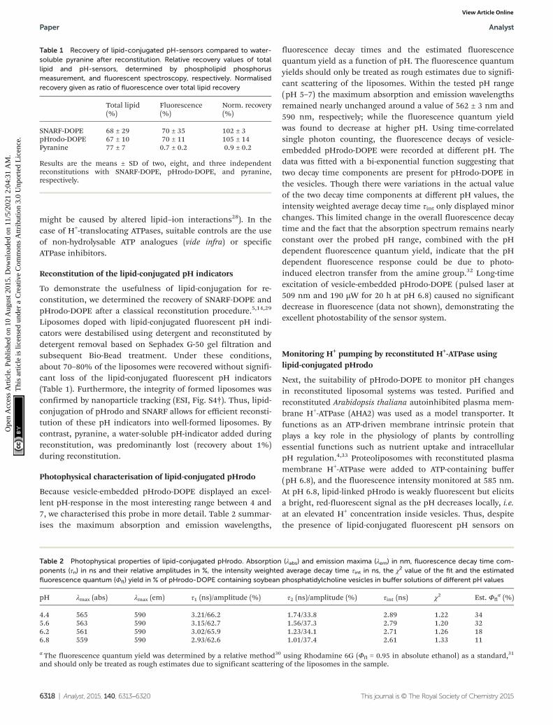
Table 1 Recovery of lipid-conjugated pH-sensors compared to water-soluble pyranine after reconstitution. Relative recovery values of totallipid and pH-sensors, determined by phospholipid phosphorusmeasurement, and fluorescent spectroscopy, respectively. Normalisedrecovery given as ratio of fluorescence over total lipid recovery
Total lipid(%)
Fluorescence(%)
Norm. recovery(%)
SNARF-DOPE 68 ± 29 70 ± 35 102 ± 3pHrodo-DOPE 67 ± 10 70 ± 11 105 ± 14Pyranine 77 ± 7 0.7 ± 0.2 0.9 ± 0.2
Results are the means ± SD of two, eight, and three independentreconstitutions with SNARF-DOPE, pHrodo-DOPE, and pyranine,respectively.
Table 2 Photophysical properties of lipid-conjugated pHrodo. Absorption (λabs) and emission maxima (λem) in nm, fluorescence decay time com-ponents (τn) in ns and their relative amplitudes in %, the intensity weighted average decay time τint in ns, the χ2 value of the fit and the estimatedfluorescence quantum (Φfl) yield in % of pHrodo-DOPE containing soybean phosphatidylcholine vesicles in buffer solutions of different pH values
pH λmax (abs) λmax (em) τ1 (ns)/amplitude (%) τ2 (ns)/amplitude (%) τint (ns) χ2 Est. Φfla (%)
4.4 565 590 3.21/66.2 1.74/33.8 2.89 1.22 345.6 563 590 3.15/62.7 1.56/37.3 2.79 1.20 326.2 561 590 3.02/65.9 1.23/34.1 2.71 1.26 186.8 559 590 2.93/62.6 1.01/37.4 2.61 1.33 11
a The fluorescence quantum yield was determined by a relative method30 using Rhodamine 6G (Φfl = 0.95 in absolute ethanol) as a standard,31
and should only be treated as rough estimates due to significant scattering of the liposomes in the sample.
Paper Analyst
6318 | Analyst, 2015, 140, 6313–6320 This journal is © The Royal Society of Chemistry 2015
Ope
n A
cces
s A
rtic
le. P
ublis
hed
on 1
0 A
ugus
t 201
5. D
ownl
oade
d on
11/
5/20
21 2
:04:
31 A
M.
Thi
s ar
ticle
is li
cens
ed u
nder
a C
reat
ive
Com
mon
s A
ttrib
utio
n 3.
0 U
npor
ted
Lic
ence
.View Article Online
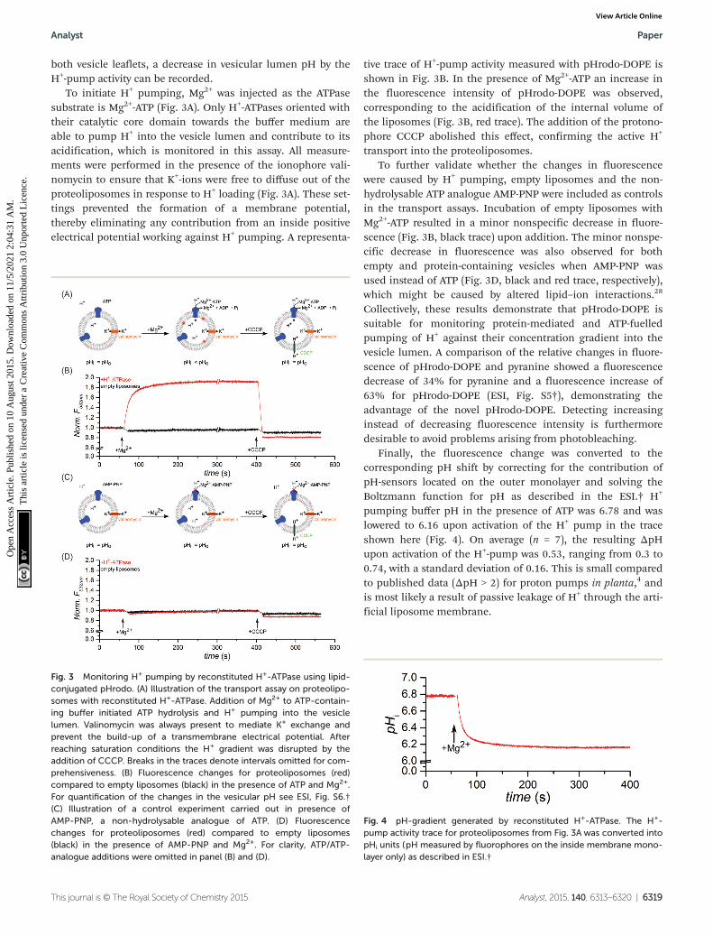
both vesicle leaflets, a decrease in vesicular lumen pH by theH+-pump activity can be recorded.
To initiate H+ pumping, Mg2+ was injected as the ATPasesubstrate is Mg2+-ATP (Fig. 3A). Only H+-ATPases oriented withtheir catalytic core domain towards the buffer medium areable to pump H+ into the vesicle lumen and contribute to itsacidification, which is monitored in this assay. All measure-ments were performed in the presence of the ionophore vali-nomycin to ensure that K+-ions were free to diffuse out of theproteoliposomes in response to H+ loading (Fig. 3A). These set-tings prevented the formation of a membrane potential,thereby eliminating any contribution from an inside positiveelectrical potential working against H+ pumping. A representa-
tive trace of H+-pump activity measured with pHrodo-DOPE isshown in Fig. 3B. In the presence of Mg2+-ATP an increase inthe fluorescence intensity of pHrodo-DOPE was observed,corresponding to the acidification of the internal volume ofthe liposomes (Fig. 3B, red trace). The addition of the protono-phore CCCP abolished this effect, confirming the active H+
transport into the proteoliposomes.To further validate whether the changes in fluorescence
were caused by H+ pumping, empty liposomes and the non-hydrolysable ATP analogue AMP-PNP were included as controlsin the transport assays. Incubation of empty liposomes withMg2+-ATP resulted in a minor nonspecific decrease in fluore-scence (Fig. 3B, black trace) upon addition. The minor nonspe-cific decrease in fluorescence was also observed for bothempty and protein-containing vesicles when AMP-PNP wasused instead of ATP (Fig. 3D, black and red trace, respectively),which might be caused by altered lipid–ion interactions.28
Collectively, these results demonstrate that pHrodo-DOPE issuitable for monitoring protein-mediated and ATP-fuelledpumping of H+ against their concentration gradient into thevesicle lumen. A comparison of the relative changes in fluore-scence of pHrodo-DOPE and pyranine showed a fluorescencedecrease of 34% for pyranine and a fluorescence increase of63% for pHrodo-DOPE (ESI, Fig. S5†), demonstrating theadvantage of the novel pHrodo-DOPE. Detecting increasinginstead of decreasing fluorescence intensity is furthermoredesirable to avoid problems arising from photobleaching.
Finally, the fluorescence change was converted to thecorresponding pH shift by correcting for the contribution ofpH-sensors located on the outer monolayer and solving theBoltzmann function for pH as described in the ESI.† H+
pumping buffer pH in the presence of ATP was 6.78 and waslowered to 6.16 upon activation of the H+ pump in the traceshown here (Fig. 4). On average (n = 7), the resulting ΔpHupon activation of the H+-pump was 0.53, ranging from 0.3 to0.74, with a standard deviation of 0.16. This is small comparedto published data (ΔpH > 2) for proton pumps in planta,4 andis most likely a result of passive leakage of H+ through the arti-ficial liposome membrane.
Fig. 3 Monitoring H+ pumping by reconstituted H+-ATPase using lipid-conjugated pHrodo. (A) Illustration of the transport assay on proteolipo-somes with reconstituted H+-ATPase. Addition of Mg2+ to ATP-contain-ing buffer initiated ATP hydrolysis and H+ pumping into the vesiclelumen. Valinomycin was always present to mediate K+ exchange andprevent the build-up of a transmembrane electrical potential. Afterreaching saturation conditions the H+ gradient was disrupted by theaddition of CCCP. Breaks in the traces denote intervals omitted for com-prehensiveness. (B) Fluorescence changes for proteoliposomes (red)compared to empty liposomes (black) in the presence of ATP and Mg2+.For quantification of the changes in the vesicular pH see ESI, Fig. S6.†(C) Illustration of a control experiment carried out in presence ofAMP-PNP, a non-hydrolysable analogue of ATP. (D) Fluorescencechanges for proteoliposomes (red) compared to empty liposomes(black) in the presence of AMP-PNP and Mg2+. For clarity, ATP/ATP-analogue additions were omitted in panel (B) and (D).
Fig. 4 pH-gradient generated by reconstituted H+-ATPase. The H+-pump activity trace for proteoliposomes from Fig. 3A was converted intopHi units (pH measured by fluorophores on the inside membrane mono-layer only) as described in ESI.†
Analyst Paper
This journal is © The Royal Society of Chemistry 2015 Analyst, 2015, 140, 6313–6320 | 6319
Ope
n A
cces
s A
rtic
le. P
ublis
hed
on 1
0 A
ugus
t 201
5. D
ownl
oade
d on
11/
5/20
21 2
:04:
31 A
M.
Thi
s ar
ticle
is li
cens
ed u
nder
a C
reat
ive
Com
mon
s A
ttrib
utio
n 3.
0 U
npor
ted
Lic
ence
.View Article Online

Conclusions
In this study we describe the synthesis of two lipid-linked pHsensors, and demonstrate their application to monitor pHchanges in reconstituted liposomal systems. The major advan-tage of this approach is that the lipid-linked pH sensorsefficiently co-reconstitute with membrane proteins into lipo-somes, thereby avoiding their loss during reconstitution. Thisapproach facilitates the study of H+ translocation by mem-brane-bound enzymes, as exemplified by the analysis of amodel plant proton pump employing lipid-conjugatedpHrodo. The excellent photostability of the pHrodo probemight be helpful in future single vesicle studies. Lipid-conju-gated SNARF (pKa 9.3) can be useful for studies in whichcation transport by ATPases is associated with H+ counter-transport, e.g. in case of Na+-translocating ATPases.34,35 Fur-thermore, the system described in this work has thesignificant benefit of detecting the pH (and ions such as Mg2+)close to the membrane bilayer which can be exploited for thedevelopment of sensor systems in biotechnology.36
In conclusion, both synthesised lipid-conjugated pHsensors open up for a wide range of biophysical and biologicalapplications.
Acknowledgements
We are grateful to Anne-Mette Bjerg Petersen for excellent tech-nical assistance. This work was supported by the DanishCouncil for Independent Research | Natural Sciences (FNU,grant number 1323-00297), the Research Centre ‘bioSYNergy’funded by the UCPH Excellence Programme for Interdisciplin-ary Research, the Villum Foundation (Project numberVKR023115) and the Danish National Research Foundationthrough the PUMPKIN Centre of Excellence (DNRF85).
Notes and references
1 J. C. Meade, C. L. Li, J. K. Stiles, M. E. Moate, J. I. Penny,S. Krishna and R. W. Finley, Parasitol. Int., 2000, 49, 309–320.
2 R. K. Nakamoto and C. W. Slayman, J. Bioenerg. Biomembr.,1989, 21, 621–632.
3 C. L. Slayman, W. S. Long and C. Y. H. Lu, J. Membr. Biol.,1973, 14, 305–338.
4 M. G. Palmgren, Annu. Rev. Plant Physiol., 2001, 52, 817–845.5 J. L. Rigaud and D. Levy, Liposomes, Pt B, 2003, 372, 65–86.6 N. R. Clement and J. M. Gould, Biochemistry, 1981, 20,
1534–1538.7 A. Holoubek, J. Večeř and K. Sigler, J. Fluoresc., 2007, 17,
201–213.8 S. Brasselet and W. E. Moerner, Single Mol., 2000, 1, 17–23.9 J. Y. Han and K. Burgess, Chem. Rev., 2010, 110, 2709–2728.10 R. Watanabe, K. V. Tabata, R. Iino, H. Ueno, M. Iwamoto,
S. Oiki and H. Noji, Nat. Commun., 2013, 4, 1631.
11 Y. Fu, M. M. Collinson and D. A. Higgins, J. Am. Chem. Soc.,2004, 126, 13838–13844.
12 X. Shi, J. Lim and T. Ha, Anal. Chem., 2010, 82, 6132–6138.13 R. Schubert, Liposomes, Pt A, 2003, 367, 46–70.14 T. Leiding, K. Górecki, T. Kjellman, S. A. Vinogradov,
C. Hägerhäll and S. P. Årsköld, Anal. Biochem., 2009, 388,296–305.
15 E. G. Bligh and W. J. Dyer, Can. J. Biochem. Physiol., 1959,37, 911–917.
16 T. White, S. Bursten, D. Federighi, R. A. Lewis andE. Nudelman, Anal. Biochem., 1998, 258, 109–117.
17 J. Schiller, J. Arnhold, S. Benard, M. Müller, S. Reichl andK. Arnold, Anal. Biochem., 1999, 267, 46–56.
18 G. Sun, K. Yang, Z. Zhao, S. Guan, X. Han and R. W. Gross,Anal. Chem., 2008, 80, 7576–7585.
19 B. Fuchs, A. Bischoff, R. Süß, K. Teuber, M. Schürenberg,D. Suckau and J. Schiller, Anal. Bioanal. Chem., 2009, 395,2479–2487.
20 M. Petković, J. Schiller, M. Müller, S. Benard, S. Reichl,K. Arnold and J. Arnhold, Anal. Biochem., 2001, 289, 202–216.
21 B. Fuchs, R. Süß and J. Schiller, Prog. Lipid Res., 2010, 49,450–475.
22 F. C. Lanfermeijer, K. Venema and M. G. Palmgren, ProteinExpression Purif., 1998, 12, 29–37.
23 G. Rouser, S. Fleische and A. Yamamoto, Lipids, 1970, 5,494–496.
24 M. Ogawa, N. Kosaka, C. A. Regino, M. Mitsunaga,P. L. Choyke and H. Kobayashi, Mol. BioSyst., 2010, 6, 888–893.
25 J. Schiller, R. Süß, B. Fuchs, M. Müller, M. Petković,O. Zschörnig and H. Waschipky, Eur. Biophys. J. Biophys.,2007, 36, 517–527.
26 S. Bassnett, L. Reinisch and D. C. Beebe, Am. J. Physiol.,1990, 258, C171–C178.
27 I. D. Johnson, Molecular Probes handbook, A guide to fluore-scent probes and labeling technologies, Life TechnologiesCorporation, 11th edition, 2010.
28 H. Binder and O. Zschornig, Chem. Phys. Lipids, 2002, 115,39–61.
29 J. L. Rigaud, D. Levy, G. Mosser and O. Lambert, Eur.Biophys. J. Biophys., 1998, 27, 305–319.
30 C. Würth, M. Grabolle, J. Pauli, M. Spieles and U. Resch-Genger, Nat. Protocols, 2013, 8, 1535–1550.
31 D. Magde, R. Wong and P. G. Seybold, Photochem. Photo-biol., 2002, 75, 327–334.
32 Q. A. Best, R. S. Xu, M. E. McCarroll, L. C. Wang andD. J. Dyer, Org. Lett., 2010, 12, 3219–3221.
33 R. Serrano, M. C. Kielland-Brandt and G. R. Fink, Nature,1986, 319, 689–693.
34 K. Soontharapirakkul and A. Incharoensakdi, BMCBiochem., 2010, 11.
35 Y. V. Balnokin, L. G. Popova, L. Y. Pagis and I. M. Andreev,Planta, 2004, 219, 332–337.
36 A. D. Robison, D. Huang, H. Jung and P. S. Cremer, Bio-interphases, 2013, 8.
Paper Analyst
6320 | Analyst, 2015, 140, 6313–6320 This journal is © The Royal Society of Chemistry 2015
Ope
n A
cces
s A
rtic
le. P
ublis
hed
on 1
0 A
ugus
t 201
5. D
ownl
oade
d on
11/
5/20
21 2
:04:
31 A
M.
Thi
s ar
ticle
is li
cens
ed u
nder
a C
reat
ive
Com
mon
s A
ttrib
utio
n 3.
0 U
npor
ted
Lic
ence
.View Article Online