lipid-based drug delivery systems in cancer therapy: what...
TRANSCRIPT

1521-0081/68/3/701–787$25.00 http://dx.doi.org/10.1124/pr.115.012070PHARMACOLOGICAL REVIEWS Pharmacol Rev 68:701–787, July 2016Copyright © 2016 by The American Society for Pharmacology and Experimental Therapeutics
ASSOCIATE EDITOR: ERIC L. BARKER
Lipid-Based Drug Delivery Systems in Cancer Therapy:What Is Available and What Is Yet to Come
Phatsapong Yingchoncharoen, Danuta S. Kalinowski, and Des R. Richardson
Molecular Pharmacology and Pathology Program, Department of Pathology, Faculty of Medicine, Bosch Institute, The University of Sydney,Sydney, NSW, Australia
Abstract . . . . . . . . . . . . . . . . . . . . . . . . . . . . . . . . . . . . . . . . . . . . . . . . . . . . . . . . . . . . . . . . . . . . . . . . . . . . . . . . . . . 703I. Introduction . . . . . . . . . . . . . . . . . . . . . . . . . . . . . . . . . . . . . . . . . . . . . . . . . . . . . . . . . . . . . . . . . . . . . . . . . . . . . . . 704II. Liposomal Drug Delivery Systems. . . . . . . . . . . . . . . . . . . . . . . . . . . . . . . . . . . . . . . . . . . . . . . . . . . . . . . . . . 705
A. Liposome Composition . . . . . . . . . . . . . . . . . . . . . . . . . . . . . . . . . . . . . . . . . . . . . . . . . . . . . . . . . . . . . . . . . 705B. Types of Liposomes . . . . . . . . . . . . . . . . . . . . . . . . . . . . . . . . . . . . . . . . . . . . . . . . . . . . . . . . . . . . . . . . . . . . 715
1. Multilamellar Vesicles. . . . . . . . . . . . . . . . . . . . . . . . . . . . . . . . . . . . . . . . . . . . . . . . . . . . . . . . . . . . . . 7152. Large Unilamellar Vesicles. . . . . . . . . . . . . . . . . . . . . . . . . . . . . . . . . . . . . . . . . . . . . . . . . . . . . . . . . 7163. Small Unilamellar Vesicles. . . . . . . . . . . . . . . . . . . . . . . . . . . . . . . . . . . . . . . . . . . . . . . . . . . . . . . . . 717
C. Lipid-core Micelles . . . . . . . . . . . . . . . . . . . . . . . . . . . . . . . . . . . . . . . . . . . . . . . . . . . . . . . . . . . . . . . . . . . . . 717III. Characteristics and Properties of Lipid-Based Nanoparticles . . . . . . . . . . . . . . . . . . . . . . . . . . . . . . . . 718
A. Morphology of Lipid-Based Nanoparticles. . . . . . . . . . . . . . . . . . . . . . . . . . . . . . . . . . . . . . . . . . . . . . . 718B. Size and Size Distribution. . . . . . . . . . . . . . . . . . . . . . . . . . . . . . . . . . . . . . . . . . . . . . . . . . . . . . . . . . . . . . 719C. Surface Charge . . . . . . . . . . . . . . . . . . . . . . . . . . . . . . . . . . . . . . . . . . . . . . . . . . . . . . . . . . . . . . . . . . . . . . . . 721D. Phase Transition Temperature . . . . . . . . . . . . . . . . . . . . . . . . . . . . . . . . . . . . . . . . . . . . . . . . . . . . . . . . . 722E. Plasma Proteins Interactions-Particle Stability and Clearance . . . . . . . . . . . . . . . . . . . . . . . . . . 723
IV. Tumor Targeting of Lipid-Based Nanoparticles . . . . . . . . . . . . . . . . . . . . . . . . . . . . . . . . . . . . . . . . . . . . . 724A. Passive Targeting . . . . . . . . . . . . . . . . . . . . . . . . . . . . . . . . . . . . . . . . . . . . . . . . . . . . . . . . . . . . . . . . . . . . . . 724B. Active Targeting . . . . . . . . . . . . . . . . . . . . . . . . . . . . . . . . . . . . . . . . . . . . . . . . . . . . . . . . . . . . . . . . . . . . . . . 725
1. Cancer Cell Targeting. . . . . . . . . . . . . . . . . . . . . . . . . . . . . . . . . . . . . . . . . . . . . . . . . . . . . . . . . . . . . . 726a. Transferrin receptor. . . . . . . . . . . . . . . . . . . . . . . . . . . . . . . . . . . . . . . . . . . . . . . . . . . . . . . . . . . . . 727b. Folate receptor. . . . . . . . . . . . . . . . . . . . . . . . . . . . . . . . . . . . . . . . . . . . . . . . . . . . . . . . . . . . . . . . . . 727c. Cell surface glycoproteins. . . . . . . . . . . . . . . . . . . . . . . . . . . . . . . . . . . . . . . . . . . . . . . . . . . . . . . . 728d. Epidermal growth factor receptor. . . . . . . . . . . . . . . . . . . . . . . . . . . . . . . . . . . . . . . . . . . . . . . . 728e. ssDNA and RNA aptamers. . . . . . . . . . . . . . . . . . . . . . . . . . . . . . . . . . . . . . . . . . . . . . . . . . . . . . 729
2. Tumoral Endothelium Targeting. . . . . . . . . . . . . . . . . . . . . . . . . . . . . . . . . . . . . . . . . . . . . . . . . . . . 729a. Vascular endothelial growth factor and its receptors, vascular endothelial
growth factor receptor-1 and vascular endothelial growth factor receptor-2. . . . . . . . 729b. The avb3 integrin.. . . . . . . . . . . . . . . . . . . . . . . . . . . . . . . . . . . . . . . . . . . . . . . . . . . . . . . . . . . . . . . 730c. Vascular cell adhesion molecule-1. . . . . . . . . . . . . . . . . . . . . . . . . . . . . . . . . . . . . . . . . . . . . . . . 730d. Matrix metalloproteinases. . . . . . . . . . . . . . . . . . . . . . . . . . . . . . . . . . . . . . . . . . . . . . . . . . . . . . . 730e. Cationic liposome targeting of tumor endothelium. . . . . . . . . . . . . . . . . . . . . . . . . . . . . . . . 731
C. Stimuli-sensitive Drug Release Strategy . . . . . . . . . . . . . . . . . . . . . . . . . . . . . . . . . . . . . . . . . . . . . . . . 7311. pH-sensitive Liposomes. . . . . . . . . . . . . . . . . . . . . . . . . . . . . . . . . . . . . . . . . . . . . . . . . . . . . . . . . . . . . 7322. Temperature-sensitive Liposomes. . . . . . . . . . . . . . . . . . . . . . . . . . . . . . . . . . . . . . . . . . . . . . . . . . . 733
V. The Cellular Uptake of Liposomal Drug Delivery Systems . . . . . . . . . . . . . . . . . . . . . . . . . . . . . . . . . . 735A. Conventional Liposomes—Negatively Charged and Neutral Liposomes . . . . . . . . . . . . . . . . . . 736B. pH-sensitive Liposomes . . . . . . . . . . . . . . . . . . . . . . . . . . . . . . . . . . . . . . . . . . . . . . . . . . . . . . . . . . . . . . . . 737
Des R. Richardson is the recipient of a National Health and Medical Research Council (NHMRC) Senior Principal Research Fellowship[1062607] and Project Grant [1060482]. Danuta S. Kalinowski is the recipient of an NHMRC RD Wright Career Development Fellowship[1083057] and NHMRC Project Grant [1048972].
Address correspondence to: Dr. Des R. Richardson, Molecular Pharmacology and Pathology Program, Department of Pathology andBosch Institute, The University of Sydney, Blackburn Building D06, Sydney, NSW 2006, Australia. E-mail: [email protected]
dx.doi.org/10.1124/pr.115.012070.
701
by guest on January 21, 2020D
ownloaded from

C. Positively Charged Liposomes . . . . . . . . . . . . . . . . . . . . . . . . . . . . . . . . . . . . . . . . . . . . . . . . . . . . . . . . . . 738D. Sterically Stabilized Liposomes. . . . . . . . . . . . . . . . . . . . . . . . . . . . . . . . . . . . . . . . . . . . . . . . . . . . . . . . . 739E. Liposome-cell Fusion . . . . . . . . . . . . . . . . . . . . . . . . . . . . . . . . . . . . . . . . . . . . . . . . . . . . . . . . . . . . . . . . . . . 739
VI. Pharmacological Characteristics and Toxicity of Lipid-based Nanoparticles. . . . . . . . . . . . . . . . . . 740A. Pharmacokinetics and Pharmacodynamics of Lipid-Based Nanoparticles. . . . . . . . . . . . . . . . . 740B. Toxicity of Lipid-Based Nanoparticles. . . . . . . . . . . . . . . . . . . . . . . . . . . . . . . . . . . . . . . . . . . . . . . . . . . 742
VII. Liposomal Formulations in the Treatment of Cancer. . . . . . . . . . . . . . . . . . . . . . . . . . . . . . . . . . . . . . . . 742A. Doxil . . . . . . . . . . . . . . . . . . . . . . . . . . . . . . . . . . . . . . . . . . . . . . . . . . . . . . . . . . . . . . . . . . . . . . . . . . . . . . . . . . 742
1. Doxorubicin.. . . . . . . . . . . . . . . . . . . . . . . . . . . . . . . . . . . . . . . . . . . . . . . . . . . . . . . . . . . . . . . . . . . . . . . . 7422. The Early Days of Liposomal Doxorubicin Formulation. . . . . . . . . . . . . . . . . . . . . . . . . . . . . . 7433. The Development of Doxil.. . . . . . . . . . . . . . . . . . . . . . . . . . . . . . . . . . . . . . . . . . . . . . . . . . . . . . . . . . 744
a. Remote loading.. . . . . . . . . . . . . . . . . . . . . . . . . . . . . . . . . . . . . . . . . . . . . . . . . . . . . . . . . . . . . . . . . 7444. Preclinical Studies of Doxil.. . . . . . . . . . . . . . . . . . . . . . . . . . . . . . . . . . . . . . . . . . . . . . . . . . . . . . . . . 7455. Clinical Studies of Doxil. . . . . . . . . . . . . . . . . . . . . . . . . . . . . . . . . . . . . . . . . . . . . . . . . . . . . . . . . . . . 7466. Side Effects and Safety of Doxil. . . . . . . . . . . . . . . . . . . . . . . . . . . . . . . . . . . . . . . . . . . . . . . . . . . . . 7467. Indications of Doxil. . . . . . . . . . . . . . . . . . . . . . . . . . . . . . . . . . . . . . . . . . . . . . . . . . . . . . . . . . . . . . . . . 746
a. AIDS-related kaposi sarcoma. . . . . . . . . . . . . . . . . . . . . . . . . . . . . . . . . . . . . . . . . . . . . . . . . . . . 746b. Recurrent ovarian cancer. . . . . . . . . . . . . . . . . . . . . . . . . . . . . . . . . . . . . . . . . . . . . . . . . . . . . . . . 747c. Metastatic breast cancer. . . . . . . . . . . . . . . . . . . . . . . . . . . . . . . . . . . . . . . . . . . . . . . . . . . . . . . . . 747d. Multiple myeloma. . . . . . . . . . . . . . . . . . . . . . . . . . . . . . . . . . . . . . . . . . . . . . . . . . . . . . . . . . . . . . . 747
8. The Shortage of Doxil and the Lack of Generic Doxil. . . . . . . . . . . . . . . . . . . . . . . . . . . . . . . . 747B. Myocet. . . . . . . . . . . . . . . . . . . . . . . . . . . . . . . . . . . . . . . . . . . . . . . . . . . . . . . . . . . . . . . . . . . . . . . . . . . . . . . . . 748
1. The Development of Myocet. . . . . . . . . . . . . . . . . . . . . . . . . . . . . . . . . . . . . . . . . . . . . . . . . . . . . . . . . 7482. Preclinical Studies of Myocet. . . . . . . . . . . . . . . . . . . . . . . . . . . . . . . . . . . . . . . . . . . . . . . . . . . . . . . . 7483. Clinical Studies of Myocet.. . . . . . . . . . . . . . . . . . . . . . . . . . . . . . . . . . . . . . . . . . . . . . . . . . . . . . . . . . 7494. Myocet versus Doxil. . . . . . . . . . . . . . . . . . . . . . . . . . . . . . . . . . . . . . . . . . . . . . . . . . . . . . . . . . . . . . . . 751
C. DaunoXome. . . . . . . . . . . . . . . . . . . . . . . . . . . . . . . . . . . . . . . . . . . . . . . . . . . . . . . . . . . . . . . . . . . . . . . . . . . . 7511. The Development of DaunoXome. . . . . . . . . . . . . . . . . . . . . . . . . . . . . . . . . . . . . . . . . . . . . . . . . . . . 751
a. Identification of lipid compositions for liposomal formulation and theselection of daunorubicin. . . . . . . . . . . . . . . . . . . . . . . . . . . . . . . . . . . . . . . . . . . . . . . . . . . . . . . . 751
b. Incorporation of daunorubicin into liposomes. . . . . . . . . . . . . . . . . . . . . . . . . . . . . . . . . . . . . 7522. Preclinical Studies of DaunoXome. . . . . . . . . . . . . . . . . . . . . . . . . . . . . . . . . . . . . . . . . . . . . . . . . . . 7523. Clinical Studies of DaunoXome.. . . . . . . . . . . . . . . . . . . . . . . . . . . . . . . . . . . . . . . . . . . . . . . . . . . . . 754
a. Clinical trials for AIDS-related kaposi sarcoma.. . . . . . . . . . . . . . . . . . . . . . . . . . . . . . . . . . 754b. Clinical trials for acute myeloid leukemia. . . . . . . . . . . . . . . . . . . . . . . . . . . . . . . . . . . . . . . . 754
3. Side Effects and Safety of DaunoXome. . . . . . . . . . . . . . . . . . . . . . . . . . . . . . . . . . . . . . . . . . . . . . 755
ABBREVIATIONS: ABV, doxorubicin, bleomycin, and vincristine; AUC, area under the curve; Av/Ap ratio, lipid vesicle to polymericnanoparticle ratio; CHEMS, 3b-hydroxy-5-cholestene 3-hemisuccinate; CMC, critical micelle concentration; cryo-TEM, cryo-transmission electronmicroscope; CSF, cerebrospinal fluid; DC-cholesterol, 3b-[N-(N9,N9-dimethylaminoethane)-carbomoyl] cholesterol; DOPC, 1,2-dioleoyl-sn-glycero-3-phosphocholine; DOPE, 1,2-dioleoyl-sn-glycero-3-phosphoethanolamine; DOTAP, dioleoyl-trimethylammonium propane; DOX, doxorubicin;DOX-NLC, doxorubicin encapsulated nanostructured lipid carrier; DPPC, 1,2-dipalmitoyl phosphatidylcholine; DPPE, dipalmitoyl phosphoe-thanolamine; DSPC, 1,2-distearoyl-sn-glycerol-3-phosphocholine; DSPE, 1,2-distearoyl-sn-glycerol-3-phosphoethanolamine; DSPE-PEG2000Da,1,2-distearoyl-sn-glycerol-3-phosphoethanolamine-N-[methoxy(polyethylene glycol)-2000]; DTX, docetaxel; DTX-NLC, docetaxel encapsulatednanostructured lipid carrier; EGFR, epidermal growth factor receptor; EMA, European Medicines Agency; EPR, enhanced permeability andretention; ESE, emulsification-solvent-evaporation; FDA, U.S. Food and Drug Administration; FITC, fluorescein isothiocyanate; HA-NLC,hyaluronic acid coated nanostructured lipid carrier; HCPT, 10-hydroxycamptothecin; HCPT-NLC, 10-hydroxycamptothecin encapsulatednanostructured lipid carrier; HIFU, high-intensity focused ultrasound; KSP, kinesin spindle protein; LCST, low critical solution temperature;LFUS, low-frequency ultrasound; LLC, Lewis lung carcinoma; L/P ratio, lipid-to-polymer mass ratio; LPN, lipid-polymer hybrid nanoparticle;LUV, large unilamellar vesicle; MDR, multidrug resistant; MLV, multilamellar vesicle; MMP, matrix metalloproteinase; MMP-2, matrixmetalloproteinase-2; MMP-9, matrix metalloproteinase-9; MPPC, 1-palmitoyl-2-hydroxy-sn-glycero-3-phosphocholine; MR-HIFU, magneticresonance guided high-intensity focused ultrasound; MSPC, 1-steroyl-2-hydroxy-sn-glycero-3-phosphocholine; MT1-MMP, membrane type1 matrix metalloproteinase; MTD, maximum tolerated dose; MVL, multivesicular liposome; NGR, Asn-Gly-Arg; NLC, nanostructured lipidcarrier; NM, neoplastic meningitis; NTA, nitrilotriacetic acid; o/w, oil-in-water; OLV-DOX, egg-derived phosphatidylcholine/egg-derivedphosphatidylglycerol/cholesterol liposomal doxorubicin formulation; PC, phosphatidylcholine; PDT, photodynamic therapy; PE, phosphatidyl-ethanolamine; PEG, polyethylene glycol; PEG-DTP-DSPE, PEG-a-aminocarbonylethyl-dithiopropionyl-DSPE; PEG-PE, polyethylene glycol-phosphatidylethanolamine conjugate; PG, phosphatidylglycerol; PI, phosphatidylinositol; PLGA, poly(lactic-co-glycolic acid); PPE, palmar plantarerythrodysthesia; PS, phosphatidylserine; PTT, phase transition temperature; PTX, Paclitaxel; PTX-NLC, paclitaxel encapsulated nano-structured lipid carrier; RES, reticuloendothelial system; Rf, Flory dimension; RGD, Arg-Gly-Asp; SLN, solid lipid nanoparticle; SUV, smallunilamellar vesicle; TATp, transactivator of transcription peptide; Tf, transferrin; TfR, transferrin receptor; ULV, unilamellar vesicle; VCAM-1,vascular cell adhesion molecule-1; VEGF, vascular endothelial growth factor; w/o, water-in-oil; w/o/w, water-in-oil-in-water.
702 Yingchoncharoen et al.

D. Marqibo . . . . . . . . . . . . . . . . . . . . . . . . . . . . . . . . . . . . . . . . . . . . . . . . . . . . . . . . . . . . . . . . . . . . . . . . . . . . . . . 7551. Vincristine.. . . . . . . . . . . . . . . . . . . . . . . . . . . . . . . . . . . . . . . . . . . . . . . . . . . . . . . . . . . . . . . . . . . . . . . . . 7552. The Development of Marqibo.. . . . . . . . . . . . . . . . . . . . . . . . . . . . . . . . . . . . . . . . . . . . . . . . . . . . . . . 7553. Preclinical Studies of Marqibo.. . . . . . . . . . . . . . . . . . . . . . . . . . . . . . . . . . . . . . . . . . . . . . . . . . . . . . 7564. Clinical Studies of Marqibo. . . . . . . . . . . . . . . . . . . . . . . . . . . . . . . . . . . . . . . . . . . . . . . . . . . . . . . . . 7565. Side Effects and Safety of Marqibo. . . . . . . . . . . . . . . . . . . . . . . . . . . . . . . . . . . . . . . . . . . . . . . . . . 757
E. DepoCyt . . . . . . . . . . . . . . . . . . . . . . . . . . . . . . . . . . . . . . . . . . . . . . . . . . . . . . . . . . . . . . . . . . . . . . . . . . . . . . . 7581. Conventional Cytarabine Treatment and Neoplastic Meningitis. . . . . . . . . . . . . . . . . . . . . . 7582. The Development of DepoCyt. . . . . . . . . . . . . . . . . . . . . . . . . . . . . . . . . . . . . . . . . . . . . . . . . . . . . . . 7583. Preclinical Studies of DepoCyt. . . . . . . . . . . . . . . . . . . . . . . . . . . . . . . . . . . . . . . . . . . . . . . . . . . . . . 7584. Clinical Studies of DepoCyt. . . . . . . . . . . . . . . . . . . . . . . . . . . . . . . . . . . . . . . . . . . . . . . . . . . . . . . . . 7595. Side Effects and Safety of DepoCyt. . . . . . . . . . . . . . . . . . . . . . . . . . . . . . . . . . . . . . . . . . . . . . . . . . 760
VIII. Advances in Liposome Technology for Cancer Therapy. . . . . . . . . . . . . . . . . . . . . . . . . . . . . . . . . . . . . . 760A. Redox-sensitive Liposomes. . . . . . . . . . . . . . . . . . . . . . . . . . . . . . . . . . . . . . . . . . . . . . . . . . . . . . . . . . 761B. Ultrasound-responsive Liposomes. . . . . . . . . . . . . . . . . . . . . . . . . . . . . . . . . . . . . . . . . . . . . . . . . . . 761C. Magnetic Liposomes. . . . . . . . . . . . . . . . . . . . . . . . . . . . . . . . . . . . . . . . . . . . . . . . . . . . . . . . . . . . . . . . 762D. Enzyme-sensitive Liposomes. . . . . . . . . . . . . . . . . . . . . . . . . . . . . . . . . . . . . . . . . . . . . . . . . . . . . . . . 762
E. Photodynamic Therapy Using Liposomes.. . . . . . . . . . . . . . . . . . . . . . . . . . . . . . . . . . . . . . . . . . . . . . . 763F. Multifunctional “SMART” Liposomes.. . . . . . . . . . . . . . . . . . . . . . . . . . . . . . . . . . . . . . . . . . . . . . . . . . . 763
IX. New Generation of Lipid Nanoparticles . . . . . . . . . . . . . . . . . . . . . . . . . . . . . . . . . . . . . . . . . . . . . . . . . . . . 764A. Solid Lipid Nanoparticles . . . . . . . . . . . . . . . . . . . . . . . . . . . . . . . . . . . . . . . . . . . . . . . . . . . . . . . . . . . . . . 764
1. Preparation of Solid Lipid Nanoparticles. . . . . . . . . . . . . . . . . . . . . . . . . . . . . . . . . . . . . . . . . . . . 764a. Preparation of solid lipid nanoparticles by high-pressure homogenization. . . . . . . . . 764b. Preparation of solid lipid nanoparticles by the microemulsion technique. . . . . . . . . . 765c. Preparation of solid lipid nanoparticles by precipitation using
solvent-evaporation technique. . . . . . . . . . . . . . . . . . . . . . . . . . . . . . . . . . . . . . . . . . . . . . . . . . . 7652. Drug Incorporation and Drug Loading Capacity. . . . . . . . . . . . . . . . . . . . . . . . . . . . . . . . . . . . . 7653. Studies of Solid Lipid Nanoparticles in Cancer Therapy. . . . . . . . . . . . . . . . . . . . . . . . . . . . . 7654. Disadvantages of Solid Lipid Nanoparticles.. . . . . . . . . . . . . . . . . . . . . . . . . . . . . . . . . . . . . . . . . 766
B. Nanostructured Lipid Carriers . . . . . . . . . . . . . . . . . . . . . . . . . . . . . . . . . . . . . . . . . . . . . . . . . . . . . . . . . 7671. Preparation of Nanostructured Lipid Carriers. . . . . . . . . . . . . . . . . . . . . . . . . . . . . . . . . . . . . . . 7672. Studies of Nanostructured Lipid Carriers in Cancer Therapy. . . . . . . . . . . . . . . . . . . . . . . . 767
C. Lipid-polymer Hybrid Nanoparticles . . . . . . . . . . . . . . . . . . . . . . . . . . . . . . . . . . . . . . . . . . . . . . . . . . . . 7681. Preparation of Lipid-polymer Hybrid Nanoparticles: The Two-step Preparation
Method. . . . . . . . . . . . . . . . . . . . . . . . . . . . . . . . . . . . . . . . . . . . . . . . . . . . . . . . . . . . . . . . . . . . . . . . . . . . . 769a. The conventional two-step method. . . . . . . . . . . . . . . . . . . . . . . . . . . . . . . . . . . . . . . . . . . . . . . 769b. The nonconventional two-step method. . . . . . . . . . . . . . . . . . . . . . . . . . . . . . . . . . . . . . . . . . . 769c. Parameters governing the two-step method. . . . . . . . . . . . . . . . . . . . . . . . . . . . . . . . . . . . . . 769
2. Preparation of Lipid-polymer Hybrid Nanoparticles: The One-step PreparationMethod. . . . . . . . . . . . . . . . . . . . . . . . . . . . . . . . . . . . . . . . . . . . . . . . . . . . . . . . . . . . . . . . . . . . . . . . . . . . . 770a. The conventional one-step method by nanoprecipitation. . . . . . . . . . . . . . . . . . . . . . . . . . 770b. Advances and modifications in lipid-polymer hybrid nanoparticle
preparation by nanoprecipitation. . . . . . . . . . . . . . . . . . . . . . . . . . . . . . . . . . . . . . . . . . . . . . . . 770c. Parameters governing nanoprecipitation. . . . . . . . . . . . . . . . . . . . . . . . . . . . . . . . . . . . . . . . . 771d. One-step method by emulsification-solvent-evaporation. . . . . . . . . . . . . . . . . . . . . . . . . . . 771e. Parameters governing emulsification-solvent-evaporation. . . . . . . . . . . . . . . . . . . . . . . . . 771
3. Studies of Lipid-polymer Hybrid Nanoparticles in Cancer Therapy. . . . . . . . . . . . . . . . . . . 772X. Conclusion. . . . . . . . . . . . . . . . . . . . . . . . . . . . . . . . . . . . . . . . . . . . . . . . . . . . . . . . . . . . . . . . . . . . . . . . . . . . . . . . . 774
Acknowledgments. . . . . . . . . . . . . . . . . . . . . . . . . . . . . . . . . . . . . . . . . . . . . . . . . . . . . . . . . . . . . . . . . . . . . . . . . . 774References . . . . . . . . . . . . . . . . . . . . . . . . . . . . . . . . . . . . . . . . . . . . . . . . . . . . . . . . . . . . . . . . . . . . . . . . . . . . . . . . . 774
Abstract——Cancer is a leading cause of death inmany countries around the world. However, the effi-cacy of current standard treatments for a variety ofcancers is suboptimal. First, most cancer treatmentslack specificity, meaning that these treatments affect
both cancer cells and their normal counterparts. Sec-ond, many anticancer agents are highly toxic, andthus, limit their use in treatment. Third, a number ofcytotoxic chemotherapeutics are highly hydrophobic,which limits their utility in cancer therapy. Finally,
The Current State of Lipid-Based Drug Delivery Systems 703

many chemotherapeutic agents exhibit short half-lives that curtail their efficacy. As a result of thesedeficiencies, many current treatments lead to sideeffects, noncompliance, and patient inconveniencedue to difficulties in administration. However, theapplication of nanotechnology has led to the developmentof effective nanosized drug delivery systems knowncommonly as nanoparticles. Among these deliverysystems, lipid-based nanoparticles, particularlyliposomes, have shown to be quite effective atexhibiting the ability to: 1) improve the selectivity ofcancer chemotherapeutic agents; 2) lower the
cytotoxicity of anticancer drugs to normal tissues,and thus, reduce their toxic side effects; 3) increasethe solubility of hydrophobic drugs; and 4) offer aprolonged and controlled release of agents. Thisreview will discuss the current state of lipid-basednanoparticle research, including the development ofliposomes for cancer therapy, different strategies fortumor targeting, liposomal formulation of variousanticancer drugs that are commercially available,recent progress in liposome technology for thetreatment of cancer, and the next generation of lipid-based nanoparticles.
I. Introduction
The application of nanotechnology in cancer, alsoknown as Cancer Nanotechnology, is an emergingfield of research involving collaborations between variousdisciplines, including biology, chemistry, engineering,and medicine. Its main goal is to develop novel technol-ogies for more advanced cancer detection, diagnosis, andtreatment (Srinivas et al., 2002; Ferrari, 2005; Nie et al.,2007; Wang et al., 2007b; Wang and Thanou, 2010). Thefield has gaineda strong support over the years because ofits potential as a solution for improving cancer therapy.The second half of the last century was characterized
by a tremendous advancement in the pharmaceuticalindustry, with much attention being given to the devel-opment of biopharmaceutics and enhanced pharmaco-kinetics (Kreuter, 2007). As a result, the idea of acontrolled and targeted drug delivery system was in-troduced for the first time. With nanotechnology becom-ing more involved in the medicinal field, such a deliverysystem was made possible in the form of submi-cron particles called nanoparticles (also known as nano-carriers or nanospheres) (Kreuter, 2007). Typically,nanoparticles are found in a size range between 100 to1000 nm, are often composed of different matrix mate-rials, and have varying surface characteristics aswell asmechanical and physicochemical properties. The appli-cation of nanoparticles in drug therapy has been in-creasingly studied in various diseases. However, manystudies have focused on the use of nanoparticles in thefield of oncology. This is because nanoparticles can bedesigned to be highly selective for tumors and allow aslow release of active anticancer agents, both of whichreduce systemic toxicity and improve the distributionand circulation time of these agents in the body.Among the available colloidal drug delivery systems,
nanoparticles prepared from natural polymers, such asphospholipids, polysaccharides, proteins, and peptides,represent the most promising formulations. Such sys-tems were proven to be more efficient than syntheticpolymers in terms of better drug loading capacity, bio-compatibility, and generate less opsonization by thereticuloendothelial system (Liu et al., 2008). Moreover,natural polymers have been proven to be more advanta-geous than synthetic polymers, because they are readily
absorbed by the human body as well as producing lesstoxic end products after degradation (Vandelli et al.,2001; Sahin et al., 2002). Therefore, nanoparticles pre-pared from naturally occurring polymers may representthe most suitable colloidal drug delivery systems forhuman use, because they are relatively safe and can beprepared efficiently (Rubino et al., 1993; Langer et al.,2003;Kommareddy andAmiji, 2005; Azarmi et al., 2006).
Liposomes, initially known as spherules, are spheri-cal lipid vesicles with a bilayered structure composed ofphospholipids (Gregoriadis, 1976a; SharmaandSharma,1997; Torchilin, 2005; Wacker, 2013). They were one ofthe first nanosized drug delivery systems ever to beproduced and also represent the first generation of lipid-based nanoparticle drug carriers. The long history ofthese particles started in 1965 when Alec D. Banghamand his colleagues published a paper on liquid crystals oflecithin (Bangham et al., 1965). It was demonstrated forthe first time that univalent cations and anionswere ableto diffuse out of spontaneously formed liquid crystals oflecithin in a similar manner as the diffusion of ionsacross a biologic membrane (Bangham et al., 1965). Theability of these lecithin liquid crystals or spherules (latercalled liposomes) to encapsulate solutes and selectivelyrelease them made such systems a suitable model forcell membrane studies. This finding led to numerousinvestigations in cell membrane physiology, including itsstructure and function (Sessa and Weissmann, 1968;Bangham et al., 1974; Klausner et al., 1980). Apart fromtheir use in cellular membrane research, the ability ofthese liquid crystals to encapsulate solutes also formedthe basis of liposomal drug delivery systems. Thisconcept was first explored by Gregoriadis and colleagues(Gregoriadis, 1976a,b, 1995) when they demonstratedthe potential use of liposomes for the delivery of enzymesand anticancer and antimicrobial drugs to cells andtissues. Since these initial studies, liposomeshave emergedas an attractive drug carrier system that could potentiallyimprove the treatment of disease.
In cancer therapy, liposomes have been demonstratedto be particularly useful. This is because liposomes arecapable of reducing the toxic side effects of chemother-apeutic agents while enhancing their antitumor effi-cacy. Generally, antitumor chemotherapeutics are quitetoxic to both cancer and normal cells, which represent a
704 Yingchoncharoen et al.

major problem as their use can be limited by theirtoxicity. However, by implementing different strategies,such as passive and active targeting (Table 1), theincorporation of chemotherapeutic agents into lipo-somes can help improve their specificity to cancer cellsand tumor tissue. Consequently, the unwanted side-effects of anticancer drugs toward normal cells andtissues can be minimized, whereas the increasedaccumulation of liposomes within tumors results inenhanced anticancer efficacy. Furthermore, becausemany chemotherapeutics require a certain concentra-tion to be efficacious, the clearance of thesemolecules bythe immune system and by bodily excretion can limittheir bioavailibility and activity. Liposomal encapsula-tion can help reduce drug clearance by the immune andrenal systems, and thus, extend the circulation time ofanticancer drugs and increase their availability to thetumor. Additionally, due to the amphiphilic proper-ties of phospholipids, liposomes are considered to be aversatile drug carrier that can encapsulate both hydro-phobic and hydrophilic drugs, improving their solubilityand stability. The encapsulation of lipophilic anticancerdrugs (e.g., anthracyclines; Fig. 1, A and B) can beachieved by the hydrophobic interaction of these mole-cules with the liposomal lipid membrane bilayer (Allen,1998) or by active loading (Gubernator, 2011). Incontrast, the encapsulation of hydrophilic chemothera-peutics (e.g., cytarabine; Fig. 1, A andB) can be achievedby entrapping these drugs within the aqueous inte-rior of the liposome (Allen, 1998). A number of existingchemotherapeutics have been incorporated into lipo-somal formulations. The chemical structures of thesechemotherapeutics and a summary of their log P andindications are shown in Fig. 1.Over the past few years, a number of liposomal
chemotherapeutic formulations have been approved bythe European Medicines Agency (EMA) and the U.S.Food andDrugAdministration (FDA) for the treatment ofvarious cancers because of the positive outcomesobservedduring clinical studies. These formulations include Doxil(Johnson & Johnson, Piscataway, NJ) (Gabizon et al.,2003b), Myocet (Teva Pharmaceutical Industries Lim-ited, Petah Tikva, Israel) (Swenson et al., 2001),DaunoXome (Galen Limited, Craigavon, U.K.) (Forssen,1997), Marqibo (Spectrum Pharmaceuticals, Henderson,NV) (Silverman and Deitcher, 2013), and DepoCyt(Sigma-Tau Pharmaceuticals, Gaithersburg, MD) (Angstand Drover, 2006). Moreover, there are several otheranticancer drug encapsulated liposome formulations cur-rently undergoing different stages of clinical trials(Tables 2–4) or awaiting approval. The growing numberof liposomal drug formulations available represents theenormous potential for the application of lipid-basednanoparticles in the treatment of cancer. This is furtherevident by the recent development of different types ofliposome technology, such as pH-sensitive liposomes,temperature-sensitive liposomes, magnetic liposomes,
multifunctional liposomes, etc. Innovations in liposometechnology have also seen the emergence of the nextgeneration of lipid-based nanoparticles, including solidlipid nanoparticles, nanostructured lipid carriers, andlipid-polymer hybrid nanoparticles, which will hopefullyovercome current drawbacks presented by liposomes.
In this review, various liposomal characteristicsand types will be discussed, including their methods ofpreparation. Additionally, important properties of lipid-based nanoparticles for cancer treatment, their routesof cellular uptake, fate within the body, and theirtoxicity will be reviewed. Moreover, various strategiesfor tumor targeting, different types of stimuli-sensitiveliposomes for cancer therapy, and the next generation oflipid-based nanoparticles drug delivery systems will bediscussed.
II. Liposomal Drug Delivery Systems
A. Liposome Composition
Liposomes are composed mainly of natural and/orsynthetic phospho- and sphingo-lipids with other mem-brane bilayer constituents, such as cholesterol andhydrophilic polymer conjugated lipids positioned ran-domly around each liposomal vesicle (Sharma andSharma, 1997). Phosphatidylcholine (PC; also known aslecithin) and phosphatidylethanolamine (PE) are themost common phospholipid found in both plants andanimals and constitute the major structural parts ofbiologic membranes (Vemuri and Rhodes, 1995). Incontrast, the membranes of liposomes and other lipid-based drug delivery systems consist mostly of PC withlittle PE present (Vemuri and Rhodes, 1995). This isbecause PEhas the ability to formnonbilayer structuresunder physiologic conditions, destabilize membranes,and induce membrane fusion (Ellens et al., 1986). Otherphospholipids, such as phosphatidylserine (PS), phos-phatidylglycerol (PG), and phosphatidylinositol (PI),can also be used in the preparation of liposomes, depend-ing on the desired liposomal characteristics (Vemuri andRhodes, 1995).
Cholesterol is also an important component in thepreparation of liposomes. Once it is incorporated intothe liposomal membrane bilayer, cholesterol arrangesitself among the phospholipid molecules with its hy-droxyl group facing toward the water phase, whereas itstetracyclic ring inserts itself between the first fewcarbons of the fatty acyl chains into the hydrocarboncore of the membrane bilayer (Vemuri and Rhodes,1995). The incorporation of cholesterol into liposomeshelps to decrease the fluidity of the liposomal mem-brane bilayer, reduce the permeability of water solublemolecules through the liposomal membrane, and im-prove the stability of the liposomal membrane in bi-ologic fluids, such as blood and plasma (Vemuri andRhodes, 1995). In the absence of cholesterol, liposomesoften interact with blood proteins, such as albumin,
The Current State of Lipid-Based Drug Delivery Systems 705

TABLE
1Spe
cificcellsu
rfacemoietiestargeted
bylipid-ba
seddr
ugde
live
rysystem
sforus
ein
canc
ertherap
y
Spe
cificCellSur
face
Moieties
Targe
tingLigan
dsCan
cerTyp
esCom
petingTissu
es(inorde
rof
highto
low
accu
mulation
)Com
men
tsReferen
ce
Transferrin
receptor
(TfR
)Holo-tran
sferrin(Tf)
Hep
atocellular
carcinom
a(H
epG2)
Liver
.sp
leen
.tumor
.lung.
kidn
ey.
hea
rtIn
vitro,
attheeq
uiva
lent
dose
ofDOX,theIC
50of
Tf-
lipo
somes
was
sign
ifican
tlylower
than
that
ofPEG-
lipo
somes
andfree
DOX.
Liet
al.(20
09)
Invivo
,the
averag
etumor
weigh
tswere:
0.33
,1.17,
and
1.38
gforTf-lipo
somes,PEG-liposom
es,an
dfree
DOX,respe
ctively.
Tumor
AUCafter96
hou
rswas
Tf-
lipo
somes
.PEG-liposom
es.
free
DOX.
Trans
ferrin
Human
smallcell
lungcancer(SBC-
3)an
ditsDOX-
resistan
tva
rian
t(SBC-3/ADM)
n/a
Invitro,
usingSBC-3
cells,
DOX
accu
mulationafter90
min
was
;3-fold
high
erforTf-lipo
somes
than
conv
ention
allipo
somes.The
IC50was
lower
forTf-
lipo
somes
compa
redwithconv
ention
allipo
somes
and
free
DOX.
Kob
ayas
hiet
al.
(200
7)
Fur
thermore,
usingtheSBC-3/ADM
cellline
,theDOX
accu
mulationafter90
min
was
;2.5-fold
high
erfor
Tf-lipo
somes
than
conv
ention
allipo
somes.The
IC50
was
lower
forTf-lipo
somes
compa
redwith
conv
ention
allipo
somes
andfree
DOX.
MurineIgG1
antihuman
TfR
scFv(5E9)
compa
redwith
tran
sferrin
Human
hea
dan
dne
ckcanc
er(JSQ-
3),hu
man
brea
stcancer(M
DA-M
B-
435),human
lung
canc
er(H
358),
human
live
rcanc
er(H
ep3B
),an
dhuman
prostate
canc
er(D
U14
5)
n/a
Invitro,
high
ertran
sfection
ofeither
luciferase
orb-gal
was
achiev
edwhe
nTfR
scFv-lipo
somes
wereus
edin
compa
risonwithconv
ention
allipo
somes
orTf-
lipo
somes
inallcanc
ercellline
s,bu
tno
tin
norm
alhu
man
fibrob
last
cells.
Xuet
al.(200
1,20
02)
Invivo
,high
erex
pression
ofp5
3was
observed
when
itwas
delive
redby
TfR
scFv-lipo
somes
compa
redwith
nontarge
tedlipo
somes.The
coad
ministrationof
either
TfR
scFv-lipo
somes-p53
orTf-lipo
some-p5
3withDTX
into
micebe
aringtumorsresu
lted
inim
prov
edan
titumor
effectsan
dsu
rvival
timethan
ano
ntarge
tedlipo
somal
form
ulationwithDTX.
Holo-tran
sferrin(Tf)
Hum
anga
stric
cancer(M
KN45
P)
Spleen.
live
r.
tumor
.kidn
eyIn
vivo
,theleve
lsof
cisp
latinin
tumor
cellsincrea
sed
sign
ifican
tlywhe
ntrea
tedwithTf-PEG-liposom
escompa
redto
non
targeted
lipo
somes,an
dfree
cisp
latin.
The
trea
tmen
tof
micebe
aringtumor
xeno
grafts
withTfR
-targe
tedform
ulation
sign
ifican
tlypr
olon
gedthesu
rvival
timeof
these
anim
alscompa
redto
nontarge
tedlipo
somes,an
dfree
cisp
latin.
Iinu
maet
al.
(200
2)
Folatereceptor
(FR)
Folate(F)
Murinelung
carcinom
a(M
109-
HiFR)an
dits
multi-dru
gresistan
tva
rian
t(M
109R
-HiFR)
n/a
Invitro,
theleve
lofD
OXin
M10
9R-H
iFRcellswas
4-to
6-fold
grea
terwhe
ntrea
tedwithF-liposom
alDOX
compa
redwithfree
DOX.The
IC50of
F-liposom
alDOX
was
compa
rableto
free
DOX,which
werebo
thlower
than
Dox
il.
Goren
etal.
(200
0)
Invivo
,theincide
nceof
tumor
grow
thin
micebe
aring
M10
9R-H
iFR
tumorswas
muc
hlower
whe
ntrea
ted
withF-liposom
alDOX
(10%
)compa
redwithDox
il(53%
)or
free
DOX
(42%
).The
tumor
weigh
tsafter
trea
tmen
twithF-liposom
es,Dox
il,an
dfree
DOX
were57
,39
7,an
d23
9mg,
resp
ective
ly.
FMou
selymph
oma
expr
essing
FR
(J64
56-FR)
n/a
Invitro¸
F-liposom
esde
mon
strateda50
-to
70-fold
increa
sein
cell-associatedfluo
rescen
cecompa
red
withno
ntarge
tedlipo
somes.
Shmee
daet
al.
(200
6)
Invivo
,theDOX
leve
lin
J645
6-FR
tumorswas
17-fold
high
erforF-liposom
escompa
redwithPEG-
lipo
somes.The
DOX
leve
lwas
also
lower
inas
citic
(con
tinued
)
706 Yingchoncharoen et al.

TABLE
1—Con
tinued
Spe
cificCellSur
face
Moieties
Targe
tingLigan
dsCan
cerTyp
esCom
petingTissu
es(inorde
rof
highto
low
accu
mulation
)Com
men
tsReferen
ce
fluid(;
2.25
-fold)
andplas
mafluid(14-fold)whe
nDOXwas
delive
redby
F-liposom
escompa
redto
PEG-
lipo
somes.
FHuman
squam
ous
celloral
carcinom
a(K
B)
n/a
Invivo
,the
circulationtimeof
F-PEG-liposom
alDOX�
PEG-liposom
alDOX
..
free
DOX.Treatmen
twith
F-PEG-liposom
alDOX
show
edasign
ifican
tlyhigh
ertumor
grow
thinhibition
andgrea
terincrea
sein
lifesp
anof
micebe
aringtumor
xeno
grafts
than
PEG-
lipo
somal
DOX
orfree
DOX.
Pan
etal.(200
3)
FHuman
squam
ous
celloral
carcinom
a(K
B)
n/a
Invitro,
F-PEG-liposom
alur
solicacid
(UA)sh
owed
;3-fold
lower
IC50an
dhigher
apop
tosisthan
PEG-
lipo
somal
UA.
Yan
get
al.(201
4b)
Invivo
,F-PEG-liposom
alUA
exhibitedgrea
terAUC
andha
lf-lifethan
free
UA
by6-fold
and9.8-fold,
resp
ective
ly.F-PEG-liposom
alUA
redu
cedtumor
volumeby
55%
compa
redwiththecontrol.Animal
lifesp
anwas
56,4
7,an
d42
days
forF-PEG-liposom
alUA,PEG-liposom
alUA,or
free
UA,resp
ective
ly.
FMurinelung
carcinom
a(M
109),
human
oral
carcinom
a(K
B),
andmou
selymph
oma(J64
56)
Empty:
live
r.
tumor
.sp
leen
For
emptylipo
somes,F-PEG-an
dPEG-liposom
esde
mon
stratedsimilar
tumor
localiza
tion
.How
ever,
thelive
ruptak
eof
FR-targe
tinglipo
somes
was
sign
ifican
tlyhigh
erthan
nontarge
tedlipo
somes.
Gab
izon
etal.
(200
3a)
DOX
load
ed:sp
leen
.tumor
.live
rFor
DOX
load
edlipo
somes,thelive
rup
take
ofbo
thform
ulations
decrea
sed,
whe
reas
thesp
leen
uptake
increa
sed.
The
uptake
ofF-PEG-liposom
esby
these
twoorga
nswas
higher
than
non
targeted
lipo
somes.
Tum
orlocaliza
tion
ofF-PEG-liposom
esincrea
sed,
but
was
not
sign
ifican
t.CD44
receptor
Hya
luronic
acid
(HA)
Mou
secolon
carcinom
a(C
-26),
mou
semelan
oma
(B16
F10
.9),high
lymetas
taticLew
islung
carcinom
a(D
122),human
aden
ocarcino
ma
(PANC-1)
Tumor
.live
r.
spleen
.kidn
eyIn
vitro,
theIC
50of
mitom
ycin
C(M
MC)ag
ains
tcanc
ercellsex
pressing
HA
receptorswas
50-to
200-fold
lower
whe
nde
live
redby
HA-liposom
escompa
red
withno
ntarge
tedlipo
somes
orfree
MMC.A
highIC
50
was
observed
incellslack
ingHA
receptors,
sugg
esting
low
cytotoxicity.
Peeran
dMarga
lit
(200
4a)
Invivo
,sign
ifican
tlyhigh
eraccu
mulationof
MMC
intumorsoccu
rred
whe
nde
live
redby
HA-liposom
es(20%
)compa
redto
nontarge
tedlipo
somes
(4%),or
free
MMC
(0.6%).HA-liposom
alMMC
sign
ifican
tly
redu
cedtumor
grow
than
dpr
olon
gedthelifesp
anof
micebe
aringtumor
xeno
grafts
compa
redwitha
nontarge
tedform
ulationan
dfree
MMC.
HA
Mou
semelan
oma
(B16
F10
.9),DOX-
resistan
tmur
ine
leuke
mia
(P38
8/ADR),mou
secoloncarcinom
a(C
-26),hu
man
aden
ocarcino
ma
(PANC-1)
Liver
.tumor
.sp
leen
.kidn
eyIn
vivo
,theorde
rof
DOX
tumor
accu
mulation
was
HA-
lipo
somes
.Dox
il.
nontarge
tedlipo
somes
.free
DOX.T
heorde
rwas
reve
rsed
intumor-freeorga
ns.A
sign
ifican
tde
crea
sein
tumor
grow
than
damarke
dincrea
sein
anim
allifesp
anwereob
served
across
differen
ttumor-typ
eswhe
ntrea
tedwithHA-
lipo
somal
DOX
compa
redwiththeothe
r3
trea
tmen
ts.4of
10micetrea
tedwithHA-liposom
alDOX
werefree
oftumor
after32
days
oftrea
tmen
t.
Peeran
dMarga
lit
(200
4b)
EGFR
Anti-E
GFR
antibo
dyHuman
brea
stcancer(M
DA-M
B-
468),human
glioblas
toma
(U87
)
After
24h:
spleen
.blood
.tumor
�live
r�
skin
Invivo
,bo
than
ti-E
GFR-liposom
esan
dnon
targeted
lipo
somes
show
edcompa
rablecirculationha
lf-life
(;21
h)an
dtumor
accu
mulation(upto
15%).
How
ever,an
ti-E
GFR-liposom
eswereintern
alized
moreefficien
tlythan
nontarge
tedlipo
somes
(92
Mam
otet
al.(20
05)
After
72h:
Spleen.
tumor
skin
.live
r.
blood
(con
tinued
)
The Current State of Lipid-Based Drug Delivery Systems 707

TABLE
1—Con
tinued
Spe
cificCellSur
face
Moieties
Targe
tingLigan
dsCan
cerTyp
esCom
petingTissu
es(inorde
rof
highto
low
accu
mulation
)Com
men
tsReferen
ce
versus
5%).Anti-EGFR-liposom
eswereab
leto
impr
ovethean
ticanc
erefficacy
ofva
riou
sdr
ugsin
micebe
aringtumorscompa
redwithno
ntarge
ted
lipo
somes
andfree
drug
s.HER2(a
mem
berof
EGFR
family)
Anti-H
ER2scFv
antibo
dyJ6
456lymph
oma,
hum
anga
stric
carcinom
a(N
87),
hum
anbrea
stcarcinom
a(SKBR-
3)
n/a
Invitro,
thebind
ingof
anti-H
ER2-lipo
somes
toHER2
expr
essing
canc
ercellswas
10-to20
-foldgrea
terthan
PEG-liposom
es.The
IC50of
DOX
was
muc
hlower
whe
nde
live
redby
anti-H
ER2-lipo
somes
compa
red
withPEG-liposom
es(;
2.8ve
rsus
;25
mM).
Shmee
daet
al.
(200
9)
Invivo
,thebind
ingof
anti-H
ER2-lipo
somes
toHER2
expr
essing
canc
ercellswas
;20
-fold
highe
rthan
PEG-liposom
es.Significant
tumor
grow
thinhibition
was
also
observed
withan
ti-H
ER2-lipo
somal
DOX,
whe
reas
PEG-liposom
alDOX
show
edno
inhibition
.VCAM-1
Anti-V
CAM
mon
oclonal
antibo
dy
Human
ovarian
canc
er(A
2780
),an
dhuman
multiplemye
loma
(Colo67
7)
Liver
.sp
leen
�kidn
ey.
tumor
.lung
Invitro,
thebind
ingof
anti-V
CAM-PEG-liposom
esto
TNF-a
activa
tedmur
ineen
dothelialcellsex
pressing
VCAM-1
was
sign
ifican
tlyhigh
erthan
non-VCAM-
targeted
lipo
somes.
Gosket
al.(200
8)
Invivo
,thetumor
accu
mulationof
anti-V
CAM-PEG-
lipo
somes
was
slightly
high
erthan
nontarge
ted
lipo
somes
inmicebe
aringhu
man
Colo67
7tumor.
How
ever,intratum
oral
localiza
tion
betw
eenthese
lipo
somes
was
differen
t.Anti-VCAM-PEG-liposom
eslocalizedto
tumor
bloodve
ssels,
while
nontarge
ted
lipo
somes
accu
mulated
withintumor
tissue
bypa
ssivediffus
ion.
Protein
tyrosine
kinas
e7(PTK7)
Sgc8ap
tamer
T-cellacute
lymph
oblastic
leuke
mia
(CCRF-
CEM)
n/a
Invitro,
sgc8-liposom
eswereab
leto
selectivelybind
totarget
cells,while
show
ingnobind
ingto
cellswitho
utthetargeted
surfacemoieties.
The
fluo
rescen
cefrom
fluo
rescein-isothiocya
nato-dex
tran
encaps
ulated
withinsgc8-liposom
eswas
also
observed
inside
target
cells,
butno
tin
non
target
cells.
Sgc8-lipo
somes
bind
tothesp
ecific
receptor,P
TK7,
which
isno
tpr
esen
ton
thesu
rfaceof
nontarge
tcells.
Kan
get
al.(20
10)
Nucleo
lin
AS14
11DNA
aptamer
Human
brea
stcancer(M
CF-7)
n/a
Invitro,
a6.6-fold
increa
sein
thefluo
rescen
ceof
MCF-7
cellswas
observed
afterincu
bation
withAS14
11-
lipo
somes
containing
uranin(tha
tbind
nucleolin)
compa
redwithno
ntarge
tedlipo
somes,su
ggesting
enha
nced
bind
ingan
dintern
alization.
The
cytotoxicity
ofDOX
was
compa
rablewhe
nde
live
red
asAS14
11-liposom
esor
free
drug
,an
dwas
highe
rthan
nontarge
tedlipo
somes.
Xinget
al.(20
13)
Invivo
,thetumor
grow
thinhibition
was
high
erfor
AS14
11-liposom
alDOXcompa
redwithanon
targeted
form
ulationdu
eto
impr
oved
bind
ingan
dintern
alization.
Early
onsetof
tumor
inhibition
byAS14
11-liposom
alDOX
was
also
observed
.VEGF
siRNA
targeting
VEGF
and/or
siRNA
targeting
kine
sinsp
indle
protein(K
SP)
Liver
canc
eran
dlive
rmetas
tases
Liver
andsp
leen
.tumor
Invivo
,lipo
somes
containing
siRNA
targetingVEGF
redu
cedtumor
hemorrh
agean
dtumor
microva
scular
dens
ityto
thesa
meex
tent
asan
ti-V
EGF
antibo
dy,
beva
cizu
mab
.Liposom
escontaining
both
VEGF
and
KSP
siRNA
couldredu
cetheex
pression
ofthesetw
oge
nesby
50%
within24
hin
micebe
aringHep
3Btumor
andpr
olon
gedthean
imal
survival
by50
%compa
redwithcontrollipo
somes.
Tab
erne
roet
al.
(201
3)
(con
tinued
)
708 Yingchoncharoen et al.

TABLE
1—Con
tinued
Spe
cificCellSur
face
Moieties
Targe
tingLigan
dsCan
cerTyp
esCom
petingTissu
es(inorde
rof
highto
low
accu
mulation
)Com
men
tsReferen
ce
Inhu
man
clinical
trials,liposom
escontaining
siRNAfor
VEGF
andKSP
werede
tected
intumor
biop
sies.
siRNA-m
ediatedmRNA
clea
vage
inlive
r,target
downr
egulation,
andan
ti-tum
oractivity
(inc
luding
completeregression
oflive
rmetas
tases)
werealso
observed
.The
selipo
somes
werewelltolerated.
avb
3integrin
Arg-G
ly-A
sppe
ptide
(RGD
peptide)
Mou
semelan
oma
(B16
F10
)an
dits
lung
metas
tasis
After
2h:liver
.sp
leen
�tumor
.lung�
kidn
eyIn
vitro,
RGD-PEG-liposom
esde
mon
strated7-fold
high
erbind
ingto
human
umbilicalve
inen
dothelial
cellscompa
redwithno
ntarge
tedlipo
somes.
Dub
eyet
al.(20
04)
After
6an
d24
h:live
r�
tumor
.sp
leen
.lung�
kidn
ey
Invivo
,the
tumor
accu
mulationof
5-fluo
rour
acil(5-FU)
delive
redby
RGD-PEG-liposom
eswas
high
erthan
that
ofno
ntarge
tedlipo
somes
orfree
drug
.RGD-
PEG-liposom
al5-FU
was
effectiveat
prev
enting
lung
metas
tasisan
dan
giog
enesis
compa
redwiththeothe
rtw
oform
ulations
tested
abov
e;5micewere
metas
tasisfree.RGD-PEG-liposom
al5-FU
also
demon
stratedbe
tter
antitumor
activity
andim
prov
edsu
rvival
timecompa
redwithno
ntarge
tedlipo
somal
5-FU
andfree
drug.
Arg-G
ly-A
sppe
ptide
(RGD
peptide)
DOX-insens
itive
murinecolon
carcinom
a(C
26)
Liver
.sp
leen
.tumor
�blood.
kidn
ey.
lung
Invitro,
RGD-PEG-liposom
essh
owed
surfacebind
ingto
human
umbilicalve
inen
dothelialcellsthat
was
;5-
fold
high
erthan
nontarge
tedlipo
somes.
Sch
iffelers
etal.
(200
3)
Invivo
,RGD-PEG-liposom
alDOX
demon
strated
antitumor
efficacy
inmicebe
aringDOX-ins
ensitive
C26
tumor.Thisform
ulationlowered
thetumor
volumeby
halfcompa
redwithno
ntarge
ted
lipo
somes,which
show
edno
effect
agains
tDOX-
insens
itivetumors.
MT1-MMP
(amem
berof
matrix
metallopr
oteina
se)
Anti-M
T1-MMP
Human
fibrosarcoma
(HT10
80)
n/a
Invitro,
anti-M
T1-MMP-PEG-liposom
esweretake
nup
moreeffectivelythan
nontarge
tedlipo
somes
(5-fold
higher)by
HT10
80cellsex
pressingMT1-MMP.
Hatak
eyam
aet
al.
(200
7)
Invivo
,an
ti-M
T1-MMP-PEG-liposom
alDOX
show
eda
moreefficaciou
san
titumor
activity
andless
side
effectsthan
nontarge
tedlipo
somal
DOX,pr
olon
ging
thesu
rvival
timeof
allm
iceun
tilthe
endof
thestud
y(5
wee
ks).Thetumor
accu
mulation
ofthesetw
oform
ulations
was
compa
rable,
theim
prov
edefficacy
ofan
ti-M
T1-MMP-PEG-liposom
alDOXcamefrom
anincrea
sein
cellular
intern
alizationby
targetingMT1-
MMP.
Aminop
eptida
seN
(amem
berof
matrix
metallopr
oteina
se)
Asn
-Gly-A
rgpe
ptide
(NGR
peptide)
Human
neuroblas
toma
(GI-ME-N
,GI-LI-
N,HTLA-230
,IM
R-32,
andSH-
SY5Y
),an
dhuman
Kap
osi
sarcom
a(K
S17
67)
n/a
Invitro,
NGR-liposom
esbo
undto
canc
ercellline
sthat
associated
them
selves
withNGR
peptide(e.g.,
KS17
67cells)
anden
dothelialcells,
butno
tto
canc
ercellsthat
didnot
interact
withNGR
peptide(e.g.,
THP-1
leuk
emia
cells).
Pas
torino
etal.
(200
3)
Invivo
,thetumor
uptake
ofNGR-liposom
alDOX
was
10-foldhigh
erthan
that
ofno
ntarge
tedlipo
somal
DOX.Rap
idtumor
regression
andmetas
tases
inhibition
was
observed
inNGR-liposom
alDOX-
trea
tedmice.
Infact,4of
6micesh
owed
nosign
oftumor,1had
.80
%tumor
mas
sredu
ction,an
dan
othe
rde
mon
strated.
90%
decrea
sein
tumor
vascular
dens
ity.
Metrono
mic
administrationof
NGR-liposom
alDOX
into
tumor-bea
ring
mice
resu
lted
incompletetumor
erad
ication.
The Current State of Lipid-Based Drug Delivery Systems 709

transferrin, macroglobulin, and high density lipopro-tein (Kirby and Gregoriadis, 1980; Damen et al., 1981;Vemuri and Rhodes, 1995; Sharma and Sharma, 1997).These proteins tend to destabilize liposomes, andthus, decrease their capacity as a drug delivery system(Kirby and Gregoriadis, 1980; Damen et al., 1981).Although cholesterol has the ability to protect liposomesfrom being destabilized by blood proteins, the loss of
liposomal phospholipids cannot be prevented com-pletely (Kirby and Gregoriadis, 1980; Damen et al.,1981).
Apart from cholesterol, a small fraction of polymerscontaining hydrophilic groups, especially polyethyleneglycol (PEG), are at times conjugated to the surface ofliposomes. PEG is often used for its stealth functions innanoparticle formulations because it is a hydrophilic
Fig. 1. (A) Line drawings of the chemical structures of common chemotherapeutic agents and (B) their Log P values and indications, includingCisplatin, Doxorubicin, Paclitaxel, Cytarabine, Vincristine, Docetaxel, Camptothecin, Topotecan, Vinorelbine, Irinotecan, Oxaplatin, Daunorubicin,and Rapamycin (Bolwell et al., 1988; Crom et al., 1994; Wall and Wani, 1995; Clarke and Rivory, 1999; Lobert et al., 2000; Screnci et al., 2000;Hirschfeld et al., 2003; Forrest et al., 2006; Pommier, 2006; Yang et al., 2006; Kelland, 2007; Kreder and Dmochowski, 2007; Cai et al., 2010;Surapaneni et al., 2012; Wilson and Lippard, 2012; Yadav and Khan, 2013; Ferrati et al., 2015; Nirmalanandhan et al., 2015; Saari et al., 2015).
710 Yingchoncharoen et al.

and flexible polymer (Bergström et al., 1994). Theconjugation of PEG to the surface of the liposomalphospholipid bilayer reduces the interaction of lipo-somes with plasma proteins through steric hindrance(Allen et al., 1985; Allen and Chonn, 1987; Gabizonand Papahadjopoulos, 1988; Allen et al., 1991b;Papahadjopoulos et al., 1991; Allen et al., 2002). As aresult, this prevents plasma proteins, such as opsonin,from adsorbing to the surface of liposomes, whichreduces opsonization and uptake of liposomes by thereticuloendothelial system (RES) (Allen et al., 1985,1991b, 2002; Allen and Chonn, 1987; Gabizon andPapahadjopoulos, 1988; Papahadjopoulos et al., 1991).The conjugation of PEG or PEGylation allows liposomesto circulate within the body for a longer period of time,extending their circulation half-life and, consequently,increasing the accumulation of liposomes within tumors(Allen et al., 1991b; Woodle, 1995; Allen et al., 2002).The stealth function of PEG was supported by tworesearch groups that demonstrated that PEGylatedliposomes exhibited up to a 10-fold increase in theircirculation half-life compared to non-PEGylated lipo-somes in a biologic environment (Klibanov et al., 1991;Lasic et al., 1991). Furthermore, Awasthi et al. (2003)demonstrated in a rabbit model that, although PEG isable to prolong the circulation half-life of liposomes, theamount observed within the circulation is dependent ontheir size. In fact, the levels of 99mTC-labeled liposomesremaining in the circulation after 24 hours decreasedwith increasing liposome size (Awasthi et al., 2003).
There are different ways in which PEG can be at-tached onto the surface of liposomes. The most commonmethod is the inclusion of PEG-lipid conjugates into thelipid bilayer of the liposome formulation (Kostarelosand Miller, 2005). Consequently, when liposomes arehydrated, PEG polymers are exposed on the outsidesurface of liposomes (Kostarelos and Miller, 2005).Other methods involve the formation of liposomalplatforms for the addition of PEG polymers. Theseinclude the postconjugation method, where PEG poly-mers are covalently attached to preformed liposomes(Wang and Thanou, 2010), and the postinsertionmethod, where preformed liposomes are incubated withPEG-lipid conjugates in an aqueous solution formingmicellar structures (Hoarau et al., 2004).
Although PEGylation may help improve the half-lifeof liposomes by prolonging their circulation time andprotecting them from RES, it is not so simple to pre-pare effective PEGylated liposomes for drug deliv-ery purposes. There are several factors that must betaken into consideration for the preparation of effectivePEGylated liposome nanocarriers. This is because eachPEG polymer chain possesses a Flory dimension (Rf),which represents the volume occupied by a singlepolymer chain (Wang and Thanou, 2010). This value isinfluenced by both the polymer chain length and, moreimportantly, PEG density on the liposomal surface
TABLE
2Liposom
alform
ulations
ofan
ticanc
erdr
ugsin
phas
eIclinical
trials
Produ
ctNam
eEncaps
ulated
Dru
gsTyp
eof
Liposom
esIn
dication
sLipid
Com
position
Particle
Size
Status
Referen
ce
nm
Alocrest
Vinorelbine
Optisom
esNSC
LC
andbrea
stcanc
ers,
non-Hod
gkin’s
lymph
oma,
Hod
gkin’sdisease
Sph
ingo
mye
linan
dch
olesterol
—Phas
eI
NCT00
3646
76a(accessedon
14/4/15);S
emple
etal.(20
05);Cattane
oet
al.(20
10);
Aroplatin
Analog
ofcisp
latin
MLVs
Adv
ancedpa
ncreatic
andcolorectal
canc
er,
malignan
tpleu
ralmesothelioma,
adva
nced
solidmaligna
ncies
DMPC
andDMPG
—Phas
eI/II
NCT00
0431
99a(accessedon
14/4/15);L
uet
al.
(200
5);Drago
vich
etal.(200
6)
ATI-11
23Docetax
elProtein
stab
ilized
lipo
somes
NSCLC,ga
stric,
panc
reatic
canc
er,an
dsoft
tissue
sarcom
aPho
spho
lipids
,ch
olesterol,
human
seru
malbu
min,
andsu
crose
60-80
Phas
eI
Mah
alinga
met
al.(201
4)
Brakiva
(TLI)
Top
otecan
Optisom
esSmallcelllung,
ovariancancers
and
adva
nced
solidtumors
Sph
ingo
mye
linan
dch
olesterol
100
Phas
eI
NCT00
7659
73a(accessedon
14/4/15);Tardi
etal.(200
0)Nan
oVNB
Vinorelbine
PEGylated
lipo
somes
Adv
ancedsolidtumors
DSPC,DSPE-PEG
2000Da,
cholesterol
98Phas
eI
Yan
get
al.(201
2b);Lin
etal.(201
3)
MCC-465
Dox
orubicin
Antibo
dyconjuga
ted
PEGylated
lipo
somes
Stomachcancer
DPPC,maleimidated
DPPE,
PEG,GAH
143
Phas
eI
Ham
aguch
iet
al.(200
4);Matsu
mura
etal.
(200
4)SGT-53
p53DNA
plas
mid
Anti-TfR
conjuga
ted
cation
iclipo
somes
Solid
tumors
DOTAP
andDOPE
114
Phas
eI
NCT00
4706
1a(accessedon
15/4/15);Xuet
al.
(200
1);C
ampet
al.(20
13);Kim
etal.(20
15)
TKM-080
301
(TKM-PLK1)
PLK1siRNA
Cationic
PEGylated
lipo
somes
Gas
trointestina
lne
uroend
ocrine
tumors,
adreno
cortical
carcinom
a,he
patocellular
carcinom
a
DSPC,DLin-M
C3-DMA,
DMG-PEG,an
dch
olesterol
80-140
Phas
eI/II
NCT01
2622
35a(accessedon
15/4/15);Tam
etal.(201
3);Leu
nget
al.(201
4)
aClinicalTrials.go
viden
tifier
DMPC,1
,2-dim
yristoyl
phosph
atidylch
oline;
DMPG,1
,2-dim
yristoyl
phosph
atidylglycerol;D
PPE,1
,2-dipalmitoy
lpho
spha
tidy
lethan
olam
ine;
GAH,h
uman
mon
oclonal
antibo
dy;N
SCLC,n
on-smallc
elllungcanc
er;D
Lin-M
C3-
DMA,h
eptatriaconta-6,9,28,31
-tetraen
-19-yl
4-(dim
ethylam
ino)bu
tanoa
te;D
MG-PEG,p
olye
thylen
eglycol-dim
yristolglycerol;TfR
,transferrinreceptor
The Current State of Lipid-Based Drug Delivery Systems 711

(Dos Santos et al., 2007; Wang and Thanou, 2010). Thedensity of PEG on the liposomal surface also influencesthe distance between each individual PEG polymer (D).For example, an increase in the PEG-lipid concentra-tion in the formulation increases surface PEG densityand, ultimately, decreases D (De Gennes, 1987). WhenD is larger than Rf, PEG polymers will reorganizethemselves and coil into a mushroom-like conformation(De Gennes, 1987). In contrast, when D is smaller thanRf, the lateral pressure between overcrowded PEGpolymers forces each polymer chain to extend into abrush conformation (De Gennes, 1987). Notably, themushroom conformation of liposomal surface PEGwas observed when the PEG concentration was below4 mol%, whereas the brush conformation was found atPEG concentrations above 4 mol% (De Gennes, 1987;Garbuzenko et al., 2005). It is the brush conformation ofPEG that is believed to prolong the circulation time ofliposomes by providing repulsive force against proteinsand other liposomes (Jeon et al., 1991; Szleifer, 1997;Gbadamosi et al., 2002). Thus, PEG chain length andconcentration plays an important role in determining:1) an effective liposome preparation; 2) the PEGconfiguration; 3) liposome size; 4) encapsulation of drug;5)membrane permeability; 6) liposomal stability; and 7)the effectiveness of PEG as a protective layer (Torchilinet al., 1994;Woodle, 1998; Vonarbourg et al., 2006;Wangand Thanou, 2010).
It has been reported that three different states existin the liposomal phospholipid bilayer containing amixture of PEG-dipalmityol phosphoethanolamine(DPPE) and PC: 1) a lamellar phase where all compo-nents exhibit some miscibility; 2) a lamellar phasewhere the components are phase separated; and 3)mixed micelles (Bedu-Addo et al., 1996a). For PEG of1,000 and 3,000 Da, DPPE-PEG and PC in liposomalbilayers were uniformly mixed at a concentration of ,5 mol% (Bedu-Addo et al., 1996a). However, phaseseparation in the form of micelle formation started tooccur at concentrations beyond 7mol%, and by 17mol%,liposomal bilayers were completely solubilized to formmicelles (Bedu-Addo et al., 1996a). A similar trend wasobserved with 5,000 Da PEG, although the three stateswere observed at different concentrations with theuniformly mixed bilayer showing phase separationbeyond 8 mol%, whereas micelle formation occurred at11mol% (Bedu-Addo et al., 1996a). For long PEG chainsof 12,000 Da, DPPE-PEG was not fully incorporatedinto the PC bilayer at any concentration (Bedu-Addoet al., 1996a). For instance, at 10 mol%, only 50% ofDPPE-PEG was incorporated. Moreover, an increase inthe concentration of DPPE-PEG resulted in the forma-tion of micelles, which coexisted with liposomes and noliposomal bilayer solubilization was observed (Bedu-Addo et al., 1996a).
The molecular weight and concentration of PEG alsoaffects the final size of the liposome, encapsulation, and
TABLE
3Liposom
alform
ulations
ofan
ticanc
erdr
ugsin
phas
eII
clinical
trials
Produ
ctNam
eEncaps
ulatedDru
gsTyp
eof
Liposom
esIn
dication
sLipid
Com
position
ParticleSize
Status
Referen
ce
nm
CPX-1
Irinotecan
and
flox
uridine(1:1)
ULVs
Adv
ancedsolidtumors,
includ
ing
colorectal
canc
erDSPC,DSPG,an
dch
olesterol
100
Phas
eII
NCT00
3618
42a(accessedon
Apr
14,2
015);Batistet
al.(200
9)Endo
TAG-1
Paclitaxe
lCationiclipo
somes
Solid
tumors
DOTAP,DOPC
180–
200
Pha
seII
Fas
olet
al.(201
2);Loh
ret
al.
(201
2);Awad
aet
al.(201
4)LEP-E
TU
Paclitaxe
lAnionic
lipo
somes
Metas
taticbrea
stcanc
erDMPC,DOPC,DSPC,ch
olesterol,
andcard
iolipin
100–
160
Pha
seII
NCT01
1909
82a(accessedon
Apr
15,2
015);Zh
anget
al.(200
5);
Sling
erland
etal.(201
3)MBP-426
Oxa
liplatin
Tf-conjug
ated
lipo
somes
Gas
tric,ga
stroesop
hage
al,
esop
hage
alad
enocarcino
mas
DSPC,DSPE-PEG
2000Da,
cholesterolan
dTf-con
juga
ted
NGPE
180
Pha
seII
Suz
ukiet
al.(200
8);va
nde
rMeel
etal.(201
3)
OSI-21
1Lurtotecan
SUVs
NSCLC,brea
st,colorectal,
ovarian,
hea
dan
dne
ckcanc
ers
Soy
PC
andch
olesterol
100
Pha
seII
Gam
ucci
etal.(200
0);Duffaud
etal.(20
04);Seide
net
al.(20
04)
aClinicalTrials.go
viden
tifier
DSPG,1
,2-distearoy
l-sn
-glycerol-3-ph
osph
o-(19-ra
c-glycerol);NGPE,N
-glutarylph
osph
atidylethan
olam
ine;
NSCLC,n
on-smallcelllungcancer;
Tf,tran
sferrin
712 Yingchoncharoen et al.

liposomal membrane permeability (Nicholas et al.,2000). It was found that the size of multilamellarvesicles (MLVs) decreased as the concentration ofDPPE-PEG5000Da conjugates increased, whereas thesize of liposomes prepared via the vesicle extrusiontechnique was independent of DPPE-PEG5000Da con-centration (Nicholas et al., 2000). Only when thetemperature was increased to 50°C, did the size of theseliposomes prepared by extrusion decrease at highDPPE-PEG5000Da (Nicholas et al., 2000). The decreasein size can be explained by the fact that the additionof PEG to the liposome surface strongly reduced theattractive van der Waals forces and increased repul-sive forces (Kenworthy et al., 1995a). Increasing thephospholipid-PEG conjugate concentration caused thedisintegration of liposome structures, resulting in agradual reduction in size, and ultimately, the solubili-zation of liposomes to form micelles (Bedu-Addo et al.,1996a; Belsito et al., 2001).
Additionally, the encapsulation of D-glucose in lipo-somes and the permeability of the liposomal mem-brane to D-glucose were both reported to be dependenton the DPPE-PEG concentration (Nicholas et al., 2000).Indeed, the encapsulation of D-glucose was found todecrease as the concentration of DPPE-PEG in-creased (Nicholas et al., 2000). However, the decreasein D-glucose encapsulation was not as dramatic forDPPE-PEG2000Da relative to DPPE-PEG5000Da. A sig-nificant decrease in encapsulation was only observedbetween 4 and 6 mol% for DPPE-PEG2000Da, whereas itwas observed at 0–2.5 mol% for DPPE-PEG5000Da
(Nicholas et al., 2000). This was believed to be becausePEG dramatically restricted the free volume insidethe liposome to carry glucose (Nicholas et al., 2000). Itwas further suggested that the percentage encapsula-tion should be comparable for liposomes with eitherPEG mushroom or brush conformation (Nicholas et al.,2000). In contrast, the membrane permeability ofliposomes to D-glucose was demonstrated to decreaseas the concentration of DPPE-PEG increased (Nicholaset al., 2000). This may be due to the fact that there isan increase in membrane bilayer disorder becausemore DPPE-PEG conjugates are added to the mem-brane, creating more defects (Nicholas et al., 2000). Inaddition, the phase transition from mushroom to brushconformation can also contribute to the increase indefect formation (Kashchiev and Exerowa, 1983;Kenworthy et al., 1995b). The maximum liposomalmembrane permeability was observed at 4 mol% ofDPPE-PEG2000Da (Nikolova and Jones, 1996), whereasit was predicted to be 1.7 mol% for DPPE-PEG5000Da
(Kenworthy et al., 1995a).In terms of liposome stability and in vivo circulation,
it was reported that 2–5 mol% of 1,2-distearoyl-sn-glycerol-3-phosphoethanolamine (DSPE)-PEG2000Da
was able to separate each particle from aggrega-tion (Dos Santos et al., 2007). The same study also
TABLE
4Liposom
alform
ulations
ofan
ti-can
cerdr
ugsin
phas
eIIIclinical
trials
Produ
ctNam
eEnc
apsu
latedDru
gsTyp
eof
Liposom
esIn
dication
sLipid
Com
position
ParticleSize
Status
Referen
ce
nm
CPX-351
Cytarab
inean
dda
unorubicin(5:1)
Bilam
ellarlipo
somes
Acu
temye
loid
leuk
emia
DSPC,DSPG
and
cholesterol
100
Pha
seIII
NCT01
6960
84a(accessedon
Apr
14,2
015);
Feldm
anet
al.(20
11);Corteset
al.(201
5)Lipop
latin
Cisplatin
PEGylated
lipo
somes
NSCLC,ga
stric,
panc
reatic,
brea
st,he
adan
dne
ckcanc
ers
PC,c
holesterol,DPPG,
DSPE-PEG
2000Da
110
Pha
seIII
Bou
lika
s(200
9);Stathop
ouloset
al.(201
0)
MM-398
(PEP02
)Irinotecan
PEGylated
lipo
somes
Metas
taticpa
ncreatic
canc
erDSPC,DSPE-PEG
2000Da,
cholesterol
100-11
0Pha
seIII
NCT01
4945
06a(accessedon
Apr
15,2
015);
Dru
mmon
det
al.(200
6);Kan
get
al.(201
5);
Roy
etal.(201
3);Saif(201
4)Thermod
oxDox
orub
icin
Lyso-lipidtempe
rature
sensitive
lipo
somes
Hep
atocellularcarcinom
aan
dbrea
stcanc
erDPPC,MSPC,
DSPE-PEG
2000Da
175
Pha
seIII
NCT00
6179
81a(accessedon
Apr
15,2
015);
May
andLi(201
3)aClinicalTrials.go
viden
tifier
DPPC,1,2-dipa
lmitoy
lph
osph
atidylch
oline;
DPPG,1,2-dipa
lmitoy
lph
osph
atidylglycerol;MSPC,m
ono-stea
royl
phosph
atidylch
oline;
NSCLC,n
on-smallcelllungcanc
er
The Current State of Lipid-Based Drug Delivery Systems 713

demonstrated that as little as 0.5% of DSPE-PEG2000Da
was able to prolong the plasma circulation of cholesterolfree liposomes in Balb/c mice (Dos Santos et al., 2007). APEG2000Da concentration of 2 mol% was believed toresult in the optimal plasma circulation time, becausethe circulation time observed for liposomes containing2 mol% of PEG2000Da was comparable to those contain-ing 5 mol% of PEG2000Da (Dos Santos et al., 2007). Inaddition, not much difference was observed in theamount of protein adsorption on liposomes betweenvarious PEG molecular weights or between PEG2000Da
at 2 and 5 mol% concentration (Dos Santos et al., 2007).Similar results were also observed in a separate studyin which the amount of protein adsorbed to the surfaceof the liposome in the presence of PEG1000Da was only2 times more than when either PEG2000Da or PEG5000Da
were present (Pozzi et al., 2014). Furthermore, it isbelieved that the presence of PEG on the surface ofliposomes, regardless of the chain length, was able toneutralize the influence of charge on circulation time(Levchenko et al., 2002). Both 750 and 5000 Da PEG-PEconjugates at 6 mol% were able to improve the circula-tion time of cationic liposomes containing stearyl-amine and anionic liposomes containing phosphatidicacid in mice (Levchenko et al., 2002). However, only5000 Da PEG-PE was able to improve the circulationtime of anionic liposomes containing PS (Levchenkoet al., 2002). Interestingly, in another investigation, itwas demonstrated that 76 2mol% of DSPE-PEG2000Da
would allow the greatest biologic stability of largeunilamellar vesciles (LUVs) to be achieved (Garbuzenkoet al., 2005). This is because at this concentration, thespecific compressibility and additive packing param-eter values for themixture of matrix lipid, cholesterol,and DSPE-PEG2000Da for all liposome compositionsreached their maximum (Garbuzenko et al., 2005).The greatest stability at 76 2 mol% DSPE-PEG2000Da
was demonstrated by the lack of liposome size andspecific turbidity reduction as the temperature andPEG concentration increased (Garbuzenko et al.,2005).Apart from PEG-phospholipid conjugates, the amphi-
philic PEG-block-poly(«-caprolactone) copolymer hasalso been incorporated into liposomal formulations toproduce stealth lipid nanoparticles (He et al., 2015).Similar to PEG-phospholipid conjugates, this alterna-tive approach also allows PEG to be attached to thesurface of liposomes. It was reported that the incorpo-ration of PEG-block-poly(«-caprolactone) of 4000 and10,000 Da resulted in liposomes that were as effective ormore superior as stealth lipid nanoparticles comparedwith liposomes composed of PEG-phospholipid conju-gates, depending on the copolymer molecular weight(He et al., 2015). PEG-block-poly(«-caprolactone) mod-ified liposomes containing Paclitaxel (PTX; Fig. 1) wereas stable as conventional PTX encapsulated PEGylatedliposomes showing minimal particle size change and
aggregation after a 48-hour incubation in 10% fetalbovine serum (He et al., 2015). However, the drugloading efficiency of PTX encapsulated PEGylatedliposomes decreasedmuchmore rapidly than that foundfor PEG-block-poly(«-caprolactone) modified liposomeswith copolymers of various molecular weight (He et al.,2015). Furthermore, modified liposomes contain-ing PEG-block-poly(«-caprolactone) of 4000 Da showeda;2-fold increase in internalization by 4T1mousemam-mary tumor cells relative to both PEGylated liposomesand liposomes containing PEG-block-poly(«-caprolac-tone) of 10,000 Da (He et al., 2015). In 4T1 mammarytumor xenografted mouse models, modified liposomescontaining PEG-block-poly(«-caprolactone) of 4000 Dathat encapsulated PTX were demonstrated to have thehighest tumor growth inhibition rate (75.1%) com-pared with other PTX loaded liposomes, includingPEGylated liposomes (56.3%) and modified liposomescontaining PEG-block-poly(«-caprolactone) of 10,000 Da(49.5%) and free PTX (32.5%) (He et al., 2015). Thismay be due to the higher rate of internalization bycancer cells when liposomes were conjugated withPEG-block-poly(«-caprolactone) of smaller molecularweight (He et al., 2015).
Despite the fact that PEGylation helps improvecirculation time and transport of liposomes to tumorsites efficiently (Wang and Thanou, 2010), the additionof PEG to the surface of liposomes may reduce thebinding and the uptake of liposomes by cancer cells dueto the steric hindrance provided by PEG. This wasdemonstrated by a decrease in the cellular binding ofshort-peptide-targeted liposomes to cancer cells in thepresence of increasing concentrations of PEG (2000 Da)(Demirgoz et al., 2008; Garg et al., 2009). Therefore,the number of PEG molecules present on the surfaceof liposomes is thought to determine the uptake of li-posomes by cancer cells (Wang and Thanou, 2010).Together, PEG chain length and PEG chain concentra-tion influence the capacity of liposomes to act as drugcarriers.
Of relevance, PEGylated liposomes have been report-ed to be recognized by anti-PEG antibodies (Wang et al.,2007a; Ishida et al., 2008; Tagami et al., 2009). Thisprocess occurs between 2 and 4 days after the firstadministration of PEGylated liposomes into the body,resulting in an accelerated blood clearance of PEGy-lated liposomes upon future administration (Ishidaet al., 2006; Judge et al., 2006; Ishida and Kiwada,2008). However, interestingly, increasing the concen-tration of the first injected dose of PEGylated liposomes(from 0.001 to 5 mmol/kg) was reported to decrease theblood clearance caused by anti-PEG antibodies duringthe second injection (Ishida et al., 2005). Furthermore,the concentration of PEG incorporated into liposomeswas reported to influence the clearance rate of theseparticles (Ishida et al., 2005). This conclusion wasreached in studies where 5mol% PEGwas incorporated
714 Yingchoncharoen et al.

into the liposome and this was injected into rats for thesecond time after the initial dose of 0.001 mmol/kg.Indeed, a marked reduction in circulation time andincrease in hepatic accumulation was observed (Ishidaet al., 2005). However, increasing the PEG concentra-tion to 10 and 15mol% in the second dose improved boththe circulation time and hepatic accumulation (Ishidaet al., 2005). No improvement in circulation time andhepatic accumulation was observed when PEG chainlength was increased from 2000 to 5000 Da (Ishidaet al., 2005). These findings may be useful in identifyingan appropriate strategy to minimize anti-PEG antibodyinduced PEGylated liposome clearance. In particular,using a higher dose during the initial injection may be amore appealing strategy than increasing the surfacePEG concentration due to the destabilizing effect ofPEG-phospholipid conjugates on the liposomal mem-brane at high mol% as mentioned above. Other stericstabilizers can also be used instead of PEG to improvethe circulation time of liposomes. For more informationon various synthetic polymers capable of extending thecirculation time of nanoparticles, please see Torchilinand Trubetskoy (1995).Alternatively, naturally derived polymers, such as
chitosan, can be used to stabilize liposomes. Chitosan isa hydrophilic biodegradable polymer of low toxicity(Guo et al., 2003). Chitosan can be incorporated ontothe surface of liposomes via an ionic interaction betweenpositively charged chitosan and negatively chargedphospholipids (Takeuchi et al., 1996). Chitosan hasbeen reported to provide liposomes with high stability(Filipovic-Grcic et al., 2001). An incubation of chitosan-coated liposomes containing fluorescein isothiocyanate(FITC)-dextran in simulated gastric fluid demonstratedthat these particles were relatively stable, retaining;75–79% of their encapsulated contents after 30 min-utes and ;25–37% after 2 hours (Filipovic-Grcic et al.,2001). In contrast, uncoated liposomes were able toretain only 11% of encapsulated FITC-dextran after30 minutes and only 4% after 2 hours (Filipovic-Grcicet al., 2001). Although chitosan can provide stericstabilization to liposomes, some leakage of encapsu-lated FITC-dextran (Filipovic-Grcic et al., 2001) andleuprolide (Guo et al., 2003) was observed. This wasthought to be due to the interaction between chitosanand the polar head groups of phospholipids on theliposomal membrane interfering with entrapment (Guoet al., 2003).
B. Types of Liposomes
Liposomes can be classified by different factors. Theycan be classified by the method of their preparation, thenumber of bilayers present within the liposome vesicle,or the vesicle size (Vemuri and Rhodes, 1995). How-ever, the most commonly known classes of liposomesare multilamellar vesicles (MLVs) and unilamellarvesicles (ULVs), which can be further classified into
large unilamellar vesicles (LUVs) and small unilamel-lar vesicles (SUVs) as shown in Fig. 2. These liposometypes are discussed in further detail below.
1. Multilamellar Vesicles. MLVs usually range insize between 0.05 and 10 mm and consist of multiplephospholipid bilayers (Fig. 2) (Sharma and Sharma,1997). They can easily be prepared and are widelystudied along with ULVs. The simplest and the mostpopular method for the preparation of MLVs is thin filmhydration (Sharma and Sharma, 1997). In this method,MLVs can be formed spontaneously by adding an excessvolume of aqueous buffer to a thin film of dry lipids at atemperature above the phase transition temperature(PTT) of lipids (Vemuri and Rhodes, 1995; Sharma andSharma, 1997). For thin film hydration, the desireddrug to be encapsulated within MLVs can either beincluded in the aqueous hydration buffer for hydrophilicdrugs or in the lipid film for lipophilic drugs (Sharmaand Sharma, 1997). Although it is easy to prepareMLVsusing thin film hydration, such a method providesrelatively poor drug encapsulation efficiency (5–15%).Therefore, the preparation procedure must be opti-mized and vesicles must be characterized carefully, as
Fig. 2. Types of liposomes classified by size and lamellarity. Based onsize and lamellarity, liposomes can be classified into 3 different types.Multilamellar vesicles (MLVs) typically range in the size between0.05 and 10 mm and consist of multiple phospholipid bilayers. Largeunilamellar vesicles (LUVs) are usually in the size .100 nm and consistof a single phospholipid bilayer. The size of LUVs is debatable, as vesiclesof size 50–100 nm had been referred to as LUVs. Small unilamellarvesicles (SUVs) are usually in the size range of 25–50 nm and, like LUVs,consist of a single phospholipid bilayer. The SUVs are prepared fromMLVs or LUVs by sonication or extrusion. In all types of liposomes,hydrophobic drugs are usually localized within the phospholipid bilayer,whereas hydrophilic drugs are usually encapsulated within the liposomecavity.
The Current State of Lipid-Based Drug Delivery Systems 715

minor changes in the preparation can result in alterna-tions to the liposomal characteristics and behavior(Vemuri and Rhodes, 1995).In the preparation ofMLVs using thin film hydration,
a thin film of lipids is preferred because this enhancesthe encapsulation efficiency of drug (Bangham, 1982).This thin film of lipid can be prepared by removing anorganic solvent from the lipid mixture under vacuumusing a rotary evaporator (Bangham, 1982). The timeallowed for the hydration of lipid film with aqueous ordrug solution also influences the amount of drugtrapped within liposomal vesicles (Olson et al., 1979).It was demonstrated by Olson et al. (1979) that lipidfilms with similar compositions could achieve greaterdrug entrapment by using a slower rate of hydrationand gentle mixing. In addition, a small proportion (10–20 mol%) of negatively charged lipids, such as PS, PI,and PG, or positively charged lipids, such as stearyl-amine, can be added to a liposome formulation toincrease the interlamellar distance between successivebilayers, and thus, increase the encapsulation capacityof MLV liposomes (Vemuri and Rhodes, 1995). Theselipids with charged moieties can also reduce vesicularaggregation after MLV synthesis (Vemuri and Rhodes,1995).As thin film hydration provides low encapsulation
efficiency, other methods of MLVs preparation withhigher encapsulation efficiency have been proposed.One method involves the hydration of the lipid in thepresence of an organic solvent using sonication or vortexmixing (Papahadjopoulos and Watkins, 1967; Gruneret al., 1985). In this method, MLVs are formed as theorganic phase is removed by passing a stream ofnitrogen through the system after sonication or vortexmixing (Papahadjopoulos and Watkins, 1967; Gruneret al., 1985). This technique has been reported to al-low up to 40% encapsulation efficiency to be achieved(Gruner et al., 1985). However, large amounts of organicsolvent are often left within the liposome formulation(Papahadjopoulos and Watkins, 1967; Gruner et al.,1985). Another method of preparation previously de-scribed involves the mixing of a preformed SUVsdispersion with an aqueous solution of the drug to beencapsulated, followed by lyophilization and rehydra-tion of the mixture (Kirby and Gregoriadis, 1984;Ohsawa et al., 1984). This technique also allows forthe formation of MLVs that achieve an encapsulationefficiency of up to 40% (Kirby and Gregoriadis, 1984;Ohsawa et al., 1984).2. Large Unilamellar Vesicles. LUVs are liposomal
vesicles consisting of a single phospholipid bilayer andare considered to be of a size greater than 100 nm (Fig.2) (Sharma and Sharma, 1997). However, the size rangeof LUVs is debatable, because some investigators pre-viously described unilamellar vesicles in a size range of50–100 nm as LUVs (Vemuri and Rhodes, 1995). UnlikeMLVs, LUVs have the ability to hold a larger volume of
solution within their cavity. Therefore, LUVs havehigher encapsulation efficiency than MLVs (Hauser,1982; Vemuri and Rhodes, 1995). As LUVs exhibit ahigh encapsulation efficiency, they are more economicalin terms of manufacturing as liposomal drug formula-tions, since a larger amount of drug can be encapsulatedwithin a smaller quantity of lipids (mg of drug per mg oflipid) (Tyrrell et al., 1976; Vemuri and Rhodes, 1995).Other than high encapsulation efficiency, LUVs alsoprovide a reproducible drug release rate, which is acrucial factor in a drug delivery system (Tyrrell et al.,1976). LUVs can be prepared by reverse-phase evapo-ration or detergent removal techniques, which arediscussed below.
In the reverse-phase evaporation technique, a water-in-oil (w/o) emulsion is formed between water andphospholipids in an excess of organic solvent by mechan-icalmeans or by sonication (Szoka and Papahadjopoulos,1978). When the organic solvent is removed from themixture under vacuum, phospholipid droplets contain-ing water are formed (Szoka and Papahadjopoulos,1978). These droplets come together to form a gel-likematrix, which transforms into a smooth paste of LUVsuspension once the organic solvent is completely re-moved (Szoka and Papahadjopoulos, 1978). The reverse-phase evaporation technique has been reported to allowa drug encapsulation efficiency of up to 60–65% to beachieved (Szoka and Papahadjopoulos, 1978). Althoughhigh encapsulation efficiency can be achieved using thereverse-phase evaporation technique, this method ex-poses drugs and biologically active molecules to organicsolvents and mechanical forces (Vemuri and Rhodes,1995). Thus, these molecules may undergo conforma-tional changes, protein denaturation, or DNA strandbreakage due to the harsh conditions used (Vemuri andRhodes, 1995).
Alternatively, LUVs can also be prepared using adetergent removal technique. In this method, phospho-lipids and a detergent are allowed to mix togetherforming micelles. The detergent is then removed fromthe preparation, which results in micelles progressivelybecoming richer in phospholipids. Subsequently, thesephospholipid-rich micelles come together to form singlebilayer vesicles (Kagawa and Racker, 1971; Milsmannet al., 1978). It is important that detergents used for thistechnique are those with high critical micelle concen-tration (CMC) in the range of 10–20mM, such as sodiumcholate, sodium deoxycholate, and octylglucoside (Vemuriand Rhodes, 1995).
In the detergent removal technique, the detergentcan be removed by different methods, such as dialysis,column chromatography, or adsorption using Bio-Beads(Bio-Rad Laboratories, Hercules, CA). The removal ofthe detergent by dialysis was first reported byMilsmann et al. (1978). In this technique, the detergentis allowed to flow throughadialysis cell fromaphospholipid-detergent mixture, resulting in a homogeneous population
716 Yingchoncharoen et al.

of single bilayer liposome vesicles ranging in size be-tween 50 and 100 nm in diameter (Milsmann et al.,1978). The use of column chromatography to removedetergents was reported by Enoch and Strittmatter(1979). Using column chromatography, Enoch andStrittmatter separated deoxycholate from thephospholipid-deoxycholate mixture by passing the mix-ture through a Sephadex (GE Healthcare, Chicago, IL)G-25 column, which yielded single layer phospho-lipid vesicles with a diameter of 100 nm. Alternatively,adsorption using Bio-Beads can be implemented toremove detergents (Gerritsen et al., 1978). It wasreported that Bio-Beads SM-2 were added directly intothe phospholipid-detergent mixture (Gerritsen et al.,1978). This allowed the detergent to bind to Bio-Beadsselectively and rapidly, separating phospholipid vesi-cles from the detergent (Gerritsen et al., 1978). How-ever, a nonionic detergent must be used in this method(Gerritsen et al., 1978).3. Small Unilamellar Vesicles. SUVs are usually
found with diameters ranging between 25 and 50 nm(Fig. 2) (Huang, 1969). They can be prepared fromMLVsor LUVs using sonication (Saunders et al., 1962; Huang,1969) or extrusion under high pressure (Hamilton et al.,1980). In the preparation of SUVs using sonication, theMLVs or LUVs suspension is sonicated under nitrogenor argon gas to reduce the size of the vesicles to the SUVsize range (Saunders et al., 1962; Huang, 1969). Bothtypes of sonication, namely a bath or a probe sonicator,can be used to generate SUVs (Vemuri and Rhodes,1995). However, bath sonication offers advantages overprobe sonication, because the preparation of SUVs canbe performed aseptically with sealed containers and thetemperature can be controlled throughout the prepara-tion (Vemuri and Rhodes, 1995). The characterization ofSUVs generated by sonication has previously demon-strated that this method is able to synthesize liposomeswith sizes ranging between 25 and 50 nm (Huang,1969). Alternatively, MLVs or LUVs can be converted toSUVs by extrusion (Hamilton et al., 1980). In thismethod, MLVs or LUVs are forced multiple timesthrough a narrow orifice under high pressure (20,000psi) at 4°C, resulting in SUVs with sizes rangingbetween 15 and 30 nm (Hamilton et al., 1980). Thistechnique allows for the reproducible production ofSUVs (Hamilton et al., 1980). However, the tempera-ture of the preparation cannot be accurately controlledusing this technique, resulting in temperature fluxesduring preparation that can affect phospholipid packingwithin the liposome (Hamilton et al., 1980).In addition, SUVs can also be prepared directly by the
solvent injection method using diethyl ether (DeamerandBangham, 1976) or ethanol (Batzri andKorn, 1973).In general, using this method, lipids dissolved in anorganic solvent are injected into an excess amount ofaqueous solution or water by a syringe-type infusionpump, forming SUVs spontaneously (Batzri and Korn,
1973; Deamer and Bangham, 1976). The organic solventis then removed from the preparation completely. Thismethod has been reported to produce SUVs with sizesbetween 50 and 200 nm (Deamer and Bangham, 1976).Similar to other liposome preparations, it is almostimpossible to completely remove organic solvents fromthe preparations when using this technique (Vemuriand Rhodes, 1995).
C. Lipid-core Micelles
Another type of nanoparticle that can be generatedfrom phospholipids are lipid-core micelles. Typically,micelles are prepared from amphiphilic copolymers,which contain both hydrophilic and hydrophobic ends(Kwon and Kataoka, 1995; Jones and Leroux, 1999;Torchilin, 2001). This characteristic of amphiphiliccopolymers gives micelles the ability to solubilize poorlysoluble drugs (Kwon and Kataoka, 1995; Jones andLeroux, 1999; Torchilin, 2001). The formation of lipid-core micelles was first observed when polyethyleneglycol-phosphatidylethanolamine conjugate (PEG-PE)mixtures of certain compositions form micelles in-stead of PEGylated liposomes after their concentra-tion exceeded a critical limit (Bedu-Addo et al., 1996b;Edwards et al., 1997). Soon after, the potential ofPEG-PE micelles as a lipid-based nanoparticle drugdelivery system was realized (Lukyanov and Torchilin,2004). Not only does PEG-PE helps prolong the circu-lation time of micelles, the use of phospholipids as ahydrophobic block of copolymers for the preparation oflipid-core micelles also make these micelles highlystable (Lukyanov and Torchilin, 2004). This increasein stability is due to the hydrophobic interaction be-tween double acyl chains of phospholipids and allowshydrophobic drugs to be solubilized (Lukyanov andTorchilin, 2004).
PEG-PE micelles can be generated spontaneously byshaking a dry PEG-PE film in the presence of anaqueous medium (Lukyanov and Torchilin, 2004). Theycan also be produced using a detergent orwatermisciblesolvent removal method (Lukyanov and Torchilin,2004). The process of micelle formation is driven bythe decrease in free energy within the system, becausehydrophobic blocks are removed from the aqueoussurroundings, whereas hydrophilic blocks form hydro-gen bondswith water (Jones and Leroux, 1999). The vander Waal interaction between hydrophobic blocks alsocontributes to the formation of a micelle core (Jones andLeroux, 1999). Generally, these particles are sphericalin shape with a size in the nanometer range (Torchilinet al., 2003). The size of PEG-PE micelles rangesbetween 7 and 35 nm depending on the molecularweight of the PEG block, with higher molecular weightsgiving larger micelles (Lukyanov and Torchilin, 2004;Torchilin, 2007a).
Similar to polymeric micelles, the stability of lipid-core micelles is determined by their CMC; that is, the
The Current State of Lipid-Based Drug Delivery Systems 717

concentration at which the copolymer chains start toassociate themselves to formmicelles (Jones andLeroux,1999). It was found that many PEG-PE conjugates haveCMCs in a range of 1025 M, which is at least 100-foldlower than those of conventional detergents (Rosenand Kunjappu, 2012). The low CMC values mean thatPEG-PE conjugates will be able to maintain theirmicellar structure under strong dilution (Lukyanovand Torchilin, 2004). Furthermore, it was reported thatmicelles prepared from DSPE-PEG2000Da and DSPE-PEG5000Da were able to maintain their size character-istics in blood plasma, even after a 48 hour incubation(Lukyanov et al., 2002). This observation suggested thatthe structure and integrity of DSPE-PEGmicelles is notaffected instantly by plasma components upon systemicadministration (Lukyanov et al., 2002).Drugs can be loaded into PEG-PE micelles by simply
mixing PEG-PE conjugates and a drug of interest in amiscible volatile organic solvent and evaporating thesolvent to form a dry PEG-PE film containing theappropriate drug (Lukyanov and Torchilin, 2004). Thedry film is then hydrated with an aqueous buffer andshaken intensively to form micelles (Lukyanov andTorchilin, 2004). The drug loading efficiency of com-pounds within lipid-core micelles is believed to becorrelated with the hydrophobicity of the compound(Lukyanov and Torchilin, 2004). The loading efficiencycan be improved by the addition of another micelle-forming compound, such as egg PC (Gao et al., 2003;Krishnadas et al., 2003). For example, it was demon-strated that;33mgofPTX/g ofmicelle-formingmaterialwas encapsulated within PEG-PE-egg PC micelles (Gaoet al., 2003) compared with only 15 mg/g in the case ofPEG-PE micelles (Gao et al., 2002).The effectiveness of PEG-PE micelles as a drug
carrier has been investigated both in vitro and in vivo.For instance, PEG-PE-egg PC micelles containing PTXwere reported to have comparable cytotoxicity as freePTX against a wide variety of different cancer celltypes (Alkan-Onyuksel et al., 1994; Krishnadas et al.,2003). The administration of PEG-PE micelles contain-ing radioactive indium-111 into mice with Lewis lungcarcinoma (LLC) or EL4 T lymphoma (EL4) tumorxenografts further revealed that these lipid-core mi-celles had a circulation half-life of 1.2–2.0 hours. Thiswas dependent on the molecular size of PEG, with thelarger PEG block increasing themicelle circulation time(Lukyanov et al., 2002). However, the circulation time ofPEG-PEmicelles in the blood was reported to be shorterthan PEGylated liposomes in the LLC tumor mousemodel (Weissig et al., 1998). This findingwas believed tobe due to the faster rate of extravasation from the bloodvessels due to their smaller size compared with lipo-somes (Weissig et al., 1998). The shorter circulationtime was associated with higher accumulation ofPEG-PE micelles than PEGylated liposomes in theLLC tumor, which has a small vasculature size cut-off
(Weissig et al., 1998). Moreover, PEG-PE micelles wereable to accumulate within LLC and EL4 tumors in micemore efficiently in comparison with normal muscletissue, suggesting that PEG-PE micelles were moreselective for tumors than normal tissues (Lukyanovet al., 2002; Lukyanov et al., 2003).
The efficiency of drug-loaded PEG-PEmicelles can beimproved by actively targeting cancer cells, whichenhances the internalization of these micelles. As anappropriate example, the monoclonal antibody 2C5 hasbeen conjugated to PEG-PE micelles to form 2C5-immunomicelles (Torchilin et al., 2003). These targetedmicelles demonstrated higher binding capacity to LLCcells, EL4 cells, and human mammary adenocarcinomacells (BT20) than nontargeted micelles (Torchilin et al.,2003). The accumulation of 2C5 PEG-PE immunomi-celles containing PTX was also investigated in the LLCtumor mouse model (Torchilin et al., 2003). It wasobserved that the accumulation of these micelles withinLLC tumors was ;30% higher than nontargeted mi-celles (Torchilin et al., 2003). Furthermore, the weightof the excised tumor was 2–3 times less in mice treatedwith 2C5 PEG-PE immunomicelles containing PTXcompared with those treated with free PTX or non-targeted PTX encapsulated micelles (Torchilin et al.,2003). Alternatively, cationic lipids can be added toPEG-PE micelles to enhance intracellular drug release(Wang et al., 2005). It was demonstrated that theaddition of cationic Lipofectin lipids to PEG-PEmicellescould destabilize the endosomal membrane, releasingmicelles containing PTX into the cytoplasm of BT20cells (Wang et al., 2005). This observation was alsoassociated with a higher cytotoxic effect of cationicPEG-PE micelles containing PTX compared with freePTX or normal PTX encapsulatedmicelles in both BT20and human ovarian carcinoma (A2780) cells (Wanget al., 2005).
Other polymers can also be used to conjugate to PE toform lipid-core micelles. For more information on lipidcore micelles, please see Lukyanov and Torchilin (2004)and Torchilin (2007a).
III. Characteristics and Properties of Lipid-Based Nanoparticles
A. Morphology of Lipid-Based Nanoparticles
Due to the most common modes of nanoparticlepreparation technology available on the market, mostnanoparticles are usually spherical (Wacker, 2013).However, this is not the case for lipid-based nano-particles, such as liposomes. For these particles, thespherical shape is not induced by the preparativeprocedure but is a result of the electrostatic interactionbetween the polar or ionogenic phospholipid head groupand the solvent medium and the nature of nonpolarlipid hydrocarbon moieties in the solvent (Banghamet al., 1965; Lasic, 1982). However, it has been shown
718 Yingchoncharoen et al.

that it is possible to alter the shape of lipid-basednanoparticles by manipulating the inner compartmentof these particles (Miyata and Hotani, 1992; Bunjeset al., 2001; Nickels and Palmer, 2003; Jores et al., 2004;Hasan et al., 2012). In liposomes encapsulating actinpolymers, the polymerization of actin led to the de-formation of spherical liposomes into dumbbell- anddisk-shape particles (Miyata and Hotani, 1992; Nickelsand Palmer, 2003). For other types of lipid-basednanoparticles, Particle Replication in Non-WettingTemplate Technology has been used to produce poly-meric core of lipid-polymer hybrid nanoparticles (LPNs)in a needle-like shape that is subsequently coatedwith phospholipids (Hasan et al., 2012). Furthermore,changes in the lipid type and composition of the lipidmixture during the preparation of solid lipid nano-particles (SLNs) and nanostructured lipid carriers(NLCs) can lead to platelet-shaped particles (Bunjeset al., 2001; Jores et al., 2004) rather than sphericalparticles (Saupe et al., 2006; Luo et al., 2011). Thesealterations in components can further lead to a phaseseparation between liquid oil and solid lipid in theNLCs, resulting in platelet-shaped particles with an oildroplet on the surface (Bunjes et al., 2001; Jores et al.,2004).In general, the shape of nanoparticles does have an
effect on their cellular uptake and circulation time(Chithrani et al., 2006; Geng et al., 2007; Grattonet al., 2008; Wang et al., 2011). It is speculated thatlipid-based nanoparticles would follow the same trendas other nanoparticles (Hasan et al., 2012). However, adirect comparison between different shapes of lipid-based nanoparticles has not been investigated. Al-though, in the case of SLNs and NLCs, changes in theshape of the particle can lead to a change in encapsu-lation efficiency (Jores et al., 2004, 2005). It has beensuggested that platelet-shaped SLNs and NLCsexhibited no advantage in terms of drug incorporationrate compared with spherical particles because of a lessthan maximum volume available for drug encapsula-tion (Jores et al., 2004, 2005).In addition, lamellarity also determines the stability,
drug loading capacity, as well as drug release propertiesof liposomes (Maestrelli et al., 2006; Wacker, 2013;Pignatello et al., 2015). A higher degree of lamellarityoften gives a more stable liposome vesicle comparedwith a vesicle with fewer layers (Tayebi et al., 2012). Itwas reported that vesicles with high lamellarity wereable to resist deformation by osmotic stress and weremore stable to changes in temperature (Tayebi et al.,2012).Lamellarity can also affect the encapsulation effi-
ciency of liposomes (Maestrelli et al., 2006; Pignatelloet al., 2015). As MLVs contain multiple phospholipidbilayers, they are generally bigger than LUVs andSUVs (Vemuri and Rhodes, 1995). However, MLVscould only provide a comparable or slightly higher
encapsulation efficiency than LUVs and SUVs, whichare significantly smaller. For instance, MLVs of 3.71 60.1 mm could entrap 75.1 6 0.7% of the ketoprofen-cyclodextrin complex, whereas LUVs of 1.85 6 0.04 mmwere able to entrap 61.6 6 0.8% of this complex(Maestrelli et al., 2006). In addition, SUVs that areonly 0.26 6 0.01 mm in size were able to encapsulate54.8 6 1.1% of the same complex (Maestrelli et al.,2006). A similar trend was also observed in liposomesencapsulating luteinizing hormone-releasing hormone(Pignatello et al., 2015). MLVs with a size larger than2 mm were able to encapsulate 78.8—81.4% of luteiniz-ing hormone-releasing hormone, whereas LUVs thatwere 241–269.5 nm in size could incorporate 66.7–77.9% of the same hormone (Pignatello et al., 2015).
Additionally, lamellarity may also have some influ-ence over the release profile of drug encapsulatedliposomes. It was reported by Maestrelli et al. (2006)that LUVs demonstrated a slower drug release profilefor the ketoprofen-cyclodextrin complex than MLVs forall time points under investigation. The authors of thispaper speculated that such results were due to theLUVs preparation method used, which allowed thegeneration of a liposomal dispersion of greater densityand viscosity (Maestrelli et al., 2006), and thus, reflect-ing the higher phospholipid concentration per givenvolume of sample.
It is noteworthy that in terms of size and size distri-bution, the morphology of nanoparticles plays a role inthe accuracy of their measurement (Wacker, 2013). Thesize and polydispersity of colloidal particles are usuallymeasured by either dynamic light scattering or laserdiffraction technology. These technologies require col-loids with a spherical shape and a known refractiveindex for the medium and particles to achieve anadequate correlation between the signal and particlesize (Ross Hallett, 1994). In fact, without prior knowl-edge of the particle shape, data obtained from dynamiclight scattering is often interpreted with an assump-tion that particles are spherically shaped (Pencer andHallett, 2003). This particular issue was observed inplatelet-shaped SLNs and NLCs in which a relativelyhigh polydispersity index was recorded for both typesof lipid nanoparticles, indicating increased inaccuracyof size and size distribution measurement (Jores et al.,2004). Hence, particle morphology determines theaccuracy of the size and size distribution of colloidalparticles.
B. Size and Size Distribution
Particle size is one of the most important parametersthat governs the biodistribution and elimination of acolloidal drug delivery system (Wacker, 2013). Unlikemicrospheres, nanosized drug delivery systems are notfiltered out by the capillary bed of the lungs after initialintravenous injection (Kreuter, 1994). However, rapidurinary excretion and renal elimination was observed
The Current State of Lipid-Based Drug Delivery Systems 719

with particles exhibiting a hydrodynamic diameterbelow 5.5 nm (Choi et al., 2007). For larger particlesthat are not subjected to renal clearance, it is believedthat these particles accumulatewithin the spleen and toa larger extent within the liver (Moghimi et al., 1993a;Moghimi et al., 1993b). Regardless of whether nano-particles are coated with a stealth component or not,particles with a hydrodynamic diameter of more than200 nm exhibit a faster rate of intravascular clearancevia splenic filtration and hepatic sequestration thanthose with a hydrodynamic diameter of less than200 nm (Moghimi et al., 1993b). There is also evidencethat nanoparticles with a diameter between 150 and250 nm are taken up by the bone marrow (Porter et al.,1992). However, bone marrow uptake was extremelylow and contributed to only 0.05–1% of total nano-particles in comparison with the liver (;60–90%) andspleen (;2–20%) (Kreuter, 1994).In the spleen, uptake of lipid-based nanoparticles,
such as liposomes, has been shown to be directly de-pendent on size (Litzinger et al., 1994; Awasthi et al.,2003). It was demonstrated by Litzinger et al. (1994) thatan increase in liposome size (from 67.8 to 338 nm)resulted in higher liposome uptake in the spleen ofC57BL/6 mice with colonic adenocarcinoma. A similartrend in splenic uptake of liposomes was also observed ina rabbit model when liposomal size was increased from136.2 to 318 nm (Awasthi et al., 2003). In fact, a directcorrelation between liposomal size and spleen uptake(R2 = 0.98) could be observed in this rabbit model(Awasthi et al., 2003).In the liver, the relationship between particle size and
hepatic uptake is less obvious for lipid particles. Forinstance, it was reported that in a healthy human liver,the hepatic sinusoidal endothelial cells contain fenes-trae with a pore size of 1076 1.5 nm in diameter (Wisseet al., 2008). This should allow particles with a diameterof no more than 100 nm to pass through and interactwith parenchymal cells (Nagayasu et al., 1999). How-ever, a study examining radioactive particles was un-able to detect small liposomes (i.e., less 100 nm indiameter) within areas consisting of parenchymal cells(Litzinger et al., 1994). In fact, it is believed that mostliposomes internalized by the liver are localized toKupffer cells, regardless of their size (Litzinger et al.,1994). In contrast, liposomes prepared from phosphati-dylserine with a size between 200 and 400 nm werefound in both hepatic parenchymal cells and Kupffercells in almost equal amounts, despite being larger thanthe endothelial fenestrae pore size (Daemen et al.,1997). Moreover, Awasthi et al. (2003) have reportedthat small and large liposomes (136.2 and 318.0 nm,respectively) were taken up more readily by the liverthan liposomes that were 165.5–275.0 nm in size. In thisstudy, larger liposomes were taken up by the liver to thegreatest extent (Awasthi et al., 2003). It is notable thatfenestrae size in the endothelium may vary between
different species (Litzinger et al., 1994; Daemen et al.,1997; Awasthi et al., 2003), and this factor is alsoimportant to consider in terms of the differences in thesizes of the nanoparticles that have been reported topass into the parenchyma of the liver.
The rapid clearance of nanoparticle drug deliverysystems, including liposomes, from the circulation bythe liver and spleen is due to the process known asopsonization, which leads to the recognition and sub-sequent uptake of nanoparticles by the reticuloendo-thelial system (RES), also known as the mononuclearphagocytic system (Owens and Peppas, 2006). Opsoni-zation is the process by which opsonin proteins presentin the blood adhere themselves to particles foreign tothe body, making these particles recognizable by phago-cytes of the RES (Owens and Peppas, 2006). Conse-quently, phagocytes sequester these particles andremove them from the circulation. However, if seques-tered particles are nonbiodegradable, this may result inthe accumulation of particles within RES organs, suchas the liver or spleen, leading to toxic side effects(Owens and Peppas, 2006). Fortunately, opsonizationcan be reduced through surfacemodification using PEG,which decreases the nonspecific binding of plasma pro-teins, including opsonin proteins, to the surface of thenanoparticles (Owens and Peppas, 2006). It was shownthat non-PEGylated liposomes that were 194 nm indiameter circulated in the blood for a shorter period oftime and were taken up more rapidly by the liver thanPEGylated liposomes of size 338 nm, despite being smaller(Litzinger et al., 1994).
The accumulation of nanoparticles in tumors isbelieved to be facilitated by highly permeable tumorblood vessels (Jain and Gerlowski, 1986; Dvorak et al.,1988). There are three possible pathways in whichnanoparticles can travel across tumor blood vessels intothe tumor interstitial space (Hashizume et al., 2000).These are intercellular openings between endothelialcells, transcellular holes, and endothelial cell fenestrae(Hashizume et al., 2000). However, the size of thesepores varies in different types of tumors, tumor micro-environments, and species (Yuan et al., 1995; Hobbset al., 1998). For instance, the functional pore size ofthe transvascular gap has been investigated based onthe size at which particles, namely sterically stabilizedliposomes and latex microspheres, can extravasate andthe upper limit where there was no particle extravasa-tion was defined (Yuan et al., 1995; Hobbs et al., 1998).It was found that the transvascular pore size cut-off forhuman colon adenocarcinoma (LS174T) xenografts was400–600 nm in diameter (Yuan et al., 1995). In contrast,murine tumor xenografts, such as murine hepatoma(HCa-1) and murine mammary carcinoma (MCa IV),have a transvascular pore size cut-off of 380–550 nmand 1200–2000 nm, respectively (Hobbs et al., 1998).Moreover, the transvascular pore size cut-off wasdifferent when the xenograft was grown in a dorsal
720 Yingchoncharoen et al.

chamber or a cranial window (Hobbs et al., 1998). Forexample, the pore size cut-off of aMCa IV tumor xenograftgrown in a dorsal chamber was 1200–2000 nm, whereasthe size cut-off was decreased dramatically to 380–550 nmwhen the same tumor was grown in cranial window(Hobbs et al., 1998). In general, it was suggested thatthe transvascular pore size cut-off for tumors grownsubcutaneously is between ;200 and 1200 nm (Hobbset al., 1998).In conclusion, nanoparticle size is an important factor
that needs to be considered carefully as it determinesthe biodistribution of the nanoparticle drug deliverysystem. Knowledge of the pore size for major sites ofnanoparticle accumulation, such as liver and spleen,and for the transvascular opening assists in determin-ing the optimal size of nanoparticles. However, otherparameters, such as particle morphology and sur-face charges, must also be taken into account, whichincreases the difficultly of the preparation of the finalnanoparticle formulation (Wacker, 2013). It should benoted that for liposomes, the deforming capacity ofliposomes due to the flexible lipid membrane may alsohelp in squeezing large particles through small in-tercellular pores (Nagayasu et al., 1999). In most cases,to achieve acceptable circulation time and tumor accu-mulation, an optimal colloidal size must be between100 and 300 nm, depending upon surface charge andother parameters (Wacker, 2013).
C. Surface Charge
As nanoparticles exhibit a high surface-to-volumeratio compared with larger particles, it is importantthat their surface characteristics be assessed andcontrolled precisely. One crucial marker used in thecharacterization of the nanoparticle surface charge isthe zeta potential (Fig. 3).When a colloidal particle develops a net charge on its
surface, a change in the distribution of ions around theparticle occurs, resulting in an increased concentrationof oppositely charged ions close to the surface of theparticle (Shaw and Costello, 1993). This new arrange-ment leads to the formation of an electrical double layeraround the charged particle (Fig. 3) (Shaw and Costello,1993). This layer consists of two parts: 1) an inner regioncalled Stern layer, where counterions are stronglyassociated with the particle; and 2) an outer regioncalled the diffuse layer, where counterions are lessstrongly associated (Fig. 3). A theoretical boundaryexists inside the diffuse layer, where ions and thecharged particle form a stable entity. As the particlemoves, ions within this theoretical boundary move withit, whereas ions beyond this boundary stay with thebulk fluid (Shaw and Costello, 1993). The potential atthis boundary is known as zeta potential, a potentialdifference between the bulk fluid and the layer of fluidcontaining oppositely charged ions that are associatedwith the particle (Fig. 3).
When the surface charge of nanoparticles is close toneutral (known as the isoelectric point), the colloidalsystem is usually unstable and agglomeration takesplace (Hunter, 1981; Shaw and Costello, 1993). Thiscolloidal instability and agglomeration can be pre-vented by increasing the net surface charge of particleswithin the colloidal system (Hunter, 1981; Shaw andCostello, 1993). This in turn increases the intercolloidalrepulsive forces between particles, preventing themfrom interacting with one another (Hunter, 1981; Shawand Costello, 1993; Wacker, 2013).
Apart from influencing the stability of the colloidalsystem, the surface charge also influences the interac-tion between nanoparticles and their environment,which ultimately controls the biodistribution of nano-particles within the body (Wacker, 2013). An increase inphagocytosis of poly(lactic-co-glycolic acid) (PLGA) mi-crospheres with a positive zeta potential by humanmacrophages was observed previously (Brandhonneuret al., 2009). In addition, liposomes with an increasingzeta potential value, regardless of the type of charge,
Fig. 3. Surface charge and zeta potential of colloidal particles. When aparticle develops a net surface charge, oppositely charged ions accumu-late around the charged particle surface. This arrangement forms anelectrical double layer around the particle consisting of an inner layercalled the Stern layer and an outer layer called the diffuse layer. Ions inthe Stern layer are strongly bound to the particle, whereas ions in diffuselayer are less strongly associated. A theoretical boundary exists withinthe diffuse layer in which ions within this boundary form a stable entitywith the particle, whereas ions beyond the boundary remain associatedwith the bulk fluid. Zeta potential is the potential difference between thebulk fluid and the layer of fluid containing oppositely charged ions that isassociated with the particle.
The Current State of Lipid-Based Drug Delivery Systems 721

were demonstrated to be removed from the system at afaster rate in mice (Levchenko et al., 2002). A similartrend was also observed in chitosan polymeric nano-particles (He et al., 2010). He et al. (2010) showed thatan increase in surface charge, either positive or nega-tive, resulted in the escalated uptake of chitosanpolymeric nanoparticles by murine macrophages. In-terestingly, He et al. (2010) further demonstrated thatwhen the absolute zeta potential values were identical,a higher percentage of positively charged nanoparticleswere phagocytosed bymacrophages compared with theirnegatively charged counterparts.As lipid-based nanoparticles, like many other nano-
particles, are generally administered systematically,the interaction between cells and nanoparticles mustbe taken into consideration. It is believed that theinside surface of blood vessels, as well as the surfaceof endothelial cells, contain many negatively chargedcomponents (Davis et al., 2008). This is because vascu-lar endothelial cells often express a sugar-rich proteincoating called the glycocalyx (van Golen et al., 2012;Mitchell and King, 2014). Structurally, the glycocalyx isa network of mostly membrane-bound proteoglycansand some glycoproteins (Fuster and Esko, 2005;Reitsma et al., 2007). It is these proteoglycans that givethe glycocalyx a negative charge, because they consist ofa protein core covalently attached to various anionicsulfated glycosaminoglycans, namely heparin sul-fate and chondroitin sulfate (Fuster and Esko, 2005;Reitsma et al., 2007). A different type of anionicglycosaminoglycan also exists in the glycocalyx calledhyaluronic acid, which does not attach itself to coreproteins but to the CD44 receptor (Fuster and Esko,2005; Reitsma et al., 2007; van Golen et al., 2012).Together, the negative charge of glycosaminoglycansand the high molecular density of the glycocalyx helpendothelial cells regulate vascular permeability andadhesive interactions between blood components, in-cluding nanoparticles, and the cell surface (Reitsmaet al., 2007; van Golen et al., 2012). Similarly, cancercells and tumor endothelial cells have also been associ-ated with the overexpression of glycosaminoglycans,including heparin sulfate, chondroitin sulfate, andhyaluronic acid (Vijayagopal et al., 1998; Park et al.,2002; Davies et al., 2004; Abid et al., 2006; Fears et al.,2006). This may influence the distribution of nano-particles and the uptake of nanoparticles by these cells(Campbell et al., 2002; Lee et al., 2002; Itano andKimata, 2008; Mitchell and King, 2014).Although it is quite certain that surface charge is
involved in the nanoparticle-cell interaction, the type ofcharge beneficial to the delivery of nanoparticles totumor site is not well understood as contradictingresults have been reported. For instance, He et al.(2010) previously demonstrated that polymeric nano-particles with a slight negative charge and a size of150 nm in diameter were able to accumulate efficiently
within H-22 mouse liver cancer tumors in vivo. Incontrast, Lee et al. (2002) showed that the addition ofcationic lipids to cisplatin (Fig. 1) encapsulated lipo-somes increased liposome uptake by human renaladenocarcinoma (ACHN) cells and murine osteosar-coma (LM8G5) cells in vitro and were more effective atreducing tumor growth in mice bearing LM8G5 tu-mors compared with neutral liposomes containing thesame anticancer drug. Moreover, Campbell et al. (2002)reported that increasing the positive charge of lipo-somes resulted in an increase in liver liposome accu-mulation and a decrease in liposome blood circulation inmice bearing a human colon adenocarcinoma (LS174T)tumor, while having no effect on tumor uptake. Asdifferent studies have given contradicting results, it isdifficult to conclude which type of charge is mostbeneficial for nanoparticle delivery. However, it hasbeen suggested that the use of slightly negativelycharged or slightly positively charged nanoparticlesmay give optimal results. This is because such nano-particles would undergo minimal self to self and self tonon-self-interactions (Davis et al., 2008).
D. Phase Transition Temperature
In contrast to polymeric nanoparticles, which arerelatively insensitive to temperature, liposomes un-dergo changes in viscosity, drug release properties,and interaction with RES upon changes in temperature(Sharma and Sharma, 1997; Wacker, 2013). This is animportant factor in liposomal drug delivery, because theencapsulation of hydrophilic molecules occurs withinthe liposomal aqueous interior surrounded by thephospholipid bilayer, whereas lipophilic molecules areincorporated into the hydrophobic core of the phospho-lipid bilayer (Allen, 1997; 1998).
Different lipids have unique PTTs and exist indifferent physical states above or below this tempera-ture (Sharma and Sharma, 1997). Below the PTT, lipidsare usually in a more structured and well-ordered orien-tation (solid gel-like phase), whereas above this temper-ature, they are usually in a liquid-crystalline (fluid)phase (Sharma and Sharma, 1997). The PTT ulti-mately determines the fluidity of liposomes (Sharmaand Sharma, 1997). By using phospholipids of differentPTT, it is possible to alter the fluidity of liposomalbilayers (Sharma and Sharma, 1997; Anderson andOmri, 2004). In the presence of high PTT lipids (PTT.37°C), liposomal bilayers are usually less fluid andless leaky at physiologic temperature (Sharma andSharma, 1997). On the other hand, in the presence ofa high concentration of low PTT lipids (PTT , 37°C),liposomal bilayers become more prone to leakage,because of their increased fluidity, allowing encapsu-lated hydrophilic drugs to escape from the liposomeinterior (Sharma and Sharma, 1997). However, hydro-phobic drugs can still remain in the membrane.Furthermore, liposomes prepared from low PTT lipids
722 Yingchoncharoen et al.

also interact more readily with macrophages in com-parison with liposomes prepared from high PTT lipids,and thus, increasing their uptake by RES (Gabizon andPapahadjopoulos, 1988).Apart from changing lipid compositions of liposomes,
the addition of different concentrations of cholesterolcan also reduce the effect of PTT of liposomes (Sharmaand Sharma, 1997). By adding high concentrations ofcholesterol, the liposomal bilayer becomes less fluid andless leaky at temperatures above their PTT, makingliposomes more stable (Sharma and Sharma, 1997;Anderson and Omri, 2004).
E. Plasma Proteins Interactions-Particle Stabilityand Clearance
Once nanoparticles are administered into the blood-stream, they can interact with plasma proteins, such ascomplement proteins (Roerdink et al., 1983; Funatoet al., 1992; Wassef and Alving, 1993; Devine et al.,1994; Szebeni et al., 1994), lipoproteins (Pownall et al.,1978; Guo et al., 1980; Surewicz et al., 1986; ComiskeyandHeath, 1990; Jian et al., 1997;Williams et al., 1998),immunoglobulins (Derksen et al., 1987; Cullis et al.,1998), and albumin (Guo et al., 1980; Sabin et al., 2009).This interaction is essential, because it determines thestability and clearance behavior of nanoparticles. Inparticular, lipid-based nanoparticles are more vulnera-ble to protein destabilization than other nanoparticles,as their interaction with certain proteins can lead toparticle deformation and leakage of the encapsulatedcontents (Guo et al., 1980; Williams et al., 1998).The interaction between lipid-based nanoparticles
and complement proteins has been observed in a studyusing hemolysis assays (Szebeni et al., 1994). Notably,the injection of either liposomes containing hemoglobinor empty liposomes into rats resulted in a significantreduction in hemolytic complement activity, whereasfree hemoglobin showed no effect (Szebeni et al., 1994).This finding suggested that liposomes interacted withcomplement proteins (Szebeni et al., 1994). The de-crease in hemolytic complement activity observed wasalso closely associated with a rise in plasma thrombox-ane B2 levels, indicating that the complement systemwas activated by liposomes (Szebeni et al., 1994). Theinteraction of lipid-based nanoparticles, such as lipo-somes, with complement proteins can lead to the uptakeof these particles by macrophages of the RES throughopsonization (Roerdink et al., 1983; Wassef and Alving,1993). For instance, it was demonstrated in vitro thatwhen galactosyl ceramide encapsulated liposomesopsonized with antigalactosyl ceramide antibodies wereadded to cultured mouse peritoneal macrophages in thepresence of guinea pig complement, the uptake ofthese liposomes by macrophages increased 5- to 10-foldrelative to nonopsonized liposomes (Roerdink et al.,1983). The uptake of liposomes opsonized with anti-bodies was reduced only when cultured macrophages
were preincubated with metabolic inhibitors, suggest-ing that phagocytosis was inhibited (Roerdink et al.,1983). The interaction between liposomes and acti-vated complement has also been shown to result in thedestabilization of liposomes (Funato et al., 1992). Itwas reported that 25% of 5(6)-carboxyfluorescein wasreleased from liposomes 15 minutes after being ex-posed to fresh rat plasma, whereas the same markerwas not released from liposomes in phosphate-bufferedsaline for at least 60 minutes (Funato et al., 1992). Therapid release of 5(6)-carboxyfluorescein was com-pletely inhibited when the plasma was preheated at56°C for 30 minutes or pretreated with the chelatingagent EDTA (Funato et al., 1992). These treatmentsare known to inactivate the complement system,because heat denatures proteins, whereas EDTA canchelate Ca2+ and Mg2+ ions, which are important forcomplement activation (Funato et al., 1992). Interest-ingly, three studies have shown that certain negativelycharged phospholipids, such as phosphatidylserineand phosphatidylinositol, were able to suppress com-plement activation (Roerdink et al., 1983; Wassef andAlving, 1993; Devine et al., 1994).
Among plasma proteins, lipoproteins and their apo-lipoproteins, in particular, are a very important factorin liposome stability, because they are able to disruptliposomes, resulting in the release of their encapsulatedcontents (Guo et al., 1980; Comiskey and Heath, 1990).It is primarily high density lipoprotein and its apolipo-proteins that are responsible for this liposomal disrup-tion (Pownall et al., 1978; Surewicz et al., 1986; Jianet al., 1997; Williams et al., 1998). The destabilizationand solubilization of liposomes was observed whenlarge multilamellar liposomes were incubated withhuman plasma apolipoprotein A-I isolated from highdensity lipoproteins at their PTT (Pownall et al., 1978;Surewicz et al., 1986). The incubation resulted in thedisappearance of a turbid dispersion of liposomes andthe formation of lipid-protein complexes, suggestingthat these particles were solubilized by the apolipopro-teins (Pownall et al., 1978; Surewicz et al., 1986). Therapid leakage of carboxyfluorescein (Guo et al., 1980)and methotrexate-g-aspartate (Comiskey and Heath,1990) from liposomes was also observed when exposedto lipoproteins and apolipoproteins. In the case ofmethotrexate-g-aspartate encapsulated anionic lipo-somes, drug leakage was reduced significantly whenlipoproteins were removed from the serum and thedegree of leakage was suggested to be dependent on thephospholipid acyl chain length (Comiskey and Heath,1990). The same study further suggested that thedestabilization of liposomes by lipoproteins may be, inpart, due to electrostatic interaction, because neutralliposomes demonstrated significantly lower leakagethan negatively charged liposomes (Comiskey andHeath, 1990). The leakage of liposomal content wasoften associated with the formation of disc-like particles
The Current State of Lipid-Based Drug Delivery Systems 723

(Guo et al., 1980). This is possible because apolipopro-teins can insert their amphipathic helical domainsinto the liposomal bilayer, disturbing the phospholipidpacking (Williams et al., 1998). This insertion, togetherwith the phospholipid solubilizing capacity of apolipo-proteins, breaks liposomes into disc-like particles,leading to leakage of liposomal content (Williamset al., 1998). The discoidal particles are then stabilizedby the amphipathic helical domains, shielding the phos-pholipid acyl chains from contact with water (Williamset al., 1998).Other plasma proteins, such as immunoglobulins and
albumin, have been reported to have an effect onliposome clearance and stability (Guo et al., 1980;Derksen et al., 1987; Sabin et al., 2009). For instance,rabbit IgG antibodies covalently attached to liposomeshave been reported to increase liposome uptake by ratKupffer cells (Derksen et al., 1987). In addition, lipo-some destabilization was reported to occur upon in-cubation with bovine serum albumin (Guo et al., 1980).However, the destabilizing effect may have been causedby the small contamination of lipoproteins within com-mercial albumin preparations (Guo et al., 1980). Re-cently, both IgG and albumin were reported to penetratethe liposomal bilayer, but liposome disruption was notobserved during the interaction (Sabin et al., 2009).For more information on liposome-protein interactions,please refer to Williams et al. (1998), Cullis et al. (1998),and Ishida et al. (2002).
IV. Tumor Targeting of Lipid-Based Nanoparticles
As conventional chemotherapeutic agents exhibitonly some tumor specificity and affect both normaland tumor cells to a certain degree, the effective doserequired for the treatment of cancer is not optimal dueto the toxicity that concurrently occurs. The tumortargeting ability of nanoparticle drug delivery systems,including lipid-based nanoparticles such as liposomes,represents an appealing strategy to increase antican-cer drug selectivity toward tumor cells while reducingtoxicity to their normal counterparts.By specifically targeting tumor cells, nanoparticles
are able to: 1) improve the drug’s pharmacokinetics andpharmacodynamics profile; 2) control and sustain therelease of drug; 3) increase the drug’s specificity towardtumor cells; 4) enhance the internalization and intra-cellular delivery of drugs; and 5) reduce the drug’ssystemic toxicity (Danhier et al., 2010). Tumor target-ing consists of two types of approach, passive targetingand active targeting, and these are described in detailbelow.
A. Passive Targeting
Passive targeting makes use of the properties of thedelivery system and the disease anatomy to specifically
accumulate the drug at a targeted site and avoidnonspecific distribution (Fig. 4) (Ganta et al., 2008).
Tumor blood vessels are different from normal bloodvessels in many ways. They are generally characterizedby abnormalities, such as high proportions of prolifer-ating endothelial cells, pericyte deficiency, and aberrantbasement membrane formation (Danhier et al., 2010).As a result of these irregularities, most tumor bloodvessels exhibit enhanced vascular permeability and areknown to be leaky (Danhier et al., 2010). It is believedthat particles in a size range between 10 and 500 nm canextravasate and accumulate inside the tumor intersti-tial space, because leaky blood vessels in most periph-eral tumors are made up of porous endothelial liningwith a pore size estimated to be between ;400 and600 nm in diameter (Yuan et al., 1995; Torchilin, 2000;2007b). Apart from enhanced vascular permeability,tumors also exhibit a nonfunctional lymphatic system,which contributes to ineffective drainage within tumortissues (Matsumura and Maeda, 1986; Danhier et al.,2010). Consequently, any nanoparticles entering tu-mors are not efficiently removed by the lymphaticsystem, causing nanoparticle accumulation within thetumor (Maeda et al., 2001; Maeda et al., 2009). Thesetwo factors together form a passive phenomenon calledthe enhanced permeability and retention (EPR) effect,which was discovered by Matsumura and Maeda andplays a major role in the passive targeting of drugs andnanoparticles (Fig. 4) (Matsumura and Maeda, 1986;Maeda et al., 2001; Maeda et al., 2009). The character-istics of tumor and tumor blood vessels, such as: 1)
Fig. 4. Passive targeting of nanoparticles. Passive targeting uses theproperties of the delivery system and the disease anatomy to specificallyaccumulate the encapsulated substance at a targeted site. Passive targetingof tumors by nanoparticles relies on a phenomenon called the enhancedpermeability and retention (EPR) effect. The EPR effect is caused by theenhanced permeability of tumor blood vessels and an ineffective lymphaticsystem. Highly permeable tumor blood vessels allow nanoparticles rangingbetween 10 and 500 nm to extravasate and accumulate within the tumorinterstitial space, whereas a dysfunctional lymphatic system preventseffective drainage within the tumor tissue. Thus, this further promotes theaccumulation of nanoparticles within the tumor.
724 Yingchoncharoen et al.

extensive angiogenesis; 2) hypervasculature; 3) incon-sistent and turbulent blood flow; and 4) slow venousreturn that leads to particle accumulation within thetumor interstitium, also help contribute to the EPReffect in passive targeting (Danhier et al., 2010).In 1987, Jain (1987) suggested that tumor interstitial
fluid pressure must be high due to the tumor architec-ture. It is now well recognized that most solid tumorsdisplay an increased interstitial fluid pressure (Danhieret al., 2010). Although vascular abnormalities enhancenanoparticle transport across tumoral blood vesselsinto the tumor interstitial space, an increased tumorinterstitial fluid pressure may impede efficient nano-particles transcapillary movement into tumors (Jain,1987). The interstitial fluid pressure is believed to behigher at the center of the tumor and is diminishedtoward the periphery (Boucher et al., 1990; DiRestaet al., 1993; Heldin et al., 2004). This pressure gradientmay result in the movement of fluid away from thecentral region of the tumor, reducing the ability ofnanoparticles to reach the inner tumor mass (Danhieret al., 2010). However, nanoparticles of greater size(larger than 10 kDa) have been shown to be less affectedby an increase in interstitial fluid pressure and areable to successfully overcome this barrier to accumulatewithin tumors (Heldin et al., 2004; Bouzin and Feron,2007). This is a result of their size as well as the highmagnitude of microvasculature pressure, which facili-tates the extravasation of nanoparticles into tumors(Jain, 1987; Heldin et al., 2004; Bouzin and Feron, 2007).The manipulation of nanoparticle size and surface
charge, as well as the addition of PEG or poly(ethyleneoxide), can further help to improve the effectiveness ofpassive targeting (Ganta et al., 2008). Nanoparticlesless than 200 nm in diameter and those with a slightpositive charge are known to preferentially accumu-late within the tumor for a longer period of time thaneither neutral or negatively charged nanoparticles (vanVlerken et al., 2007; Ganta et al., 2008). In addition, thesurface modification of nanoparticles with either PEGor poly(ethylene oxide) also allows for an increase innanoparticle circulation time by reducing opsonin ad-hesion and opsonization, and thus, lowering nanopar-ticle recognition by RES (Ganta et al., 2008). Thisultimately increases nanoparticle accumulation in solidtumors, as shown by stealth PEGylated doxorubicin-loaded liposomes (Doxil) (Noble et al., 2004) andpoly(ethylene oxide)-modified poly(«-caprolactone) nano-particles containing tamoxifen (Shenoy and Amiji, 2005).Once passive targeting is completed, namely when
the delivery system has accumulated passively withinthe tumor site, the active targeting process can thenpotentially occur (Bae, 2009).
B. Active Targeting
Apart from passive targeting, nanoparticle drug de-livery systems can be modified to be more selective
toward cancer cells by means of active targeting. Inactive targeting, specific ligands (Table 1) recognized bycells at the disease site are coupled to the surface ofnanoparticles (Figs. 5 and 6), and thus, allowing them tointeract specifically with tumor cells.
Themost common approach for active targeting is theuse of a ternary structure composed of: a ligand or anantibody as a targeting moiety, polymers or lipids as acarrier, and an active pharmaceutical (Fig. 5) (Choet al., 2008). When preparing ternary structures, somefactors relating to the targeting moiety used must beconsidered to generate an effective delivery system.First, a cellular receptor and its ligand should exhibitproperties that make them suitable as a tumor-specifictarget. For example, the receptor should be overex-pressed on cancer cells and not expressed on normal
Fig. 5. Uptake of nanoparticles via receptor-mediated endocytosis. In theprocess of receptor-mediated endocytosis, one of the ligands on thesurface of the nanoparticle binds to its cell surface receptor forming aligand-receptor complex. Consequently, the plasma membrane forms aninvagination surrounding the ligand-receptor complex preparing it forcellular internalization. This process is often assisted by clathrin orcaveolin proteins, depending on the type of ligands and receptors, andresults in the formation of an endocytic vesicle. The resulting vesicleis coated with either clathrin or caveolin. Once dissociated from themembrane, the coated endocytic vesicle travels through the cytoplasm tofuse with an early endosome, also known as a sorting endosome. As theearly endosome matures, vacuolar ATPase pumps are recruited to pumpH+ ions into its lumen decreasing the pH to ;5–6. The decrease in pHfacilitates the dissociation of the ligand from its receptor by causingconformational changes to the receptor. The reduced pH also facilitatesthe release of the encapsulated drug from the nanoparticle by partialdegradation. At this point, the receptor and its ligand will either getrecycled back to the plasma membrane or continue along the endolyso-somal pathway for lysosomal degradation in a late endosome. However,this depends on the receptor and ligand under consideration. If thereceptor and its ligand get recycled, they will be sorted to a recyclingendosome, which travels to the plasma membrane. Such a receptor andligand may be of limited use to drugs requiring intracellular accumula-tion for therapeutic action. If the receptor and its ligand continue alongthe endolysosomal pathway, it will enter into the multivesicular body,which matures into the late endosome. In the process of maturation froman early endosome into a late endosome, the pH decreases further to ;5,allowing more encapsulated drug to be released from the nanoparticle.Finally, the late endosome fuses with the lysosome where its content isdegraded by the lysosomal enzymes in the acidic pH. This further allowsmore encapsulated drug to be released from the nanoparticle.
The Current State of Lipid-Based Drug Delivery Systems 725

cells (Cho et al., 2008). Second, the chosen receptorshould be expressed homogeneously on the surface of alltargeted cancer cells (Cho et al., 2008; Danhier et al.,2010). Third, once the ligand binds to its receptor, theligand-receptor complex should not be discharged intothe blood circulation (Cho et al., 2008). Finally, theligand-receptor complex must be internalized into thetargeted cell after binding (Cho et al., 2008). Thesefactors are important criteria for active targeting,because they determine the effectiveness of the receptorand its ligand as a candidate for such targeting.The internalization of a ligand-receptor complex usu-
ally occurs via receptor-mediated endocytosis assistedby proteins, namely caveolin-1 or clathrin (Fig. 5). In theprocess of internalization, the plasma membrane formsan invagination enveloping the ligand-receptor complexforming an endocytic vesicle (Bareford and Swaan,2007). The vesicle then enters into the cytoplasm, whereit fuses with an early or sorting endosome (Barefordand Swaan, 2007). As the early endosome matures,vacuolar ATPase pumps are recruited to the endosomefor the purpose of transporting H+ ions into the endo-some decreasing the endosomal pH to as low as ;5.0(Clague et al., 1994; Lee et al., 1996; Nishi and Forgac,2002).The increased acidity within the early endosome can
causemany receptors to change conformation, and thus,allows associated ligands to be released (Rudenko et al.,2002; Kamen and Smith, 2004). At this point, thereceptor and its ligand will either get recycled back tothe plasma membrane or continue along the endoly-sosomal pathway for lysosomal degradation in a lateendosome depending on the receptor/ligand combina-tion under consideration (Lakadamyali et al., 2006;Bareford and Swaan, 2007). For instance, the trans-ferrin receptor together with its ligand, the low-densitylipoprotein receptor, and the folate receptor are believedto be recycled back to the plasma membrane (Morgan,1981; Anderson et al., 1982; Dautry-Varsat et al., 1983;Hopkins and Trowbridge, 1983; Paulos et al., 2004). Incontrast, low-density lipoprotein, the epidermal growthfactor receptor (EGFR) togetherwith its ligand, and a-2-macroglobulin are transported to multivesicular bodies,which mature into late endosomes for lysosomal degra-dation (Carpenter and Cohen, 1979; Van Leuven et al.,1980; Goldstein et al., 1985). It is important to note thatthe exact mechanism by which various types of endocy-tosis occur still remains unclear and requires furtherinvestigation.For active targeting drug delivery, the final destina-
tion of the receptors and ligands may not be the maindeterminant of the efficacy of delivered drugs. Rather,their effectiveness depends more heavily on the chem-ical properties of the drugs, such as net ionic charge, logP value, and amphiphilicity (Weijer et al., 2015). Forinstance, the recycling of receptors and ligands to theplasma membrane may be of limited use to certain
drugs, which require intracellular accumulation fortherapeutic action (Bareford and Swaan, 2007). How-ever, the same receptors and ligands may be beneficialfor lipophilic drugs, namely lipophilic photosensitizersused for photodynamic therapy (PDT). These agentswill likely remain within the lipid environment of theplasma membrane because of their lipophilicity andcause membrane disruption (Weijer et al., 2015).
Although the active targeting feature of nanoparticlesis an attractive strategy in delivering drugs into tumorcells, their internalization pathway and final destina-tion are still unclear and require more studies ascontradicting results have been reported (Turek et al.,1993; Paulos et al., 2004; Chang et al., 2009). Forinstance, Turek et al. (1993) showed that transferrin-coated colloidal gold particles followed the same path-way as transferrin and transferrin receptor throughclathrin-dependent, receptor-mediated endocytosis.In contrast, Chang et al. (2009) demonstrated thattransferrin-coated PLGA nanoparticles were internal-ized via caveolin-assisted, receptor-mediated endocyto-sis within the blood-brain barrier. These investigatorsdemonstrated that a caveolae inhibitor (filipin) was ableto inhibit the uptake of transferrin-coated PLGA nano-particles but not empty nanoparticles and albumin-coated nanoparticles (Chang et al., 2009). Turek et al.(1993) also showed that folate-protein-conjugated col-loidal gold particles resided mainly in multivesicularbodies with some particles localized to secondary lyso-somes and cytoplasm after 6 hours. However, Pauloset al. (2004) observed that only ;10–25% of 111Inlabeled folate-conjugated diethylene triamine penta-acetic acid-ethylenediamine-g was internalized by can-cer cells, suggesting that themajority of folate conjugateseither remained on the cell surface or were recycledthrough cell interior without unloading. This is possiblebecause the intracellular folate content regulates thereceptor-mediated accumulation of folate (Kamen andCapdevila, 1986).
In active targeting, there are two cellular targets inwhich nanoparticles can be directed to: 1) cancer cells,and 2) tumoral endothelium (Fig. 6) (Danhier et al.,2010).
1. Cancer Cell Targeting. The aim of cancer celltargeting is to improve the cellular uptake of nano-particles into these cells (Fig. 6). In this strategy, it isthe enhanced cellular internalization rather than theenhanced tumor accumulation that improves the ef-ficacy of nanoparticles (Kirpotin et al., 2006). Thiscellular targeting will also cause a direct cell kill fromthe release of an encapsulated drug rather than starv-ing cancer cells of nutrients and oxygen by blockingtumor vasculature (Pastorino et al., 2006). The follow-ing list of receptors and specific cell surface moieties(Table 1) are capable of being internalized and havebeen studied extensively with nanoparticles for activecancer cell targeting (Danhier et al., 2010), namely,
726 Yingchoncharoen et al.

transferrin receptor, folate receptor, cell surface glyco-proteins, epidermal growth factor receptor (EGFR), andssDNA and RNA aptamers.a. Transferrin receptor. Transferrin is a serum
glycoprotein involved in the transport of iron through-out the body and into cells (Richardson and Ponka,1997). To deliver iron to the cell, transferrin binds to thetransferrin receptor, which subsequently internalizestransferrin into the cell via receptor-mediated endocy-tosis. As iron is vital to cellular proliferation, can-cer cells exhibit a higher level of surface transferrinreceptors than normal cells (Richardson and Baker,1990; Richardson and Baker, 1992; Trinder et al., 1996;Kalinowski and Richardson, 2005). It is believed thatthere may be up to a 100-fold higher level of expressionof transferrin receptor on the surface of cancer cells thanon normal cells (Prost et al., 1998; Shinohara et al.,2000; Gomme et al., 2005; Daniels et al., 2006). Due toits surface accessibility, ability to internalize, and itssignificant role in human cancer pathology, the trans-ferrin receptor makes an appealing target for cancertreatment. Thus, the conjugation of transferrin to nano-particles as an active targeting strategy has beeninvestigated (Chang et al., 2009; Ulbrich et al., 2009).The effectiveness of transferrin receptor targeting of
cancer cells has been investigated both in vitro andin vivo for its use in liposomal formulations (Table 1).In this case, Ishida et al. (2001a) demonstrated thattransferrin conjugated to PEGylated liposomes becamebound more readily to mouse colon carcinoma (colon 26)
cells than nonconjugated PEGylated liposomes by al-most ;14-fold (;1.4 versus ;0.1 mmol lipid/1 � 106
cells). The increased binding was reduced almost com-pletely in the presence of free transferrin, suggestingthat this ligand competitively inhibited receptor-mediated endocytosis of transferrin conjugated lipo-somes (Ishida et al., 2001a). Liposomal doxorubicin(DOX; Fig. 1) conjugated to transferrin was also in-vestigated in multidrug-resistant human small celllung cancer SBC-3/ADM for its ability to target trans-ferrin receptors (Kobayashi et al., 2007). In comparisonwith the free drug and nontargeted liposomal DOX,the transferrin conjugated formulation demonstrated asignificantly higher cellular DOX accumulation over thecourse of a 90-minute incubation (Kobayashi et al.,2007). Furthermore, transferrin conjugated liposomalDOX was also 3.6-fold more cytotoxic than free DOX,suggesting that transferrin receptor targeting has thepotential to overcome multidrug resistance in cancercells (Kobayashi et al., 2007). The transferrin receptoralso shows some potential as a targeting moiety inovercoming the blood-brain barrier (Soni et al., 2005).The intravenous administration of technetium 99mTc-DTPA encapsulated liposomes conjugated with trans-ferrin into normal albino rats demonstrated a 17-foldand 13-fold higher uptake from brain capillary endothe-lial cells compared with free 99mTc-DTPA and nonconju-gated 99mTc-DTPA encapsulated liposomes, respectively(Soni et al., 2005).
b. Folate receptor. The folate receptor is a well-known tumor marker that binds strongly to its sub-strate, namely the vitamin folic acid, which is requiredfor the synthesis of nucleotide bases. Apart from folicacid, the folate receptor also exhibits a high affinity forfolate-drug conjugates and folate-grafted nanocarriers(Cho et al., 2008; Danhier et al., 2010). Like the trans-ferrin receptor, the folate receptor internalizes folatevia receptor-mediated endocytosis (Cho et al., 2008;Danhier et al., 2010).
In a study by Saba et al. (2009), it was demonstratedthat the folate receptorwas expressed in 45% of patientswith primary squamous cell carcinoma of the head andneck and in 40% of patients with corresponding lymphnode metastases, whereas 10 normal bone marrowsamples from these patients did not show any sign offolate receptor expression. Furthermore, two isoforms ofthe folate receptor, namely the a- and b-isoforms, arebelieved to be present on the surface of most cancer cells(Low and Kularatne, 2009). Approximately 40% ofhuman cancers were found to overexpress the a-isoform,whereas the b-isoform was identified on the surface ofmalignant cells of hematopoietic origin (Low andKularatne, 2009). As the folate receptor was foundto be expressed abundantly on the surface of cancercells, this receptor represents another attractive targetfor nanoparticle drug delivery systems (Table 1). In fact,Yang et al. (2014b) reported that human oral cancer
Fig. 6. Active targeting of nanoparticles to cancer tumors. In activetargeting, specific ligands recognized only by cells at the disease site arecoupled on to the surface of nanoparticles, allowing them to interactspecifically with these cells. Active targeting can only occur once passivetargeting is completed. This means it can only take place afternanoparticles have accumulated passively at the disease site. For thetreatment of cancer, there are two cellular targets in which nanoparticlescan be directed to via active targeting, namely, cancer cells and tumoralendothelium. The targeting of cancer cell aims at improving the uptakeof nanoparticles by these cells. In contrast, the targeting of tumoralendothelium aims to kill cancer cells indirectly by starving them ofoxygen and nutrients.
The Current State of Lipid-Based Drug Delivery Systems 727

KB cells incubated with folate receptor-targeted lipo-somes containing calcein exhibited a 31-fold higherfluorescence intensity in comparison to an incubationwith nontargeted calcein containing liposomes. Theseresults suggested that more folate receptor-targetedliposomes were taken up by cells than nontargetedliposomes (Yang et al., 2014b). Furthermore, Balb/c nudemice xenografted with KB tumors and treated with folatereceptor-targeted liposomes containing the novel antican-cer drug, ursolic acid, were reported to have the longestlifespan (56 days) relative to those treated with free drug(42 days) or the nontargeted formulation (47 days) (Yanget al., 2014b).c. Cell surface glycoproteins. As mentioned previ-
ously, tumor cells have been associated with the over-expression of cell surface glycoproteins, such aschondroitin sulfate and hyaluronic acid (Lee et al.,2002; Itano and Kimata, 2008; Mitchell and King, 2014).Moreover, glycoproteins expressed on the surface oftumor cells usually contain carbohydrate moieties thatare different from those found on normal cells (Bies et al.,2004). Lectins, which are proteins of nonimmunologicorigin, can be used to specifically recognize and bind tocarbohydratemoieties of these plasmamembrane-boundglycoproteins (Bies et al., 2004; Minko, 2004; Danhieret al., 2010). This is because the specificity of lectins forsugar residues of glycoproteins can be as high as those ofenzymes and substrates (Bies et al., 2004; Minko, 2004;Danhier et al., 2010). On the other hand, certain lectinsare also expressed on the cell surface (Perillo et al., 1998).For instance, galectin-1 and galectin-3 were observed tobe upregulated on the surface of colon cancer cells (Lotanet al., 1991; Ohannesian et al., 1994; Schoeppner et al.,1995; Kayser et al., 2001, 2002; Hittelet et al., 2003).This represents a potential use in colon cancer-specificdrug delivery (Minko, 2004). There are two approaches inwhich lectins can be used to develop active-targetingnanoparticles: 1) direct lectin targeting—lectins arecoupled on to the surface of nanoparticles as targetingmoieties to direct the particles to cell-surface carbohy-drates and 2) reverse lectin targeting—carbohydratemoieties are incorporated on to the surface of nano-particles to target lectins (Bies et al., 2004; Minko, 2004;Cho et al., 2008; Danhier et al., 2010). The lectin, wheatgerm agglutinin, was previously investigated as a target-ing drug carrier protein for acid labile DOX against thesolid cancer cell line human colon carcinoma (Caco-2)(Wirth et al., 1998). DOX conjugated to wheat germagglutinin showed a binding capacity to colon carcinomacells that was 4.5-fold higher than normal humancolonocytes and lymphoblastic MOLT-4 cells (Wirthet al., 1998). The antiproliferative effect of DOX whenconjugated to lectin was also higher in Caco-2 cells thanin MOLT-4 cells (Wirth et al., 1998).Alternatively, glycoproteins can also be used to tar-
get their receptors on the cell surface. For instance,the hyaluronic acid receptor has been reported to be
overexpressed in several cancer cell types, includinghuman breast epithelial cancer cells (Bourguignonet al., 2000), ovarian cancer cells (Catterall et al.,1999), colon cancer cells (Yamada et al., 1999), and lungcancer (Matsubara et al., 2000). By using this targetingstrategy, N-(2-hydroxypropyl)methacrylamide linkedto hyaluronic acid (which binds to the hyaluronicacid receptor) was conjugated with DOX to examineits specificity and cytotoxicity against various solidcancer-derived cell lines (Luo et al., 2002). By imple-menting hyaluronic acid conjugation, the cytotoxicity ofDOX was higher against human breast cancer (HBL-100), ovarian cancer (SKOV-3), and colon cancer (HCT-116) cells compared with the nontargeted DOX formu-lation (Luo et al., 2002). This strategy of hyaluronic acidreceptor targeting also demonstrated minimal toxicityagainst normal mouse NIH 3T3 fibroblast cells (Luoet al., 2002).
In addition, hyaluronic acid conjugated to DOXencapsulated liposomes was shown to be internalizedby B16F10 murine melanoma cells overexpressing theCD44 receptor more avidly than normal CV-1 Africangreen monkey kidney cells (Eliaz and Szoka, 2001). TheIC50 of DOX when encapsulated in hyaluronic acidconjugated liposomes (0.6 mM) was also found to besignificantly lower than that of nontargeted liposomalDOX (.172 mM) and free DOX (6.4 mM) after 24 hours(Eliaz and Szoka, 2001). This improved specificity andanticancer activity was also reflected in tumor-bearingmice. Additionally, hyaluronic acid conjugated lipo-somes increased the tumor accumulation of DOXcompared with nontargeted liposomes, PEGylated lipo-somes, and free DOX (Peer and Margalit, 2004b)(Table 1). Hyaluronic acid conjugation to liposomesfurther demonstrated enhanced antitumor efficacy, re-ducing tumor growth rate in mice and increased theirlifespan significantly compared with other treatments(Peer and Margalit, 2004b). These improvements wereobserved across various tumors, namely mouse mela-noma (B16F10.9), DOX-resistant murine leukemia(P388/ADR), mouse colon carcinoma (C-26), and humanadenocarcinoma (PANC-1) (Peer and Margalit, 2004b).These results suggest that hyaluronic acid may beuseful in drug delivery by active targeting.
d. Epidermal growth factor receptor. EGFR belongsto a family of tyrosine kinase receptors known as theErbB family (Scaltriti and Baselga, 2006). Upon itsactivation, EGFR stimulates multiple processes im-portant for tumor growth and progression, includingproliferation, angiogenesis, invasion, and metastasis(Scaltriti and Baselga, 2006). EGFR is often overex-pressed in different types of cancer, especially in breastcancer (Witton et al., 2003; Abd El-Rehim et al., 2004;Bossuyt et al., 2005). It was reported that EGFR wasoverexpressed in 15–20% of breast carcinomas (Wittonet al., 2003; Abd El-Rehim et al., 2004), whereas humanepidermal receptor-2 (HER-2) was found to be highly
728 Yingchoncharoen et al.

expressed in 14–91% of breast cancer patients (Bossuytet al., 2005). Other solid tumors that express or over-express EGFR include colorectal cancer, non-small celllung cancer, squamous cell carcinoma of head and neck,ovarian cancer, kidney cancer, pancreatic cancer, andprostate cancer (Danhier et al., 2010; Lurje and Lenz,2009). In fact, the specificity of anti-HER-2 scFv anti-body fragment conjugated to PEGylated liposomescontaining DOX to HER-2 has been studied bothin vitro and in vivo (Table 1) (Shmeeda et al., 2009).The in vitro binding of HER-2-targeted liposomes was10- to 20-fold higher than nontargeted liposomes in twosolid cancer-derived cell lines, HER-2 expressing breastcancer (SKBR-3) and human gastric carcinoma (N-87)(Shmeeda et al., 2009). The HER-2-targeted liposomalDOX formulation was also significantly more cytotoxicto cancer cells than the nontargeted formulation, bothin vitro and also in vivo in the J6456 ascitic lymphomamouse model (Shmeeda et al., 2009). The greater in vivocytotoxicity of the HER-2-targeted formulation againstthis hematologic cancer model may be due to a highrelative association of this formulation to HER-2 express-ing tumor cells compared with the nontargeted formula-tion (Shmeeda et al., 2009).e. ssDNA and RNA aptamers. Aptamers are ssDNA
or RNA oligonucleotides that can be conjugated on tothe surface of nanoparticles (Deshpande et al., 2013).Aptamers can interact with their target molecules withhigh affinity and specificity via electrostatic interac-tions, hydrogen bonding, and hydrophobic interactions(Deshpande et al., 2013). The use of aptamers for activetargeting can be advantageous in that they are smalland stable molecules, which can be easily synthesizedand modified for conjugation as well as for improvedtarget specificity (Kang et al., 2010; Deshpande et al.,2013).Liposomes conjugated to sgc8 aptamers have been
developed to target hematologic cancers, such as leuke-mia CCRF-CEM cells (Shangguan et al., 2007a;Shangguan et al., 2007b; Kang et al., 2010; Deshpandeet al., 2013). It is believed that the sgc8 aptamer can bindto its target, protein tyrosine kinase 7, in these cells withhigh affinity (Table 1) (Kang et al., 2010; Deshpandeet al., 2013). Through the use of flow cytometry, it wasdemonstrated that aptamer-conjugated liposomes couldbind to target cells and subsequently release fluorescein-isothiocyanato-dextran within 30 minutes of incubation(Kang et al., 2010).It is worth mentioning that the targeting of hemato-
logic cancer cells may require the use of other target-ing ligands apart from those described above. This isbecause blood cells possess their own specific markersthat may not be present on other cell types. Some of theligands capable of targeting hematologic cancers thathave been investigated with lipid nanoparticles includeCD19 and/or CD20 (Lopes de Menezes et al., 1998;Sapra andAllen, 2004; Laginha et al., 2005a; Cheng and
Allen, 2008), CD74 (Lundberg et al., 2004; Mao et al.,2013), CD33 (Simard and Leroux, 2010), very lateantigen-4 (Kiziltepe et al., 2012), and Apo2-ligand/TNF-related apoptosis-inducing ligand (De Miguel et al.,2013).
2. Tumoral Endothelium Targeting. In 1971, JudahFolkman (1971) suggested that the growth of tumorscould be suppressed by preventing tumors from formingnew blood vessels. This suggestion forms the basis of thedesign of nanomedicines that actively target tumorendothelial cells (Lammers et al., 2008). In contrast todirectly targeting cancer cells, the targeting of tumoralendothelium aims to kill cancer cells by deprivingcancer cells of oxygen and nutrients. In this strategy,ligand-targeted nanoparticles bind to and kill angio-genic blood vessels (Fig. 6), which in turn kills cancercells that these blood vessels support in terms ofproviding nutrients. By inhibiting blood supply totumors, the size and metastatic capabilities of tumorscan be reduced (Danhier et al., 2010). The activetargeting of the tumoral endothelium is also advanta-geous in that it can solve the issue of insufficient drugdelivery to hypovascular tumors to some extent as: 1)the tumor vascular networks are more accessible tocirculating nanoparticles than cancer cells localized inthe tumoral interstitial matrix; 2) the extravasation ofnanoparticles is not required to reach the targeted site;3) the binding of ligands to their receptors is possibleimmediately after intravenous injection; 4) endothelialcells are genetically more stable than cancer cells andthe risk of endothelial cells developing drug resistanceis lower; 5) it has been estimated that the destruction ofone endothelial cell could lead to the death of around100 neoplastic cells, whereas the targeting of cancercells requires the direct eradication of both accessibleand inaccessible tumor cells to be considered effective;6) the targeting of tumor vascular endothelium canachieve therapeutic efficacy without having to pene-trate deep into the tumor, therefore, it is not heavilyaffected by tumor interstitial hypertension and does notrequire highly perfused or vascularized blood vessels;and 7) most endothelial cell markers are expressedacross a wide range of tumor-types, and thus, broadapplication is possible (Denekamp and Hobson, 1982;Denekamp, 1984; Danhier et al., 2010).
The following proteins are the main candidates fortumoral endothelium targeting, namely, vascular endo-thelial growth factor (VEGF) and its receptors, VEGFR-1 and VEGFR-2; avb3 integrin; vascular cell adhesionmolecule-1 (VCAM-1); and matrix metalloproteinases(MMPs) (Table 1). Alternatively, cationic liposomes canbe used to target the negatively charged surface oftumor endothelial cells via an electrostatic interaction(Campbell et al., 2009; Weijer et al., 2015).
a. Vascular endothelial growth factor and its recep-tors, vascular endothelial growth factor receptor-1 andvascular endothelial growth factor receptor-2. VEGFand
The Current State of Lipid-Based Drug Delivery Systems 729

its receptors are essential to tumor angiogenesis andneovascularization (Carmeliet, 2005). The upregulationof VEGF is influenced by tumor hypoxia and oncogenes(Carmeliet, 2005). This subsequently increases theexpression of VEGF receptors on tumor endothelialcells.There are two main approaches for the targeting of
angiogenesis via the VEGF pathway that have beeninvestigated. The first approach involves the targetingof VEGFR-2, which will reduce VEGF-binding throughinduction of endocytosis of VEGFR-2. In contrast, thesecond approach involves the targeting of VEGF, whichwill inhibit ligand-binding to the VEGFR-2 receptor(Shadidi and Sioud, 2003; Carmeliet, 2005; Byrne et al.,2008).It was demonstrated in mice bearing Hep3B tumor
xenografts that the administration of liposomes con-taining VEGF siRNA was able to reduce both tumorhemorrhage and microvascular density to the sameextent as the anti-VEGF antibody, bevacizumab(Tabernero et al., 2013). Furthermore, liposomes con-taining both VEGF and kinesin spindle protein (KSP)siRNAwere able to suppress the expression of these twogenes by 50% within 24 hours (Table 1) (Taberneroet al., 2013). This response improved the mean animalsurvival time by 50% compared with control liposomes(Tabernero et al., 2013). The antitumor efficacy ofVEGFand KSP siRNA liposomes was also observed in aclinical study in which 1 patient attained a completeresponse for over 26 months and 3 patients achievedstable disease at all anatomic sites for ;8–12 months(Tabernero et al., 2013).b. The avb3 integrin. This protein is an endothelial
cell receptor for extracellular matrix proteins, includingfibrinogen (fibrin), vibronectin, thrombospondin, osteo-pontin, and fibronectin (Desgrosellier and Cheresh,2010). The avb3 integrin protein is highly expressed intumor cells and angiogenic endothelial cells but not inresting endothelial cells in most normal organs (Byrneet al., 2008). This distribution of expression is becauseavb3 integrin is important for the calcium-dependentpathway involved in endothelial cell migration (Nisatoet al., 2003; Byrne et al., 2008). Cyclic or linearderivatives of the Arg-Gly-Asp (RGD) oligopeptide havebeen studied extensively to bind to endothelial avb3
integrin, which may be exploited for tumoral endothe-lium targeting (Hood et al., 2002; Desgrosellier andCheresh, 2010).The conjugation of the RGD oligopeptide to PEGy-
lated liposomes resulted in a ;5-fold greater binding tohuman umbilical vein endothelial cells relative to non-targeted liposomes (Table 1) (Schiffelers et al., 2003).The inclusion of DOX into this liposomal formulationwas also able to improve its therapeutic efficacy againstthe DOX-insensitive C26 tumor mouse model comparedto nontargeted liposomes (Schiffelers et al., 2003). De-spite the fact that RGD conjugated liposomal DOX
demonstrated a comparable tumor accumulation asnontargeted formulations and exhibited an even higherliver accumulation, it was able to reduce the tumorvolume by half, whereas the nontargeted formulationsdid not show any effects against DOX-insensitivetumors at all (Schiffelers et al., 2003).
c. Vascular cell adhesion molecule-1. VCAM-1 is animmunoglobulin-like trans-membrane glycoproteinthat is expressed on the surface of tumor endothelialcells (Danhier et al., 2010). VCAM-1 is vital to theangiogenesis process, because it promotes cell-to-celladhesion (Danhier et al., 2010). It was reported pre-viously that the overexpression of VCAM-1 was ob-served in various solid cancers, including lung andbreast cancer, melanoma, renal cell carcinoma, gastriccancer, and nephroblastoma (Dienst et al., 2005).
In vitro, PEGylated immunoliposomes targetingVCAM-1 were reported to bind to TNF-a activatedmurine endothelial cells overexpressing VCAM-1 withhigh specificity under both static and simulated bloodflow conditions compared with nontargeted liposomes(Table 1) (Gosk et al., 2008). In mice bearing hu-man multiple myeloma (Colo677) tumor xenografts,the tumor accumulation of radiolabeled anti-VCAM-1PEGylated immunoliposomes was only slightly higherthan that of nontargeted liposomes (Gosk et al., 2008).However, there was a difference in terms of the intra-tumoral localization between the two formulations(Gosk et al., 2008). Although nontargeted liposomesaccumulated within tumor tissue by passive diffusion,the anti-VCAM-1 PEGylated immunoliposomes werelocalized to tumor blood vessels via VCAM-1 targeting(Gosk et al., 2008).
d. Matrix metalloproteinases. MMPs are a family ofzinc-dependent endopeptidases that mediate the deg-radation of the extracellular matrix (Vihinen et al.,2005). MMPs are important in angiogenesis andmetastasis because they are involved in endothelialcell invasion and migration, the formation of capillarytubes, and in the recruitment of accessory cells (Geniset al., 2006). Membrane type 1 matrix metalloprotei-nase (MT1-MMP) has been found on endothelial cells ofvarious solid tumors, including malignant lung, gas-tric, colon, and cervical carcinoma, as well as ingliomas and melanomas (Genis et al., 2006). Anotherwell-known member of the MMP family is aminopep-tidase N\CD13, a metalloproteinase that removesamino acids from the unblocked N-terminal segmentsof peptides or proteins (Saiki et al., 1993). Aminopep-tidase N\CD13 also acts as an endothelial cell surfacereceptor that facilitates cancer cell invasion anddegradation of extracellular matrix during metastasisboth in vitro and in vivo (Saiki et al., 1993). It wasreported that Asn-Gly-Arg (NGR) oligopeptide exhibitsthe ability to bind to aminopeptidase (Pasqualini et al.,2000), and this represents a strategy for targetingMMPs.
730 Yingchoncharoen et al.

In fact, it was reported that liposomes conjugated toNGR peptides were only able to bind to human Kaposisarcoma cells (KS1767) or endothelial cells that couldspecifically associate with the NGR peptide (Table 1)(Pastorino et al., 2003). In contrast, liposomes conju-gated to NGR peptides were unable to bind to THP-1cells, which did not interact with the NGR peptide(Pastorino et al., 2003). The study of NGR peptideconjugated liposomal DOX has also been investigatedin mice bearing neuroblastoma tumor xenografts(Pastorino et al., 2003). In these animal models, NGRconjugated liposomal DOX demonstrated a 10-foldhigher accumulation of DOX in the tumor than thenontargeted formulation (Pastorino et al., 2003). Fur-thermore, treatment with NGR conjugated liposomalDOX also resulted in rapid tumor regression andmetastases inhibition in tumor-bearing mice comparedwith nontargeted liposomal DOX (Pastorino et al.,2003). It was reported that 4 of 6 mice treated withNGR conjugated liposomal DOX showed no signs oftumor (Pastorino et al., 2003). The remaining two micewere reported to have .80% reduction in tumor massor .90% decrease in tumor vascular density (Pastorinoet al., 2003). Interestingly, metronomic administrationof NGR conjugated liposomal DOX into tumor-bearingmice further improved the efficacy of the liposomalformulation and resulted in complete tumor eradication(Pastorino et al., 2003).e. Cationic liposome targeting of tumor endothelium.
Upon intravenous administration, nanoparticles willfirst interact with the glycocalyx on the surface ofendothelial cells before reaching the interstitial space.As mentioned in section III.C, this layer is made up ofnegatively charged proteoglycans and glycosamino-glycans and regulates the vascular permeability andadhesive interactions between macromolecules andthe cell surface. The exact composition and expressionof each component of the glycocalyx varies greatlydepending on the tissue type and the pathology present(Campbell et al., 2009). For instance, a member of thedermatan sulfate proteoglycan class, namely endocan,was demonstrated to be preferentially expressed in theendothelial lining of human non-small cell lung cancer(NCI-H1437), rat glioma (C6), and human renal cellcarcinoma (786-0) tumor xenografts in athymic nudemice (Abid et al., 2006). In contrast, only a low level ofexpression of endocan mRNA was observed in normalmouse FVB embryos (Abid et al., 2006). The differentialexpression of syndecans and glypican, which are mem-bers of the heparin sulfate proteoglycan family, havealso been investigated (Davies et al., 2004). In this case,it was demonstrated that syndecan-1 was expressed onthe surface of epithelial and stromal cells of benign andborderline tumors, as well as in ovarian adenocarci-nomas, but not innormal ovarian cells (Davies et al., 2004).In contrast, syndecan-2, 3, 4, and glypican-1 expressionwere detected in normal, benign, and malignant ovarian
tumors (Davies et al., 2004). Furthermore, syndecan-2was found highly expressed in mouse solid cancergliomas (GL261) cells and may be involved in cellmotility and formation of capillary tube-like structuresduring angiogenesis ofMvECmouse brainmicrovascularendothelial cells (Fears et al., 2006). The overall expres-sion and the expression of different types of proteogly-cans have also been observed to vary between normaland cancerous tissues (Vijayagopal et al., 1998). The totalproteoglycan content of breast adenocarcinoma tissuewas demonstrated to be significantly higher than normalbreast tissue (Vijayagopal et al., 1998). The levels ofchondroitin sulfate in cancerous tissue were found to be32.2% higher than in normal tissue, whereas the expres-sion of dermatan sulfate and heparin sulfate weredetected to be 18.5 and 29.6% lower in cancerous tissuethan in normal tissue, respectively (Vijayagopal et al.,1998).
It is believed that cationic liposomes can associatewith the negatively charged glycocalyx of angiogenicendothelial cells by electrostatic interaction (Weijeret al., 2015). This is supported by the evidence thatcationic liposomes have the tendency to accumulatemore extensively with tumor vessels (;25–28% of theadministered dose) than with normal vessels (;4% ofthe administered dose) (Campbell et al., 2002). Simi-larly, angiogenic endothelial cells of the RIP-Tag2tumor mouse model demonstrated up to a 33-foldincrease in the uptake of cationic liposomes composedof dioleoyl-trimethylammonium propane (DOTAP) andcholesterol (at a ratio of 55:45 mol%) compared withnormal endothelial cells (Thurston et al., 1998). Theassociation of cationic liposomes with a negativelycharged surface may also be facilitated by the over-expression of anionic glycoproteins, such as sialic acid-rich glycoproteins, in the tumor endothelium (Diazet al., 2009; Weijer et al., 2015).
The antitumor efficacy of chemotherapeutic-loadedcationic liposomes has been shown to be quite promising(Strieth et al., 2004; Eichhorn et al., 2007). Cationicliposomes encapsulating PTX (called MBT-0206) havedemonstrated efficacy at reducing tumor perfusion andvascular diameter, as well as slowing the growth ofA-Mel-3 melanoma in mouse models (Strieth et al.,2004). This formulation is composed of DOTAP, 1,2-dioleoyl-sn-glycero-3-phosphocholine (DOPC) and PTXat a concentration of 0.1, 0.094, and 0.006 mmol, re-spectively (Strieth et al., 2004). Furthermore, a cationicliposomal formulation of camptothecin (EndoTAG-2)composed entirely of DOTAP was also able to greatlyreduce the microvessel density and tumor perfusion inmouse models of LLC-1 by up to 50% (Eichhorn et al.,2007).
C. Stimuli-sensitive Drug Release Strategy
To enhance the effectiveness of passive and activetargeting in delivering anticancer agents to a tumor
The Current State of Lipid-Based Drug Delivery Systems 731

site, a specific stimuli-sensitive drug release strategycan be incorporated into nanoparticles. This strategyallows encapsulated drugs to be released from nano-particles in the presence of their stimuli (Ganta et al.,2008). The acidic environment usually observed intumors, as well as in lysosomes and endosomes, repre-sents one of the most attractive strategies for stimuli-sensitive drug release.1. pH-sensitive Liposomes. Due to defects in their
mitochondrial respiratory chain and hypoxic nature,tumors generally exhibit a lower extracellular pH thannormal tissue (Feron, 2009). This is due to their metab-olism heavily relying on glycolysis to produce energyand is known as theWarburg effect (Warburg, 1956a,b).In the tumor microenvironment where glycolysis ishighly active, lactate is constantly being produced as abyproduct of pyruvate to generate nicotinamide adenine(NAD+) required by various glycolytic enzymes. Conse-quently, lactate is transported out of tumor cells viamonocarboxylate transporters to avoid lactate cytotox-icity and maintain a high metabolic activity (Dimmeret al., 2000; Feron, 2009). The process of lactate elim-ination progressively increases the acidity of the tumorextracellular space as the monocarboxylate transporteralso simultaneously exports one proton out of tumorcells with each lactate molecule (Dimmer et al., 2000;Cardone et al., 2005). It has been shown that theextracellular pH of tumor tissues is between 6.0 and 7.0,whereas the extracellular pH of normal tissues and theblood is approximately 7.4 (van Sluis et al., 1999;Cardone et al., 2005).By using pH-sensitive lipids and polymers, nano-
particles that release encapsulated drugs in the pres-ence of low pH within tumors, as well as in endosomesand lysosomes, can be prepared (Shenoy et al., 2005a,b).As liposomes are versatile drug delivery systems,pH-sensitive liposomes have been investigated.Originally, PE and its derivatives were often used in
the preparation of pH-sensitive liposomes together withamphiphilic compounds containing an acidic group thatacts as a stabilizer at neutral pH (Ellens et al., 1985; Liuand Huang, 1989; Duzgunes et al., 2001; Torchilin,2005; Karanth and Murthy, 2007). Unlike other phos-pholipids, PE consists of a small head group that isweakly hydrated and which occupies a low molecularvolume relative to its acyl chain (Cullis and de Kruijff,1979; Seddon et al., 1983). This characteristic gives PE acone shape instead of a cylinder shape observed inbilayer stabilizing phospholipids and impedes the for-mation of a lamellar phase (Cullis and de Kruijff, 1979;Seddon et al., 1983). In contrast, the cone shape of PEmolecules favor the formation of a strong intermo-lecular interaction between the amine and phosphategroups of the phospholipid polar heads (Karanth andMurthy, 2007). As a result, PE molecules have a strongtendency to possess an inverted hexagonal phase aboveits PTT (Karanth and Murthy, 2007).
The incorporation of amphiphilic molecules contain-ing an acidic group, which can be protonated, in betweenPEmolecules can promote electrostatic repulsion allow-ing a bilayer structure to be formed, and ultimately, thegeneration of liposomes under physiologic pH andtemperature (Duzgunes et al., 1985; Lai et al., 1985).Although stable liposomes containing PE can be pre-pared under physiologic pH, destabilization of lipo-somes can occur when the negatively charged acidicgroups of amphiphilic molecules are protonated at lowpH (Duzgunes et al., 1985; Lai et al., 1985; Torchilinet al., 1993; Karanth andMurthy, 2007). This is becauseunder this latter condition, amphiphiles lose theirbilayer stabilizing capacity, allowing PE moleculesto acquire their inverted hexagonal phase (Duzguneset al., 1985; Lai et al., 1985; Torchilin et al., 1993;Karanth and Murthy, 2007). It is the tendency of PE toform an inverted hexagonal phase in the absence ofsufficient bilayer stabilizing agents at low pH that givesliposomes containing PE their pH-sensitive property(Torchilin et al., 1993).
Various other pH-sensitive polymers and strategieshave been used to exploit the acidic microenvironmentof tumors. For example, succinylated poly(glycidol),PEG derivatives containing carboxyl groups, werereported previously to promote the fusion of liposomesunder acidic conditions (Kono et al., 1994; Kono et al.,1997). The incorporation of these PEG derivatives intoliposomes composed of egg yolk-PC demonstrated en-hanced calcein leakage from liposomes, liposomal ag-gregation and fusion as the pH decreased (Kono et al.,1994). Moreover, these liposomes containing succiny-lated poly(glycidol) were relatively stable under normalphysiologic conditions at pH 7.4 (Kono et al., 1994). Alipid mixing assay further revealed that egg yolk-PCliposomes bearing succinylated poly(glycidol) fusedwith endosomal and/or lysosomal membranes (Konoet al., 1997). These studies suggested that these lipo-somes delivered their contents into the cytoplasm byfusing with the endosomal membrane once they wereinternalized by cells via endocytosis (Kono et al., 1997).Additionally, an examination of egg yolk-PC liposomesbearing succinylated poly(glycidol) in normal CV-1renal cells also demonstrated that once internalized,these liposomes were able to deliver calcein into thecytoplasm more efficiently than liposomes made up ofegg yolk-PC only (Kono et al., 1997).
The use of pH-sensitive liposomes has shown somepromising results for their future application in cancertherapy. For instance, pH-sensitive octylamine graftedpolyaspartic acid was incorporated into cytarabineencapsulated liposomes (Wang et al., 2014). This newliposomal formulation was compared with conventionalnon-pH-sensitive cytarabine encapsulated liposomesand standard cytarabine (Fig. 1) for its antitumor effectand cytotoxicity in both human HepG2 hepatoma cellsand normal human liver L02 cells (Wang et al., 2014).
732 Yingchoncharoen et al.

The new formulation was found to be more effective atkilling cancer cells than both the non-pH-sensitiveliposomal formulation and standard cytarabine at allconcentrations tested after 48 hours (Wang et al., 2014).In contrast, the non-pH-sensitive liposomal formulationof cytarabine and standard cytarabine were morecytotoxic to normal liver cells than the pH-sensitiveliposomal formulation of cytarabine, with the latterformulation showing 100% cell viability at all concen-tration tested after 48 hours (Wang et al., 2014). Similarresults were observed when PEG-poly(monomethylitaconate)-CholC6 copolymer was incorporated intorapamycin encapsulated liposomes (Ghanbarzadehet al., 2014). This formulation was able to deliverrapamycin (Fig. 1) to HT-29 colon cancer cells moreefficiently than conventional liposomes, while show-ing less cytotoxicity to human umbilical vein endothe-lial cells than the latter formulation (Ghanbarzadehet al., 2014). In addition, the incorporation of eitheroleyl alcohol or dimethyldioctadecylammonium bro-mide into liposomes composed of egg-PC, 3b-hydroxy-5-cholestene 3-hemisuccinate (CHEMS) and Tween-80was also reported to demonstrate pH sensitivity underan acidic environment (Shi et al., 2002; Sudimack et al.,2002). Both formulations were able to efficiently retainencapsulated calcein within liposomes at pH = 7.4,while undergoing rapid calcein release and liposomedestabilization upon acidification (Shi et al., 2002;Sudimack et al., 2002). Furthermore, both types ofliposomes (i.e., those containing either oleyl alcohol ordimethyldioctadecylammonium bromide) showed bet-ter retention of their pH sensitivity in 10% serumcompared with pH-sensitive liposomes composed of1,2-dioleoyl-sn-glycero-3-phosphoethanolamine(DOPE) (Shi et al., 2002; Sudimack et al., 2002). Theconjugation of folate to these two formulations for thetargeting of the folate receptor in human oral cancer-derived KB cells also demonstrated improved cyto-plasmic delivery of cytosine-b-D-arabinofuranoside incomparison with folate receptor-targeted non-pH-sensitive liposomes (Shi et al., 2002; Sudimack et al.,2002). This was reflected in a 17-and 11-fold increase incytotoxicity of cytosine-b-D-arabinofuranoside when en-capsulated in folate receptor-targeted liposomes contain-ing either oleyl alcohol or dimethyldioctadecylammoniumbromide, respectively, compared with folate receptor-targeted non-pH-sensitive liposomes (Shi et al., 2002;Sudimack et al., 2002).Other strategies reported previously that can be
employed to target the acidic tumor environment includethe incorporation of fusogenic peptide together withencapsulated substances and zinc oxide-liposome nano-complexes. It was reported that the coencapsulationof both pH-dependent fusogenic peptide (diNF-7) anddiphtheria toxin A chain into non-pH-sensitive immu-noliposomes targeting the EGFR could induce fusionand leakage of encapsulated contents at low pH
(Mastrobattista et al., 2002). Consequently, the desta-bilization of liposomes observed led to an increase incytosolic delivery of liposomal contents (Mastrobattistaet al., 2002). Furthermore, the coencapsulation of diNF-7 and diphtheria toxin A chain resulted in increasedcytotoxicity of EGFR-targeted immunoliposomes againstovarian carcinomaOVCAR-3 cells compared with immu-noliposomes without the diNF-7 peptide (Mastrobattistaet al., 2002). More recently, zinc oxide nanoparticleswere incorporated into the liposomal bilayer, resulting inthe formation of zinc oxide-liposome nanocomplexes(Tripathy et al., 2015). This complexation gave liposomesa pH-sensitive property, as zinc oxide nanoparticles aredissociated under the acidic condition of lysosomes, andthus, releasing the encapsulated content from the lipo-somes (Tripathy et al., 2015). These nanocomplexesencapsulating daunorubicin were found to be signifi-cantly more cytotoxic to alveolar adenocarcinoma A549cells than conventional daunorubicin (Fig. 1) encapsu-lated liposomes and also than the unencapsulated drug,because they rapidly released their contents upon lyso-somal localization (Tripathy et al., 2015).
2. Temperature-sensitive Liposomes. The concept oftemperature-sensitive liposomes was first introduced in1978 by Yatvin et al. (1978) who reported that therelease of the encapsulated content from liposomescould be achieved by applying heat to the target site tocreate a mild local hyperthermia. These liposomes wereobserved to be stable under normal physiologic temper-ature, while becoming leaky at higher temperature(Yatvin et al., 1978). In cancer treatment, the use oftemperature-sensitive liposomes would involve localheating of the tumor site to trigger liposomal drugrelease (Deshpande et al., 2013). Furthermore, thehyperthermic condition would also enhance the liposo-mal drug delivery to tumors by increasing vascularperfusion and extravasation of liposomes across tumoralblood vessels (Ponce et al., 2006). These enhancementswere observed in tumor-bearing mice, where the up-take of non-temperature-sensitive liposomes increasedby ;2- to 4-fold in heated tumors in comparison withnonheated tumors (Kong et al., 2000; 2001). A 2- to 16-fold increase in liposome delivery to heated tumors wasalso observed in cats with soft tissue sarcomas relative tononheated tumors (Matteucci et al., 2000).
As phospholipid bilayers can undergo phase transi-tion, such as gel-to-liquid crystalline and lamellar-to-hexagonal transition, it is possible for them to becomehighly permeable to water soluble molecules (Gantaet al., 2008). This allows certain phospholipids to be usedin the generation of temperature-sensitive liposomes.Often, 1,2-dipalmitoyl phosphatidylcholine (DPPC) isused as the main component of temperature-sensitiveliposomes, as this phospholipid undergoes gel-to-liquidcrystalline phase transition at 41°C and causes theleakage of encapsulated substances (Yatvin et al., 1978;Jeong et al., 2009).
The Current State of Lipid-Based Drug Delivery Systems 733

Initially, temperature-sensitive liposomes preparedby Yatvin et al. (1978) included DPPC together with 1,2-distearoyl-sn-glycerol-3-phosphocholine (DSPC) at aratio of 7:1 (Yatvin et al., 1978). Consequently, theinclusion of DSPC increased the PTT of the liposomalmembrane from 41°C to 43–45°C, where the maximumcontent release from liposomes was observed (Yatvinet al., 1978; Yatvin et al., 1981; Mills and Needham,2005; Marsh, 2013). The content release of this formu-lation was reported to be slightly improved over non-temperature-sensitive liposomes (Mills and Needham,2005). However, the release was still too slow to be usedfor therapeutic purposes (Mills and Needham, 2005).Moreover, the temperature required to trigger releasewas marginally higher than that achievable clinically(Kong and Dewhirst, 1999). As a result, micelle-forminglysolipids, such as 1-palmitoyl-2-hydroxy-sn-glycero-3-phosphocholine (MPPC) and 1-steroyl-2-hydroxy-sn-glycero-3-phosphocholine (MSPC), have been incorpo-rated into temperature-sensitive liposome formulationsto increase the permeability of the liposomal membraneto their encapsulated contents (Needham et al., 2000;Needham et al., 2013).Liposomal DOX composed of DPPC, MPPC, and
DSPE-PEG2000Da, at a molar ratio of 90:10:4, was foundto release ;45% of the encapsulated drug within20 seconds of being exposed to a temperature of 42°C(Needham et al., 2000). In contrast, liposomes com-posed of pure DPPC released 20% of their encapsulatedDOX over 1 hour at 42°C, whereas non-temperature-sensitive liposomes demonstrated no release at thesame temperature (Needham et al., 2000). Liposomescontaining MPPC were also more superior than thetraditional temperature-sensitive liposomes composedof DPPC, hydrogenated soy-PC, cholesterol, and DSPE-PEG2000Da (molar ratio 100:50:30:6), which released40% of their contents after 30 minutes at 42°C(Needham et al., 2000). Furthermore, the trigger tem-perature for the release of DOX from temperature-sensitive liposomes containing MPPC was found to beas low as 39–40°C (Needham et al., 2000).In mice bearing human squamous cell carcinoma
(FaDu) xenografts, temperature-sensitive MPPC lipo-somal DOX demonstrated essentially no antitumoractivity at 34°C (Needham et al., 2000). However, at atumor temperature of 42°C, this liposomal DOX formu-lation showed dramatic anticancer activity, with all11 mice in this group achieving a complete responseand none showed any cancer regrowth. Animals inthis group also remained tumor-free for up to60 days (Needham et al., 2000). In comparison,non-temperature-sensitive liposomes also showed someantitumor activity at 42°C (Needham et al., 2000).However, the efficacy of this formulation was not asgreat in extending the tumor growth inhibition time ofthese animals relative to the temperature-sensitiveliposomes (Needham et al., 2000). Similarly, the inclusion
of MSPC into the liposomal formulation led to a rapidrelease of liposomal contents under mild hyperthermicconditions (Needham et al., 2013). In fact, incorporationof MSPC into the liposomal formulation at 5.0, 7.4, 8.5,and 9.3 mol% accelerated the initial DOX release rate,with the 8.5 and 9.3 mol% formulations releasing 80% oftheir content within 4 and 3 minutes, respectively(Needham et al., 2013).
The rapid release of encapsulated content fromtemperature-sensitive liposomes containing lysolipidscan be explained by the fact that these single chainlipids can increase the fluidity of the liposomal mem-brane and reduce the PTT slightly by ;1°C (Needhamet al., 2013). More importantly, lysolipids can formporous defects that are stabilized by DSPE-PEG2000Da
within the liposomal bilayer along the grain boundarybetween solid and liquid lipid at the PTT (Needhamet al., 1997; Zhelev, 1998; Mills and Needham, 2005;Needham et al., 2013).
The use of MSPC in the preparation of temperature-sensitive liposomes has demonstrated to be quiteeffective at inducing rapid liposomal drug release undermild hyperthermic conditions. This led to the develop-ment of ThermoDox, a temperature-sensitive liposomalDOX formulation. ThermoDox (Celsion Corpora-tion, Lawrenceville, NJ) is composed of DPPC:MSPC:DSPE-PEG2000Da at 86.5:9.7:3.8 mol% (Needham et al.,2013). In rabbits bearing the Vx2 tumor, the use ofThermoDox resulted in a 3- to 4-fold increase in DOXlevels within heated tumors compared to nonheatedtumors and an 8-fold increase in comparison to freeDOX (Ranjan et al., 2012). Similarly, mice bearingfibrosarcoma FSA-1 tumor xenografts were found tohave a 10-fold higher DOX level when administered asThermoDox to hyperthermic tumors compared withcontrol tumors at 37°C (Ponce et al., 2007). The higherDOX concentration also resulted in enhanced antitumoractivity in mice treated with ThermoDox and hyper-thermia (Ponce et al., 2007). Currently, ThermoDox isbeing investigated in phase II clinical trials for breastcancer and colorectal liver metastases and also in phaseIII trials for the treatment of hepatocellular carcinoma(May and Li, 2013). For more information on Thermo-Dox, please refer to Grull and Langereis (2012) andMayand Li (2013).
There are many ways in which mild tumor hyper-thermia (;39–42°C) can be generated to trigger drugrelease from temperature-sensitive liposomes. Thisincludes implementing: 1) radio frequency heating viaan array of antennas (van der Zee et al., 2000) or acatheter (Wood et al., 2012); 2) focused microwaves(Hauck et al., 2006); 3) infrared laser (Salomir et al.,2005); and 4) ultrasound (Deckers et al., 2008; Frenkel,2008). More recently, high-intensity focused ultrasound(HIFU) has emerged as a promising technology fornoninvasively applying heat to deep-seated tumors byfocusing multiple ultrasound waves at the focal point
734 Yingchoncharoen et al.

(Grull and Langereis, 2012). This results in the de-position of high acoustic intensity and subsequent localheating of the tumor (Grull and Langereis, 2012). Theinduction of hyperthermia by ultrasound is associatedwith the absorption of acoustic energy by the fluids andtissues and occurs as a result of a rise in power densitywhenHIFU is focused on the target tissue (Ahmed et al.,2015). For more information, please refer to Lu et al.(1996), Huber et al. (2001), and Leighton (2007).HIFU has been used in combination with mag-
netic resonance imaging for magnetic resonance guidedHIFU (MR-HIFU) treatment (Grull and Langereis,2012). For temperature-sensitive liposomal drug de-livery, this strategy would allow the heating of apredefined volume of tumor to be more controlled andprecise (Grull and Langereis, 2012). This is becausemagnetic resonance imaging can map in detail thetreatment field and monitor near-real time tempera-ture changes (Grull and Langereis, 2012). The benefitsand problems of this technology were apparent inanimal studies using ThermoDox, where DOX levelswithin the heated tissue were significantly highercompared with nonheated tissue and free DOX (Ponceet al., 2007; Ranjan et al., 2012; Staruch et al., 2012).However, partial thermal ablation was also observedin the heated tissue, which is believed to be due toimperfect temperature control (Ranjan et al., 2012). It isworth noting that MR-HIFU will be used in phase IIclinical trials investigating the use of ThermoDox forthe treatment of prostate cancer metastases to bone(May and Li, 2013). For more information on HIFU,please refer to Grull and Langereis (2012) and May andLi (2013).Apart from the incorporation of lysolipids to generate
temperature-sensitive liposomes, polymers with a lowcritical solution temperature (LCST) have also beeninvestigated as an alternative composition for prepar-ing this type of stimuli-sensitive liposomes (Ganta et al.,2008). When polymers with a LCST are incorporatedinto liposomes, they remain hydrated below the LCSTand stabilize the liposomes (Ganta et al., 2008). How-ever, above the LCST, these polymers become dehy-drated and destabilize the liposomal membrane, causingthe release of their encapsulated contents (Ganta et al.,2008).By using polymers with a LCST that is slightly above
normal physiologic temperature, liposomes with ther-mosensitive properties can be prepared (Sawant andTorchilin, 2010). Polymers that have been investigatedfor their use in temperature-sensitive liposome prep-arations include poly(N-isopropylacrylamide) (Konoet al., 2002), poly(N-iso isopropylacrylamide-co-acryl-amide) (Han et al., 2006) and Pluronic F127 (Chandaroyet al., 2001). However, poly(N-isopropylacrylamide) isnot biodegradable, and therefore, can result in un-wanted side effects associated with polymer accumula-tion (Sawant and Torchilin, 2010). Please see a review
byKono (2001) for detailed information on temperature-sensitive polymer modified liposomes.
The use of pH-sensitive and temperature-sensitivedrug release is considered to be an effective stimuli-responsive strategy, which shows promising results forclinical use in cancer therapy. However, other stimuli-sensitive liposomes also exist and are further discussedin section VIII.
V. The Cellular Uptake of Liposomal DrugDelivery Systems
Thedelivery of liposomal-encapsulated drugs into cellsis influenced by various parameters. The contents ofliposomes are transported into cells by different mecha-nisms depending on liposome phase state, lipid compo-sitions, cell-type, surface properties and stability (Fig. 7).
By varying lipid composition, liposomes of differentcharges and other properties can be synthesized(Vemuri and Rhodes, 1995). For instance, neutralliposomes can be prepared from neutral phospholipids,such as PC and PE, whereas anionic liposomes can beprepared from phospholipids, such as PS, PG, phospha-tidic acid, and PI, which are negatively charged (Milleret al., 1998; Wu et al., 2003). Additionally, positivelycharged lipids, such as stearylamine, can be added toliposomal formulations and will give rise to cationicliposomes (Senior et al., 1991; Miller et al., 1998). Formore information on cationic lipids, please refer toCampbell et al. (2009). It is also worth considering that3b-[N-(N’,N’-dimethylaminoethane)-carbomoyl] choles-terol (DC-cholesterol) can also be used in the prepara-tion of positively charged liposomes (Broekgaardenet al., 2014, 2015). The use of DC-cholesterol can bequite beneficial because this modified cholesterol isgenerally less toxic in comparison with other cationiclipids (Choi et al., 2004). Moreover, liposomes contain-ing DC-cholesterol were shown to be internalizedreadily by both cancer cells (Broekgaarden et al.,2014, 2015) and tumor endothelial cells (Thurstonet al., 1998), even when liposomes were PEGylated.Furthermore, the addition of PEG or stimuli-sensitivecomponents (e.g., pH-sensitive) can also make theliposomal membrane more hydrophilic, resulting insteric stability and stealth properties or make themmore vulnerable to various stimuli for triggered drugrelease as discussed in sections II.A, IV.C, and VIII.Together, these modifications can influence the proper-ties of liposomes and the mechanism by which differenttypes of liposomes are taken up by cells (Gabizon andMartin, 1997; Sharma and Sharma, 1997; Allen, 1998;Gabizon et al., 2003b).
As surface modifications for active targeting havealready been discussed above in terms of the cellularuptake mechanism (section IV.B), the following sec-tion will focus on the internalization of liposomes withother surface properties, including negatively charged
The Current State of Lipid-Based Drug Delivery Systems 735

liposomes, neutral liposomes, pH-sensitive liposomes,cationic liposomes, and sterically stabilized liposomes.
A. Conventional Liposomes—Negatively Charged andNeutral Liposomes
It is believed that negatively charged or anionicliposomes enter cells by the process of clathrin-coatedpit endocytosis, where the subsequent release of liposo-mal contents occurs via the acidification of endosomesupon fusion with the lysosome (Fig. 7). This concept wassupportedby several studies (Poste andPapahadjopoulos,1976; Straubinger et al., 1983; Dijkstra et al., 1984).When endocytosis was inhibited by cytochalasin B or
a combination of azide and deoxyglucose, Poste andPapahadjopoulos (1976) found that the uptake of neg-atively charged liposomes made up of PS, DSPC, and1,2-dipalmitoyl-sn-glycero-3-phosphocholine in a ratioof 1:4.5:4.5 by noncancerous 3T3 fibroblast cells wasreduced by 80–90%, suggesting that these liposomesentered cells primarily via endocytosis. This findingwasfurther supported by Straubinger et al. (1983), whoreported the observation of a punctate fluorescencepattern in cultured normal CV-1 renal cells upon their
incubation with negatively charged liposomes com-posed of PS:cholesterol (2:1) containing the fluorescentdye, calcein, and colloidal gold. This observation sug-gested that negatively charged liposomes could beendocytosed into cells (Straubinger et al., 1983). Con-sequently, thin section electron micrographs of colloidalgold-containing liposomes showed that such liposomeswere bound to coated pits or localized to intracellularcoated vesicles, suggesting that anionic liposomes areendocytosed via the coated pit pathway (Straubingeret al., 1983).
A similar result was also reported by Dijkstra et al.(1984) in which the uptake of negatively chargedliposomes prepared from PC:cholesterol:PS (4:5:1) wasexamined using normal rat Kupffer cells. In this study,it was discovered that anionic liposomes containing thefluorescently tagged markers, FITC-dextran or -horse-radish peroxidase, were internalized by rat Kupffercells through endocytosis (Dijkstra et al., 1984). More-over, upon inhibition by metabolic inhibitors, the up-take of these liposomes by Kupffer cells was reduceddramatically (Dijkstra et al., 1984). In addition toproving that anionic liposomes were endocytosed bycells, both Straubinger et al. (1983) and Dijkstra et al.(1984) also demonstrated that following endocytosis,liposomes were localized to lysosomes. This was shownby thin section electron microscopy (Straubinger et al.,1983) and by the intracellular degradation of liposomal-encapsulated albumin in lysosomes after their uptake(Dijkstra et al., 1984). These studies provided furtherevidence that negatively charged liposomes are endo-cytosed by cells as their localization to lysosomes canonly occur after endocytosis (Straubinger et al., 1983;Dijkstra et al., 1984).
The uptake of negatively charged liposomes intocancer cells was expected to follow the same route asobserved in normal cells. The endocytosis of anionicliposomes was demonstrated using liposomes composedof 1,2-dioleolyl-sn-glycero-3-PC and negatively charged1,2-dioleolyl-sn-glycero-3-PS (11:1 and 4:1 ratio) inHeLa cells derived from a human ovarian carcinoma(Miller et al., 1998). These liposomes contained thepH-dependent fluorescent dye 1-hydroxypyrene-3,6,8-trisulfonic acid that was able to differentiate liposomesat normal pH (bound to cell surface) and at low pH(within endosomes and lysosomes) (Miller et al., 1998).These studies demonstrated that upon incubation ofthese cells with anionic liposomes, a change in fluores-cence was observed between extracellular liposomesand those within endosomes and lysosomes (Milleret al., 1998).
Apart from negatively charged liposomes, neutralliposomes have also been suggested to enter cells viaendocytosis (Fig. 7). It was found that the uptake ofneutral PC liposomes by normal 3T3 cells was inhibitedby 80–90% when the cells were treated with theendocytosis inhibitor, cytochalasin B, or a combination
Fig. 7. Cellular uptake of different types of liposomes. Conventionalliposomes (negatively charged and neutral) are taken up by cells via theprocess of clathrin-coated pit endocytosis. This process generates aclathrin-coated endosome, which undergoes acidification and fusion withlysosomes. In the presence of lysosomal enzymes and low pH, liposomeswithin the clathrin-coated endosome become destabilized and eventuallyget degraded by lysosomal enzymes (represented by dashed lines),releasing their contents into the cytoplasm. Additionally, pH-sensitiveliposomes follow the same uptake pathway as conventional liposomes.However, these liposomes became destabilized (represented by dashedlines) and degraded once the endosome is acidified releasing their contentinto the cytoplasm. Positively charged or cationic liposomes can entercells by two pathways. That is, via either clathrin-coated pit endocytosisor via membrane fusion. However, the primary route of entry for cationicliposomes is via clathrin-coated pit endocytosis similar to conventionalliposomes. Although they are able to extravasate through the endothe-lium layer of small blood vessels, including those of tumors, stericallystabilized liposomes are generally not readily taken up by cells because ofsteric hindrance and the decreased hydrophobic interaction between theparticle and the cell surface. Instead, these liposomes slowly release theircontents into the interstitial fluid, which then enter cells via diffusion orpinocytosis.
736 Yingchoncharoen et al.

of azide and deoxyglucose (Poste and Papahadjopoulos,1976). Similarly, Jansons et al. (1978) demonstratedthat preincubation of mouse leukemia L1210 cells withthe metabolic inhibitor, azide, significantly reduced theuptake of neutral liposomes composed of PC:cholesterol(4:3) by these cells. This observation suggested that cellsinternalized neutral liposomes primarily via endocyto-sis. The endocytosis of neutral liposomes was furtherconfirmed by Pires et al. (1999) in an attempt to studythe fusion activity of liposomes of different surfacecharges to monocytic THP-1 cells. Pires et al. (1999)demonstrated that liposomes made up of PC:PE dis-played low cell-binding ability and extremely limitedcell fusion, which suggested that neutral liposomes areinternalized mainly by an endocytic mechanism. Al-though the exact endocytosis mechanism responsiblefor the uptake of neutral liposomes is still unclear, it hasbeen speculated that such amechanismmay involve thebinding of liposomes to a trypsin-sensitive site (Paganoand Takeichi, 1977). In fact, it was demonstrated thatthe interaction of neutral PC liposomes to Chinesehamster V79 fibroblasts could only be dissociated inthe presence of trypsin (Pagano and Takeichi, 1977).
B. pH-sensitive Liposomes
It is believed that pH-sensitive liposomes are in-ternalized more effectively than non-pH-sensitive lipo-somes (Schroit et al., 1986; Chu et al., 1990). This is dueto the fact that liposomes containing PE are prone toaggregation as a result of poor hydration of PE headgroups, which results in liposomes with high ability toadhere to cells (Chu and Szoka, 1994; Collins, 1995).Once pH-sensitive liposomes bind to cells, they areinternalized via endocytosis and are kept within earlyendosomes, which mature into late endosomes (Collins,1995; Karanth and Murthy, 2007). At this point, theacidic environment within the endosomes can destabi-lize pH-sensitive liposomes, causing these particles torelease their contents (Collins, 1995; Yoshimura et al.,1995; Karanth andMurthy, 2007). It was demonstratedthat liposomes composed of DOPE, CHEMS, andDSPE-PEG2000Da at a ratio of 6:4:0.3 were unable to enterinto monocytic human THP-1 cells derived from acutemonocytic leukemia when treated with a mixture ofmetabolic inhibitors, such as antimycin, sodium fluo-ride, and sodium azide (Simoes et al., 2001). Thisobservation suggested that these pH-sensitive lipo-somes were internalized mainly by endocytosis (Simoeset al., 2001). Furthermore, incubation of THP-1 cellswith either chloroquine, bafilomycin A1, or ammoniumchloride that inhibit the acidification of endosomes wasable to prevent the intracellular release of the encap-sulated contents from liposomes composed of DOPE:CHEMS:DSPE-PEG2000Da (Slepushkin et al., 1997;Simoes et al., 2001). These studies further indicatedthat acidic conditions are involved in the destabilizationof pH-sensitive liposomes that leads to the intracellular
release of liposomal contents (Slepushkin et al., 1997;Simoes et al., 2001).
Three mechanisms have been proposed for the in-tracellular delivery of encapsulated content frompH-sensitive liposomes (Collins, 1995; Ropert et al.,1996; Karanth andMurthy, 2007): 1) the destabilizationof pH-sensitive liposomes, resulting in the perturbationof the endosomal membrane via pore formation and therelease of the encapsulated content into the cytoplasm;2) the diffusion of encapsulated contents across theendosomal membrane into the cytoplasm follow-ing destabilization of liposomes; and 3) the fusion ofliposomes with the endosomal membrane, leading tothe release of liposomal contents into the cytoplasm.However, it has been suggested that the first and secondhypotheses are the most widely accepted mechanismsof intracellular delivery of pH-sensitive liposomes(Karanth and Murthy, 2007). This was demonstratedby the differential destabilization of liposomes undervarious conditions when composed from different typesof PE, namely DOPE, egg-PE, and phosphatidyletha-nolamine prepared by transesterification of egg-PC(Ellens et al., 1985; Ellens et al., 1986). It was shownthat below their PTT, these liposomes formed aggre-gates and induced leakage at pH = 4.5 or at pH = 9.5 inthe presence of calcium ions, whereas no aggregationand leakage at pH = 9.5 was observed in the absence ofcalcium ions (Ellens et al., 1986). It was also reportedthat this destabilization of liposomes might be duepartly to liposome/liposome contact (Ellens et al.,1986). On the other hand, only a small extent of fusion(lipid mixing and mixing of liposomal contents) wasobserved at pH = 4.5, which increased slightly at pH =9.5 in the presence of calcium ions (Ellens et al., 1986).These results suggested that membrane destabiliza-tion and liposomal leakage could occur under an acidicenvironment, whereas additional factors, such asalkalinity (pH = 9.5) and calcium ions, may be requiredto initiate liposomal fusion (Ellens et al., 1986). It isworth noting that both liposomal leakage and lipidfusion were found to be temperature dependent, in-creasing as the temperature rose toward the PTT(Ellens et al., 1986). However, no fusion was observedabove the PTT, which was believed to be due to theformation of the inverted hexagonal phase of PE (Ellenset al., 1986).
In addition to the physicochemical characteristics of PEthat facilitate the destabilization of pH-sensitive lipo-somes and their intracellular delivery of encapsulatedcontents, proteinsmay also be involved in these processes.By using lipid mixing assays to monitor the fusion ofendosomes with liposomes composed of DOPC and DOPEat a molar ratio of 6:4, it was reported that the treatmentof endosomes with trypsin inhibited the interaction ofendosomal vesicles with liposomes (Vidal and Hoekstra,1995). This suggested that endosomal-associated proteinsmay be involved in the destabilization of pH-sensitive
The Current State of Lipid-Based Drug Delivery Systems 737

liposomes, as well as their fusion with endosomes (Vidaland Hoekstra, 1995).
C. Positively Charged Liposomes
Positively charged or cationic liposomes are believedto enter cells via two mechanisms, endocytosis ormembrane fusion (Fig. 7). However, endocytosis isbelieved to be the primary route of entry for theseliposomes. In a study by Jansons et al. (1978), it wasshown that the uptake of PC:cholesterol:stearylamine(4:3:1) cationic liposomes by L1210 cells was about20-fold higher than either neutral or anionic liposomes.Furthermore, their uptake was dramatically reducedwhen the cells were pretreated with 5 mM sodium azideand, to a greater extent, when the cells were pretreatedwith both 5mMsodiumazide and 20mM2-deoxyglucose(Jansons et al., 1978). The reduction in the uptake ofcationic liposomes after inhibition of energy metabolism,provided evidence that positively charged liposomesentered cells to a major extent by an endocytic mecha-nism. Friend et al. (1996) also demonstrated in differ-ent cell types that the majority of cationic liposomesentered cells via endocytosis using transmission elec-tron microscopy. It was observed that complexes of goldparticles and liposomes prepared from synthetic cationicdioleyloxypropyl-trimethylammonium chloride and dio-leoyl-PE were associated with the cell surface, clathrin-coat assembly, plasma membrane invagination, andmaturing endocytic vesicles, indicating clathrin-coatedpit-mediated endocytosis (Friend et al., 1996).Despite the fact that the majority of positively
charged liposomes enter cells by endocytosis, the mem-brane fusion pathway of these liposomes remains anattractive route of entry, especially for DNA delivery,into cells. The use of cationic liposomes for cellu-lar delivery of DNA was first described by Felgneret al. (1987). Felgner and colleagues demonstrated thatliposomes prepared from cationic dioleyloxypropyl-trimethylammonium chloride and dioleoyl-PE wereable to form complexes with DNA strands exhibiting100% encapsulation (Felgner et al., 1987) comparedwith 12% achieved by conventional liposomes (Cuddand Nicolau, 1985). By using fluorescent lipids, Felgneret al. (1987) also observed a spontaneous fusion of DNA-liposome complexes with the plasma membrane, lead-ing to both the uptake and the expression of the DNA.Furthermore, the fluorescence intensity was foundto increase over time, with fluorescent lipid diffusingthroughout intracellular membrane compartments(Felgner et al., 1987). Interestingly, when dioleoyl-PEwas replaced with dioleoyl-PC during liposome prepa-ration, only punctate fluorescence was observed, sug-gesting that liposomes were either adsorbed to the cellsurface or endocytosed rather than undergoing fusion(Felgner et al., 1987). These contrasting results could beexplained by the fact that PE is capable of destabilizingthe bilayermembrane because of its tendency to form an
inverted hexagonal phase, as discussed in section IV.Cabove. In contrast, PC tends to form a lamellar phasethat is able to maintain the bilayermembrane structure(Cullis and de Kruijff, 1979; Duzgunes et al., 1981;Duzgunes et al., 1985; Karanth and Murthy, 2007).
This initial work by Felgner et al. (1987) was furtherinvestigated by Pires et al. (1999), who studied theinteraction of cationic liposomes with monocytic THP-1leukemia cells. It was demonstrated in Pires’s studythat after the binding of cationic DOTAP:dioleoyl-PE (1:1) liposomes to THP-1 cells, there was extensive fusionof the liposomes with the plasma membrane as mea-sured by the fluorescence lipid mixing assay. In con-trast, membrane fusion could not be observed whenDOTAP:dioleoyl-PC (1:1) liposomes were examined,although they were observed to bind to the cell surfaceto the same extent as the former formulation (Pireset al., 1999). Moreover, Pires et al. (1999) also showedthat when DNA-liposome complexes were formed, theirtransfection activity was dependent upon the net chargeexhibited by the complexes. When DNA-liposome com-plexes exhibited a net positive or neutral charge, anincrease in transfection activity was observed (Pireset al., 1999). However, when these complexes exhibiteda net negative charge, a decrease in transfection activitywas observed (Pires et al., 1999). Although extensivemembrane fusion of cationic liposomes was visible inPires et al. (1999), there was a significant decrease inDNA transfection when cells were treated with endocy-tosis inhibitors, suggesting that endocytosis was theprimary route of DNA delivery.
In addition, it was previously suggested that theprocess of membrane fusion by cationic liposomes mightbe a sequential event, where fusion only occurred afterliposomes were internalized by cells (Noguchi et al.,1998). This concept was first explored by Wrobel andCollins (1995), where they proposed that cationic lipo-somes fused with the endosomalmembrane after crossingthe plasma membrane via endocytosis and only then wastheir contents released into the cytoplasm. By using thefluorescence lipidmixing assay,Wrobel andCollins (1995)demonstrated that the binding of cationic DOTAP:dioleoyl-PE liposomes to HepG2 and CHO D cells wasinsufficient for the liposomes to undergo membranefusion. However, changes in the fluorescence of the lipidswere observed when endosomes were formed and hadentered the cytoplasm, suggesting that lipid mixing hadoccurred between liposomes and the endosomal mem-brane, leading to membrane fusion (Wrobel and Collins,1995).
Confocal laser scanningmicroscopy studies investigat-ing the transfection of NIH3T3, COS-7, and HeLa cellswith cationic cholesteryl-3b-carboxyamidoethylenedi-methylamine:dioleoyl-PE liposomes and fluorescein-conjugated antisense oligonucleotide complexes furthersupported the idea of a multiple-step process (Noguchiet al., 1998). In all cell types, the transfection using these
738 Yingchoncharoen et al.

complexes was completely blocked by nigericin, whichacts to dissipate the pH gradient across the endosomalmembrane, although these complexes were presentinside the endosomes (Noguchi et al., 1998). It can bespeculated that acidification may have neutralized thenet negative charge on the DNA-liposome complex toenable fusion with the endosomal membrane (Noguchiet al., 1998). Furthermore, liposome complexes wereshown to be absent in all cell types when the endocytosisinhibitor wortmannin was added to the cells (Noguchiet al., 1998). These results suggested that membranefusion may occur after endosome formation via endocy-tosis. Thus, there may be more than one step involved inDNA transfection by cationic liposomes (Noguchi et al.,1998).Studies examining the targeting of tumor endothelial
cells by cationic liposomes also showed that they aretaken up by these cells via endocytosis (Thurston et al.,1998). Electron microscopic analysis of the associationbetween gold-labeled cationic liposomes and tumorendothelial cells revealed that;53% of these liposomeswere internalized into endosomes and multivesicularbodies after 20 minutes (Thurston et al., 1998). In-terestingly, it is believed that there are some vasculardomains lacking negatively charged surface proteogly-cans and glycoprotein potentially involved in endocyto-sis and transcytosis (Vincent et al., 1988; Campbellet al., 2009). These areas of low negative charge can beobserved on the luminal surface of capillary endothe-lium, particularly on the surface of plasmalemmalvesicles, which facilitate the internalization of circulat-ing molecules, including liposomes (Simionescu et al.,1981; Ghinea and Simionescu, 1985; Simionescu et al.,1985; Campbell et al., 2009).
D. Sterically Stabilized Liposomes
The interaction between sterically stabilized lipo-somes and cells has also received extensive investiga-tion. Sterically stabilized liposomes composed of eithermonosialoganglioside (Gm1) or PEG modified polymershave been shown to be able to extravasate through theendothelium of small blood vessels, including those oftumors (Huang et al., 1992) and Kaposi sarcoma-likelesions in mice (Huang et al., 1993). However, it wasobserved by electron microscopy that Gm1-modifiedliposomes containing gold particles were not taken upby C-26 mouse colon carcinoma cells, because no goldparticles were visible in the cytoplasm of these cells(Huang et al., 1992).It is believed that the addition of Gm1 or PEG can
reduce the uptake of liposomes by cells in general,because these polymers bind to cells and induce adecrease in cell surface hydrophobicity (Vertut-Doiet al., 1996). This was evident by: 1) a 45% inhibitionin the uptake of the fluid-phase endocytosis marker,sulforhodamine B, in J774 cells in the presence of PEG-cholesterol-containing liposomes or free PEG-cholesterol
(Vertut-Doi et al., 1996); 2) a reduction in the uptake ofGm1 and PEG-PE containing liposomes by cultured bonemarrowmacrophages (Allen et al., 1991a); 3) a reductionin the uptake of liposomes containing PEG-PE by HeLacells (Miller et al., 1998); and 4) a reduction in cellu-lar binding of short peptide-targeted liposomes to CT26.WT, HCT116, and RKO colon cancer cells and LNCaPprostate cancer cells in the presence of increasing con-centrations of PEG on the liposome surface (Demirgozet al., 2008; Garg et al., 2009). These observationssuggest that sterically stabilized liposomes deliver phar-maceutics into cells by slowly releasing their contentsextracellularly instead of entering cells by the endocyto-sis pathway (Fig. 7). However, the extent by whichsterically stabilized liposomes are taken up by cancercells can be influenced by the size of the liposome(Broekgaarden et al., 2014). In fact, more efficient uptakeof PEGylated liposomes containing zinc phthalocyaninefor PDT by human extrahepatic cholangiocarcinoma-derived Sk-Cha1 cells was observed when liposomes ofsmaller diameter were used. This increased uptakeresulted in enhanced cell death post-PDT treatment(Broekgaarden et al., 2014).
E. Liposome-cell Fusion
Some early studies on liposomes suggested that it ispossible for these particles to fuse with the cell mem-brane under appropriate conditions and this repre-sents an alternative cellular internalization pathway(Papahadjopoulos et al., 1973; Pagano et al., 1974; Posteand Papahadjopoulos, 1976). Upon metabolic inhibitionby cytochalasin B or a combination of sodium azide anddeoxyglucose, it was reported that the uptake of lipo-somes composed of PS and PC was reduced by only 30–40% (Poste and Papahadjopoulos, 1976). This suggestedthat liposomes were able to interact with the cellmembranebyanothermechanismapart fromendocytosis,possibly by liposomal fusion (Poste and Papahadjopoulos,1976). Furthermore, using electron microscopy and radio-active markers, it was demonstrated that liposomes couldassociate themselves with the cell membrane. Addition-ally, some of the radiolabeled liposomal phospholipidswere also found incorporated within the cell membrane(Poste and Papahadjopoulos, 1976).
It is possible that the fusion of liposomes with theplasma membrane may occur via bilayer fusion inter-mediates, namely by hemifusion structures and fusionpores (Lentz et al., 2000; Chernomordik and Kozlov,2003; 2008). Hemifusion structures occur as a result ofthe connection between the outer leaflets of adjacentmembrane bilayers, whereas the inner leaflets remainseparated (Chernomordik and Kozlov, 2008). Hemi-fusion has been demonstrated experimentally to allowlipid mixing without content mixing or the mixing ofouter leaflets but not the inner leaflets (Chernomordiket al., 1987; Chanturiya et al., 1997; Chernomordik andKozlov, 2008). It was reported that hemifusion is a
The Current State of Lipid-Based Drug Delivery Systems 739

transient event, which may result in either the dissoci-ation of connecting membranes into two separateentities or the formation of fusion pores (Chanturiyaet al., 1997; Lentz et al., 2000; Chernomordik andKozlov, 2008).In contrast to hemifusion, fusion pores represent a
connection betweenmergingmembranes involving boththe outer and the inner leaflets (Chernomordik andKozlov, 2008). The formation of fusion pores allows theaqueous compartments initially separated by adjacentmembrane bilayers to come in contact and undergomixing (Chanturiya et al., 1997; Chernomordik andKozlov, 2008). This process has been studied by electro-physiological approaches and monitoring the mixing ofaqueous contents and/or lipids of the inner leaflets(Chanturiya et al., 1997). These investigations alsoreported that it is possible for fusion pores to close andthat the edges of these pores consist of the polar headgroups of phospholipids (Chernomordik et al., 1987).The formation of the fusion pore after the interaction oftwo liposomes has been shown to result in the mixing ofthe encapsulated contents (Papahadjopoulos et al.,1973). Considering this observation, it can be specu-lated that the interaction of the liposome with theplasma membrane and the subsequent formation ofthe fusion poremay result in the release of the liposomalcontents into the cytoplasm.It has been suggested that the tendency of liposomes
to undergo fusion with cellular membranes may bedependent on lipid compositions, the molecular shapesof lipids making up the liposomes, and the degree ofinter-bilayer contact (Chernomordik and Kozlov, 2008).Studies by Papahadjopoulos et al. (1973), as well asPoste and Papahadjopoulos (1976), demonstrated thatliposomes composed purely of PS, or PS in a 1:9 mixturewith PC, could induce cell-cell fusion of 3T3, L929, andBHK21 cells, whereas pure PC liposomes were unableto cause such fusion (Papahadjopoulos et al., 1973).Furthermore, PG:DPPC (1:9 ratio) liposomes were ableto induce cell-cell fusion to a greater extent than PG:DSPC (1:9 ratio) liposomes (Papahadjopoulos et al.,1973). In this study, it was suggested that the cell-cellfusion may have taken place because of the fusion ofliposomes to the plasmamembrane of two adjacent cellsforming a bridge between these cells (Papahadjopouloset al., 1973).Moreover, liposomes possessing membranes in the
fluid-phase state may be taken up by cells via mem-brane fusion, whereas liposomes with membranes inthe gel-phase state are likely to enter cells via endocy-tosis (Poste and Papahadjopoulos, 1976). The molecularshape of phospholipids could also play a role in facili-tatingmembrane fusion. For instance, it is believed thatPE with an inverted cone shape can induce hemifusion,while inhibiting fusion pore formation (ChernomordikandKozlov, 2008). In contrast, lyso-PCwith a cone shapecan promote fusion pore formation but not hemifusion
(Chernomordik and Kozlov, 2008). Finally, anotherfactor that can induce membrane fusion is the distancebetween the two adjacent membranes. These mem-branesmust have sufficiently close inter-bilayer contactfor fusion to occur (Chernomordik and Kozlov, 2008).This condition can be promoted by a direct dehydrationof the membrane, which could drive the membranesthat do not merge spontaneously into close proximitywith one another (Yang and Huang, 2003). For moreinformation on membrane fusion, please refer to re-views by Wilschut and Hoekstra (1986), Lentz et al.(2000), Chernomordik andKozlov (2003), andChernomordikand Kozlov (2008).
VI. Pharmacological Characteristics andToxicity of Lipid-based Nanoparticles
A. Pharmacokinetics and Pharmacodynamics ofLipid-Based Nanoparticles
Regardless of the types of liposomes, the tissues andorgans responsible for the uptake of these lipid particlesare relatively similar. In cancer therapy, the sites ofliposome accumulation, apart from the tumor, includethe liver, spleen, kidney, lung, skin, and muscles.
The organ distribution and tumor uptake of MLVscontaining annamycin (size ;1.88 mm) compared withfree annamycin and DOX has been investigated in theB16 melanoma tumor mouse model (Zou et al., 1993).The majority of these liposomes were found in liver .spleen � kidney . lung . tumor . heart . brain �plasma (Zou et al., 1993). Compared with free annamy-cin, the MLV formulation of annamycin resulted in2-fold higher levels of the drug in the liver, plasma, andtumor (Zou et al., 1993). However, its distributionwithin the brain was 2-fold lower than that of freeannamycin (Zou et al., 1993). In all organs, the distri-bution of the MLV formulation of annamycin wassignificantly higher than that of free DOX (Zou et al.,1993). For instance, the spleen and tumor uptake ofMLVs containing annamycin was 9- and 10-fold higherthan that of free DOX (Zou et al., 1993). This variation indrug distribution might have been the result of thedifference in physicochemical properties between thesetwo drugs (e.g., the higher lipophilicity of annamycinrelative to DOX) and the use of the liposomal drugdelivery system in the case of annamycin (Zou et al.,1993).
In a clinical study investigating the pharmacology ofnegatively chargedMLVs composed of dimyristoylphos-phatidylcholine and dimyristoylphosphatidylcholine (7:3 molar ratio), these liposomes were labeled with 99mTcand administered intravenously into 7 patients withvarious cancers (Lopez-Berestein et al., 1984). It wasreported that 99mTc labeled MLVs exhibited a biphasichalf-life (a = ;5.53 minutes and b = ;289 minutes)(Lopez-Berestein et al., 1984). Similar to the in vivo studymentioned above, the majority of these radiolabeled
740 Yingchoncharoen et al.

liposomes accumulated within highly vascularizedorgans rich in RES cells, such as the liver (;44.5%),spleen (;25.5%), and lung (;14.5%) after 24 hours(Lopez-Berestein et al., 1984). Cumulative urinaryexcretion was found to be 13.4% after 24 hours, andno adverse side effects were observed in patients uponMLV administration (Lopez-Berestein et al., 1984).Although MLVs are able to improve the therapeuticefficacy of anticancer drugs, they may be of limitedoverall use in the clinics. This is suggested becausesignificant, but manageable, toxicity is still presentin an MLV formulation of the cisplatin analog, cis-bis-neodecanoato-trans-R,R-1,2-diaminocyclohexaneplatinum(II) (Perez-Soler et al., 1990; Lu et al., 2005).Another disadvantage of MLVs is that after adminis-tration, they have limited access to the systemiccirculation, as demonstrated by the 17- to 49-foldhigher exposure of MLVs containing cis-bis-neodeca-noato-trans-R,R-1,2-diaminocyclohexane platinum(II)in the peritoneal relative to the plasma compartment(Verschraegen et al., 2003). In addition, MLVs gener-ally exhibit a short circulation half-life due to the rapidclearance by a complement-mediated process (Szebeniet al., 1999; Szebeni et al., 2000).As SUVs and LUVs are both unilamellar vesicles, the
difference between the two liposomes is essentiallytheir size. PEGylated unilamellar vesicles labeledwith 99mTc were found to be distributed in the liver .spleen.muscle. skin. kidney of the rabbit (Awasthiet al., 2003). As mentioned above, the accumulation ofunilamellar vesicles within various organs is influ-enced by their size as well as charge (see section III.Band III.C). In fact, it is believed that larger liposomesabove ;210 nm have higher accumulation within theliver and spleen compared with smaller particles(Awasthi et al., 2003). The half-life of PEGylated LUVsof 318 nm in size was also found to be significantlyshorter than that of PEGylated SUVs of size 136 nm(8.9 versus 21.7 hours, respectively) (Awasthi et al.,2003). This was confirmed by an in vivo pharmacolog-ical study on various liposomal DOX formulations,including Doxil, administered to monkeys (Mamidiet al., 2010). Compared with SUVs like Doxil (size =;102 nm) at an equivalent dose, the LUV formulationof DOX (size = 336 nm) demonstrated a significantlyshorter half-life (78.1 versus 45.1 hours, respectively)and a lower area under the curve (AUC; by almost4-fold), while showing a 4-fold higher clearance ratethan Doxil (Mamidi et al., 2010). Greater toxicity ofDOX was also associated with the LUV formulation ofDOX when prepared at 196 and 336 nm in diameter(Mamidi et al., 2010).Furthermore, it was observed in mice bearing
Yoshida sarcoma tumors that the uptake of unilamellarvesicles by RES was size dependent. An increasein the size of liposomes from SUVs (39–43 nm) toLUVs (317–394 nm) resulted in an increase in liposome
uptake by the RES from 35.9 to 73.7% (Uchiyama et al.,1995). The rapid clearance of LUVs may be due to thehigh extent of complement activation by larger particles(Bradley et al., 1998; Szebeni and Alving, 1999). Inaddition, it is believed that the half-life of unilamellarvesicles also follows a biphasic model similar to MLVsas observed with Doxil and the liposomal formula-tion of docetaxel (DTX; Fig. 1) (Gabizon et al., 1994;Immordino et al., 2003). Interestingly, the pharmaco-logical characteristics of SUVs and long-circulatingmonosialoganglioside (Gm1) grafted SUV formulationsof annamycin have been compared with MLV formula-tions of this drug (Zou et al., 1995). Although allformulations demonstrated a biphasic half-life in theB16 melanoma mouse model, the distribution andterminal half-life of SUVs (1.73 and 64.3 hours, re-spectively) and Gm1 grafted SUVs (7.33 hour and128 hour, respectively) were significantly higher thanthat of MLVs (1.2 and 30.5 hours, respectively) (Zouet al., 1995). The plasma AUC of SUV and Gm1 graftedSUV formulations of annamycin were also higher thanthe MLV formulation by 2-fold and 3-fold, respectively(Zou et al., 1995). In tumors, the annamycin concen-tration and the AUC was much greater when deliveredas either SUVs or Gm1 grafted SUVs in comparisonwith MLVs (Zou et al., 1995). Only within the liver andkidney was the annamycin concentration significantlyhigher when delivered as MLVs relative to SUVs, andGm1 grafted SUVs, which suggested rapid elimination(Zou et al., 1995).
Another type of lipid-based nanoparticle, lipid mi-celles, are made solely of polymer-phospholipid conju-gates. These nanoparticles are believed to exhibitsimilar long-circulating properties as PEGylated lipo-somes due to the presence of amphiphilic polymers onthe outer shell of the particles (Lukyanov and Torchilin,2004). In fact, lipid micelles prepared from PEG-PEhave a circulation half-life that ranges from 1.2 to2 hours, depending on the molecular weight of PEGpolymer (Lukyanov et al., 2002). Furthermore, theselipidmicelles also demonstrated higher accumulation inLLC and EL4 T-lyphoma tumors in mouse models thanin muscle tissue (Lukyanov et al., 2002). However, incomparison with PEGylated liposomes, the circulationtime of PEG-PE micelles in mice bearing LLC tumorswas significantly shorter after the first 4 hour post-injection (Weissig et al., 1998). Thismay be explained bythe more rapid vascular extravasation of micellesthan PEGylated liposomes because of their smaller sizecompared with liposomes (;5–50 versus ;100 nm)(Weissig et al., 1998). Moreover, PEG-PE micellesdemonstrated significantly higher levels of accumula-tion within LLC tumors 23 hours postinjection com-pared with PEGylated liposomes, suggesting that thesmall sized PEG-PE micelles were able to travel acrossblood vessels into the tumor more efficiently (Weissiget al., 1998). Interestingly, the accumulation of lipid
The Current State of Lipid-Based Drug Delivery Systems 741

micelles and PEGylated liposomes within the liver andspleen after 23 hours was comparable (Weissig et al.,1998). It is worth mentioning that LLC tumors arecharacterized by a small vasculature cutoff size, whichrepresents a challenge for liposomes (Hobbs et al., 1998;Weissig et al., 1998). Thus, lipid micelles may representan alternative drug delivery system that has a potentialto overcome this problem (Lukyanov and Torchilin,2004).Upon examination of those liposomal formulations
that are commercially available or that have success-fully entered into clinical trials for the treatment ofcancer (see section VII and Tables 2–4), it is evident thatunilamellar vesicles of size ;60–200 nm are the mosteffective lipid-based drug delivery systems for cancertherapy. This is because they demonstrate high tumoraccumulation, while being taken up by organs of RESand normal tissues to a lesser extent compared withother types of liposomes as mentioned above. Thisresults in lower levels of toxicity mediated by the drugdelivery system (Bradley et al., 1998). However, intumors with small vasculature cutoff size, where lipo-somes have limited excess, lipid micelles may representa useful alternative drug delivery system.
B. Toxicity of Lipid-Based Nanoparticles.
It is widely argued that basic phospholipid bilayers donot constitute any proteins, and therefore, they are notimmunogenic (Szebeni et al., 2011). However, this is notthe case against innate immunity that provides anonspecific, first-line of defense against foreign objectsentering the body (Szebeni et al., 2011). This is becauselipid-based nanoparticles, particularly liposomes andlipidmicelles, resemble the size and shape of pathogenicmicroorganisms, which the innate immune systemtargets (Szebeni et al., 2011). It has been known formany years that lipid-based nanoparticles, includ-ing liposomes and lipid micelles, can activate thecomplement system through both classic and alterna-tive pathways (Szebeni, 1998, 2001; Szebeni et al., 1998,2001, 2003). The activation of the complement systemby these particles is often associated with complementactivation-related pseudoallergy, an acute hypersensi-tivity or infusion reaction (Szebeni et al., 1999, 2011;Szebeni, 2005). It is believed that all types of liposomescan activate the complement system. However, SUVscomposed of neutral lipids are the least immunogenic(Szebeni et al., 2011). The following factors have beenobserved to enhance complement activation in vitro(Szebeni et al., 1999, 2000): 1) positive or negativecharge; 2) an increase in size from 70 to 300 nm; 3)heterogeneity in size and composition of liposomes; 4)liposomal aggregation; 5) presence of high cholesterolconcentrations in liposomes (.71 mol%); and 6) theincorporation of anionic PEG-PE, but not neutral PEG-propandiol distearoyl ester (Szebeni et al., 1999, 2000).
In the clinical setting, the most common symptoms ofcomplement activation-related pseudoallergy includeflushing, rash, shortness of breath, dyspnea, chest pain,back pain, and distress (Szebeni et al., 2011). Thesesymptoms often occur during the first treatment andcan be delayed to a certain extent by premedication(Szebeni et al., 2011). However, it is also possible thatthese hypersensitivity symptoms may occur duringsubsequent treatments (Szebeni et al., 2011). Althoughno specific method of prevention of these adversereactions has been studied, a "kitchen sink" approachcan be used to reduce or prevent these pseudoallergicreactions. This involves using a variety of premedica-tions, including antihistamines and corticosteroids,and/or a reduction in the administration rate of theliposomal drug (Gabizon and Muggia, 1998; Lenz,2007). If the reaction is severe, discontinuation of drugadministrationwith orwithout supportive therapy (e.g.,fluids and bronchodilators) may be required (Lenz,2007). For example, premedication with dexametha-sone, slowing the administration rate and the dilution ofliposomes with a larger infusion volume were sufficientto reduce the pseudoallergic reactions observed uponDoxil administration (Gabizon and Muggia, 1998). Formore information, please refer to excellent reviews bySzebeni (2001) and Szebeni et al. (2002, 2011).
VII. Liposomal Formulations in the Treatmentof Cancer
A. Doxil
1. Doxorubicin. DOX (Fig. 1) is an anticancer drug ofthe anthracycline family (Blum and Carter, 1974).Like other members within this family, it is producedby Streptomyces bacteria (Streptomyces peucetius var.caesius) (Blum and Carter, 1974). DOX interacts withnucleic acids of dividing cells by two mechanisms (Yanget al., 2014a). First, it inhibits the synthesis of DNA andRNA by intercalating between base pairs of the nucleicacid strand with high affinity, and thus, preventingrapidly dividing cancer cells from undergoing replica-tion and transcription (Yang et al., 2014a). Second, itinhibits the topoisomerase IIa enzyme, impeding theability of supercoiled DNA from unfolding, and thus,further blocking DNA replication and transcription(Gewirtz, 1999). Apart from interacting with nucleicacids, DOX can also form iron complexes to generateredox-active free radicals that are detrimental tonucleic acids, proteins, and membrane lipids (Keizeret al., 1990).
The redox activity of DOX may contribute partially tothe drug’s high cumulative dose-dependent cardiotoxicity,because cardiac muscle is enriched with mitochondria,which containahigh level of anionicdiphosphatidylglycerol(cardiolipin) that interacts strongly with DOX(Goormaghtigh et al., 1980). This can lead to lipid perox-idation within cardiac tissue (Keizer et al., 1990).
742 Yingchoncharoen et al.

Similarly, as hepatocytes also contain high levels ofmitochondria, it is possible that the redox activity ofDOX may cause lipid peroxidation within the liver,leading to hepatotoxicity (Keizer et al., 1990; Bagchiet al., 1995; Damodar et al., 2014). The redox activity ofDOX within the liver was supported by an increase inmalondialdehyde, as well as hepatic enzymes involvedin free radical metabolism, such as superoxide dismu-tase, glutathione peroxidase, and catalase (Kalenderet al., 2005). However, the severity of DOX-inducedhepatotoxicity may not be as severe as the cardiotox-icity. This may be because of the fact that semiquinoneradicals of DOX tend to react with oxygen to form lessharmful superoxide radicals in the liver, whereas theyreact preferentially with hydrogen peroxide to formhighly reactive hydroxyl radicals in cardiac tissue (Nohland Jordan, 1983). In addition, the lower level ofNADPH cytochrome P-450 reductase in sarcosomes ofthe cardiac tissue compared to liver microsomes canlead to an inefficient reduction of DOX to its semi-quinone form, which may contribute to the preferentialreaction of DOX with hydrogen peroxide in the heart(Nohl and Jordan, 1983).Typically, the standard treatment of cancers using
conventional DOX is performed by an intravenousinfusion at a dose between 10 and 60 mg/m2, with anupper accumulative dose of 550 mg/m2 to reduce its sideeffects and toxicities (Skeel and Khleif, 2011; Eassonand Pointon, 1985). Other side effects of DOX, apartfrom cardiotoxicity, include myelosuppression, nauseaand vomiting, stomatitis, alopecia, local tissue damage,and hyperpigmentation of skin (Peng et al., 2005;Takimoto and Calvo, 2008; Skeel and Khleif, 2011).Although DOX exhibits a high level of toxicity, it was
still selected as a candidate for liposomal drug deliverysystems because of its effectiveness against cancers(Barenholz, 2012b). DOX is considered to be one of themost effective anticancer drugs ever discovered withactivity against a broad range of cancer types, includingboth hematologic cancers (leukemia and lymphoma)and solid cancers (breast, uterine, ovarian, and lungcancers) (Weiss, 1992). Until today, it remains one of themost effective first line treatments for cancer (Weiss,1992). Other practical reasons that lead to the selec-tion of DOX for a liposomal drug delivery system areits distinct absorbance and fluorescence properties(Barenholz, 2012b). With a fluorescence excitationwavelength of 480 nm and a fluorescence emissionwavelength of 550 nm, the easy and accurate quantifi-cation of DOX concentration, its chemical degrada-tion, its state of aggregation, and changes in its localenvironment due to the difference in its fluorescenceexcitation and emission spectra, can be examined(Karukstis et al., 1998). The fluorescence properties ofDOX also enable the drug’s DNA-binding activity to bedetected through the fast quenching of the fluorescentDOX (Barenholz, 2012b). Altogether, the molecular
characteristics of DOX allow its pharmacokinetic andbiodistribution to be studied over a long period of time(Gabizon et al., 1991; Amselem et al., 1993a).
2. The Early Days of Liposomal DoxorubicinFormulation. The first generation of liposomal DOXthat led to the later development of Doxil was initiallyprepared in the 1980s by Alberto Gabizon and Yechez-kel Barenholz (Barenholz, 2012a). This negativelycharged, medium-size multilamellar liposomal DOXformulation prepared from low Tm egg-derived PC,negatively charged egg-derived PG, and cholesterol(OLV-DOX) was able to enter into the “first in man”clinical trial in 1989 (Gabizon et al., 1989). Notably, OLV-DOX reduced DOX toxicity by lowering the peak level ofthe free drug and changing the drug’s biodistribution,leading to a higher maximum tolerated dose (MTD)compared with conventional DOX (Gabizon et al., 1989).Despite these pharmacological improvements, OLV-DOXwas not granted a further clinical trial as a result of itsinferior therapeutic efficacy (Gabizon et al., 1989).
Through studies focusing on the pharmacokineticsand biodistribution of OLV-DOX, it was concluded thatthe ineffectiveness of OLV-DOX was a result of twoseparate issues with the formulation: 1) the rapidclearance of OLV-DOX by hepatic and splenic RES,while the accumulation of OLV-DOX in the tumorwas avoided; and 2) the plasma clearance of fast re-leased free DOX from liposomes (Gabizon et al., 1991;Amselem et al., 1993a). It was believed that the highclearance level of OLV-DOX was contributed to by itsinferior physicochemical characteristics. These charac-teristics included: 1) the drug localization within theliposome membrane bilayer instead of being encapsu-lated inside the liposome interior, leading to rapidrelease upon a large dilution, such as during an intra-venous infusion to humans (Amselem et al., 1993a,b;Barenholz and Cohen, 1995; Barenholz, 2003); 2) thehigh level of negative charge on the liposome due to theincorporation of a high concentration of PG into liposomebilayer, which may increase uptake by RES and inducecomplement activation (Gabizon and Papahadjopoulos,1988; Szebeni et al., 2007; Szebeni et al., 2011); and 3) thelarge size of OLV-DOX (200–500 nm) inhibited theextravasation of liposomes into tumoral tissue, and thus,decreased the EPR effect (Hwang, 1987).
Although the development of OLV-DOX for humanuse was a failure, the inferior characteristics of OLV-DOX did shed some light on the improvements thatcould be made for the development of a new generationof liposomal DOX, namely Doxil. Doxil was a productdeveloped by Liposome Technology Inc., which changedits name to Sequus and was eventually owned byJohnson & Johnson (Barenholz, 2012b). For a moreeffective delivery system, Doxil was designed to be inthe nano-scale (;100 nm) range to take advantage ofthe EPR effect and the permeable tumoral blood vessles.However, this approach posed some challenges, because
The Current State of Lipid-Based Drug Delivery Systems 743

nanosized liposomes exhibit an extremely small vol-ume, whereas DOX required a relatively high dosage toachieve therapeutic efficacy (10–50 mg/m2). Moreover,the low solubility of DOX also limited the use ofconventional drug loading methods. It was rationalizedthat an appropriate nanosized liposome must be able toextend the plasma circulation time compared withOLV-DOX to improve its efficacy. As a result, a remoteloading technique and sterically stabilized liposomeswere developed by Liposome Technology Inc. for themanufacture of Doxil (Barenholz, 2012b).3. The Development of Doxil.a. Remote loading. In remote loading (also known as
active loading), a transmembrane gradient of ammo-nium sulfate, [(NH4)2SO4]liposome.. [(NH4)2SO4]medium,is used as a driving force for the efficient and stableloading of amphipathic weak base drugs, such asdoxorubicin-NH2 (DOX-NH2), into preformed nano-liposomes (Fig. 8) (Haran et al., 1993; Bolotin et al.,1994; Lasic et al., 1995). This strategy involves pre-paring nanosized liposomes that exhibit a trans-membrane concentration gradient consisting of a highintraliposomal concentration of (NH4)2SO4 and alow extraliposomal concentration of (NH4)2SO4 (Haranet al., 1993). Depending on the pH, intraliposomal(NH4)2SO4 can exist in the ionized form and the ionizedspecies (NH4
+ and SO422) usually exhibit very low
permeability coefficients. Therefore, these ions eitherdo not, or very slowly, permeate through the liposomallipid bilayer (Haran et al., 1993). Only upon baseexchange between NH4
+ and DOX-NH2 during drugloading will the unionized ammonia molecules (NH3) beformed, which can then traverse across the liposomallipid bilayer. The counterion, SO4
2-, also plays animportant role in the drug loading process. This anionregulates the state of aggregation and precipitation/crystallization of DOX inside the liposome by forming a(DOX-NH3)2SO4 salt, which can be observed as long and
fiber-like crystals within liposomes (Fig. 9). In doing so,SO4
22 controls the efficiency and stability of bothremote loading and drug release (for more informationon remote loading see: Barenholz, 2007; Barenholz andHaran, 1994; Bolotin et al., 1994; Haran et al., 1993;Lasic et al., 1995).
By using remote loading, it is possible to encapsulatemore than 90% of DOX within liposomes. However, theefficiency of this loading technique and the stability ofliposomal DOX, such as Doxil, depend on the followingfactors (Barenholz, 2012b):
• The large difference in permeability coefficient ofthe uncharged NH3 (1021 cm/s) and the anionicSO4
2- (.10212 cm/s).• The initial pH gradient having a higher concen-
tration of intraliposomal H+ than extraliposomalH+.
• The low solubility of the (DOX-NH3)2SO4 salt,which dramatically reduces the intraliposomalosmotic pressure and, therefore, helps preventliposomes from collapsing (Fig. 9).
• The asymmetry of the DOX partition coefficient(Kp) with higher extraliposomal Kp than intra-liposomal Kp.
Not only was Doxil designed as a nanoscale particle, itwas also engineered to be sterically stabilized in orderto extend its circulation time within human plasma.To improve the short plasma half-life of OLV-DOX,DSPE-PEG2000Da was incorporated into the lipid bila-yer of Doxil. According to Barenholz (2012b), whoplayed a key role in the design and development ofDoxil, this idea came from the work of Frank Davis,Abraham Abuchowski, and colleagues (Abuchowskiet al., 1977). These investigators conjugated PEG toproteins (Abuchowski et al., 1977) and later demon-strated that PEGylation helped reduce the immunoge-nicity of therapeutics aswell as improve their safety and
Fig. 8. Remote loading of DOX into Doxil liposomes using (NH4)2SO4 gradient. The remote loading of DOX into a Doxil liposome uses atransmembrane concentration gradient consisting of a high intraliposomal concentration of (NH4)2SO4 and a low extraliposomal concentration of(NH4)2SO4 as a driving force for DOX loading. In this process, the ammonium salt of DOX (DOX-NH3
+) donates H+ to form an amphipathic weak baseDOX-NH2, which can diffuse across the phospholipid bilayer of the liposome. Within the liposome, (NH4)2SO4 dissociates to form NH4
+ and SO42-. Then
NH4+ undergoes a base exchange with DOX-NH2 to form NH3 and DOX-NH3
+ within the liposome. Two molecules of DOX-NH3+ quickly precipitate or
crystallize with SO42- to form the (DOX-NH3)2SO4 salt inside the liposome cavity, whereas NH3 diffuses across liposome membrane into the external
medium.
744 Yingchoncharoen et al.

efficacy (Veronese, 2001; Veronese and Pasut, 2005).Through experimentation with PEG polymers of differ-ent sizes (350–15000 Da) and some consideration re-garding the metabolism of PEGylated lipids and theirkidney excretion rate, a 2000 Da PEG residue was thepolymer of choice for the development of Doxil(Barenholz, 2012b). The addition of PEG polymers toDoxil was believed to reduce nonspecific protein-bindingand cell interaction, allowing liposomes to stay in thecirculation for an extended period of time (Barenholz,2012b). The current formulation of Doxil is composed ofhydrogenated soy-PC:cholesterol:DSPE-PEG2000Da at aratio of 56.4: 38.3:5.3 (Mamidi et al., 2010).4. Preclinical Studies of Doxil. Through a number of
animal studies, it is believed that Doxil liposomes canescape highly permeable tumor blood vessels and sub-sequently accumulate within the tumor as intact lipo-somes (Huang et al., 1992; Working et al., 1994; Yuanet al., 1994; Vaage et al., 1997).Within the tumor tissue,Doxil liposomes travel and distribute themselves acrossthe tumor mass by diffusion in a similar manner asmacromolecules (Gabizon et al., 2003b). In contrast, freeDOX is distributed throughout the body, but its move-ment toward the tumor is restricted by the tumorinterstitial hypertension, which decreases the freedrug’s ability to enter tumor tissue (Jain, 1987; Jainet al., 2007). Unfortunately, Doxil’s mechanism ofdrug release and its internalization are still unclear.
However, two mechanisms have been proposed to beresponsible for DOX release and the internalization ofDoxil (Barenholz, 2012b): 1) intact Doxil liposomes aretaken up by cells followed by intracellular drug release;and/or 2) DOX is released extracellularly into the tumorinterstitial fluid, which is then taken up by cells as thefree drug. Factors that may cause the release of DOXfrom Doxil include the collapse of the (NH4)2SO4
gradient and the liposomal destabilization of Doxil byphospholipases that hydrolyze phospholipids (Huanget al., 1992; Working et al., 1994; Mouritsen andJorgensen, 1998). However, the latter factor may onlybe partially involved, because the presence of choles-terol in the liposome bilayer can dramatically de-crease phospholipase activity (Mouritsen and Jorgensen,1998). Interestingly, it was recently suggested thatammonia produced at the tumor site by glutaminolysismay enhance the release of DOX from Doxil (Silvermanand Barenholz, 2015). This may also represent a newstrategy for stimuli-responsive drug release, as the level ofammonia observed in the tumor was much higher than inthe plasma. For more details, please see Silverman andBarenholz (2015).
Doxil treatment has been shown to be more superiorthan free DOX in terms of its pharmacokinetic andpharmacodynamic properties in vivo (Lu et al., 2004;Laginha et al., 2005b; Anders et al., 2013). In micebearing murine hepatocarcinoma (H22) solid tumorxenografts, a single dose of 5 mg/kg Doxil was able toprolong the elimination half-life of DOX, extending theduration from;24 hours (observed with a single dose of5 mg/kg free DOX) to ;46 hours (Lu et al., 2004).Furthermore, the AUC upon Doxil treatment wasshown to be 6.8-fold higher than free DOX after a single5 mg/kg dose of each treatment (Lu et al., 2004). Ateither a 5 or 10 mg/kg dose administered intravenously,Doxil was demonstrated to be more effective than freeDOX, showing a significantly higher tumor inhibitionrate in H22 hepatocarcinoma-bearing mice after 2 and7 days (Lu et al., 2004). Another pharmacokinetic studycomparing Doxil to free DOX has also been reportedusing murine mammary carcinoma (4T1)-bearing mice(Laginha et al., 2005b). This study measured theamount of DOX released from Doxil (at an equivalentDOX dose of 9 mg/kg) in comparison with free DOX (at asingle dose of 9 mg/kg) (Laginha et al., 2005b). It wasreported that although ;49% of DOX was bioavailablefrom Doxil compared with ;95% of free DOX, the AUCofDOX in the tumor available from a single dose of Doxil(measured 7 days after administration) was 87-foldhigher than that of a single dose of free DOX (measured24 hours after administration) (Laginha et al., 2005b).Hence, these studies demonstrated the far greater half-life and distribution of Doxil compared to free DOXwithin the animal. In fact, Doxil was demonstrated toincrease the half-life of DOX from 25 hours observed for“free DOX” to 151 hours and reduce the clearance rate
Fig. 9. Cryotransmission electron microscopy (cryo-TEM) of commercialDoxil liposomes. In a commercial Doxil formulation, remote loadingproduces doxorubicin sulfate [(DOX-NH3)2SO4] crystals within PEGy-lated nanoliposomes. These crystals are clearly shown by cryo-TEM aslong and fiber-like structures within liposomes. In addition, it can also beseen from cryo-TEM that (DOX-NH3)2SO4 rods come into contact with theliposome membrane, slightly changing the shape of spherical liposomes.Reprinted with permission from Elsevier from Barenholz (2012b).
The Current State of Lipid-Based Drug Delivery Systems 745

from 89 g/h/kg for free DOX to 1.5 g/h/kg for Doxil(Laginha et al., 2005b).For the purpose of studying breast cancer brain
metastases, athymic mice were inoculated intracere-brally with invasive human breast adenocarcinomaMDA-MB-231-BR-luciferase-expressing cells and thentreated with either Doxil or free DOX (Anders et al.,2013). It was reported that Doxil improved the serumand intracranial tumor AUC of DOX in these mice incomparison to free DOX by 1500- and 20-fold, respec-tively (Anders et al., 2013). Doxil also improved themedian survival time of these intracranial tumor-bearing mice compared with free DOX, in this case, by32 versus 23.5 days (Anders et al., 2013). Additionally,the toxicity of Doxil in vivo was shown to be less thanfreeDOX. In fact, intravenous administration of a singledose of Doxil (10 mg/kg) to H22 hepatocarcinoma-bearing mice led to no significant toxicity to the gastricmucosa, intestinal mucosa, and heart muscle (Lu et al.,2004). However, severe damage was observed when freeDOX of an equivalent dose was used, most likelybecause of the higher level of this drug in these tissues(Lu et al., 2004).5. Clinical Studies of Doxil. The “first in human”
clinical trial for Doxil aimed to study the plasmapharmacokinetics and the accumulation of DOX admin-istered asDoxil comparedwith freeDOX (Gabizon et al.,1994). This study clearly demonstrated that a signifi-cantly higher level of DOX was present in both tumorcells and tumor interstitial fluid after administration ofDoxil compared with free DOX (Gabizon et al., 1994).Moreover, using cationic ion exchangers, it was shownthat more than 98% of the plasma DOX remainedassociated with liposomes after the intravenous admin-istration of Doxil (Gabizon et al., 1994), indicating theimproved stability of Doxil over the former OLV-DOXformulation. It was also demonstrated that the elimi-nation time of Doxil followed a biexponential curvehaving a half-life of 2 and 45 hours, with most of thedrug being eliminated after the second half-life(Gabizon et al., 1994). In addition, the clearance rateof DOX derived fromDoxil was shown to bemuch slowerthan that of free DOX (0.1 l/h for Doxil compared with45 l/h for free DOX) as well as its volume of distribution,which was lower for Doxil (4 liters) compared with freeDOX (254 liters) (Gabizon et al., 1994). As expected,DOX metabolites derived from the urine of patientstreated with Doxil were identical to those from patientstreated with free DOX. However, the total amount ofmetabolites from daily urinary excretion was signifi-cantly lower in patients treated with Doxil (Gabizonet al., 1994).The improvement of Doxil over conventional DOX
was also indicated by the levels of the drug inmalignanteffusions, which was 4–16 times higher than that of freeDOX (Gabizon et al., 1994). Furthermore, the tumoralDOX levels peaked between 3 and 7 days after the
administration of Doxil, suggesting the exposure oftumor cells to the drug was much higher and for alonger period (Gabizon et al., 1994, 2003b; Solomon andGabizon, 2008). Overall, Doxil with PEG modificationand remote loading exhibited more superior character-istics than free DOX in a number of clinical trials (seesection VII.A.7 below). For more information on Doxil’sperformance, see the review by Gabizon et al. (2003b).
6. Side Effects and Safety of Doxil. Although thedevelopment of Doxil significantly reduced the cardio-toxicity of DOX, allowing a higher accumulated doseand extended treatment duration to be achieved, twoside effects typically not observed with free DOX areapparent after Doxil treatment. The first and moredominant side effect is grade 2 or 3 desquamatingdermatitis, alsoknownaspalmarplantar erythrodysthesia(PPE) or “foot and hand syndrome,” which appears asredness, tenderness, and peeling of the skin (SolomonandGabizon, 2008). The severity of PPE increases withdosage and is more pronounced after treatment inter-vals of 3 weeks rather than 4 weeks. Thus, the latterinterval time is used to lessen PPE symptoms, be-cause there is no solution to counteract this side effect(Solomon and Gabizon, 2008). The second side effect isan infusion-related reaction, which is a common sideeffect of nano-scale drug delivery systems and appearsas flushing and shortness of breath. This Doxil-inducedinfusion reaction is a complement activation-relatedpseudoallergy, an acute hypersensitivity, which can bereduced by slowing the infusion rate and by appropri-ate premedication (Solomon and Gabizon, 2008;Barenholz, 2012b). Although Doxil shows minimalhepatotoxicity during clinical trials (Northfelt et al.,1998; Gordon et al., 2001; O’Brien et al., 2004;Orlowski et al., 2007), interestingly, changes in size(from 102 to 196 and 336 nm) and composition (fromhydrogenated soy-PC:cholesterol: DSPE-PEG2000Da
56.4:38.3:5.3 to 52.7:38.3:9) of the Doxil formulationhave resulted in significantly elevated liver enzymes inthe blood of mice, indicating liver damage (Mamidiet al., 2010).
7. Indications of Doxil. Due to the improved thera-peutic index profiles reported during phase III clinicaltrials, Doxil was approved by the FDA and/or EMA forthe following cancer types:
a. AIDS-related kaposi sarcoma. Doxil was ap-proved for the treatment of AIDS-related Kaposi sar-coma in 1995 after showing some improvements in bothselectivity and activity at lower doses compared withfree DOX (Northfelt et al., 1996). Doxil was able to bedeposited in the skin lesions of Kaposi sarcoma at levelsbetween 5.2- to 11.4-fold higher than that of normal skinowing to the EPR effect, and thus, Doxil showed highspecificity for tumor tissue (Northfelt et al., 1996).There were equivalent or higher response rates andless toxic side effects observed in AIDS-related Kaposisarcoma patients treated with Doxil compared with
746 Yingchoncharoen et al.

patients receiving combination therapy with conven-tional chemotherapeutic agents (DOX, bleomycin, andvincristine) (Krown et al., 2004). The objective responserate for Doxil treatment was also found to be nearlydouble when compared with combination therapy(Krown et al., 2004). A subtoxic Doxil dose of 20 mg/m2
was recommended to be safe and effective for thetreatment AIDS-related Kaposi sarcoma (Solomon andGabizon, 2008).b. Recurrent ovarian cancer. The use of Doxil for the
treatment of recurrent ovarian cancer was approved in1998. A superior efficacy and safety profile were ob-served when Doxil was compared with a standardtreatment (topotecan; Fig. 1) during a phase III clinicaltrial (Gordon et al., 2001). It was demonstrated that theoverall response rate for Doxil was 19.7% comparedwith 17.0% for topotecan. The overall survival time forDoxil-treated patients was also significantly increasedto 108 weeks from 71.1 weeks observed in topotecan-treated patients (Gordon et al., 2004). Furthermore, animprovement was also demonstrated in patients classi-fied as platinum sensitive, whose tumors reappearedmore than 6 months after chemotherapy completion. Inthis group, a survival time of 9 months was achieved, animprovement of 54% in overall survival relative totopotecan (Thigpen et al., 2005).Additionally, combination therapy using platinum-
based drugs andDoxil has been reported to be beneficialfor recurrent ovarian cancer patients. In fact, a combi-nation of carboplatin and Doxil was shown to improvethe response rate and increase the duration of response(du Bois et al., 2006). As there were no overlappingtoxicities, the combination therapy between Doxil andplatinum drugs was well tolerated and might evendecrease the incidence of PPE (Lyass et al., 2001).c. Metastatic breast cancer. The use of Doxil in the
treatment of metastatic breast cancer was approved in2003 after showing a significant result during a multi-center phase III trial (O’Brien et al., 2004). In this trial,the effectiveness of Doxil (50 mg/m2 every 4 weeks) wascompared with free DOX treatment (60 mg/m2 every3 weeks). Despite the lower dose intensity (Doxil doseintensity: 12.5 mg/m2 per week versus free DOX:20 mg/m2 per week), response rates, including bothcomplete and partial response, median duration ofresponse, and median overall survival were relativelysimilar betweenDoxil and free DOX (i.e., 33 versus 38%,7.3 versus 7.1 months, and 21 versus 22 months, re-spectively). A more positive result was observed inDoxil’s safety profile, where a significantly lower riskof cardiac events and congestive heart failure wasobserved with Doxil treatment (O’Brien et al., 2004).Furthermore, a reduction in myelosuppression, alope-cia, and nausea was also found in Doxil-treated patients(O’Brien et al., 2004).d. Multiple myeloma. Approved in 2007, Doxil in
combination with vincristine and dexamethasone
demonstrated equivalent efficacy and improved safetyprofile, showing less cardiotoxicity compared with thestandard DOX combination for this disease (i.e., DOX,vincristine, and dexamethasone) (Rifkin et al., 2006).The former combination also required less hospitaliza-tion, no central venous catheter, and reduced toxic sideeffects (alopecia and severe leucopenia) (Rifkin et al.,2006).
8. The Shortage of Doxil and the Lack of GenericDoxil. In 2011, Ben Venue Laboratories, the solesupplier of Doxil, made the decision to shut down itsBedford site in Ohio temporarily because of the lack ofGood Manufacturing Practices cited by the FDA(Barenholz, 2012b). However, the shutdown was madepermanent in 2013 because of legal and financialreasons. As a result, the supply of Doxil has been scarcesince 2011, with thousands of people still waiting forDoxil treatment. Fortunately, in 2013 the FDA tookaction to temporarily approve a generic version of Doxilcalled Lipodox, supplied by Sun Pharmaceutical In-dustries Ltd (Mumbai, India) (Gaspani and Milani,2013).
Recently, one study demonstrated that Lipodox didnot show equivalent efficacy as Doxil in patients for thetreatment of ovarian cancer (Smith et al., 2015). In thisstudy, data from ovarian cancer patients receivingLipodox (from 21st February 2012–1st March 2013)was compared with data from ovarian cancer patientsreceiving Doxil (from 1st January 1996–30th June2006). For every three patients receiving Doxil (n =120), their data were compared with the results fromone patient treated with Lipodox (n = 40) by matchingtheir age, stage of cancer, dose of liposomal DOX,platinum sensitivity, and number of prior treatments.It was found that the overall response rate of ovariancancer patients treated with Lipodox was 4.3% com-pared with 18% for those treated with Doxil, whereasthe mean time to progression was 4.1 6 2.8 months forthe Lipodox-treated group and 6.2 6 7.2 months for theDoxil-treated group (Smith et al., 2015).
An indirect comparison between two separate clinicaltrials examining the efficacy of Lipodox and Doxil alsodemonstrated the lower anticancer activity of Lipodox(Gordon et al., 2001; Chou et al., 2006). In a study byChou et al. (2006), 29 recurrent platinum-resistant/refractory ovarian cancer patients were treated withLipodox at a dose of 45 mg/m2 for an average of 4.6cycles. This study reported a median progression freesurvival of 5.4 months. On the other hand, a study byGordon et al. (2001) reported a progression free survivalof 7.2 months for recurrent epithelial ovarian carci-noma patients treated with Doxil. In this investigation,239 patients were given Doxil at a dose of 50 mg/m2 forsix cycles (Gordon et al., 2001).
The inferior efficacy of Lipodox relative to Doxilmay be due to the lower uptake of Lipodox in tumortissue compared with Doxil (Smith et al., 2016). This
The Current State of Lipid-Based Drug Delivery Systems 747

explanation was suggested by a preclinical study ofhuman ovarian cancer (SKOV3-GFP-LUC) tumor-bearing mice treated with either Lipodox or Doxil at adose of 5 and 10 mg/kg for three cycles (Smith et al.,2016). It was found that the intratumoral concentrationof Lipodox was lower than that of Doxil at both the5 mg/kg (1.0–25.5 ng/ml versus 2.7–42.2 ng/ml) and10 mg/kg (2.9–35.6 ng/ml versus 2.0–76 ng/ml) doses(Smith et al., 2016). Furthermore, compared with Doxil,there was a 15.7% and 21.3% decrease in the efficacy ofLipodox at both 5 and 10 mg/kg, respectively (Smithet al., 2016). This suggested that the lower efficacy ofLipodox observed might have been the result oflower tumor accumulation (Smith et al., 2016). It isworth noting that although both the preclinical anddata comparison studies have suggested the loweranticancer activity of Lipodox compared to Doxil, adirect comparative clinical study between Lipodoxand Doxil must be performed to ensure a more accurateassessment.Despite the fact that the patent protection of Doxil
in the United States ended in early March 2010(Barenholz, 2012b), there is still a surprising lack ofgeneric PEGylated liposomal DOX formulations ap-proved by the FDA and EMA. The reason for the lackof generic Doxil may be because of the complexity ofthe approvals required by the authorities. In fact,generic Doxil does not only need to meet the require-ments for the approval of generic low molecularweight drugs and biologicals, but it must also meetthe physical and physiochemical requirements forliposomal products (Barenholz, 2012b).
B. Myocet
Considering the success of liposome technology,Sopherion Therapeutics developed a non-PEGylatedliposomal DOX formulation, named Myocet. This agenthas been approved by the EMA and Health Canada forthe treatment of metastatic breast cancer in combina-tion with cyclophosphamide (Batist et al., 2001; Chanet al., 2004).1. The Development of Myocet. The current formu-
lation ofMyocet is composed of egg PC and cholesterol inthe ratio of 55:45mol% (Swenson et al., 2001). Similar toDoxil, the encapsulation of DOX inMyocet is carried outby remote loading. However, the preparation of Myocetuses a pH gradient rather than the (NH4)2SO4 gradientas a driving force for DOX remote loading (Fig. 10)(Swenson et al., 2001). In this approach, sodium car-bonate is added to an aqueous suspension of proton-richliposomes containing citrate buffer, and thus, an acidicenvironment inside the liposomes (pH = 4) is createdwith a neutral pH environment outside the liposomes(pH = 7.8) (Swenson et al., 2001). Once added to thesuspension, DOX is forced to accumulate within theliposomes by the lower intraliposomal pH. This condi-tion allows more DOX to be loaded into the liposomes
that is well beyond its solubility limit (Swenson et al.,2001). The accumulation of DOXwithin the liposomes isfurther facilitated by the unique complexation of DOXwith citrate anions, which forms bundle-like structureswithin the liposomes (Fig. 11). This complexation alsoreduces the rate at which DOX diffuses from the acidicliposomes (Li et al., 2000).
The exact interaction between citrate anions andDOX inside Myocet remains unclear. However, it iscertain that the multivalency of the citrate anion iscrucial for DOX complexation, because similar DOXbundle formations were observed in di-anionic sulfatecontaining liposomes (Lasic et al., 1992) but not inliposomes containing mono-anionic species (Li et al.,1998). By using pH gradient remote loading, it ispossible to encapsulate more than 95% of DOX insideliposomes of size;150 nm with a final DOX/lipid molarratio of 0.27 (Swenson et al., 2001). Upon physicalcharacterization of Myocet, DOX appears as bundlesof fibers inside the liposomes, where DOX monomersstack to form each fiber (Fig. 11A). These fibers arebelieved to be flexible, because cryoelectron microscopyrevealed them to be in different geometries, such asstraight, curved, and circulars (Fig. 11, B and C) (Liet al., 1998).
2. Preclinical Studies of Myocet. The higher efficacyof Myocet over conventional DOX is thought to be due toan increase in its circulation time and tumor accumu-lation, as well as a reduction in DOX toxicity. In fact, aninjection of 14C-labeled DOX into male beagle dogs aseither Myocet or the free drug demonstrated thatMyocet localized less readily to normal tissue and waseliminated in the bile much slower than free DOX(Potchoiba et al., 1996). The radioactivity of 14C-labeled DOX also revealed that the AUC of Myocet in
Fig. 10. Remote loading of DOX into Myocet liposomes using a pHgradient. In contrast to Doxil, Myocet uses a pH gradient as a drivingforce to actively load DOX into liposomes. In this approach, sodiumcarbonate buffer creates a neutral pH environment (pH = 7.8) outside theliposome, whereas citrate buffer creates an acidic environment (pH = 4)inside the liposome. Outside the liposome, DOX-H+ is deprotonated toDOX, which can diffuse through liposomal membrane into the liposomecavity. Within the liposome, the acidic pH protonates DOX back to DOX-H+, which interacts with citrate anions to form the DOX-citrate salt thatprecipitates into a crystal.
748 Yingchoncharoen et al.

myocardial tissues of beagle dogs to be ;67% of thatfound for conventional DOX-treated animals, suggest-ing that Myocet would potentially cause less cardiotox-icity (Potchoiba et al., 1996).
The reduction in cardiotoxicity after Myocet treat-ment was demonstrated in another study in whichbeagle dogs were administered intravenously witheither Myocet or free DOX at a dose of 1.5 mg/kg every3 weeks for a total of 8 cycles (Kanter et al., 1993). It wasshown that all dogs treated with free DOX exhibitedhistologic evidence of anthracycline-induced cardiotox-icity by the end of the treatment (157–164 days) (Kanteret al., 1993). In contrast, none of the dogs in the Myocet-treated group showed any signs of microscopic ormacroscopic damage to the heart by the end of thetreatment (Kanter et al., 1993). Furthermore, dogstreated with free DOX were found to be five times morelikely to experience vomiting and diarrhea comparedwith those treated with Myocet (Kanter et al., 1993).Alopecia and gastrointestinal bleeding were also pre-sent in the free drug-treated group, whereas none wasobserved in the Myocet-treated group (Kanter et al.,1993). This improved safety may be due to the loweraccumulation of DOX in normal tissue when adminis-tered as Myocet, because normal tissues do not possesspermeable blood vessels from which the liposomes canescape (Kanter et al., 1993).
The improved anticancer activity of Myocet in com-parison with free DOXwas observed inmice bearing theSC115 Shionogi mouse mammary tumor (Mayer et al.,1990a). In this comparative study, tumor-bearing micewere administered with either Myocet or free DOX at6.5mg/kg (theMTD for freeDOX)weekly for 3weeks. Atthis dose, DOX levels within the tumor after Myocettreatment increased from 2.6 mg/g of tissue after 1 hourto 5.5 mg/g of tissue after 24 hours (Mayer et al., 1990a).In contrast, the DOX levels within the tumor for theconventional free drugwas 2.0mg/g of tissue after 1 hourand remained constant over 24 hours (Mayer et al.,1990a). In addition, there was an increase in tumorgrowth inhibition observed in mice treated with Myocetcompared with free DOX (Mayer et al., 1990a).
A further increase in the administration dose ofMyocet to 13 mg/kg (the MTD for Myocet) weekly for3 weeks resulted in greater tumor accumulation of DOXand tumor growth inhibition. It was found that at thisdose, the DOX tumor concentration was 5.7 mg/g oftissue after 1 hour and 10.2 mg/g of tissue after 24 hours(Mayer et al., 1990a). Moreover, this treatment led to asignificant reduction in tumor weight, from 5 to 0.5 g(Mayer et al., 1990a). Complete tumor regression wasalso observed in 25% of mice treated with 13 mg/kg ofMyocet, which lasted over the 50-day study period(Mayer et al., 1990a).
3. Clinical Studies of Myocet. The advantage ofMyocet over conventional DOX has been demonstratedpreviously for the treatment of metastatic breast
Fig. 11. Cryotransmission electron microscopy (cryo-TEM) of Myocetliposomes. The unique complexation of DOX with citrate anions duringthe remote loading of DOX into Myocet liposomes results in the formationof bundles of DOX fibers within the liposome. (A) The arrow shows theend of a DOX fibrous bundle with each fiber within the bundle being ;3–3.5 nm apart. (B) The flexibility of these DOX fibers is demonstrated bytheir ability to exist as fibrous bundles in different geometries, includingU-shape, circle, and straight. (C) The arrow shows striated regions inDOX fibrous bundles that repeat approximately every 50 nm. Reprintedwith permission from Elsevier from Li et al. (1998).
The Current State of Lipid-Based Drug Delivery Systems 749

cancer. The plasma level of total DOX was shown to besubstantially higher for Myocet than for conventionalDOX, whereas peak plasma levels of free DOX wereshown to be lower forMyocet than for conventional DOX(Swenson et al., 2001). Three pivotal phase III clinicaltrials evaluatingMyocet for the treatment of metastaticbreast cancer reported the improved safety of DOXwhen administered as Myocet with patients showing alower incidence of cardiotoxicity, whereas the thera-peutic efficacy ofMyocet remained comparable to that ofconventional DOX (Batist et al., 2001; Harris et al.,2002; Chan et al., 2004).In a study by Batist et al. (2001), metastatic breast
cancer patients were given either a combination ofcyclophosphamide (600 mg/m2) and Myocet (60 mg/m2)or conventional DOX (60 mg/m2) for a median of6 cycles/patient. Both groups showed comparable anti-tumor activity with a response rate of 43%. Additionally,the combination of cyclophosphamide with Myocet orconventional DOX resulted in a median time to progres-sion of 5.1 versus 5.5months, amedian time to treatmentfailure of 4.6 versus 4.4 months, and a median survivaltime of 19 versus 16 months, respectively (Batist et al.,2001). Although the therapeutic efficacy of Myocet andconventionalDOXwas similar, therewere fewer patientsin the Myocet-treated group that developed cardiotox-icity compared with the group receiving conventionalDOX treatment (6 versus 21%, respectively) (Batist et al.,2001). None of the patients treated with Myocet sufferedcongestive heart failure, although five cases were foundin the conventional DOX-treated patients (Batist et al.,2001). The estimated median cumulative lifetime dose ofDOX at which cardiotoxicity first appears was reportedto be.2220mg/m2 forMyocet comparedwith 480mg/m2
for conventional DOX (Batist et al., 2001). Finally,patients receiving Myocet also showed fewer incidenceof grade 4 neutropenia compared with conventionalDOX-treated patients (61 versus 75%, respectively)(Batist et al., 2001).A study onMyocet as amonotherapy for the treatment
of metastatic breast cancer was also investigated (Harriset al., 2002). In this study, 75 mg/m2 of Myocet (n = 108)was compared with a dose of 75 mg/m2 of conventionalDOX (n = 116) for amedian of four cycles. Both treatmentgroups demonstrated an overall response rate of 26%anda similar time to disease progression of 3.8 months forthe Myocet-treated group versus 4.3 months for theconventional DOX-treated group (Harris et al., 2002).The survival time was reported to be 16 months forpatients receiving Myocet and 20 months for patientsreceiving conventional DOX (Harris et al., 2002). Fur-thermore, it was shown in the study that cardiac eventssufficient to remove patients from the trial were morethan twice as likely in patients receiving conventionalDOX than in those receiving Myocet (29 versus 13%,respectively). Nine cases of congestive heart failure werefound in the conventional DOX-treated group, whereas
only two were found in the Myocet-treated group (Harriset al., 2002). In addition to lower cardiotoxicity, Myocet-treated patients also showed fewer incidences of grade3 or 4 infections and grade 3 or 4 nausea/vomiting (Harriset al., 2002). Similar to the previous mentioned clinicaltrial by Batist et al. (2001), the estimated mediancumulative lifetime dose of DOX at which cardiotoxicityfirst appeared was higher for Myocet than for conven-tional DOX (785 versus 533 mg/m2, respectively) (Harriset al., 2002).
As patients with high risk factors for cardiotoxicityhave a greater possibility of suffering cardiac damagewhen treated with conventional DOX, it is important thatthey are considered for Myocet treatment. In light ofthis, a subset analysis of high risk patients (namely thosewith previousDOX exposure or history of cardiac disease)was performed in individuals treated withMyocet (Batistet al., 2006). This analysis included 68 patients whohad been previously treated with adjuvant conven-tional DOX. Although these patients received a highercumulative dose of DOX in the Myocet-treated group(308 mg/m2) compared to the conventional DOX-treatedgroup (225 mg/m2), the incidence of cardiac events wasstill significantly lower in the Myocet-treated grouprelative to conventional DOX (22 versus 39%, respec-tively). The median cumulative lifetime dose at whichcardiotoxicity first appeared was 780 mg/m2 for Myocetversus 580 mg/m2 for convention DOX (Batist et al.,2006). Furthermore, antitumor efficacy was shown to besignificantly higher in the Myocet-treated group with aresponse rate of 31 versus 11% in conventional DOX-treated group (Batist et al., 2006). The median timeto treatment failure was also higher in Myocet-treatedgroup than the conventional DOX-treated group(4.2 versus 2.1 months, respectively). However, therewas no difference in the survival time between bothtreatment groups (Batist et al., 2006).
In another phase III clinical trial evaluating Myocetfor the treatment of metastatic breast cancer, patientswere randomized to receive either Myocet (75 mg/m2) orepirubicin (75 mg/m2), a stereoisomer of DOX known tobe less cardiotoxic (Chan et al., 2004). Both groupswere treated in combination with cyclophosphamide(600 mg/m2) for a maximum of 8 cycles. Myocet wasreported to be more effective than epirubicin in thisstudy, showing a significantly longer time to progression(7.7 versus 4.4 months, respectively) and time to treat-ment failure (5.7 versus 4.4 months, respectively). Therewere no significant differences observed between theMyocet or epirubicin groups regarding the overall re-sponse rate (46 versus 39%, respectively) or overallsurvival time (18.3 versus 16 months, respectively)(Chan et al., 2004). Both the Myocet and epirubicintreatment groups also showed a lower incidence ofcardiotoxicity with only 11.8 versus 10.2%, respectively,showing asymptomatic impairment in the left ventricu-lar ejection fraction (Chan et al., 2004). No clinical
750 Yingchoncharoen et al.

congestive heart failure was observed in both groups.However,Myocet in combinationwith cyclophosphamidedid exhibit increased hematologic toxicity comparedwithepirubicin showing a significantly higher incidence ofgrade 4 neutropenia (87 versus 67%, respectively),although the frequency of prolonged or febrile grade4 neutropenia was similar in both groups (Chan et al.,2004). This may be because of the difficulty in finding acomparable equivalent dose between the two drugs.4. Myocet versus Doxil. No direct comparative stud-
ies between Myocet, Doxil, and conventional DOX havebeen conducted in humans to date. This may be becauseeach of these formulations exhibits strikingly differentDOX pharmacokinetics (Swenson et al., 2001; Leonardet al., 2009), and thus, making it difficult to determine acomparable effective dose for each formulation. How-ever, Myocet (;46.7 l/h) is believed to have a slowerclearance rate than conventional DOX (;5.1 l/h), but isnot as slow as Doxil (Leonard et al., 2009). Due to itslong circulation time and low clearance rate, Doxil isable to penetrate into skin tissues readily, making itmore effective for the treatment of AIDS-related Kaposisarcoma (Gordon et al., 1995; Swenson et al., 2001).However, as previously mentioned, Doxil has beenobserved to result in PPE as a side effect (Gordonet al., 1995). This problem is rarely observed in Myocet-treatedmetastatic breast cancer patients (,0.5%), whichmay be due to the fact that non-PEGylated liposomes arephagocytosed by mononuclear phagocytes more fre-quently than PEGylated liposomes (Sparano and Winer,2001).
C. DaunoXome
1. The Development of DaunoXome.a. Identification of lipid compositions for liposomal
formulation and the selection of daunorubicin.DaunoXome is a liposomal formulation of daunorubicin(Fig. 1) (Forssen, 1997), a member of anthracyclinefamily that is similar to DOX (Forssen, 1997). LikeDOX, the anticancer activity of daunorubicin is due to:1) its ability to inhibit DNA synthesis by DNA in-tercalation and/or inhibition of DNA polymerase activ-ity; 2) interference with topoisomerase to induce DNAdamage; and 3) generation of free radicals by redoxreactions that can cause molecular damage, such aslipid peroxidation. Interestingly, it was reported thatdaunorubicin is more effective at inhibiting DNA thanRNA synthesis compared with DOX (Bremerskov andLinnemann, 1969). Apart from nucleic acid and oxida-tive damage that contributes to daunorubicin’s antican-cer activity, it was demonstrated that daunorubicin canalso induce cancer cell apoptosis by initiating sphingo-myelin hydrolysis, which subsequently produces cer-amide (Jaffrezou et al., 1996). Currently, conventionaldaunorubicin is primarily used for the treatment ofacute myeloid leukemia in contrast to DOX, which is
implemented for the treatment of various solid tumors(Gewirtz, 1999).
The reason that daunorubicin was selected over otheranthracyclines for the DaunoXome liposomal formula-tion was due to its enhanced stability in aqueoussolution (Bosanquet, 1986) and its significant activityagainst various solid tumors (Hortobagyi, 1997) withtherapeutic efficacy comparable to that of DOX(Michieli et al., 1993; Iwasaki et al., 1995; Nagasawaet al., 1996). Furthermore, daunorubicin is less cardio-toxic than DOX on a cumulative basis after prolongedadministration (Forssen, 1997). DaunoXome was ap-proved by the FDA and EMA in 1996 for the treatmentof AIDS-related Kaposi sarcoma (Gill et al., 1996). Itwas also approved by the FDA for the treatment of acutemyeloid leukemia in 2008 (Latagliata et al., 2008).
The development of DaunoXome began with theidentification of suitable liposome compositions andphysical characteristics that would allow for the max-imum uptake of the encapsulated therapeutic bytumors (Forssen, 1997). This identification step wasconducted by an in vivo screening approach developedbyMauk and colleagues (Hwang andMauk, 1977;Maukand Gamble, 1979b; Mauk et al., 1980). In this ap-proach, a radioactive gamma emitter, indium-111(111In), was actively loaded into liposomes (Fig. 12),enabling the investigators to assess the stability andrelease of the encapsulated content from liposomes as afunction of the formulation (Mauk and Gamble, 1979a).The radioactive indium was actively loaded into a pre-formed liposome by incorporating ionophores (A23187)into the liposomal membrane bilayer, allowing 111In3+
ions to pass through the membrane (Fig. 12).Once inside the liposome, 111In3+ ions were trapped
within the liposomal cavity upon complexation with theweak chelating agent, nitrilotriacetic acid (NTA) (Maukand Gamble, 1979a). Although NTA could trap 111In3+
within the liposome, only weak binding between 111In3+
and NTA occurred, and thus, allowing the high affinityassociation of 111In3+ with biologic macromolecules tooccur (Mauk and Gamble, 1979a). By using activeloading, .90% of 111In3+ could be encapsulated withinliposomes. As 111In3+ binds strongly to tissues and isknown to have minimal redistribution (Hwang et al.,1982), the total 111In3+ released from liposomes into thetargeted tissue can be estimated with accuracy.
From extensive in vivo screening of different liposo-mal formulations, liposomes composed of DSPC andcholesterol in a 2:1 molar ratio and with a diameterbetween 40 and 80 nm were the most effective formu-lation for the delivery of encapsulated 111In to tumortissues in vivo (Proffitt et al., 1983b; Turner et al., 1988;Williams et al., 1988). These DSPC:cholesterol lipo-somes were able to deliver encapsulated 111In to tumorcells selectively in a number of murine tumor models,including the mammary adenocarcinomas, EMT-6 and16C, B16 melanoma, Lewis lung carcinoma, P1798
The Current State of Lipid-Based Drug Delivery Systems 751

lymphosarcoma, sarcoma 180, and colon carcinoma51 (Proffitt et al., 1983a,b; Patel et al., 1985). Thisliposomal formulation was subsequently developed intoan 111In-based tumor imaging agent (VesCan), whichcould image a wide range of solid neoplasms (Presantet al., 1988, 1990). In particular, Kaposi sarcomademonstrated the highest level of 111In uptake of alltumors investigated (Presant et al., 1990). It wassuggested that the high uptake of 111In in Kaposisarcoma lesions might be due to their highly vascular-ized nature, which allowed small particles, such asliposomes, to extravasate readily (Wu et al., 1993;Brown et al., 1996). As a result of the high accumulationof liposomal 111In within Kaposi sarcoma lesions, thedevelopment of DaunoXome using DSPC:cholesterol(2:1 molar ratio) began.Apart from delivering liposomal contents efficiently,
liposomes prepared from a DSPC:cholesterol (2:1 mo-lar ratio) formulation also posed other advantages(Forssen, 1997). First, liposomal membranes composedof these lipid compositions exhibit a high PTT (Forssen,1997). This means that, under physiologic or ambientconditions, DSPC:cholesterol liposomes remain stableand are more resistant to the leakage of their entrappedcontents. Second, having only two key components in aliposomal formulation simplifies the manufacturingconditions and formulation parameters suitable forboth the active ingredients and the carrier (Forssen,1997). Third, as the in vivo delivery mechanism of theseliposomes depends mostly upon the physiologic proper-ties of tumors and their highly vascularized nature,these liposomes can target a wide range of solid tumorsand are less affected by targeting problems, such asantigenic drift (Forssen, 1997). Although antigenic driftusually occurs in viruses where the virus undergoes
mutations in the antigenic epitopes (antibody bindingsite) to escape recognition by antibodies and cytotoxicT-lymphocytes, tumors have also been reported to use asimilar process as one of the mechanisms to avoid theimmune system (Bai et al., 2003). For more detail,please see Bai et al. (2003).
b. Incorporation of daunorubicin into liposomes.Initially, two approaches were investigated for thepreparation of liposomal daunorubicin (Forssen, 1997).The first approach involved incorporating daunorubicininto the membrane bilayer of liposomes as an associatedcomplex between the drug and negatively charged 1,2-distearoyl-sn-glycero-3-phospho-(19-rac-glycerol). On theother hand, the second approach involved encapsulatingdaunorubicin within the liposome interior in the form ofa citrate salt similar to that used with Myocet (Forssen,1997; Swenson et al., 2001). Although both approachesdemonstrated some improvements over the free drug,the encapsulation of daunorubicin within the liposomecavity was shown to be more effective in terms ofbiodistribution and antitumor activity during animalstudies than the former membrane-associated approach(Forssen et al., 1992).
The poorer activity of membrane-bound daunorubicinliposomes could be explained by the lower serumstability of this formulation compared with the liposo-mal encapsulated daunorubicin, which was most likelydue to the high exposure of the drug to serum proteinsand its leakage from the membrane bilayer (Forssenet al., 1992). In contrast, when the drug was encapsu-lated within the liposome interior, it became lessaccessible to the liposomal membrane, making it lesslikely to leak or interact with serum proteins (Forssenet al., 1992).
The difference in serum stability between the twoformulations was investigated in fetal calf serum at37°C. Liposomal encapsulated daunorubicin demon-strated a significantly higher retention time overmembrane-bound daunorubicin liposomes (Forssenet al., 1992). It was shown in this study thatmembrane-bound daunorubicin liposomes releasedmore than one-third of the drug after 3 hours ofincubation, whereas the release of only 2% of liposomalencapsulated daunorubicin was observed after 50 hours(Forssen et al., 1992). Furthermore, the encapsulationof daunorubicin within the liposome interiormeant thatno anionic phospholipid was required, and thus,resulted in enhanced tumor uptake, because less neg-atively charged liposomes were shown to be moreeffective at delivering drugs to tumors (Juliano andStamp, 1975; Gregoriadis, 1988; Krasnici et al., 2003;Jung et al., 2009).
2. Preclinical Studies of DaunoXome. Once it wasestablished that the formulation in which liposomeencapsulated daunorubicin within its interior was themost effective delivery approach, preclinical studies
Fig. 12. Remote loading of 111In into liposomes. The ionophore A23187 isinserted into the lipid bilayer of liposomes. The incorporation ofionophores into liposomal membrane allows 111In3+ ions to cross themembrane into the liposome cavity. The weak chelator, nitrilotriaceticacid, inside the liposome then traps 111In by forming a weak complex with111In3+ ions.
752 Yingchoncharoen et al.

were investigated to evaluate the efficacy, toxicity, andpharmacokinetic profile of this formulation.The examination of the in vitro cytotoxicity ofDaunoXome
and conventional daunorubicin against P1798 tumorcells demonstrated that the relative potency of thesetwo forms of daunorubicin varied widely depending onthe incubation time (Forssen et al., 1996). For incubationperiods under 8 hours, DaunoXome was observed to beless cytotoxic than free daunorubicin. This could beexplained by the high membrane permeability of theconventional drug, allowing it to become fully availableimmediately after it was added to the culture medium.However, after a longer incubation period, DaunoXomewas more cytotoxic than conventional daunorubicin(Forssen et al., 1996). A similar result was observed byconfocal microscopy imaging of P1798 cells incubatedwith either free daunorubicin or DaunoXome (Forssenet al., 1996).It was suggested that DaunoXome is internalized by
cells via endocytosis (Forssen, 1997) and subsequentlyenters the lysosomal compartment, like other nano-particles (Harding et al., 1991; Turek et al., 1993; Naitoet al., 1996; Nam et al., 2009). The study of DaunoXome’smechanism of uptake was examined by Forssen (1997).In these studies, the indigo dye precursors, 5-bromo-4-chloro-3-indolyl-b-D-galactopyranoside or 5-bromo-4-chloro-3-indolyl-b-D-phosphate, were encapsulated withinDSPC:cholesterol liposomes and used to investigatethe mechanism of uptake of these liposomes into cells.These precursors, when not encapsulated within lipo-somes, were poorly taken up by cells both in vitroand in vivo. Furthermore, when activated by the lyso-somal enzymes b-galactosidase (for 5-bromo-4-chloro-3-indolyl-b-D-galactopyranoside) or alkaline phosphatase(for 5-bromo-4-chloro-3-indolyl-b-D-phosphate), theseagents form an insoluble blue-violet dye (Tsou et al.,1972). When liposomes containing these precursors wereexamined in tissue culture, a compartmentalized blue-violet dye was observed within the cytoplasm (Forssen,1997). In contrast, this dye formation was absent whenthe same compounds in their free formswere used to treatthese cells (Forssen, 1997). Thus, these observations wereconsistent with liposome endocytosis followed by lyso-somal fusion.Two murine models bearing P1798 lymphosarcoma
and mammary adenocarcinoma MA16C xenograftswere used in the preclinical animal studies of DaunoXome(Forssen et al., 1992). In the fast growing P1798lymphosarcoma animal model, the single-dose efficacyof DaunoXome and conventional daunorubicin werecompared. At equivalent daunorubicin doses,DaunoXomegenerated a significantly higher median survival timethan the conventional drug at all doses investigated(Forssen et al., 1992). The maximum median survivaltime of tumor-bearing mice achieved for the conven-tional drug-treated group was 18.5 days at 30 mg/kg,whereas a dose of 40 mg/kg became too toxic, reducing
the median survival time to 12.5 days (Forssen et al.,1992). There were no long-term survivors observed in theconventional drug-treated group. In comparison, themaximum median survival time for the DaunoXome-treated mice at 30 mg/kg was 21.5 days. Moreover, therewere three long-term survivors observed (.60 days) inthis group (Forssen et al., 1992).
In the slow growing mammary adenocarcinomaMA16C model, tumor-free long-term survivors wereobserved inmice treatedwith conventional daunorubicinat a dose of 10 and 20 mg/kg, with one and four survi-vors observed, respectively (Forssen et al., 1992). Theremaining mice in this group either developed detect-able tumors or died of drug toxicity at higher doses(Forssen et al., 1992). In the DaunoXome-treated group,the occurrence of tumor-free long-term survivors wasmuch more frequent. Forty-nine mice in this groupwere treated with DaunoXome at doses ranging from2 to 35 mg/kg, with only three mice having smallresidual tumors after treatment (two mice at 2 mg/kgand one at 20 mg/kg). In contrast, the other remaining46 mice were observed to be tumor free (Forssen et al.,1992). Additionally, an apparent complete cure wasobserved in DaunoXome-treated mice at 25 mg/kg,with all mice becoming tumor free at this dose (Forssenet al., 1992).
The direct cytotoxic effect of DaunoXome againsttumors in vivo was also demonstrated by a significantreduction in tumor volume of DaunoXome-treated micein comparison with conventional drug-treated mice(Forssen et al., 1992). In the conventional drug-treatedgroup, regardless of the dose level, each establishedtumor eventually grew to a size of .500 mg (Forssenet al., 1992). In contrast, of the 40 mice treated withDaunoXome at doses between 15 and 30 mg/kg, 14 ofthem were observed to have tumors that regressed anddisappeared completely. Based on these data, it was sug-gested that the tumor growth suppression of DaunoXomeat 2 mg/kg was approximately equivalent to the effectobserved with conventional daunorubicin at 15 mg/kg(Forssen et al., 1992).
The effectiveness of DaunoXome over conventionaldaunorubicin can be explained in terms of alteredpharmacokinetics. Significant differences in the phar-macokinetics of these two formulations were observedin plasma and also using P1798 solid tumor xenografts(Forssen et al., 1992). The plasma level of daunorubicin-eq (daunorubicin fluorescent equivalent; parent drugplus fluorescent metabolites) produced by DaunoXomeafter 1 hour was 268 mg/ml, a 185-fold increase over thelevel produced by the conventional drug (i.e., 1.4mg/ml).Additionally, the plasma AUC value of DaunoXomewas227-fold higher than the conventional drug (Forssenet al., 1992). The daunorubicin-eq levels in tumors1 hour postinjection of the conventional drug was9.6 mg/g. On the other hand, DaunoXome produced anaccumulation of daunorubicin-eq within the tumor that
The Current State of Lipid-Based Drug Delivery Systems 753

continued to increase 8 hour postinjection, peaking at100 mg/g (Forssen et al., 1992).The tissue daunorubicin AUC ratios for DaunoXome
and the conventional drug were also compared (Forssenet al., 1992). The comparison demonstrated a 10-foldincrease in the AUC ratio produced by DaunoXome intumor tissue relative to conventional daunorubicin(Forssen et al., 1992). In contrast, the AUC ratios ofDaunoXome for hepatic, splenic, and brain tissue in-creased slightly, with no significance difference beingfound relative to conventional daunorubicin (Forssenet al., 1992). In addition, a slight decrease in the AUCratio was observed in both the heart and lungs relativeto daunorubicin. Hence, as shown by these results,DaunoXome was able to accumulate daunorubicinwithin tumor tissues at a higher level than other tissuetypes compared with conventional daunorubicin (Forssenet al., 1992). From preclinical data, it was concluded thatDaunoXome operated through targeted delivery, asshown by a 10-fold increase in the tumor AUC valuein comparison with a 0.4- to 1.6-fold change observed innormal tissues (Forssen et al., 1992). This finding dem-onstrated the improved tumor selectivity of DaunoXomeover free daunorubicin.3. Clinical Studies of DaunoXome.a. Clinical trials for AIDS-related kaposi sarcoma.
The therapeutic efficacy and safety of DaunoXome hasalso been investigated in human trials. The effective-ness of DaunoXome in humans was observed to besimilar to the results found in preclinical studies andwas well tolerated (Money-Kyrle et al., 1993; Presantet al., 1993). In phase I/II dose escalation clinical studiesinvolving 22 patients with AIDS-related Kaposi sar-coma, an overall response rate (including both partialand complete responses) of 55% was noted at the doselevels of 50 and 60 mg/m2 every fortnight (Gill et al.,1995). In another human study, Presant et al. (1993)investigated the efficacy and toxicity of DaunoXome bytreating HIV-associated Kaposi sarcoma of poor prog-nosis patients (n = 25) with DaunoXome at 40 mg/m2
every 2 weeks. At this dosage, 2 patients demonstratedcomplete remission (8.3%), whereas 13 patients had apartial remission (54.2%), including 5 of 11 patientsdiagnosedwithDOX-resistantKaposi sarcoma (Presantet al., 1993). The side effects of DaunoXome were welltolerated, with myelosuppression being the most com-mon adverse event. Vomiting, stomatitis, and alopeciawere rare and mild, whereas no serious cardiotoxicitywas observed in any patients (Presant et al., 1993).A similar result was also achieved in phase II clinical
studies investigating the efficacy and toxicity ofDaunoXome(Money-Kyrle et al., 1993). In this study, 11 advancedAIDS-related Kaposi sarcoma patients with a life expec-tancy of at least 8 weeks were given DaunoXome at40 mg/m2 every 2 weeks. By the end of the study,1 patient died before receiving any treatment, whereas4 of 10 patients had a partial response (Money-Kyrle
et al., 1993). However, 2 of these 4 patients with a partialresponse were subsequently observed to have a relapse.The remaining 6 patients had stabilized disease, whichlasteduntil the end of the trial (Money-Kyrle et al., 1993).This study also demonstrated that liposomal daunoru-bicin, DaunoXome, was generally well tolerated with nosign of cardiotoxicity. However, nausea, vomiting, sto-matitis, alopecia, and neutropenia were evident (Money-Kyrle et al., 1993). In addition, there was no evidence ofserious complications, such as hepatotoxicity.
A more comprehensive, randomized phase III clinicaltrialwas conductedbyGill et al. (1996).Theaimof this trialwas to compare the therapeutic efficacy of DaunoXome tothe standard regimen ofDOX, bleomycin, and vincristine(ABV) for the treatment of AIDS-related Kaposi sar-coma. This involved treating 232 AIDS-related Kaposisarcoma patients with either DaunoXome (40 mg/m2) ora combination of DOX (10 mg/m2), bleomycin (15 U), andvincristine (1 mg; ABV). Patients were given the treat-ment every 2 weeks until: 1) a complete response wasachieved; 2) unacceptable toxicity was observed; or 3) thedisease continued to progress. Interestingly, the resultswere comparable between DaunoXome and the ABVcombination. The overall response rate was 25% (3 com-plete responses and 26 partial responses of 116 patients)for the DaunoXome-treated group and 28% (1 completeresponse and 30 partial responses of 111 patients) for theABV-treated group (Gill et al., 1996). In general, therewas no significant difference between the two treatmentsin terms of their response rate, survival time, andmedian time to disease progression. However, toxicitiesinduced by the two treatments were significantly differ-ent (Gill et al., 1996). The ABV-treated group had ahigher incidence of alopecia and neuropathy, whereasthe DaunoXome-treated group exhibited a higher in-cidence of grade 4 neutropenia (Gill et al., 1996). Inaddition, the cardiac functions of all patients werenormal,with neither treatment group showing any signsof anthracycline-associated cardiotoxicity (Gill et al.,1996).
b. Clinical trials for acute myeloid leukemia.DaunoXome was approved for the treatment of acutemyeloid leukemia by the FDA in 2008. Latagliata et al.(2008) conducted a randomized phase III clinical trial in301 patients aged over 60 years diagnosed with acutemyeloid leukemia to compare the efficacy of DaunoXomeversus daunorubicin. Patients received either DaunoX-ome (45 mg/m2, days 1–3) plus cytarabine (100 mg/m2,days 1–7 by continuous infusion) or daunorubicin(80 mg/m2, days 1–3) plus cytarabine (100 mg/m2, days1–7 by continuous infusion). Patients with a completeresponse received a further course of the assignedtreatment, followed by a randomized treatment ofcytarabine plus trans retinoic acid or no treatment. Of148 patients receiving DaunoXome, 73 (49.3%) achieveda complete response, 47 (31.8%) were resistant to thetreatment and 28 (18.9%) died during the study
754 Yingchoncharoen et al.

(Latagliata et al., 2008). On the other hand, of 153 pa-tients receiving daunorubicin, 78 (51.0%) achieved acomplete response, 55 (35.9%) were resistant to thetreatment, and 20 (13.1%) died during the study. Inpatients demonstrating a complete response, those re-ceiving DaunoXome showed a higher rate of early deathsrelative to those receiving conventional daunorubicin(12.5 versus 2.6% at 6 months, respectively), but a lowerincidence of relapse beyond 6 months (59 versus 78% at24 months, respectively). In conclusion, although Dau-noXome performance may be inferior to daunorubicin interm of its short-term efficacy, its long-term anticanceractivity was slightly greater than daunorubicin, becauseit improved both overall survival and disease freesurvival time (Latagliata et al., 2008).3. Side Effects and Safety of DaunoXome.
Overall, DaunoXome provides an effective and safealternative to conventional drugs for the treatment ofAIDS-related Kaposi sarcoma and acute myeloid leuke-mia. It has demonstrated a significant rate of tumorregression even in patients who have failed prior chemo-therapy (Gill et al., 1996). Side effects of DaunoXomehave been shown to be milder than free daunorubicin,with no evidence of anthracycline-related cardiotoxicity.Furthermore, because of the minimal myelosuppressionobserved with DaunoXome, anti-HIV therapies, such asazidothymidine, can be used together with DaunoXome,further improving patient treatment (Forssen and Ross,1994; Forssen, 1997).
D. Marqibo
Marqibo is a liposomal formulation of vincristinesulfate. It was approved by the FDA in 2012 under theaccelerated program for the treatment of adult pa-tients with Philadelphia chromosome-negative acutelymphoblastic leukemia after a second or furtherrelapse, or whose disease progressed after two ormore antileukemia therapies (Silverman andDeitcher,2013). The composition of Marqibo liposomes consistedof sphingomyelin and cholesterol, which were designedto: 1) enhance the loading and retention of vincristine;2) improve the circulation time of encapsulated vin-cristine; 3) increase the extravasation of vincristineinto tumors; and 4) sustain vincristine release into thetumor interstitium (Zhigaltsev et al., 2005; Johnstonet al., 2006).1. Vincristine. Vincristine (Fig. 1) is a cell-cycle
dependent anticancer drug that has been commerciallyavailable on the market since 1963 (Silverman andDeitcher, 2013). It directly binds to tubulin, causingmicrotubule depolymerization, M-phase arrest, andapoptosis in cells undergoing mitosis (Silverman andDeitcher, 2013). The most effective treatment strategyfor vincristine occurs when cancer cells are exposed to ahigh concentration of vincristine over a long period oftime. Unfortunately, this is unachievable because vin-cristine also induces autonomic and peripheral sensory-
motor polyneuropathy, which is due to a blockage inaxon transport and axon degradation caused by non-functional microtubules (Moore and Pinkerton, 2009).As a result of its dose-limiting toxic side effects, vincris-tine dosage is usually restricted to 2 mg/m2 (Moore andPinkerton, 2009). Hence, vincristine sulfate liposomalinjection (i.e., Marqibo) was developed to improve thepharmacokinetic and pharmacodynamic profiles ofvincristine.
2. The Development of Marqibo. Early studies in-vestigating the liposomal formulation of vincristineexamined the encapsulation of the drug within eggPC/cholesterol or DSPC/cholesterol liposomes, both at alipid composition ratio of 55:45 (Mayer et al., 1990b;Boman et al., 1995; Waterhouse et al., 2005). Theloading of vincristine into these liposomes was con-ducted by remote loading using a pH gradient, wherethe intra- and extraliposomal pH were 4.0 and 7.5,respectively (Mayer et al., 1990b). Although remoteloading was able to encapsulate a high concentration ofvincristine with an encapsulation efficiency of 98% inboth liposome formulations, the release of vincristinefrom these liposomes was not as prolonged and sus-tained as expected (Mayer et al., 1990b). In fact, the eggPC/cholesterol liposomal formulation of vincristineshowed a release of 96% of the entrapped content intowhole blood after just 24 hours at 37°C (Mayer et al.,1993). Only a small improvement was observed whenegg PC was replaced by DSPC, with ;80% of vincris-tine being released after 24 hours at 37°C (Mayer et al.,1993). A similar result was observed in vivo, whereencapsulated vincristine escaped the liposomes rap-idly into the plasma after an intravenous injection intomice inoculated with L1210 leukemia (Mayer et al.,1993).
However, a significant improvement in terms ofprolonged vincristine release was observed when DSPCwas substituted with sphingomyelin. Such substitutionled to a reduction in both the liposomal vincristineleakage rate and hydrolysis rate by 1.42-fold and 100-fold in vitro, respectively (Webb et al., 1995) and a;75%retention of encapsulated vincristine within liposomes24 hours after an intravenous injection intomice (Bomanet al., 1994, 1995; Webb et al., 1995; Johnstone et al.,2001; Krishna et al., 2001). Furthermore, the meanparticle size of ;100 nm helped prolong the circulationtime of liposomal vincristine (Peixoto Júnior et al., 2009;Thomas et al., 2009; Silverman and Deitcher, 2013). Anegligible level of bovine and human plasma proteinbinding was also observed on the surface of liposo-mal vincristine during in vitro studies, which wasprobably due to the neutral charge of the sphingomye-lin/cholesterol composition and a tight lipid arrangement(Webb et al., 1995; Oja et al., 1996). Thisminimal proteinbinding may have further extended the circulation timeof sphingomyelin/cholesterol liposomes (Webb et al.,1995). As such, the sphingomyelin/cholesterol (55:45)
The Current State of Lipid-Based Drug Delivery Systems 755

liposomal formulation was used to encapsulate vincris-tine in the production of Marqibo.3. Preclinical Studies of Marqibo. Preclinical stud-
ies demonstrated Marqibo to be more effective than freevincristine without imposing additional toxicities bymeans of prolonged plasma circulation time, increaseddrug penetration, and increased plasma drug concen-tration (Kanter et al., 1994; Webb et al., 1998). Asignificantly higher AUC and a lower total vincristineclearance and volume of distribution compared withstandard vincristine were observed across mice, rats,and dog models treated with Marqibo, suggesting thatthe encapsulated drug remained within the body for alonger period of time (Kanter et al., 1994). This wassupported by the delay in vincristine excretion in ratsinjected with Marqibo. In fact, a radiolabeled mass-balance study in rats demonstrated that 90% ofMarqiboadministered was detected in the urine and feces morethan 72 hours after the initial administration (Kanteret al., 1994), a delay of 12–48 hours compared withstandard vincristine (Castle et al., 1976).In addition, Webb et al. (1998) demonstrated that
vincristine concentration in the blood and plasma wereat least 2-fold higher in mice treated with Marqibo(2 mg/kg of encapsulated vincristine) than in thosegiven unencapsulated vincristine at an identical dose.There was also at least a 10-fold higher drug concen-tration accumulated within lymph nodes, heart, kidney,liver, skin, small intestine, and spleen in mice treatedwith liposomal vincristine than in those treated withfree drug 1 day after administration (Webb et al., 1998).The study further showed that the increased drugexposure as a result of liposomal encapsulation wasnot associated with increased drug toxicity. It wasreported that mice bearing a P388 ascitic tumor givenMarqibo (2, 3, or 4 mg/kg) had a survival rate of at least50% in all treatment groups, with a 4 mg/kg dose beingthe most effective (8–9 of 10 mice survived past 60 daysdepending on drug:lipid ratio). In contrast, mice treatedwith free vincristine at an identical dose displayed onlya 33–38% survival rate (Webb et al., 1998).By using the liposomal formulation, vincristine was
proven to be more efficacious and tolerable than itsstandard formulation (Shan et al., 2006; Silvermanand Deitcher, 2013). In these studies, mice bearingNamwala tumor xenografts were injected intravenouslywith 1.0, 1.5 and 2.5 mg/kg of Marqibo and comparedwith those injected with 0.5, 1.0, and 1.5 mg/kg ofstandard vincristine (Silverman and Deitcher, 2013).The mice injected with Marqibo demonstrated greatertumor suppression than those given standard vincris-tine, with the Marqibo dose of 2.5 mg/kg showing thehighest activity. The MTD for Marqibo was 2.5 mg/kg, adose unachievable by standard vincristine treatment,where aMTD of 1.5mg/kg was observed (Silverman andDeitcher, 2013). A similar result was reported using theLX-1 human small-cell lung carcinoma xenograft mouse
model, in which Marqibo was shown to have greaterantitumor activity than its free drug formulation at theequivalent dose of 1.0 mg/kg (Shan et al., 2006). Thisstudy also investigated the extravasation kinetics andaccumulation of liposomal encapsulated vincristineusing intravital microscopy imaging. Notably, signifi-cantly faster extravasation of liposomes was observed intumor blood vessels than in normal tissues after a singledose of fluorescently labeled liposomal encapsulatedvincristine (Shan et al., 2006). Furthermore, the accu-mulation of liposomal vincristine within the intersti-tium was ;70-fold higher in tumor tissue than innormal tissues at 1 hour and remained greater evenafter 48 hours (Shan et al., 2006).
Silverman and Deitcher (2013) summarized the effectof Marqibo over standard vincristine in 18 animaltumor models representing 11 different cancer typesto show the superior anticancer activity of Marqibo. Inall cases, Marqibo showed equivalent or higher anti-cancer activity than standard vincristine for the samedose level. The dose-dependent antitumor activity ofMarqibo was observed in 13 of 18 models. Three modelswere not sensitive to either Marqibo or standard freedrug, whereas one was highly sensitive to both treat-ments (Silverman and Deitcher, 2013).
4. Clinical Studies of Marqibo. As observed inanimal models, Marqibo administered to humans dem-onstrated a similar improvement over standard vincris-tine (Thomas et al., 2009; Yan et al., 2012; O’Brien et al.,2013). Indeed, higher doses of Marqibo were able to beadministered to patients compared with standardvincristine (Hagemeister et al., 2013; O’Brien et al.,2013). As vincristine in excess of 2 mg has beenassociated with severe neuropathy, a standard vincris-tine dose of 1.5 mg/m2 has a dose cap of 2.0 mg per singleadministration, regardless of the total body surface area(Haim et al., 1994). This helps limit the amount ofvincristine exposed to patients with a total body surfacearea.1.33m2 (Raj et al., 2013). In contrast, this dose capwas not assigned to the liposomal encapsulated vincris-tine formulation Marqibo during two clinical studies(Hagemeister et al., 2013; O’Brien et al., 2013). It wasalso identified in two phase I clinical studies that thedose-limiting toxicities and MTD of Marqibo monother-apy were 2.8 and 2.4 mg/m2, respectively, whereasthe dose-limiting toxicities and MTD of Marqibo incombination with dexamethasone were 2.4 and2.25 mg/m2, respectively (Gelmon et al., 1999; Thomaset al., 2009).
Liposomal encapsulation of vincristine was also re-cently reported to improve the pharmacokinetics of thisdrug. A single dose injection of 2.0 mg/m2 Marqibo intopatients with solid tumors demonstrated an increase inthe plasma AUC and a reduction in the clearance rate ofencapsulated vincristine compared with patients treat-ed with a single 2.0 mg/m2 dose of standard vincristine(Yan et al., 2012). However, the concentration-dependent
756 Yingchoncharoen et al.

efficacy of Marqibo was not significantly different inthis study when theMarqibo dose was changed to 1.5 or2.3 mg/m2.Further positive results with Marqibo treatment in
humans were reported in clinical trials conducted forits approval (Thomas et al., 2006; O’Brien et al., 2013).In a phase II clinical trial for Marqibo monotherapy(Thomas et al., 2006), 16 adult patients with recurrentor refractory acute lymphocytic leukemia were givenliposomal encapsulated vincristine/sodium phosphatemixture (concentration of 0.16 mg/ml) via an intrave-nous infusion over 60 minutes at a dose of 2 mg/m2. Theadministration was repeated every 2 weeks providedthat there was no sign of rapid disease progression ordose-limiting toxicities observed in each patient. Of14 patients eligible for evaluation, 2 patients (14%) metthe overall objective response criteria. This resultconsisted of one complete response after three doses ofliposomal encapsulated vincristine and one partial re-sponse after two doses (Thomas et al., 2006).In the landmark phase I trial ofMarqibo that led to its
approval by the FDA, some positive responses wereobserved in adult patients with advanced relapsed orrefractory Philadelphia chromosome-negative–acutelymphoblastic leukemia (O’Brien et al., 2013). Sixty-five patients were treated with Marqibo on a weeklybasis at a dose of 2.25 mg/m2 with no dose capping viaintravenous infusion over 60 minutes (O’Brien et al.,2013). The primary efficacy end point was defined as theproportion of patients who achieved a complete re-sponse or a complete response with incomplete recoveryof peripheral blood neutrophil counts or platelet counts.The overall response rate obtained was 35%, with 20%achieving either a complete response or a completeresponse with incomplete recovery (O’Brien et al.,2013). Complete response/complete response with in-complete recovery was observed in 25% of patients withan untreated relapse and 14% of patients with a relapsepreviously refractory to single- or multi-agent antileu-kemic therapy (O’Brien et al., 2013). Median completeresponse/complete response with incomplete recoveryduration was 23 weeks, with 12 patients proceeding tohematopoietic cell transplantation after Marqibo treat-ment, whereas 5 patients were long-term survivors(O’Brien et al., 2013).Apart from being studied as a monotherapy, Marqibo
had been investigated in combination with variousanticancer drugs. For example, a phase I multi-centerstudy assessed Marqibo in combination with dexameth-asone for the treatment of relapsed or refractoryacute lymphoblastic leukemia (Thomas et al., 2009). Inthis investigation, 36 patients were administeredwith Marqibo (i.v.) weekly at 1.5, 1.825, 2.0, 2.25, or2.4 mg/m2, whereas dexamethasone (40 mg) was givenon days 1 through 4 and on days 11 through 14 of each4-week cycle (Thomas et al., 2009). This study reportedthe complete response rate of 29% for the 14 patients
who underwent therapy as their first salvage attempt(Thomas et al., 2009).
Furthermore, a phase II clinical trial examining thelong-term effects of Marqibo in multidrug therapywhere conventional vincristine was replaced withMarqibo also demonstrated positive results in patientswith hematologic cancer (Hagemeister et al., 2013).This study included 72 patients with untreated aggres-sive non-Hodgkin lymphoma, 60 of whom had diffuselarge B-cell lymphoma. Patients received a combinationof cyclophosphamide, DOX,Marqibo (2.0mg/m2withoutdose cap), and prednisone with or without rituximab viaintravenous infusion for six cycles. Furthermore, pa-tients with disease regression were prescribed up toeight cycles, whereas patients with no enlargement oflymph node larger than 5 cm were subjected to threecycles followed by local radiotherapy (Hagemeisteret al., 2013). The overall response rate was 96%(69/72), including 65 (90%) complete responses, 2 (3%)with unconfirmed complete responses, and 2 (3%) withpartial responses. The median progression free survivaland overall survival were not reached at median fol-low up of 8 and 10.2 years, respectively. In addition,the 5 and 10 year progression free survival and over-all survival were 75 and 63, 87 and 77%, respec-tively (Hagemeister et al., 2013). Apart from this study,Marqibo is also currently being investigated as amultidrug therapy (prednisone with or without ritux-imab) in a phase III clinical trial for elderly pa-tients with untreated diffuse large B-cell lymphoma(Hagemeister et al., 2013).
5. Side Effects and Safety of Marqibo. Despitehigher vincristine exposure in Marqibo compared withstandard vincristine, Marqibo treatment was welltolerated in most patients (Thomas et al., 2006, 2009;Hagemeister et al., 2013; O’Brien et al., 2013). Thomaset al. (2006) reported two patients with grade 1 periph-eral neuropathy after 2 and 4 doses of Marqibo.However, both patients had a history of vincristine-related peripheral neuropathy. They also reported onepatient with grade 2 orthostasis and intermittentheadaches (Thomas et al., 2006). O’Brien et al. (2013)reported that of 65 patients, 86% had neuropathy-associated adverse events, with 23% reported to begrade 3 peripheral neuropathy-related events. In addi-tion, one grade 4 peripheral neuropathy-related adverseevent was also observed in this study. The high incidenceof adverse side effects could be explained by the fact that77% of these patients had reported neuropathy-relatedsymptoms before the study (O’Brien et al., 2013). Apartfrom neuropathy-related side effects, grade 1–2 consti-pation, nausea, and vomiting were observed in 34, 22,and 11% of patients, respectively (O’Brien et al., 2013).
In combination therapy studies, patients treated withMarqibo and dexamethasone were reported to exhibitconstipation (67%), fatigue (61%), peripheral neuropa-thy (55%), anemia (50%), and pyrexia (50%), most of
The Current State of Lipid-Based Drug Delivery Systems 757

which were of grade 1–2 (Thomas et al., 2009). Further-more, clinically relevant toxicities of grade 3–4 wereonly observedwhen theMTD of 2.25mg/m2was reached(Thomas et al., 2009). Similarly, combination therapy ofMarqibo with multiple anticancer agents was alsoreported to be well tolerated, despite the exposure ofup to 35 mg of liposomal encapsulated vincristine(Hagemeister et al., 2013). All nervous system-relatedadverse events in this latter study were of grade 1–2magnitude, except for grade 3 peripheral neuropathythat was reported in 2 of 72 patients. No patientsreported grade 4 neuropathy-related adverse events(Hagemeister et al., 2013). Constipation of any gradewas reported in 15% of patients with no patient report-ing grade 3–4 constipation (Hagemeister et al., 2013).
E. DepoCyt
DepoCyt (also known as DTC 101) is the liposomalformulation of the anticancer drug cytarabine (Fig. 1)(Murry and Blaney, 2000; Angst and Drover, 2006). Itwas approved by the FDA in 2000 for the intrathecalinjection treatment of lymphomatous meningitis (Dagheret al., 2004). To understand why the development ofDepoCyt was necessary, it is important to understandthe pathology of neoplastic meningitis (NM) and itscurrent form of treatment.1. Conventional Cytarabine Treatment and Neoplas-
tic Meningitis. NM, also known as leptomeningealmetastases, is characterized by the infiltration ofleptomeninges by cancerous cells (Little et al., 1974;Olson et al., 1974; Theodore and Gendelman, 1981).Once inside the leptomeninges, cancerous cells usecerebrospinal fluid (CSF) as a mean to metastasizethroughout the neuraxis (Chamberlain and Corey-Bloom, 1991). NM most often occurs in patients withprimary hematologic malignancies, solid tumors of thebreast and lungs, and melanoma (Kaplan et al., 1990).The main obstacle for the treatment of NM has alwaysbeen the poor penetration of chemotherapeutic agentsadministered systemically across the blood-brain bar-rier into the CSF (Angst and Drover, 2006). As a result,the treatment of NMmay involve radiation therapy andintrathecal chemotherapy (Shapiro et al., 1977). Al-though radiation therapy is effective in the treatment ofleukemic meningitis, its long-term efficacy in othertypes of NM is uncertain (Murry and Blaney, 2000).Furthermore, craniospinal radiation has been shown tocause acute toxicity, such as myelosuppression, as wellas long-term neurologic and neuroendocrine complica-tions (Murry and Blaney, 2000). Consequently, intra-thecal administration of the antimetabolites cytarabineand methotrexate has been chosen as a preferredstandard treatment of NM (Murry and Blaney, 2000;Angst and Drover, 2006).The cytotoxic effects of both cytarabine and metho-
trexate are cell-cycle specific and only occur during thesynthesis of DNA (Graham and Whitmore, 1970). The
optimal therapeutic efficacy of these cell-cycle-specificagents requires cancer cells to be exposed to low ormoderate concentrations of these compounds for aprolonged period of time (Graham and Whitmore,1970). However, such treatment is limited by the shorthalf-life of these chemotherapeutic agents in the CSF(Bleyer et al., 1978; Zimm et al., 1984). Typically, toachieve the effective drug concentration and to solve theproblem of their short half-life, cytarabine and metho-trexate are administered by frequent intrathecal injec-tions or a continuous infusion, both of which pose therisk of infections and are inconvenient as well as timeconsuming for patients (Angst and Drover, 2006). Thus,a sustained release delivery system generating a min-imum cytotoxic drug concentration over an extendedperiod of time after an initial intrathecal injection maysolve the problems presented in the current standardtreatment of NM (Angst and Drover, 2006).
2. The Development of DepoCyt. DepoCyt is essen-tially cytarabine encapsulated in multivesicular lipo-somes (MVLs) generated by DepoFoam technologyderived from a water-in-oil-in-water (w/o/w) doubleemulsification process (Mantripragada, 2002). Depo-Foam MVLs containing cytarabine are microscopicspherical particles usually ranging between 3 and30 mm in size (Murry and Blaney, 2000; Angst andDrover, 2006). Each particle is composed of numerousnonconcentric polyhedral aqueous compartments sepa-rated by a lipid bilayer membrane made of lipids, suchas dioleoyl PC, dipalmitoyl PG, cholesterol, and triolein(Fig. 13) (Murry and Blaney, 2000; Angst and Drover,2006). It is possible to imagine MVLs as architecturallyanalogous to aggregated soap bubbles (Angst andDrover, 2006). As MVLs are much larger than typicalunilamellar or multilamellar liposomes, they provide amuch greater drug loading capacity. Typical DepoFoamMVLs are composed of;4% lipid and 96% water, whichare suitable for encapsulating hydrophilic drugs, suchas cytarabine (Murry and Blaney, 2000). Although thecytarabine encapsulated MVLs (i.e., DepoCyt) are notconsidered nanoparticles, they are worth discussing,because they represent a different type of liposome thatis capable of encapsulating hydrophilic drugs as well assensitive molecules, such as proteins, and provide analternative mean for sustained release.
3. Preclinical Studies of DepoCyt. The evidence forDepoCyt providing sustained drug release was firstdemonstrated in animal studies (Kim and Howell,1987a,b; Kim et al., 1987b). Studies in mice demon-strated that when DepoCyt was injected subcuta-neously or intraperitoneally, the serum half-life ofcytarabine was significantly longer than that foundwith conventional cytarabine (Kim and Howell,1987a,b; Kim et al., 1987b). For example, after a singlesubcutaneous injection of DepoCyt into BDF1 miceinoculated with L1210 leukemia cells, the half-lifeof cytarabine was 4 days (96 hours) compared with
758 Yingchoncharoen et al.

10 minutes when unencapsulated cytarabine was ad-ministered (Kim and Howell, 1987b). Furthermore, asingle dose of DepoCyt was demonstrated to be curativeto systemic leukemia in these mice (Kim and Howell,1987b). A similar result was observed in mice adminis-tered with DepoCyt via the intraperitoneal route,showing a terminal half-life of 21 hours versus 16 min-utes for the unencapsulated cytarabine (Kim andHowell, 1987a).The lateral ventricular intrathecal administration of
DepoCyt into Sprague-Dawley rats demonstrated anincrease in the CSF terminal half-life of cytarabine by afactor of 55 (from 2.7 to 148 hours) and resulted in themaintenance of cytarabine levels above the minimalcytotoxic CSF level of 0.1 mg/ml for at least 14 days (Kimet al., 1987a). Rhesus monkeys (Macaca mulatta) havealso been used to study the pharmacokinetics of Depo-Cyt (Kim et al., 1993b). In this study, six rhesusmonkeys of similar weight (9–12 kg) were injected witha single dose of DepoCyt (2 mg/monkey) via an intra-lumbar route, and one, also of similar weight, wasinjected with a single dose of conventional unencapsu-lated cytarabine (2 mg/monkey). Both DepoCyt andunencapsulated cytarabine displayed a biexponentialelimination pattern (Kim et al., 1993b). The cytarabineterminal half-life of 156 hours was observed after theintrathecal administration of DepoCyt, whereas a ter-minal half-life of only 0.74 hour was observed forunencapsulated cytarabine (Kim et al., 1993b). In
addition, the cytarabine concentration was maintainedabove the minimal cytotoxic concentration of 0.1 mg/mlfor a longer period of time after DepoCyt administrationcompared with unencapsulated cytarabine administra-tion (Kim et al., 1993b).
4. Clinical Studies of DepoCyt. Due to the positiveresults observed in animal studies, clinical trials focus-ing on the antitumor activity, pharmacokinetics, andsafety of these multivesicular liposomes encapsulatingcytarabine were investigated further in patients withNM secondary to various hematologic and nonhemato-logic malignancies. In a phase I clinical trial, ninepatients with leptomeningeal metastasis were given1–7 cycles of DepoCyt (cytarabine 10 mg/ml) into thelateral ventricle using anOmmaya reservoir (Chamberlainet al., 1993). DepoCyt was administered such thatthe final quantity of cytarabine ranged from 25 to125 mg/dose. It was reported in this study that theventricular concentration of free cytarabine releasedfrom DepoCyt into CSF exhibited a biexponential half-life with an initial half-life of 7.2 6 1.7 hours and aterminal half-life of 1406 49 hours. Five of six patientswho were eligible for CSF cytologic evaluation re-sponded well to the treatment, with the CSF beingcleared of malignant cells within 3 weeks after initialtreatment (Chamberlain et al., 1993). The duration ofthe response was found to be between 2 to greaterthan 14 weeks (median of 11 weeks), whereas the doselimiting toxicities and MTD were identified to be at aDepoCyt dose of 125 and75mg, respectively (Chamberlainet al., 1993).
The second clinical study conducted by the samegroup of investigators further demonstrated the im-proved pharmacokinetics of cytarabine in the form of aDepoCyt injection (Kim et al., 1993a). In this secondinvestigation, 12 patients received an increasing dose ofDepoCyt by mean of a single intraventricular injectionor lumbar intrathecal injection every 2–3 weeks for 15–27 cycles. Therapeutic ventricular CSF concentration ofcytarabine was maintained for 9 6 2 days after theintraventricular injection (Kim et al., 1993a). On theother hand, therapeutic intralumbar concentration ofcytarabine was maintained for up to 14 days after theintralumbar injection, with 7 of 9 patients receivingDepoCyt via this route exhibiting some cytologic re-sponses (Kim et al., 1993a). This response was definedas two consecutive negative CSF cytology examinationsat least 1 week apart. Although both routes were able tomaintain cytarabine concentrations above the minimalcytotoxic level for about 14 days, intralumbar adminis-tration was shown to result in higher cytarabine CSFconcentrations (Kim et al., 1993a).
A pivotal randomized trial of 28 patients with lym-phomatous meningitis was conducted to examine theefficacy and safety of DepoCyt in comparison withconventional cytarabine (Glantz et al., 1999b). Fromthis trial, it was demonstrated that DepoCyt injection
Fig. 13. Morphology of DepoCyt liposomes prepared using DepoFoamTechnology. The production of DepoCyt uses DepoFoam technology,which generates multivesicular liposomes (MVLs). Each DepoCyt MLV iscomposed of numerous non-concentric polyhedral aqueous compartmentsseparated by a lipid bilayer membrane. The arrangement of the MVLgives the whole DepoCyt particle a resemblance of aggregated soapbubbles. DepoCyt MLVs are microscopic spherical particles that usuallyrange in size between 3 and 30 mm.
The Current State of Lipid-Based Drug Delivery Systems 759

(50 mg) every 2 weeks was more effective than aconventional 50 mg injection of cytarabine every2 weeks. The elimination of malignant cells from CSFwas significantly better in DepoCyt-treated patientsrelative to conventional cytarabine (71 versus 15%,respectively) and the time to neurologic progression ofdisease (79 versus 42 days, respectively) as well assurvival time (100 days versus 63 days, respectively)were observed to be longer (Glantz et al., 1999b).Additionally, patients receiving DepoCyt were observedto be more functional during their daily activities withno sign of cumulative toxicity present. However, Depo-Cyt treatment was frequently associated with transientsymptoms of arachnoiditis, such as headache (27 versus2%) and nausea/vomiting (9 versus 2%) relative tocytarabine.Two other studies involving larger numbers of par-
ticipants were also investigated previously (Jaeckleet al., 2001, 2002). In the first study examining 51 pa-tients diagnosed with NM due to breast cancer, patientswere given a 50 mg intrathecal injection of DepoCyt for1month followed by an additional 3months of therapy ifpatients responded to the initial treatment (Jaeckleet al., 2001). Of 43 patients cytologically evaluated, 28%showed no sign of malignant cells in the CSF, whereasthe median time to neurologic progression was 49 daysand median survival time was 88 days. In the secondstudy, 110 patients diagnosed with NM were given thesame treatment as the first study above (Jaeckle et al.,2002). Similarly, 19 of 70 patients cytologically evalu-ated (27%) showed some responses, whereas themediantime to neurologic progression andmedian survival timewere 55 days and 95 days, respectively (Jaeckle et al.,2002). Only mild headache (4–11%) and arachnoiditis(6–19%) were observed throughout both studies (Jaeckleet al., 2001, 2002).Apart from being investigated in comparison with
conventional cytarabine, DepoCyt was also previouslycompared with an alternative drug, methotrexate, inpatients with NM secondary to solid tumors (Glantzet al., 1999a). On an intent-to-treat basis, 8 of 31 pa-tients (26%) receiving DepoCyt compared with 6 of30 (20%) patients receiving methotrexate, demon-strated some responses. The median time to diseaseprogression was 58 days for DepoCyt-treated patientscompared with 30 days for those treated with metho-trexate. Median survival time was also found to behigher for patients receiving DepoCyt compared withthose receiving methotrexate (105 versus 78 days),although the improvement was not significant (Glantzet al., 1999a).5. Side Effects and Safety of DepoCyt. The toxic side
effects associated with DepoCyt are similar to those ofconventional cytarabine. In the phase I clinical study,the predominant toxic side effects of DepoCyt observedat the MTD of 75 mg were symptoms associated withdrug-induced chemical arachnoiditis (Chamberlain
et al., 1993). These symptoms included grade 1–2 fever(50%), grade 1–2 headache (38%), grade 1–2 back and/orneck pain (38%), and grade 1–2 nausea or vomiting(25%). Less than 15% of patients had grade 3–4 nauseaor vomiting. The observed side effects of DepoCyt weretransient, occurring typically on day 2 to day 4 and werenot apparent on day 1 and days 5 to 7. These toxic-ities were confirmed in two larger studies (Jaeckleet al., 2001, 2002). Both studies (51 and 110 patients)reported symptoms of headache, nausea, vomiting, andarachnoiditis, predominately at a grade 1–2 magnitude(Jaeckle et al., 2001, 2002). All side effects were alsogenerally transient, resolving after the end of thetreatment cycle they occurred in. In all studies, thetoxic side effects could be ameliorated by a concomi-tant systemic administration of dexamethasone 2–4 mgtwice daily for 5 days, beginning on the day of initialDepoCyt administration (Chamberlain et al., 1993;Jaeckle et al., 2001, 2002).
DepoCyt is currently formulated as a sterile suspen-sion in NaCl 0.9% weight/volume solution for intrathe-cal injection. It is available in a ready-to-use, single-usevial, with each vial containing 50mg cytarabine in 5mLof NaCl solution (Murry and Blaney, 2000). A singledose of DepoCyt administered intrathecally at 50 and75 mg was shown to elevate the levels of cytarabineabove itsminimum cytotoxic concentration (.0.1mg/mL)for 2 weeks (Murry and Blaney, 2000). Thus, the recom-mended DepoCyt dosage for adults is 50 mg everyfortnight. In addition, as DepoFoam particles are gener-ally denser than the suspending medium, DepoCyt mustbe resuspended by gently shaking the vial immediatelybefore injection (Murry and Blaney, 2000).
VIII. Advances in Liposome Technology forCancer Therapy
As liposomal drug delivery systems have proven to bequite effective at enhancing the efficacy and safety ofchemotherapeutic agents for cancer treatment, manynovel liposomal formulations are currently being in-vestigated, some of which have already entered latestage clinical trials (Slingerland et al., 2012; May andLi, 2013; Oberoi et al., 2013; Wicki et al., 2015). Themost recent example is ThermoDox, a temperature-sensitive liposomal formulation of DOX, which is cur-rently being investigated in a phase III clinical trial forhepatocellular carcinoma and a phase II clinical trial forbreast cancer and colorectal liver metastases (May andLi, 2013).
Other examples include liposomal formulations of plat-inum drugs (Oberoi et al., 2013), such as: 1) Lipoplatin(Regulon Inc., Mountain View, CA), a liposomal cis-platin formulation, that has received orphan drugstatus from the EMA for the treatment of pancreaticadenocarcinoma and is also being investigated in phaseII/III clinical trials for different cancer-types (Boulikas,
760 Yingchoncharoen et al.

2009); 2) Lipoxal (Regulon Inc., Mountain View, CA), aliposomal formulation of oxaliplatin (Fig. 1), that hasbeen under investigation for advanced gastrointestinalcancer in a phase I clinical trial (Stathopoulos et al.,2006); and 3) MBP-426 (Mebiopharm Co. Ltd, Tokyo,Japan), a transferrin conjugated liposomal formulationof oxaliplatin, currently undergoing a phase II clinicaltrial for solid tumors (for more information on liposomalformulation of platinum drugs see Oberoi et al., 2013).Tables 2–4 provide information regarding liposomaldrug formulation, in terms of product name, drug,liposome-type, indications, lipid compositions, particlesize, and status in phase I (Table 2), phase II (Table 3),and phase III (Table 4) clinical trials.Additionally, new generations of liposomes have also
been developed over the last 40 years. Investiga-tions focusing on the development of newer liposo-mal technologies, including redox-sensitive liposomes,ultrasound-responsive liposomes, magnetic liposomes,enzyme-sensitive liposomes, liposomes for photody-namic therapy (PDT) and multifunctional “SMART”liposomes are discussed below.A. Redox-sensitive Liposomes. A high redox poten-
tial difference exists between the intracellular compart-ment and extracellular space due to the abundantamount of reducing agents, such as glutathione, withincells compared with extracellular fluid (Schafer andBuettner, 2001). The distinction in redox potentialbetween the two compartments may be exploited byredox-sensitive liposomes. Disulfide bonds can be usedas a linker to conjugate specific ligands onto the surfaceof liposomes or it can be used as a disulfide bridge toprepare thiolated lipid, which can be incorporated intoliposomes (Ganta et al., 2008). Upon arrival into in-tracellular compartment, ligands conjugated by disul-fide bond or thiolated liposomes can be destabilized bythe reduction of the disulfide bond by glutathione,resulting in the release of the encapsulated contents(Ganta et al., 2008).Disulfide-mediated redox-sensitive liposomes are
generally prepared by using a mixture of standardphospholipids and a small amount of lipids containingdisulfide bonds linking their hydrophobic and hydro-philic regions together (Ganta et al., 2008). The couplingof PEG2000Da to DSPE via 3,39dithiopropionate resultedin a DSPE-PEG2000Da derivative containing a disul-fide bond, known as PEG-a-aminocarbonylethyl-dithiopropionyl-DSPE, or PEG-DTP-DSPE (Kirpotinet al., 1996). When PEG-DTP-DSPE was incorporatedinto DOPE liposomes, liposomal fusion and the rapidand complete release of encapsulated contents wasobserved after thiolytic cleavage of PEG from liposomes(Kirpotin et al., 1996). Similarly, anti-CD19-targetedliposomes composed of DOPE, CHEMS and PEG-DTP-DSPE exhibiting both pH and redox sensitivity alsodemonstrated improved DOX delivery and anticanceractivity against B-lymphoma cells compared with non-
pH-sensitive liposomes (Ishida et al., 2001b). Thein vivo study of these liposomes in mice bearing B-celllymphoma also showed their improved antitumor ac-tivity over non-pH-sensitive anti-CD19-targeted lipo-somes, despite having a faster rate of drug releaseand higher clearance (Ishida et al., 2001b). AnotherDSPE-PEG2000Da derivative containing the disulfidebond linkage was prepared using dithiobenzylurethane(Zalipsky et al., 1999). This derivative is believed to bemore effective than PEG-DTP-DSPE because it does notrequire potent thiolytic agents for its cleavage (Zalipskyet al., 1999). The cleavage of the dithiobenzylurethanelinkage by cysteine, a mild thiolytic agent, was reportedto be relatively efficient, resulting in a release of dyefrom DOPE liposomes within 60 minutes (Zalipskyet al., 1999). For more information, please refer toSawant and Torchilin (2010), Ganta et al. (2008), andSaito et al.(2003).
Although enhanced drug delivery by redox-sensitiveliposomes may be promising, the cleavage of theseliposomes requires high concentration of reducingagents, which may not be present in some patho-logic tissues (Sawant and Torchilin, 2010). In addi-tion, the reduction of redox-sensitive liposomes may notbe optimal, because many liposomes enter cells viaendosomes, whereas the reduction process takes placewithin the cytosol (Sawant and Torchilin, 2010). There-fore, redox-sensitive liposomes must escape from theendosome first before destabilization (Sawant andTorchilin, 2010).
B. Ultrasound-responsive Liposomes. Ultrasound canbe classified into low frequency and high frequencyultrasound (Ahmed et al., 2015). High frequency ultra-sound is often associated with thermal effects and can beused to trigger drug release from temperature-sensitiveliposomes as described above in section IV.C.2 (Ahmedet al., 2015). In contrast, low frequency ultrasound(LFUS) is associated with mechanical effects, such asoscillation and cavitation (Nyborg, 2001). Acoustic cavi-tation occurs as a result of the interaction betweenacoustic waves and gas bubbles (Leighton, 2007). Whenultrasound excites gas bubbles, they oscillate in responseto acoustic pressure (Ahmed et al., 2015). When acousticpressure is increased further, the oscillation becomesnonlinear (Ahmed et al., 2015). This can lead to theincrease in size of the gas bubble and their eventualcollapse (Ahmed et al., 2015). It was shown that LFUScan be used to trigger the release of liposomal contentsmechanically.
A study on the release of DOX from Doxil afterexposure to LFUS (20 kHz) for 30 minutes reportedthat 85% of DOX was released from Doxil in salinesolution, and 61% was released in human plasma(Schroeder et al., 2009b; Ahmed et al., 2015). In contrast,exposure to 30 minutes of higher frequency ultra-sound (1 MHz) resulted in the release of 58% of DOXfrom Doxil in saline and 5% release in human plasma.
The Current State of Lipid-Based Drug Delivery Systems 761

A further reduction in DOX release was observed when3 MHz ultrasound was used (Schroeder et al., 2009b;Ahmed et al., 2015). In mice bearing J6456 murinelymphoma tumors, exposure to LFUS for 2 minutesresulted in the release of nearly 70% of cisplatin fromPEGylated liposomes (hydrogenated soybean PC:cho-lesterol:DSPE-PEG2000Da 51:44:5 mol%) comparedwith the release of ,3% of cisplatin from PEGylatedliposomes without LFUS (Schroeder et al., 2009a).Furthermore, when these liposomes were injectedintravenously into mice bearing C26 colon adenocarci-noma tumors followed by LFUS 24 hours postadminis-tration, they demonstrated more efficacious antitumoractivity than free cisplatin and PEGylated liposomalcisplatin without LFUS (Schroeder et al., 2009a). Formore information on ultrasound-responsive liposomes,please refer to reviews by Ahmed et al. (2015) andSchroeder et al. (2009b).As liposomes contain no gas, they are only partially
sensitive to ultrasound (Ahmed et al., 2015). Recently,liposomes with echogenic properties were developedthat contain a gas phase or an emulsion that is vapor-izable, making them more responsive to ultrasound(Ahmed et al., 2015). For instance, liposomes containinga liquid emulsion of perfluorocarbons, called eLipo-somes, have been investigated (Javadi et al., 2012;Lattin et al., 2012; Javadi et al., 2013). These liposomescan be ruptured by reducing the local pressure belowthe vapor pressure of the perfluorocarbon emulsion,allowing it to vaporize and making the liposomesensitive to ultrasound (Ahmed et al., 2015). However,more studies on these eLiposomes are needed becausetheir design still requires optimization. For more in-formation on these liposomes, please refer to Javadiet al. (2012, 2013) and Lattin et al. (2012).Alternatively, liposomal DOX containing perfluorobu-
tane microbubbles has been investigated in comparisonwith normal liposomal DOX formulations (Lentackeret al., 2010). This microbubble-liposome system wasdemonstrated to increase BLM melanoma cell deathin vitro by 2-fold when exposed to ultrasound (1 MHz)compared with standard liposomal DOX (Lentacker et al.,2010). This was suggested to be due to the sonoporation ofthe cell membrane by the implosion of microbubbles uponultrasound exposure, causing an increase in the cellularuptake of DOX (Lentacker et al., 2010).C. Magnetic Liposomes. The use of magnetic lipo-
somes is a relatively recent approach in targeted drugdelivery using amagnet as ameans of targeting (Nobutoet al., 2004). In this strategy, liposomes are loaded witha drug and a ferromagnetic material and are infusedinto the subject intravenously. The subject’s tissueaffected by cancer is subsequently placed between twopoles of a magnet to direct nanoparticles to the targetedsite. Generally, iron oxide nanoparticles of size less than10 nm, namely, magnemite and magnetite, are incor-porated into liposomes to achieve themagnetic property
(Sawant and Torchilin, 2010; Deshpande et al., 2013).In one study, hamsters bearing a limb osteosarcomawere intravenously administered with magnetic lipo-somes encapsulating DOX, and the limb was subse-quently placed between two poles of a 0.4 Tesla magnet(Nobuto et al., 2004). After 60 minutes, a 4-fold in-crease in the tumor drug concentration was observed(Nobuto et al., 2004). Alternatively, the magnet canbe implanted within the tumor. Using the sameosteosarcoma-bearing hamster model, this strategydemonstrated enhanced accumulation of DOX encap-sulated magnetic liposomes in the tumor vasculatureand improved inhibition of tumor growth (Kubo et al.,2001). This strategy also demonstrated a 25-fold in-crease in the accumulation of 99mTc-albumin loadedmagnetite-containing liposomes within the kidneyimplanted with a magnet (Babincova et al., 2000).However, because iron oxide nanoparticles must beincorporated into magnetic liposomes together withthe encapsulated drug, it is important that the in-corporation of these particles does not affect the efficacyof the entrapped drug (Sawant and Torchilin, 2010). Itwas reported that the drug loading efficiency of mag-netic liposomes was affected at high magnetite concen-trations, and thus, the incorporation of these iron oxidenanoparticles decreased drug encapsulation (Dandamudiand Campbell, 2007).
It is also possible tomodifymagnetic liposomes to givethem temperature-sensitive and active targeting prop-erties. For example, magnetic nanoparticles andDOX have been incorporated into temperature-sensitive liposomes composed of DPPC, cholesterol,DSPE-PEG2000Da, and DSPE-PEG2000Da-folate at amolar ratio of 80:20:4.5:0.5 (Pradhan et al., 2010). Whenthese liposomes were targeted to KB and HeLa cellcultures by a magnetic field, the cellular uptake of DOXwas increased substantially compared with Doxil, non-magnetic folate receptor-targeted liposomal DOX,and free DOX (Pradhan et al., 2010). Furthermore, theapplication of hyperthermia (at 42.5 and 43.5°C) and amagnetic field synergistically increased the antitumoractivity of these folate receptor-targeted temperature-sensitivemagnetic liposomes containing DOX (Pradhanet al., 2010).
D. Enzyme-sensitive Liposomes. Stimuli-sensitive li-posomes can be generated to exploit the expression ofvarious enzymes within the tumor (Deshpande et al.,2013). Enzymes that have been found to be overex-pressed within tumor tissue include MMPs, phospholi-pase A2, alkaline phosphatase, transglutaminase, andPI-specific phospholipase (Arias, 2011). In the presenceof these catalytic proteins, enzyme-sensitive liposomescan be engineered to destabilize and release theirencapsulated contents at the tumor site (Arias, 2011).A linker that is cleavable by enzymes that are overex-pressed in tumors can be used to modify the liposomes(Danhier et al., 2010; Zhu et al., 2012).
762 Yingchoncharoen et al.

For instance, a matrix metalloproteinase 2 (MMP-2)-cleavable octapeptide linker composed of Gly-Pro-Leu-Gly-Ile-Ala-Gly-Gln has been used to conjugatePEG2000Da with DSPE and incorporated into liposomalformulations (Zhu et al., 2012). It was reported thatthese MMP-2-sensitive DSPE-PEG2000Da conjugateswere incorporated into liposomes modified with cell-penetrating peptide, namely transactivator of tran-scription peptide (TATp), and antinucleosome antibody2C5 (Zhu et al., 2012). Consequently, MMP-2-sensitiveDSPE-PEG2000Da acts as a steric shield for theseliposomes, which become detached in the presence ofhigh concentrations of MMP-2 enzymes, leading to theexposure of other surface functionalities (Zhu et al.,2012). During in vitro studies, Gly-Pro-Leu-Gly-Ile-Ala-Gly-Gln octapeptides were cleaved in the presence ofMMP-2, removing the PEG shield from liposomes andexposing other active-targeting moieties (Zhu et al.,2012). Subsequently, the enhanced cellular internali-zation of these liposomes by mouse breast cancer cells(4T1) was observed (Zhu et al., 2012).Additionally, a lipopeptide generated by the conjuga-
tion of matrix metalloproteinase-9 (MMP-9) substratepeptide to a fatty acid chain, has been incorporated intoliposomes (Banerjee et al., 2009; Deshpande et al.,2013). These liposomes were reported to rapidly releasea significant amount of encapsulated contents in breastcancer cells (MCF-7) expressing high levels of MMP-9,while slowly releasing their contents in colorectaladenocarcinoma cells (HT-29) with low levels of MMP-9 (Banerjee et al., 2009).
E. Photodynamic Therapy Using Liposomes.
Another use for liposomes is in PDT for the treatmentof superficial tumors. In this treatment, light-sensitivephotosensitizers are exposed to light of appropriatewavelength, which then mediates the generation ofreactive oxygen species that compromise and kill cancercells (Weijer et al., 2015). Liposomes can be used toencapsulate these photosensitizers, acting as both thedrug carrier and the enhancer, which results in manyadvantages (Weijer et al., 2015), including: 1) the pre-vention of these molecules from aggregating withinaqueous solution, including biologic fluids (Dhami andPhillips, 1996; Damoiseau et al., 2001; Garcia et al.,2011); 2) the use of a lower dose of photosensitizersencapsulated within liposomes due to the higher pay-load of liposomes relative to the unencapsulated formula-tion; 3) the minimization of photosensitizer accumulationwithin the skin, and thus, reducing phototoxicity andincreasing photosensitizer bioavailability within the tu-mor (Jori, 1990; Derycke and de Witte, 2004); 4) thecoencapsulation of a chemotherapeutic agent togetherwith the photosensitizer within a single liposomal drugdelivery system, further enhancing antitumor efficacy;and 5) the modification of photosensitizer encapsulated
liposomes to improve target specificity similar to otherliposomal formulations.
Cetyl-polyethyleneimine-modified liposomes contain-ing a benzoporphyrin derivative have been examined inantiangiogenic PDT (Takeuchi et al., 2004). Theseliposomes were found to be effectively internalized byhuman umbilical vein endothelial cells, where theybecame localized to the intranuclear region or mito-chondria (Takeuchi et al., 2004). In animal models,photofrin encapsulated liposomes demonstrated betterefficacy against human gastric cancer than unencapsu-lated photofrin during PDT (Igarashi et al., 2003).Moreover, improved PDT results were observed in nudemice bearing human adenocarcinoma tumors due to theenhanced accumulation of dimyristoyl-PC liposomescontaining SIM01 porphyrin derivatives (Bourre et al.,2003). Finally, a commercial liposomal formulation ofthe benzoporphyrin derivative monoacid ring A wasfound to be active against sarcomas in mice (Ichikawaet al., 2004). For further reading on the use of liposomesin PDT, please refer to reviews by Weijer et al. (2015)and Campbell et al. (2009).
F. Multifunctional “SMART” Liposomes.
The stimuli-sensitive properties of PEG polymercoatings can be used to prepare a multifunctionalnanoparticle drug delivery system (Sawant and Torchilin,2010). In this concept, the ideal multifunctional“SMART” nanoparticles must be able to accumulateprecisely within the desired tissue or organ and thenpenetrate the target cells specifically to unload theircontents (Sawant and Torchilin, 2010). The accumula-tion of multifunctional nanoparticles within targettissue/organ can be achieved by passive targeting (e.g.,the EPR effect) or by active targeting using specificantibodies (Kale and Torchilin, 2007; Sawant andTorchilin, 2010). On the other hand, their intracellulardelivery can be achieved by using certain internalizableligands, such as folate, transferrin, or peptides (Kaleand Torchilin, 2007; Sawant and Torchilin, 2010). Inliposomes, this system consists of multiple functionalmoieties attached to the liposomal surface that areshielded by the PEG coating under normal conditionsbut become exposed in the presence of certain stimulithat remove the PEG coating (Sawant and Torchilin,2010). Furthermore, each functional moiety attachedmust be able to perform its task once it is exposed to aspecific stimulus after the PEG coating is detached(Sawant and Torchilin, 2010).
For example, PEGylated immunoliposomes capableof penetrating cells were prepared previously (Sawantet al., 2006). These liposomes consisted of cell-penetrating peptide, TATp, pH-sensitive PEG-PEconjugates, and PEG-PE conjugated to monoclonalantibodies specific to myosin and cancer cells, namely,the antimyosin antibody 2G4 and the antinucleosomeantibody 2C5, respectively (Sawant et al., 2006). It is
The Current State of Lipid-Based Drug Delivery Systems 763

believed that under normal pH, the nonselective cell-penetrating property of TATp would be shielded by theprotective layer of pH-sensitive PEG, and thus, allowingthese liposomes to travel in circulation without inter-acting and penetrating nontargeted cells (Sawant et al.,2006; Kale and Torchilin, 2007). Moreover, the incorpo-ration of PEG-PE-antibody conjugates into these mul-tifunctional liposomes further allows for the specificbinding of these liposomes to the target site (Sawantet al., 2006). In the presence of an acidic environment(e.g., within tumor), the pH-sensitive PEG-PE conju-gates are hydrolyzed, exposing the TATp functionalmoieties to targeted cells to enhance the cellularinternalization of these multifunctional liposomes(Sawant et al., 2006). It was reported in an in vitrostudy that at pH = 7.5–8.0, such liposomes demon-strated an enhanced specific binding with antibodysubstrates, but showed minimal internalization by cells(Sawant et al., 2006). However, at lower pH (pH = 5.0–6.0), liposomes lost their protective PEG shell uponhydrolysis of hydrazone bond and were subsequentlytaken up by cells effectively (Sawant et al., 2006). Such amultifunctional approach represents an important che-motherapeutic delivery strategy that deserved furtherattention.
IX. New Generation of Lipid Nanoparticles
A. Solid Lipid Nanoparticles
In the early 1990s, a new class of lipid particle drugcarrier was developed called solid lipid nanoparticles(SLNs), which are also known as lipospheres or solidlipid nanospheres (Muller et al., 2000b; Wong et al.,2007). They are submicron particles ranging in sizebetween 50 and 100 nm prepared from lipids thatremain in the solid state at room and body temperature(Fig. 14) (Wong et al., 2007). In this case, the solid lipidis used as a matrix material for drug encapsulation andcan be selected from a variety of lipids, includingmono-,di-, or triglycerides; glyceride mixtures; and lipid acids.The lipid matrix is then stabilized by biocompatiblesurfactants (Wong et al., 2007). SLNs can be producedby a number of techniques, including high-pressurehomogenization, microemulsion, and precipitation oflipid particles by solvent evaporation. The preparationof SLNs is discussed briefly below. For more informa-tion on the preparation of SLNs, please refer to reviewsby Muller et al. (2000a,b) and Mehnert and Mader(2001).Apart from offering the advantages of physical sta-
bility, protection of labile drug degeneration, controlledrelease, and easy preparation (Wissing et al., 2004),SLNs can also be produced through economical large-scale production and exhibit fewer drug storage andleakage problems compared with other nanoparticles,such as liposomes (Heath, 1988; Muller et al., 2000a,b;Wong et al., 2007). Moreover, toxicity and acidity issues
associated with biodegradable polymers (Smith, 1986;Muller et al., 1996) are not observed in SLNs, becausethe lipids used to prepare SLNs are more biocompatibleand biodegradable than polymeric materials (Mulleret al., 2000b).
1. Preparation of Solid Lipid Nanoparticles.a. Preparation of solid lipid nanoparticles by high-
pressure homogenization. The preparation of SLNs byhomogenization can be divided into two basic produc-tion methods, namely the hot and cold homogenizationtechnique (Schwarz et al., 1994; Muller et al., 2000b). Inboth techniques, the drug is dissolved in a lipid that hasbeen heated to a temperature 5–10°C above its meltingpoint. However, in the hot homogenization technique,the mixture of the molten lipid and drug is dispersed inan aqueous surfactant solution of the same temperatureto form a pre-emulsion (Muller et al., 2000b). The pre-emulsion is then homogenized using a piston-gaphomogenizer. The homogenization process produces ahot oil-in-water (o/w) nanoemulsion as a result, which isallowed to cool to room temperature to recrystallize thelipid and leads to SLN formation (Muller et al., 2000b).In the hot homogenization technique, it is importantthat lipid recrystallization occurs once the molten lipidhas cooled down to room temperature (Muller et al.,2000b). The hot homogenization technique can beused for the preparation of SLNs containing moder-ately temperature-sensitive drugs, because the exposureto high temperature is relatively short. However, forhighly temperature-sensitive agents, the cold homogeni-zation technique must be used (Muller et al., 2000b).
In the cold homogenization technique, the mixture ofmolten lipid and the drug is cooled down to form the solidlipid, which is ground further to form microparticles of a
Fig. 14. Structure of solid lipid nanoparticles (SLNs). SLNs aresubmicron particles that range between 50 and 100 nm in size, whichare prepared from lipids that remain solid at room temperature and bodytemperature. The solid lipid is used as a matrix material in whichhydrophobic drugs can be stored. The lipid matrix is then stabilized bybiocompatible surfactants, which in this case, are phospholipid and/orlipid-PEG.
764 Yingchoncharoen et al.

size between 50 and 100 mm (Mehnert and Mader,2001). Lipid microparticles are then dispersed in a coldsurfactant solution to form a presuspension. Subse-quently, this presuspension is homogenized at roomtemperature or lower (Mehnert and Mader, 2001). Thecavitation forces produced by the high-pressure ho-mogenizer are sufficient to break the lipid microparti-cles directly into SLNs (Mehnert andMader, 2001). Forthe cold homogenization technique, it is important thatthe difference between the melting point of the lipidand the homogenization temperature is large enoughto prevent the lipid melting within the homogenizer(Muller et al., 2000b).b. Preparation of solid lipid nanoparticles by the
microemulsion technique. To produce SLNs by themicroemulsion technique, the lipid used must be solidat room temperature and the microemulsion processmust be performed at temperatures higher than themelting point of the lipid (Gasco, 1993; Muller et al.,2000b). In this process, the lipid is melted to a temper-ature above its melting point, and a mixture of water,cosurfactants, and surfactants at the same temperatureis added to the molten lipid under mild stirring (Gasco,1993; Muller et al., 2000b). The ratio of surfactant tolipid for microemulsion formulation must be correct toform a transparent, thermodynamically stable system(Mehnert andMader, 2001). The microemulsion formedis then dispersed in cold water (2–3°C) with mildstirring to ensure that small particles are formed uponprecipitation (Boltri et al., 1993; Gasco, 1997).By using the microemulsion technique, it is possible
to scale-up the production of SLNs for commercial use(Muller et al., 2000b). The scale-up requires preparing amicroemulsion in a large, temperature-controlled tank,which is then pumped into a cold water tank in theprecipitation step (Muller et al., 2000b). However,parameters such as the temperature of the microemul-sion and the water, as well as the mixing conditionsmust be maintained constantly to achieve SLNs ofidentical characteristics.c. Preparation of solid lipid nanoparticles by
precipitation using solvent-evaporation technique.Similar to polymeric nanoparticles, SLNs can also beprepared by the precipitation method using the solvent-evaporation technique (Sjostrom and Bergenstahl,1992; Sjostrom et al., 1993). This method requires theuse of an organic solvent to dissolve the lipid. Theorganic phase is then emulsified in an aqueous phase,and the organic solvent is subsequently allowed toevaporate (Siekmann and Westesen, 1996). The evapo-ration of the organic solvent results in the precipitationof the lipid to form nanoparticles (Siekmann and West-esen, 1996). However, the disadvantages of this methodinvolve difficulties in removing organic solvent, whichcan be toxic, and scale-up problems associated with theproduction of SLNs using the precipitation method(Mehnert and Mader, 2001).
2. Drug Incorporation and Drug Loading Capacity.The drug loading capacity of SLNs is determined by thefollowing parameters: 1) the solubility of the drug in themolten lipid; 2) the miscibility of the melted drug andmolten lipid; 3) the chemical and physical structures ofsolid lipid matrix; and 4) the polymorphic state of thelipid material (Muller et al., 2000b).
To achieve adequate drug loading in SLNs, theencapsulated drug must be sufficiently soluble in themolten lipid. As solubility decreases with decreasingtemperature, it is also necessary for the drug to havesufficiently high solubility to avoid separation from themolten or solid lipid (Mehnert and Mader, 2001).Solubilizers can be added to improve drug solubility inthe molten lipid (Muller et al., 2000b). Additionally, thepresence of mono- and diglycerides in the lipid mixtureused to prepare the SLN matrix can further improvedrug solubilization (Muller et al., 2000b).
The nature of lipid, including its chemical andphysical structure, also plays an important role in drugloading capacity. Lipids that form particles with aperfect crystalline lattice matrix will cause the expul-sion of the drug (Westesen et al., 1997). In contrast, acomplex lipid mixture consisting of different types oflipids and fatty acid chain lengths produces imperfectcrystalline lattices within the SLN matrix, and thus,will accommodate more drugs within the matrix as aresult of lattice imperfections (Muller et al., 2000b).
As the degree of crystallization of the lipid matrixdetermines the drug loading capacity of SLNs, lipidcrystal polymorphismmay also play a crucial role in thedrug loading efficiency of SLNs (Muller et al., 2000b).The crystallization of the lipids in nanoparticles isdifferent from that of the bulk material (Westesenet al., 1993). Although the lipids in nanoparticlesrecrystallize partially into a less stable a-crystallineform, the bulk lipid usually recrystallizes preferentiallyand rapidly into the more stable b-crystalline form viab-modification (Westesen et al., 1993). With increasinglevels of the stable b-crystalline form within SLNs, thecrystalline lipid lattice becomes more perfect and thenumber of imperfections decreases (Westesen et al.,1993, 1997). Thus, fewer drug molecules can be accom-modated within SLN matrices with higher levels of theb-crystalline form, which promotes drug expulsion andthe lowering of the drug loading capacity (Bunjes et al.,1996; Westesen et al., 1997). On the other hand, anincrease in thea-crystalline formwithin SLNs improvesthe drug loading capacity of these particles (Westesenet al., 1997).
3. Studies of Solid Lipid Nanoparticles in CancerTherapy. A number of anticancer drug classes havebeen encapsulated within SLNs and examined bothin vitro and in vivo since the early 1990s, includ-ing anthracyclines, taxanes, camptothecins, etoposide,flurodeoxyuridine, and retinoic acid (Cavalli et al.,1993; Yang et al., 1999; Wang et al., 2002; Lim et al.,
The Current State of Lipid-Based Drug Delivery Systems 765

2004; Serpe et al., 2004; Harivardhan Reddy et al.,2005).Animal studies have shown that SLNs can increase
the AUC of encapsulated drugs by 3- to 20-fold, as wellas significantly extending the half-life of the encapsu-lated agent compared with the corresponding free drug(Yang et al., 1999; Zara et al., 1999; Fundaro et al., 2000;Wang et al., 2002; Zara et al., 2002). Moreover, thestealth form of SLNs that are generated by PEGylationfurther improves the AUC and the half-life of encapsu-lated drugs to a greater extent than non-stealth SLNs(Fundaro et al., 2000; Zara et al., 2002). The cytotoxicityof cholesteryl butyrate, DOX, and PTX encapsulatedSLNs were previously investigated in the human co-lorectal HT-28 cancer cell line (Serpe et al., 2004). SLNscontaining cholesteryl butyrate and DOX demonstratedhigher cytotoxicity than their conventional drug formu-lations, showing lower IC50 values (Serpe et al., 2004).Similarly, PTX encapsulated SLNs exhibited highercytotoxicity than an equivalent amount of the conven-tional PTX formulation (Serpe et al., 2004).To overcome resistance to anticancer agents,
P-glycoprotein inhibiting chemosensitizers have beenincorporated into SLNs (Fischer et al., 1998; Krishnaand Mayer, 2000; Ugazio et al., 2002; Planting et al.,2005). The incorporation of chemosensitizers into SLNscould potentially improve the potency, safety, andspecificity of these compounds (Krishna and Mayer,2000). Additionally, their incorporation may reduce theinteraction between chemosensitizers and cytotoxicdrugs observed in conventional formulations whenadministered simultaneously (Fischer et al., 1998;Planting et al., 2005). Previously, the chemosensitizercyclosporin-A was successfully encapsulated withinSLNs (Ugazio et al., 2002). Such a delivery systemwas shown to release cyclosporin-A slowly over a longperiod of time with less than 4% of the chemosensitizerreleased after 120 minutes (Ugazio et al., 2002).The enhanced specificity by active targeting has been
investigated in a number of nanoparticles for manyyears (Chang et al., 2009; Low and Kularatne, 2009;Ulbrich et al., 2009). Recently, this strategy wasassessed on SLNs encapsulating anticancer drugs(Stevens et al., 2004). Folate receptor-targeting SLNscontaining the PTX-prodrug were shown to improveboth the uptake and cytotoxicity of this agent in cellsexpressing the folate receptor compared with nontar-geted SLNs (Stevens et al., 2004). This improvementwas also observed in vivo, where folate receptor-targeted SLNs containing the PTX-prodrug signifi-cantly increased tumor growth inhibition and animalsurvival time relative to nontargeted SLNs and PTX inCremophor EL (BASF SE, Lugwigshafen, Germany)(Stevens et al., 2004).Similar to liposomes, SLNs have been investigated
for their potential use in gene delivery (Tabatt et al.,2004a,b), because it is believed that these particles
should be at least as effective as liposomes in deliveringgenetic material (Wong et al., 2007). Both SLNs andliposomes incorporating the same cationic lipid weredemonstrated to have similar transfection efficiencyin vitro in Cos-1 monkey kidney fibroblast-like cells(Tabatt et al., 2004a). Furthermore, alterations in thelipid composition between the two delivery systems wereable to influence the transfection efficiency (Tabatt et al.,2004a). In addition, low toxicity and high transfectionefficiency could be achieved by selecting combinations oftwo-tailed cationic lipids and matrix lipids (Tabatt et al.,2004b). Hence, there is a great possibility that SLNdelivery system may be used in gene therapy for cancermanagement (Wong et al., 2007).
4. Disadvantages of Solid Lipid Nanoparticles.Although SLNs provide many advantages over existingnanoparticle drug delivery systems, they do possesssome drawbacks, particularly low encapsulation ofhydrophilic drugs and nonuniform drug release (Wonget al., 2007). SLNs exhibit poor encapsulation of hydro-philic drugs as the drug to be encapsulated must beadequately dissolved within the melted lipid droplets toachieve high drug loading during SLN preparation(Mehnert and Mader, 2001). The addition of organiccounterions during SLN preparation to form ionic pairswith charged drug molecules has been proposed tosolve this problem (Cavalli et al., 1993, 2000). Anotherstrategy involves the formation of polymer-lipid hybridnanoparticles (PLNs), in which ionic drugs are electro-statically neutralized by polymer counterions and thedrug-polymer complexes are subsequently incorporatedinto lipids for nanoparticle preparation (Fig. 15) (Wonget al., 2004, 2006; Li et al., 2006;Wong et al., 2006). Sucha strategy has been shown to improve the encapsulationefficiency of ionic drugs, such as DOX-HCl and verap-amil HCl from 20 to .80% (Wong et al., 2004, 2006).PLNs are not to be confused with lipid-polymer hybridnanoparticles discussed below (see section IX.C).
The issue of nonuniform drug release by SLNs isrelated to the burst release or burst effect, which is thefast initial release of a large dose of the encapsulated drugfollowed by slow incomplete release (zur Muhlen et al.,1998). Burst release can create serious complicationsbecause of the cytotoxicity of anticancer drugs (Wonget al., 2007). Such behavior is attributed to the unevendistribution of the drug within SLNs due to the perfectlipid crystal structure that forms the SLN matrix(Westesen et al., 1993; Siekmann and Westesen, 1994;Bunjes et al., 1996), which lacks free space to accommo-date large quantities of drug molecules. Thus, themajority of the drug is concentrated at or near thesurface of the particles, which can then diffuse awayrapidly into the surrounding medium, resulting in theburst release effect (zur Muhlen et al., 1998). Thisproblem can be avoided by: 1) reducing the surfactantconcentration; 2) cooling the lipid emulsion quickly toallow nanoparticles to be made up mainly of solid drug;
766 Yingchoncharoen et al.

and/or 3) by selecting lipid compositions that do not formperfect crystal lattices, such asmono-, di-, or triglyceridesof different chain lengths (Wissing et al., 2004).
B. Nanostructured Lipid Carriers
Nanostructured lipid carriers (NLCs) are the secondgeneration of lipid nanoparticles with SLNs being thefirst generation. NLCs were developed in 1999/2000 byMuller et al. (2002) to improve the burst releaseproblem observed with SLNs. NLCs are lipid nano-particles composed of a solid lipid matrix incorporatedwith liquid lipid or oil (Fig. 16) (Iqbal et al., 2012). Thesolid lipidmatrix immobilizes the drug and prevents theparticles from coalescing with one another, whereas theliquid oil droplet within the solid matrix increases thedrug loading capacity of the particles (Fig. 16) (Iqbalet al., 2012). As a result, the mixture of lipids enablespoorly structured lipid crystals to be formed within theparticles, allowing more drugs to be encapsulatedevenly and preventing rapid drug diffusion from thesurface of the particles (Fig. 16).1. Preparation of Nanostructured Lipid Carriers.
NLCs can be produced by several methods, most ofwhich are adopted from polymeric nanoparticle prep-arations. These methods include high-pressure ho-mogenization (Stecova et al., 2007; Huang et al., 2008;Ruktanonchai et al., 2009); microemulsion (Doktoro-vova et al., 2010; Souza et al., 2011); phase inversion(Souto et al., 2007); emulsification by sonication (Das andChaudhury, 2011); emulsification-solvent-evaporation(ESE) (Lin et al., 2010); solvent diffusion and solventinjection/solvent displacement (Schubert and Muller-Goymann, 2003); and the membrane contactor method(Charcosset et al., 2005). However, the most preferredmethod of NLC preparation is high-pressure homoge-nization (Iqbal et al., 2012).2. Studies of Nanostructured Lipid Carriers in Cancer
Therapy. As NLCs are a relatively new development,
not many studies have been focused on their use withanticancer drugs. Docetaxel encapsulated NLCs (DTX-NLCs) were developed for in vitro assessment againstthree human cancer cell lines (HepG2, SKOV3, andA549) and one murine B16 malignant melanoma cellline (Liu et al., 2011). In comparison with Duopafei(Qilu Pharmaceutical Co. Ltd, Jinan, China; thestandard formulation of Docetaxel), DTX-NLCs weredemonstrated to have a significantly lower IC50 valuethan Duopafei in all cell lines and were more cytotoxicagainst A549 cells by inducing a higher level ofapoptosis and G2/M arrest (Liu et al., 2011). This studyfurther investigated the in vivo anticancer activity ofDTX-NLCs against Duopafei in Kunmingmice bearingmurine B16 malignant melanoma xenografts (Liuet al., 2011). Consequently, DTX-NLCs were reportedto be more effective than Duopafei in vivo, and showeda higher inhibition rate at doses of 10 and 20 mg/kg(62.69 and 90.36% compared to the control, respec-tively) relative to Duopafei at 10 mg/kg, whichexhibited an inhibition rate of 42.7% of the control(Liu et al., 2011).
PTX- and DOX-loaded NLCs, as well as NLCs thathave been surface modified with folic acid and stearicacid have also been investigated against cancer cells,including multi-drug resistant (MDR) cells (Zhanget al., 2008b). Indeed, PTX-NLCs demonstrated a highlevel of cytotoxicity in MCF-7 and MCF-7/ADR cells,whereas DOX-NLCs only showed a high level ofcytotoxicity against MCF-7/ADR cells (Zhang et al.,2008b). The ability of PTX-NLCs and DOX-NLCs toreverse MDR was 34.3- and 6.4-fold greater than thefree drugs, respectively (Zhang et al., 2008b). A similartrend in cytotoxicity was also observed using SKOV3and SKOV3-TR30 cells, where DOX-NLCs and PTX-NLCs were 2.2- and 31.0-fold more effective at re-versing drug resistance, respectively, than the freedrugs (Zhang et al., 2008b). In addition, the surface
Fig. 15. Incorporation of hydrophilic drugs into SLNs using polymers. One of the strategies for incorporating hydrophilic drugs into SLNs is to usecharged polymers. In this procedure, the ionic form of the hydrophilic drug is electrostatically neutralized by counterions on the polymer. The drug-polymer complexes are subsequently incorporated into lipids for SLN preparation. This strategy gives rise to polymer-lipid hybrid nanoparticles(PLNs), which are not to be confused with lipid-polymer hybrid nanoparticles (LPNs).
The Current State of Lipid-Based Drug Delivery Systems 767

modification of NLCs with folic and stearic acidfurther enhanced the cytotoxicity of PTX and DOXagainst cancer cells, includingMDR cells (Zhang et al.,2008b).Additionally, PTX has been incorporated into hyalur-
onic acid-coated NLCs (HA-NLCs) to improve nano-particle specificity (Yang et al., 2013). It wasdemonstrated that PTX was released slowly fromPTX-loaded HA-NLCs and these nanoparticles weremore cytotoxic than standard PTX in three cancer celllines (i.e., the B16, CT26, and HCT116 cell lines)(Yang et al., 2013). In vivo studies further showed thatPTX-loaded HA-NLCs exhibited a higher anticancerefficacy and were better tolerated by B16-bearingKumming mice compared to conventional PTX. Thiscould be due to the prolonged circulation time of PTX-loaded HA-NLCs and the increased accumulation ofthese nanoparticles in the tumor (Yang et al., 2013).PEG-NLCs have also been prepared to encapsulate
10-hydroxycamptothecin (HCPT) and compared withfree HCPT and HCPT encapsulated NLCs (HCPT-NLCs) (Zhang et al., 2008c). During in vitro studies,such PEGylated particles were able to accumulatewithin human lung adenocarcinoma epithelial A549cells and, during in vivo studies, had longer circulationtimes and decreased uptake by the RES comparedwith free HCPT and unmodified HCPT-NLCs (Zhanget al., 2008c). Moreover, PEGylated HCPT-NLCsdemonstrated superior antitumor efficacy againstA549 lung cancer xenografts in vivo compared to
free HCPT and unmodified HCPT-NLCs (Zhanget al., 2008c).
C. Lipid-polymer Hybrid Nanoparticles
Finally, the most recent development in the field oflipid nanoparticles is lipid-polymer hybrid nanopar-ticles (LPNs). LPNs combine the characteristics of bothpolymeric nanoparticles and liposomes. They consist ofthree components (Fig. 17) (Hadinoto et al., 2013): 1) apolymeric core in which pharmaceuticals are encapsu-lated; 2) a lipid layer surrounding the polymeric core,which prevents the encapsulated content from leakageand slows down polymer degradation by limiting in-ward water diffusion; and 3) an outer lipid-PEG stealthlayer, which helps prolong the in vivo circulation ofnanoparticles by reducing opsonization and providingsteric stabilization. When lipid-PEG is added, its hy-drophobic tail usually extends into the inner lipid layer,whereas its hydrophilic PEG head group projects outinto the aqueous environment (Zhang et al., 2008a). Dueto its hybrid architecture, LPNs show high structuralintegrity, stability, and controlled release owing to theirpolymeric core, whereas its lipid/lipid-PEG layer pro-vides biocompatibility and bioavailability (Chan et al.,2009).
LPNs can be prepared by the original two-stepmethodor a more recently developed one-step procedure. Thesetwo methods and the parameters governing each arediscussed below (for more details see Hadinoto et al.,2013).
Fig. 16. Morphology of nanostructured lipid carriers (NLCs) and their improvements over SLNs. NLCs are lipid nanoparticles composed of solid lipidmatrix immersed in liquid lipid (oil) droplets. The solid lipid is used as a matrix to immobilize the drug and prevent particles from coalescing with oneanother, whereas the liquid lipid increases the drug loading capacity. The mixture of lipids of different phases also allows an imperfect lipid crystallattice to be formed within NLCs, in contrast to SLNs, where a solid lipid matrix is almost perfect, forcing encapsulated drugs to the surface of theparticle. As a result, more drugs can be encapsulated within NLCs, preventing rapid drug release from the surface of the particles, which is observedwith SLNs.
768 Yingchoncharoen et al.

1. Preparation of Lipid-polymer Hybrid Nanopar-ticles: The Two-step Preparation Method.a. The conventional two-step method. The conven-
tional two-step method of LPNs preparation generallyinvolves the mixing of preformed polymeric nanopar-ticles with preformed lipid vesicles, which leads to theadsorption of lipid vesicles to polymeric nanoparticlesdue to an electrostatic interaction (Hadinoto et al.,2013). The polymeric nanoparticles used for preparingLPNs can be generated by nanoprecipitation (Thevenotet al., 2007), ESE (Mieszawska et al., 2012), or high-pressure homogenization (Fenart et al., 1999). Thepreformed polymeric nanoparticles are then added toeither a dried thin lipid film prepared by dissolving thelipid in an organic solvent and evaporating the solvent(Thevenot et al., 2007) or to preformed lipid vesiclesprepared by hydrating a thin lipid film (Messerschmidtet al., 2009). Subsequently, the LPNs are generated byvortexing or ultrasonicating the mixture of polymer/lipid suspension at a temperature higher than thegel-to-liquid transition temperature of the lipid(Hadinoto et al., 2013). Once LPNs are generated, thesuspension is subjected to ultracentrifugation to sep-arate LPNs from the unused lipid (Hadinoto et al.,2013). In terms of their size distribution, a monodis-persed population of LPNs can be prepared by extrud-ing an LPN suspension through a porous membrane(Barichello et al., 1999; Sengupta et al., 2005;Messerschmidt et al., 2009) or by using a high-pressure homogenizer (Fenart et al., 1999; De Miguelet al., 2000).
b. The nonconventional two-step method. The non-conventional two-step method of preparing LPNs hasbeen developed by various groups (Hitzman et al., 2006;Li et al., 2010b; Hasan et al., 2012). For example,Hitzman et al. (2006) dispersed polymeric nanoparticles(400–500 nm in size) in a dichloromethane solutioncontaining lipid, which was then subjected to spraydrying to form lipid-coated polymeric nanoparticles.Another strategy involved the use of Particle Replica-tion in Non-Wetting Templates technology to preparepolymeric nanoparticles, which were stabilized by apoly(vinyl alcohol) coating (Hasan et al., 2012). Thispoly(vinyl alcohol) coating was subsequently replacedwith a lipid-coating to form LPNs (Hasan et al., 2012).
c. Parameters governing the two-step method. In thetwo-step method, the parameters governing the physi-cal characteristics of LPNs are: 1) size homogeneity ofthe preformed lipid vesicles; 2) charge on the lipidformulation; 3) ionic strength of the continuous phasein which the lipid vesicles and polymeric nanoparticlesare dispersed; 4) the lipid vesicle-to-polymeric nano-particle ratio (Av/Ap); and 5) the presence of lipid-PEG(Hadinoto et al., 2013).
Lipid vesicles of a small and uniform size allow smallmonodispersed LPNs to be formed (Hadinoto et al.,2013). This can be achieved by extruding lipid vesiclesthrough a membrane before the addition of lipid vesiclesto a preformed polymeric nanoparticles suspension(Troutier et al., 2005). LPN monodispersity can also beinfluenced by the net charge of the lipid formulation. Theuse of lipidswith opposite charge produces LPNs that are
Fig. 17. Morphology of lipid-polymer hybrid nanoparticles (LPNs). LPNs exhibit characteristics of both polymeric nanoparticles and liposomes. Theyconsist of three components: 1) a polymeric core made of polymers, such as PLGA or PLA, in which drugs are encapsulated; 2) a phospholipid layersurrounding the polymeric core, which prevents the encapsulated content from leakage and slows down polymer degradation by limiting inward waterdiffusion; and 3) an outer lipid-PEG stealth layer, which helps prolong the in vivo circulation time of LPNs and sterically stabilizes the particles.
The Current State of Lipid-Based Drug Delivery Systems 769

prone to aggregation due to electrostatic interactions.However, minimal LPN aggregation can be achieved byusing lipids of the same charge (Troutier et al., 2005).Other than the charge on lipid formulations that governsthe aggregation of LPNs, the Av/Ap ratio significantlyaffects the stability and the aggregation of LPNs(Troutier et al., 2005). At a high Av/Ap ratio, lipid vesiclescan act as a stabilizer to prevent LPNs from aggregating.However, at a lowAv/Ap ratio, an incomplete lipid coatingof preformed polymeric nanoparticles occurs, exposingparts of the LPN polymeric interior to the surroundings(Troutier et al., 2005). Aggregation can then take place asa consequence of the charge on the polymeric core beingopposite to the charge on the lipid coating (Troutier et al.,2005).The presence of lipid-PEG also plays a role in the
colloidal stability of LPNs. However, this is dependenton the chain length of the PEG moiety (Thevenot et al.,2007). It was demonstrated that despite having a lowerPEG-PE concentration and zeta potential, LPNs with alonger PEG chain exhibited a higher colloidal stabilityin NaCl solution than LPNs with a shorter PEG chainand a higher zeta potential (Thevenot et al., 2007).2. Preparation of Lipid-polymer Hybrid Nanopar-
ticles: The One-step Preparation Method. In contrastto the two-step method, the more recently developedone-step method involves mixing polymer with a lipidsolution, which leads to self-assembly of lipid moleculesaround the polymer core by either nanoprecipitation orESE forming the LPNs (Hadinoto et al., 2013). The one-stepmethod is thought to bemore efficient than the two-step method because it does not require preformedpolymeric nanoparticles and lipid vesicles. Therefore,the one-step method requires less time and energy thanthe two-step method (Hadinoto et al., 2013). In the one-step method, lipid is used as a stabilizer to produceLPNs rather than as an ionic/nonionic surfactant usedin the preparation of polymeric nanoparticles by nano-precipitation or ESE (Hadinoto et al., 2013).a. The conventional one-stepmethod by nanoprecipitation.
According to Hadinoto et al. (2013), conventional nano-precipitation involves dissolving the polymer and thesubstance to be encapsulated in a water-miscible or-ganic solvent (e.g., acetone) and adding it dropwise intolipid and/or a lipid-PEG aqueous dispersion undercontinuous stirring. Such an addition causes the polymerto precipitate forming nanoparticles. Simultaneously,lipid molecules within the water will self-assemblearound the polymeric nanoparticles because of thehydrophobic interaction in which the hydrophobic tailsof lipid molecules interact with the polymer core,whereas the hydrophilic heads face the aqueous sur-roundings (Hadinoto et al., 2013). As a result, LPNs areformed and stabilized by the lipid. This process can bemademore efficient by heating the lipid and/or the lipid-PEG solution to ;65–70°C during lipid dispersion inorder to form a homogeneous lipid mixture (Hadinoto
et al., 2013). The resultant LPNs can be recovered bycentrifugation at high speed after the solvent has beenevaporated.
b. Advances and modifications in lipid-polymerhybrid nanoparticle preparation by nanoprecipitation.The rapid nanoprecipitation process was recently de-veloped by Fang et al. (2010). This process involvessupplying a high and uniform energy input via bathsonication to enable rapid LPN assembly. This devel-opment was able to increase the productivity of nano-precipitation by 20-fold compared with the conventionalmethod (Fang et al., 2010). This method also reducedthe time required for solvent evaporation, becauseit used a smaller amount of organic solvent, whichevaporated quickly during the self-assembly process.The size monodispersity of LPNs was optimized bychanging the concentration ratio between differentcomponents leading to a low LPN polydispersity index(PDI) of ;0.08 (Fang et al., 2010).
The use of a microchannel during nanoprecipitationcan also improve the size homogeneity of LPNs, asdemonstrated recently by Valencia et al. (2010). Thisapproach allowed the mixing of lipids and polymers tobe precisely controlled at a microscale, leading tohomogeneous polymer nucleation, which in turn pro-duced LPNs of uniform size (Valencia et al., 2010).Additionally, the precise microscale mixing also en-abled a uniform lipid coating of the polymer core(Valencia et al., 2010). The mixing of the aqueous andorganic phase was further improved by using a micro-mixing structure, called the Tesla mixer, in conjunctionwith the microchannel (Valencia et al., 2010). Thiswould enable a complete displacement of the organicphase to occur at a shorter time scale than the timerequired to form the polymer core, resulting in theformation of homogeneous polymeric nanoparticles(Valencia et al., 2010). Furthermore, the mixer allowedthe two processes, that is, minimum lipid coating tostabilize the polymer core and polymer core formation,to take place within the same time scale as one another(Valencia et al., 2010). These processes would occurprovided that the lipid-to-polymermass ratio (L/P ratio)was optimized, which would lead to stable monodis-persed LPNs (Valencia et al., 2010; Hadinoto et al.,2013).
In a study by Kim et al. (2012), the microchannelapparatus used for preparing LPNs (Valencia et al.,2010) was improved upon by using a three-inlet setup.This consisted of the organic phase going through thecentral inlet, while the aqueous phase was injectedthrough two inlets on either side (Kim et al., 2012).This setup enabled a three-dimensional flow to gen-erate a symmetrical microvortex, which led to thedispersion of large aggregates into smaller particles,resulting in a 200-fold improvement in LPN through-put comparedwith the original method (Valencia et al.,2010; Kim et al., 2012). A larger-scale microfluidic
770 Yingchoncharoen et al.
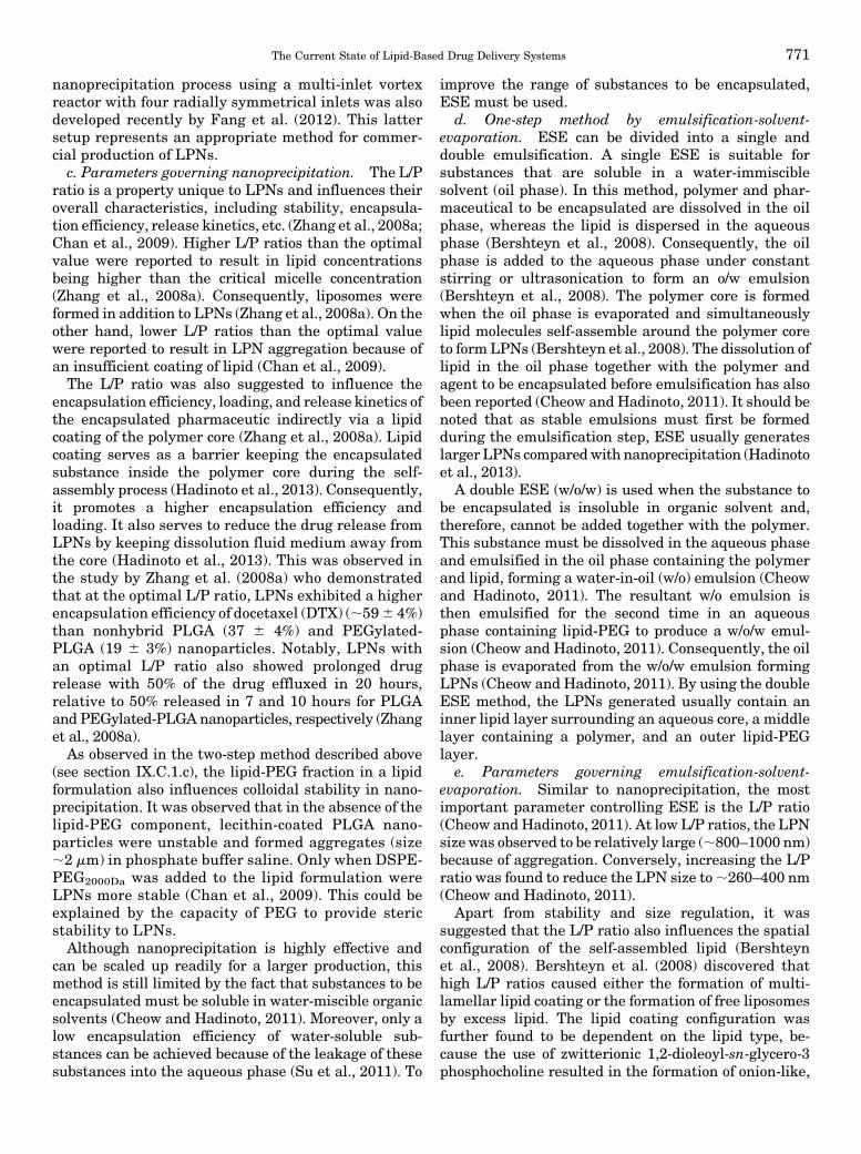
nanoprecipitation process using a multi-inlet vortexreactor with four radially symmetrical inlets was alsodeveloped recently by Fang et al. (2012). This lattersetup represents an appropriate method for commer-cial production of LPNs.c. Parameters governing nanoprecipitation. The L/P
ratio is a property unique to LPNs and influences theiroverall characteristics, including stability, encapsula-tion efficiency, release kinetics, etc. (Zhang et al., 2008a;Chan et al., 2009). Higher L/P ratios than the optimalvalue were reported to result in lipid concentrationsbeing higher than the critical micelle concentration(Zhang et al., 2008a). Consequently, liposomes wereformed in addition to LPNs (Zhang et al., 2008a). On theother hand, lower L/P ratios than the optimal valuewere reported to result in LPN aggregation because ofan insufficient coating of lipid (Chan et al., 2009).The L/P ratio was also suggested to influence the
encapsulation efficiency, loading, and release kinetics ofthe encapsulated pharmaceutic indirectly via a lipidcoating of the polymer core (Zhang et al., 2008a). Lipidcoating serves as a barrier keeping the encapsulatedsubstance inside the polymer core during the self-assembly process (Hadinoto et al., 2013). Consequently,it promotes a higher encapsulation efficiency andloading. It also serves to reduce the drug release fromLPNs by keeping dissolution fluid medium away fromthe core (Hadinoto et al., 2013). This was observed inthe study by Zhang et al. (2008a) who demonstratedthat at the optimal L/P ratio, LPNs exhibited a higherencapsulation efficiency of docetaxel (DTX) (;596 4%)than nonhybrid PLGA (37 6 4%) and PEGylated-PLGA (19 6 3%) nanoparticles. Notably, LPNs withan optimal L/P ratio also showed prolonged drugrelease with 50% of the drug effluxed in 20 hours,relative to 50% released in 7 and 10 hours for PLGAand PEGylated-PLGA nanoparticles, respectively (Zhanget al., 2008a).As observed in the two-step method described above
(see section IX.C.1.c), the lipid-PEG fraction in a lipidformulation also influences colloidal stability in nano-precipitation. It was observed that in the absence of thelipid-PEG component, lecithin-coated PLGA nano-particles were unstable and formed aggregates (size;2 mm) in phosphate buffer saline. Only when DSPE-PEG2000Da was added to the lipid formulation wereLPNs more stable (Chan et al., 2009). This could beexplained by the capacity of PEG to provide stericstability to LPNs.Although nanoprecipitation is highly effective and
can be scaled up readily for a larger production, thismethod is still limited by the fact that substances to beencapsulated must be soluble in water-miscible organicsolvents (Cheow and Hadinoto, 2011). Moreover, only alow encapsulation efficiency of water-soluble sub-stances can be achieved because of the leakage of thesesubstances into the aqueous phase (Su et al., 2011). To
improve the range of substances to be encapsulated,ESE must be used.
d. One-step method by emulsification-solvent-evaporation. ESE can be divided into a single anddouble emulsification. A single ESE is suitable forsubstances that are soluble in a water-immisciblesolvent (oil phase). In this method, polymer and phar-maceutical to be encapsulated are dissolved in the oilphase, whereas the lipid is dispersed in the aqueousphase (Bershteyn et al., 2008). Consequently, the oilphase is added to the aqueous phase under constantstirring or ultrasonication to form an o/w emulsion(Bershteyn et al., 2008). The polymer core is formedwhen the oil phase is evaporated and simultaneouslylipid molecules self-assemble around the polymer coreto form LPNs (Bershteyn et al., 2008). The dissolution oflipid in the oil phase together with the polymer andagent to be encapsulated before emulsification has alsobeen reported (Cheow and Hadinoto, 2011). It should benoted that as stable emulsions must first be formedduring the emulsification step, ESE usually generateslarger LPNs comparedwith nanoprecipitation (Hadinotoet al., 2013).
A double ESE (w/o/w) is used when the substance tobe encapsulated is insoluble in organic solvent and,therefore, cannot be added together with the polymer.This substance must be dissolved in the aqueous phaseand emulsified in the oil phase containing the polymerand lipid, forming a water-in-oil (w/o) emulsion (Cheowand Hadinoto, 2011). The resultant w/o emulsion isthen emulsified for the second time in an aqueousphase containing lipid-PEG to produce a w/o/w emul-sion (Cheow and Hadinoto, 2011). Consequently, the oilphase is evaporated from the w/o/w emulsion formingLPNs (Cheow and Hadinoto, 2011). By using the doubleESE method, the LPNs generated usually contain aninner lipid layer surrounding an aqueous core, a middlelayer containing a polymer, and an outer lipid-PEGlayer.
e. Parameters governing emulsification-solvent-evaporation. Similar to nanoprecipitation, the mostimportant parameter controlling ESE is the L/P ratio(Cheow and Hadinoto, 2011). At low L/P ratios, the LPNsize was observed to be relatively large (;800–1000 nm)because of aggregation. Conversely, increasing the L/Pratio was found to reduce the LPN size to;260–400 nm(Cheow and Hadinoto, 2011).
Apart from stability and size regulation, it wassuggested that the L/P ratio also influences the spatialconfiguration of the self-assembled lipid (Bershteynet al., 2008). Bershteyn et al. (2008) discovered thathigh L/P ratios caused either the formation of multi-lamellar lipid coating or the formation of free liposomesby excess lipid. The lipid coating configuration wasfurther found to be dependent on the lipid type, be-cause the use of zwitterionic 1,2-dioleoyl-sn-glycero-3phosphocholine resulted in the formation of onion-like,
The Current State of Lipid-Based Drug Delivery Systems 771

multilamellar stacks of lipid coating. In contrast, themixture of zwitterionic and cationic lipids led to atypical core-shell LPN structure with a single lipidlayer (Bershteyn et al., 2008).As the L/P ratio controls the LPN size and lipid
coating, it indirectly influences the encapsulation effi-ciency in a similar manner observed in the nanopreci-pitation method (Liu et al., 2010b). Typically, high L/Pratios give rise to smaller LPNs, leading to a lowerability to encapsulate drugs (Liu et al., 2010b). At thesame time, the presence of a high level of lipid on thesurface of the polymer core due to high L/P ratios canincrease the surface pressure and, consequently, re-duces drug leakage (Liu et al., 2010b). Therefore, LPNswith high encapsulation efficiency can be obtained byoptimizing the L/P ratio.As observed in the two-step method and also nano-
precipitation, the lipid-PEG fraction in a lipid formu-lation also influences the colloidal stability of theresulting LPNs in ESE (Chu et al., 2011). A lower massratio of lipid-PEG to polymerwas demonstrated to causeaggregation, reducing the fraction of LPNs in the sub-50 nm size (Chu et al., 2011). In contrast, a higher massratio increased the LPN fraction in the sub-50 nm sizefrom less than 20% to as high as 85% (Chu et al., 2011).Finally, another factor governing the ESE process is
the ionic interaction between the drug and the lipid(Cheow and Hadinoto, 2011). This parameter is uniqueto ESE. It was discovered by Cheow and Hadinoto(2011) that an unsuccessful emulsification processoccurred, causing the LPN formation to fail whenoppositely charged lipid and drug components wereused. However, this problem could be solved by addingcounterionic surfactants to the emulsion prior to LPNformation. In addition, the drug loading and releasekinetics were shown to be dependent on drug lipophi-licity (Cheow and Hadinoto, 2011). A higher drugloading and a slower drug release were observed withhighly lipophilic drugs as a result of strong hydrophobicinteractions between the drug and the lipid layer(Cheow and Hadinoto, 2011).It is worthmentioning that generally the ESEmethod
results in a higher encapsulation efficiency than nano-precipitation, which is due to the larger size of LPNsproduced by the ESEmethod (Hadinoto et al., 2013). Byusing nanoprecipitation, the encapsulation efficiency ofDTX in 50–60 nm LPNs was 20% (Chan et al., 2009),whereas 200–300 nm LPNs prepared using the ESEmethod were able to encapsulate up to ;60% of PTX(Liu et al., 2010b).3. Studies of Lipid-polymer Hybrid Nanoparticles in
Cancer Therapy. A single-drug delivery system usingLPNs has been studied against various cancer cell-typesin vitro, such as breast, prostate, lung, liver, andcervical cancer (Hitzman et al., 2006; Zhang et al.,2008a; Chan et al., 2009; Liu et al., 2010b; Chu et al.,2011). For instance, Liu et al. (2010b) encapsulated PTX
within LPNs, which led to a sustained release of thedrug and a 6- to 7-fold greater in vitro cytotoxicityagainst MCF-7 breast cancer cells after a 24 and72-hour incubation, respectively, compared with stan-dard taxol. In addition, LPNs exhibited cellular uptakeafter a 4-hour incubation that was ;70% higher thannonhybrid polymeric nanoparticles (Liu et al., 2010b).These LPNs released 100% of PTX within 7 dayscompared with 30% released by polymeric nanopar-ticles over the same period (Liu et al., 2010b).
More attention has been given to the capacity andpotential of LPNs as a combinatorial drug deliverysystem (Sengupta et al., 2005; Aryal et al., 2010, 2011,2012; Wang et al., 2010a,b; Kong et al., 2013). By usingvarious strategies, it is possible to incorporate multipledrugs within LPNs (Fig. 18). For instance, differentdrugs can be conjugated to polymers or lipids separatelybefore LPN preparation (Fig. 18, A and B) or encapsu-lated within preformed polymeric nanoparticles andlipid vesicles in the case of the two-step method (Fig.18C). Alternatively, a second drug can be conjugated tothe surface of LPNs postpreparation (Fig. 18D). The useof LPNs as a combinatorial drug delivery system wouldimprovemultidrug therapy by overcoming difficulties inobtaining a precise level of therapeutic exposure causedby variations in the pharmacokinetic and biodistribu-tion of different drugs (Hadinoto et al., 2013).
Sengupta et al. (2005) incorporated the anticancerdrug DOX and antiangiogenesis drug combretastatininto LPNs via a two-step method. Combretastatin wasincorporated into the lipid layer of LPNs, whereas DOXwas loaded into the polymeric core. This arrangementallowed an instant release of combretastatin to collapsethe tumoral vasculature, trapping DOX-encapsulatedpolymeric nanoparticles within the tumor (Senguptaet al., 2005). Such LPNs were taken up rapidly by tumorvasculature within 5 hours after initial exposure andwere retained within the tumor for at least 24 hours(Sengupta et al., 2005). In vivo studies using these LPNsalso demonstrated greater tumor growth inhibitionand a longer lifespan of mice bearing B16/F10 mela-noma or Lewis lung carcinoma compared with DOX andcombretastatin-loaded liposomes (Sengupta et al.,2005).
By using the nanoprecipitation technique, DOX andthe radiotherapeutic agents Indium-111 or Yttrium-90have also been successfully incorporated into LPNs(Wang et al., 2010a). The resulting LPNs, called Chemo-Rad, were taken up rapidly by LNCaP prostate cancercells within 45 minutes and exhibited a higher in vitrocytotoxicity than LPNs containing only one of theagents (Wang et al., 2010a). No interference to theencapsulation or the sustained release of DOX by theradiotherapeutics was observed using these LPNs, with100% of DOX being released within 7 days (Wang et al.,2010a). Furthermore, stimuli-responsive combinatorialLPNs were developed by Kong et al. (2013), who
772 Yingchoncharoen et al.

generated camptothecin-loaded LPNs containing mag-netic Fe3O4 nanoparticles. Upon the application of aremote radio frequency magnetic field, more than 90%of the encapsulated drug was released within 46 hours,and a greater growth inhibition of MT2 cancer cells wasobserved compared with nonhybrid magnetic nanopar-ticles (Kong et al., 2013).Active targeting strategies have also been examined
using LPNs. The conjugation of folic acid onto thesurface of LPNs containing DTX was shown to increasetheir uptake by;54% compared with unmodified LPNsafter only 2 hours of incubation with MCF-7 breastcancer cells (Liu et al., 2010a). This increased uptakeled to enhanced in vitro cytotoxicity by;51% comparedwith the unmodified counterpart (Liu et al., 2010a).
Similar results were observed in HeLa cells, bothin vitro and in vivo, when folic acid-conjugated LPNscontaining PTX were tested in comparison with theirunmodified counterparts (Zhao et al., 2012). Othertargeting ligands that have been examined includeaptamers (Zhang et al., 2008a), antibody (Hu et al.,2010), transferrin (Zheng et al., 2010), and peptides(Chan et al., 2010, 2011).
Apart from the incorporation of cytotoxic chemothera-peutic agents into LPNs as a single or combinatorial drugtransport system with/without active targeting, LPNshave been used for the delivery of genes (Li et al., 2010a;Zhong et al., 2010), siRNA (Shi et al., 2011; Hasan et al.,2012; Yang et al., 2012a), and diagnostic imaging agents(Kandel et al., 2011; Mieszawska et al., 2012).
Fig. 18. Various strategies for combinatorial drug delivery using LPNs. (A) Separate covalent conjugation of different drugs (drug X and Y) to thepolymer and phospholipid precursors before LPN preparation. For instance, drug X can be attached to the polymer, whereas drug Y can be attached tophospholipid molecules or vice versa. (B) Separate covalent conjugation of different drugs to polymer precursors prior to preparation. (C) Fusion ofpreformed polymeric nanoparticles and lipid vesicles via a two-step method of preparation. Different drugs are encapsulated within polymericnanoparticles and liposomes separately before particle fusion. (D) Conjugation of the second drug (drug Y) to the surface of LPNs postpreparation. Thefirst drug (drug X) is encapsulated within the polymeric core during LPN preparation.
The Current State of Lipid-Based Drug Delivery Systems 773

X. Conclusion
The development of nanoparticle drug delivery sys-tems, such as lipid-based nanoparticles, and particularlyliposomes, has emerged as a promising strategy for thetreatment of cancer. Since their discovery in the 1960s,liposome technology has advanced rapidly from beingused as an apparatus for cell membrane research tobecoming a versatile drug carrier. Similar to other typesof nanosized drug delivery systems, liposomes can im-prove the specificity and efficacy of chemotherapeutics,while reducing their toxic side effects bymeans of passiveand active targeting. However, lipid-based nanoparticlesaremore advantageous over other nanoparticles becauseof the more biocompatible and biodegradable nature oftheir constituents relative to synthetic polymers found inother types of nanoparticles. Moreover, the amphiphilicproperties of phospholipids that make up liposomesallow these nanoparticles to encapsulate both hydropho-bic and hydrophilic anti-cancer agents.Presently, a variety of liposomal encapsulated anti-
cancer drugs are clinically approved and commerciallyavailable, while many more formulations are beinginvestigated in different stages of clinical trials or areawaiting approval.Moreover, the development of variousliposome technologies, such as different types of stimuli-sensitive liposomes and multifunctional “SMART” lipo-somes, has also demonstrated the promising potential ofliposomes as effective anticancer agent delivery systems.Furthermore, advances in liposomal design have seenthe emergence of the next generation of lipid-basednanoparticles, including lipid micelles, solid lipidnanoparticles, nanostructured lipid carriers, and lipid-polymer hybrid nanoparticles. Such innovations arebelieved to be capable of overcoming current drawbacksobserved in liposome technology. However, more stud-ies are still required to optimize their capacity as drugdelivery systems. Together with other forms of targeteddrug delivery systems, liposomes and lipid-based nano-particles will help improve the efficacy and safetyprofile of anticancer agents and, more importantly,the fate of cancer patients.
Acknowledgments
Phatsapong Yingchoncharoen would like to dedicate this review toMrs. Sujira Yingchoncharoen, “my loving mother, my first teacher,and my best friend”, who died from lung cancer with leptomeningealmetastases on the 30th of April 2014. Phatsapong Yingchoncharoenalso thanks the Yingchoncharoen family, Thaveesakvilai family,Leehacharoenkul family, Uthayaratana family, Thongkongtoon fam-ily, and Ms. Kulvarong Kijtanasopa for their support through thishardship.
Authorship Contributions
Wrote or contributed to the writing of the manuscript: Yingchoncharoen,Kalinowski, and Richardson.
ReferencesAbd El-Rehim DM, Pinder SE, Paish CE, Bell JA, Rampaul RS, Blamey RW, RobertsonJF, Nicholson RI, and Ellis IO (2004) Expression and co-expression of the members of
the epidermal growth factor receptor (EGFR) family in invasive breast carcinoma. BrJ Cancer 91:1532–1542.
Abid MR, Yi X, Yano K, Shih S-C, and Aird WC (2006) Vascular endocan is prefer-entially expressed in tumor endothelium. Microvasc Res 72:136–145.
Abuchowski A, McCoy JR, Palczuk NC, van Es T, and Davis FF (1977) Effect ofcovalent attachment of polyethylene glycol on immunogenicity and circulating lifeof bovine liver catalase. J Biol Chem 252:3582–3586.
Ahmed SE, Martins AM, and Husseini GA (2015) The use of ultrasound to releasechemotherapeutic drugs from micelles and liposomes. J Drug Target 23:16–42.
Alkan-Onyuksel H, Ramakrishnan S, Chai H-B, and Pezzuto JM (1994) A mixedmicellar formulation suitable for the parenteral administration of taxol. Pharm Res11:206–212.
Allen C, Dos Santos N, Gallagher R, Chiu GN, Shu Y, Li WM, Johnstone SA, JanoffAS, Mayer LD, and Webb MS, et al. (2002) Controlling the physical behavior andbiological performance of liposome formulations through use of surface graftedpoly(ethylene glycol). Biosci Rep 22:225–250.
Allen TM, Austin GA, Chonn A, Lin L, and Lee KC (1991a) Uptake of liposomes bycultured mouse bone marrow macrophages: influence of liposome composition andsize. Biochim Biophys Acta 1061:56–64.
Allen TM and Chonn A (1987) Large unilamellar liposomes with low uptake into thereticuloendothelial system. FEBS Lett 223:42–46.
Allen TM, Hansen C, Martin F, Redemann C, and Yau-Young A (1991b) Liposomescontaining synthetic lipid derivatives of poly(ethylene glycol) show prolonged cir-culation half-lives in vivo. Biochim Biophys Acta 1066:29–36.
Allen TM (1997) Liposomes. Opportunities in drug delivery. Drugs 54 (Suppl 4):8–14.Allen TM (1998) Liposomal drug formulations. Rationale for development and whatwe can expect for the future. Drugs 56:747–756.
Allen TM, Ryan JL, and Papahadjopoulos D (1985) Gangliosides reduce leakage ofaqueous-space markers from liposomes in the presence of human plasma. BiochimBiophys Acta 818:205–210.
Amselem S, Cohen R, and Barenholz Y (1993a) In vitro tests to predict in vivo per-formance of liposomal dosage forms. Chem Phys Lipids 64:219–237.
Amselem S, Gabizon A, and Barenholz Y (1993b) A large-scale method for thepreparation of sterile and non-pyrogenic liposomal formulations of defined sizedistributions for clinical use, in Liposome Technology: Liposome Preparation andRelated Techniques (Gregoriadis G ed) pp 501–525, CRC Press, Boca Raton.
Anders CK, Adamo B, Karginova O, Deal AM, Rawal S, Darr D, Schorzman A, SantosC, Bash R, and Kafri T, et al. (2013) Pharmacokinetics and efficacy of PEGylatedliposomal doxorubicin in an intracranial model of breast cancer. PLoS One 8:e61359.
Anderson RG, Brown MS, Beisiegel U, and Goldstein JL (1982) Surface distributionand recycling of the low density lipoprotein receptor as visualized with antireceptorantibodies. J Cell Biol 93:523–531.
Anderson M and Omri A (2004) The effect of different lipid components on the in vitrostability and release kinetics of liposome formulations. Drug Deliv 11:33–39.
Angst MS and Drover DR (2006) Pharmacology of drugs formulated with DepoFoam:a sustained release drug delivery system for parenteral administration usingmultivesicular liposome technology. Clin Pharmacokinet 45:1153–1176.
Arias JL (2011) Drug targeting strategies in cancer treatment: an overview.Mini RevMed Chem 11:1–17.
Aryal S, Hu C-MJ, Fu V, and Zhang L (2012) Nanoparticle drug delivery enhancesthe cytotoxicity of hydrophobic-hydrophilic drug conjugates. J Mater Chem 22:994–999.
Aryal S, Hu C-MJ, and Zhang L (2010) Combinatorial drug conjugation enablesnanoparticle dual-drug delivery. Small 6:1442–1448.
Aryal S, Hu C-MJ, and Zhang L (2011) Polymeric nanoparticles with precise ratio-metric control over drug loading for combination therapy.Mol Pharm 8:1401–1407.
Awada A, Bondarenko IN, Bonneterre J, Nowara E, Ferrero JM, Bakshi AV, Wilke C,and Piccart M; CT4002 study group (2014) A randomized controlled phase II trialof a novel composition of paclitaxel embedded into neutral and cationic lipidstargeting tumor endothelial cells in advanced triple-negative breast cancer(TNBC). Ann Oncol 25:824–831.
Awasthi VD, Garcia D, Goins BA, and Phillips WT (2003) Circulation and bio-distribution profiles of long-circulating PEG-liposomes of various sizes in rabbits.Int J Pharm 253:121–132.
Azarmi S, Tao X, Chen H, Wang Z, Finlay WH, Löbenberg R, and Roa WH (2006)Formulation and cytotoxicity of doxorubicin nanoparticles carried by dry powderaerosol particles. Int J Pharm 319:155–161.
Babincová M, Altanerová V, Lampert M, Altaner C, Machová E, Srámka M,and Babinec P (2000) Site-specific in vivo targeting of magnetoliposomes usingexternally applied magnetic field. Z Naturforsch C 55:278–281.
Bae YH (2009) Drug targeting and tumor heterogeneity. J Control Release 133:2–3.Bagchi D, Bagchi M, Hassoun EA, Kelly J, and Stohs SJ (1995) Adriamycin-inducedhepatic and myocardial lipid peroxidation and DNA damage, and enhanced ex-cretion of urinary lipid metabolites in rats. Toxicology 95:1–9.
Bai X-F, Liu J, Li O, Zheng P, and Liu Y (2003) Antigenic drift as a mechanism fortumor evasion of destruction by cytolytic T lymphocytes. J Clin Invest 111:1487–1496.
Banerjee J, Hanson AJ, Gadam B, Elegbede AI, Tobwala S, Ganguly B, Wagh AV,Muhonen WW, Law B, and Shabb JB, et al. (2009) Release of liposomal contents bycell-secreted matrix metalloproteinase-9. Bioconjug Chem 20:1332–1339.
Bangham AD (1982) Preparation of liposomes and methods for measuring theirpermeabilities, in Technique in Life Science - Technique in Lipid and MembraneBiochemistry Part II (Hesketh TR, Kornberg HL, Metcalfe JC, Northcote DH,Pogson CI, and Tipton KF eds) pp 1–25, Elsevier/North-Holland, Amsterdam.
Bangham AD, Hill MW, and Miller NGA (1974) Preparation and use of liposomes asmodels of biological membranes, inMethods in Membrane Biology, Vol. 1 (Korn EDed) pp 1–68, Springer US, Boston.
Bangham AD, Standish MM, and Watkins JC (1965) Diffusion of univalent ionsacross the lamellae of swollen phospholipids. J Mol Biol 13:238–252.
774 Yingchoncharoen et al.

Bareford LM and Swaan PW (2007) Endocytic mechanisms for targeted drug de-livery. Adv Drug Deliv Rev 59:748–758.
Barenholz Y (2003) Relevancy of drug loading to liposomal formulation therapeuticefficacy. J Liposome Res 13:1–8.
Barenholz Y (2007) Amphipathic weak base loading into preformed liposomes havinga transmembrane ammonium ion gradient: From the bench to approved DOXIL, inLiposome Technology: Entrapment of drugs and other materials into liposomes(Gregoriadis G ed) pp 1-25, CRC Press, Boca Raton, FL.
Barenholz Y (2012a) Doxil® - the first FDA-approved nano-drug: from an idea to aproduct, in Handbook of Harnessing Biomaterials in Nanomedicine: Preparation,Toxicity, and Applications (Peer D ed) pp 335–398, Pan Stanford Publishing,Singapore.
Barenholz Y (2012b) Doxil®–the first FDA-approved nano-drug: lessons learned. JControl Release 160:117–134.
Barenholz Y and Cohen R (1995) Rational design of amphiphile-based drug carriersand sterically stabilized carriers. J Liposome Res 5:905–932.
Barenholz Y and Haran G (1994) inventors, Yissum Research Development Companyof the Hebrew University of Jerusalem, assignee. Methods of amphipathic drugloading in liposomes by ammonium ion gradient. U.S. patent US5316771. 1992 Dec18.
Barichello JM, Morishita M, Takayama K, and Nagai T (1999) Encapsulation ofhydrophilic and lipophilic drugs in PLGA nanoparticles by the nanoprecipitationmethod. Drug Dev Ind Pharm 25:471–476.
Batist G, Gelmon KA, Chi KN, Miller WH Jr, Chia SK, Mayer LD, Swenson CE,Janoff AS, and Louie AC (2009) Safety, pharmacokinetics, and efficacy of CPX-1liposome injection in patients with advanced solid tumors. Clin Cancer Res 15:692–700.
Batist G, Harris L, Azarnia N, Lee LW, and Daza-Ramirez P (2006) Improved anti-tumor response rate with decreased cardiotoxicity of non-pegylated liposomaldoxorubicin compared with conventional doxorubicin in first-line treatment ofmetastatic breast cancer in patients who had received prior adjuvant doxorubicin:results of a retrospective analysis. Anticancer Drugs 17:587–595.
Batist G, Ramakrishnan G, Rao CS, Chandrasekharan A, Gutheil J, Guthrie T, ShahP, Khojasteh A, Nair MK, and Hoelzer K, et al. (2001) Reduced cardiotoxicity andpreserved antitumor efficacy of liposome-encapsulated doxorubicin and cyclo-phosphamide compared with conventional doxorubicin and cyclophosphamide in arandomized, multicenter trial of metastatic breast cancer. J Clin Oncol 19:1444–1454.
Batzri S and Korn ED (1973) Single bilayer liposomes prepared without sonication.Biochim Biophys Acta 298:1015–1019.
Bedu-Addo FK, Tang P, Xu Y, and Huang L (1996a) Effects of polyethyleneglycolchain length and phospholipid acyl chain composition on the interaction ofpolyethyleneglycol-phospholipid conjugates with phospholipid: implications inliposomal drug delivery. Pharm Res 13:710–717.
Bedu-Addo FK, Tang P, Xu Y, and Huang L (1996b) Interaction of polyethyleneglycol-phospholipid conjugates with cholesterol-phosphatidylcholine mixtures: stericallystabilized liposome formulations. Pharm Res 13:718–724.
Belsito S, Bartucci R, and Sportelli L (2001) Lipid chain length effect on the phasebehaviour of PCs/PEG:2000-PEs mixtures. A spin label electron spin resonanceand spectrophotometric study. Biophys Chem 93:11–22.
Bergström K, Osterberg E, Holmberg K, Hoffman AS, Schuman TP, Kozlowski A,and Harris JH (1994) Effects of branching and molecular weight of surface-bound poly(ethylene oxide) on protein rejection. J Biomater Sci Polym Ed 6:123–132.
Bershteyn A, Chaparro J, Yau R, Kim M, Reinherz E, Ferreira-Moita L, and IrvineDJ (2008) Polymer-supported lipid shells, onions, and flowers. Soft Matter 4:1787–1791.
Bies C, Lehr C-M, and Woodley JF (2004) Lectin-mediated drug targeting: historyand applications. Adv Drug Deliv Rev 56:425–435.
Bleyer WA, Pizzo PA, Spence AM, Platt WD, Benjamin DR, Kolins CJ, and PoplackDG (1978) The Ommaya reservoir: newly recognized complications and recom-mendations for insertion and use. Cancer 41:2431–2437.
Blum RH and Carter SK (1974) Adriamycin. A new anticancer drug with significantclinical activity. Ann Intern Med 80:249–259.
Bolotin EM, Cohen R, Bar LK, Emanuel N, Ninio S, Barenholz Y, and Lasic DD(1994) Ammonium sulfate gradients for efficient and stable remote loading ofamphipathic weak bases into liposomes and ligandoliposomes. J Liposome Res 4:455–479.
Boltri L, Canal T, Esposito PA, and Carli F (1993) Lipid nanoparticles: evaluation ofsome critical formulation parameters. Proc Int Symp Control Release Bioact Mater20:346–347.
Bolwell BJ, Cassileth PA, and Gale RP (1988) High dose cytarabine: a review. Leu-kemia 2:253–260.
Boman NL, Bally MB, Cullis PR, Mayer LD, and Webb MS (1995) Encapsulation ofvincristine in liposomes reduces its toxicity and improves its anti-tumor efficacy. JLiposome Res 5:523–541.
Boman NL, Masin D, Mayer LD, Cullis PR, and Bally MB (1994) Liposomal vin-cristine which exhibits increased drug retention and increased circulation lon-gevity cures mice bearing P388 tumors. Cancer Res 54:2830–2833.
Bosanquet AG (1986) Stability of solutions of antineoplastic agents during prepara-tion and storage for in vitro assays. II. Assay methods, adriamycin and the otherantitumour antibiotics. Cancer Chemother Pharmacol 17:1–10.
Bossuyt V, Fadare O, Martel M, Ocal IT, Burtness B, Moinfar F, Leibl S,and Tavassoli FA (2005) Remarkably high frequency of EGFR expression in breastcarcinomas with squamous differentiation. Int J Surg Pathol 13:319–327.
Boucher Y, Baxter LT, and Jain RK (1990) Interstitial pressure gradients in tissue-isolated and subcutaneous tumors: implications for therapy. Cancer Res 50:4478–4484.
Boulikas T (2009) Clinical overview on Lipoplatin: a successful liposomal formulationof cisplatin. Expert Opin Investig Drugs 18:1197–1218.
Bourguignon LY, Zhu H, Shao L, and Chen YW (2000) CD44 interaction with tiam1promotes Rac1 signaling and hyaluronic acid-mediated breast tumor cell migra-tion. J Biol Chem 275:1829–1838.
Bourré L, Thibaut S, Fimiani M, Ferrand Y, Simonneaux G, and Patrice T (2003) Invivo photosensitizing efficiency of a diphenylchlorin sensitizer: interest of a DMPCliposome formulation. Pharmacol Res 47:253–261.
Bouzin C and Feron O (2007) Targeting tumor stroma and exploiting mature tumorvasculature to improve anti-cancer drug delivery. Drug Resist Updat 10:109–120.
Bradley AJ, Devine DV, Ansell SM, Janzen J, and Brooks DE (1998) Inhibition ofliposome-induced complement activation by incorporated poly(ethylene glycol)-lipids. Arch Biochem Biophys 357:185–194.
Brandhonneur N, Chevanne F, Vié V, Frisch B, Primault R, Le Potier M-F, and LeCorre P (2009) Specific and non-specific phagocytosis of ligand-grafted PLGA mi-crospheres by macrophages. Eur J Pharm Sci 36:474–485.
Bremerskov V and Linnemann R (1969) Some effects of daunomycin on the nucleicacid synthesis in synchronized L-cells. Eur J Cancer 5:317–330.
Broekgaarden M, de Kroon AI, Gulik TM, and Heger M (2014) Development andin vitro proof-of-concept of interstitially targeted zinc- phthalocyanine liposomesfor photodynamic therapy. Curr Med Chem 21:377–391.
Broekgaarden M, Kos M, Jurg FA, van Beek AA, van Gulik TM, and Heger M (2015)Inhibition of NF-kB in tumor cells exacerbates immune cell activation followingphotodynamic therapy. Int J Mol Sci 16:19960–19977.
Brown LF, Tognazzi K, Dvorak HF, and Harrist TJ (1996) Strong expression ofkinase insert domain-containing receptor, a vascular permeability factor/vascularendothelial growth factor receptor in AIDS-associated Kaposi’s sarcoma and cu-taneous angiosarcoma. Am J Pathol 148:1065–1074.
Bunjes H, Drechsler M, Koch MH, and Westesen K (2001) Incorporation of the modeldrug ubidecarenone into solid lipid nanoparticles. Pharm Res 18:287–293.
Bunjes H, Westesen K, and Koch MH (1996) Crystallization tendency and poly-morphic transitions in triglyceride nanoparticles. Int J Pharm 129:159–173.
Byrne JD, Betancourt T, and Brannon-Peppas L (2008) Active targeting schemesfor nanoparticle systems in cancer therapeutics. Adv Drug Deliv Rev 60:1615–1626.
Cai S, Thati S, Bagby TR, Diab H-M, Davies NM, Cohen MS, and Forrest ML (2010)Localized doxorubicin chemotherapy with a biopolymeric nanocarrier improvessurvival and reduces toxicity in xenografts of human breast cancer. J ControlRelease 146:212–218.
Camp ER, Wang C, Little EC, Watson PM, Pirollo KF, Rait A, Cole DJ, Chang EH,and Watson DK (2013) Transferrin receptor targeting nanomedicine deliveringwild-type p53 gene sensitizes pancreatic cancer to gemcitabine therapy. CancerGene Ther 20:222–228.
Campbell RB, Fukumura D, Brown EB, Mazzola LM, Izumi Y, Jain RK, Torchilin VP,and Munn LL (2002) Cationic charge determines the distribution of liposomesbetween the vascular and extravascular compartments of tumors. Cancer Res 62:6831–6836.
Campbell RB, Ying B, Kuesters GM, and Hemphill R (2009) Fighting cancer: from thebench to bedside using second generation cationic liposomal therapeutics. J PharmSci 98:411–429.
Cardone RA, Casavola V, and Reshkin SJ (2005) The role of disturbed pH dynamicsand the Na+/H+ exchanger in metastasis. Nat Rev Cancer 5:786–795.
Carmeliet P (2005) VEGF as a key mediator of angiogenesis in cancer. Oncology 69(Suppl 3):4–10.
Carpenter G and Cohen S (1979) Epidermal growth factor. Annu Rev Biochem 48:193–216.
Castle MC, Margileth DA, and Oliverio VT (1976) Distribution and excretion of(3H)vincristine in the rat and the dog. Cancer Res 36:3684–3689.
Cattaneo AG, Gornati R, Sabbioni E, Chiriva-Internati M, Cobos E, Jenkins MR,and Bernardini G (2010) Nanotechnology and human health: risks and benefits. JAppl Toxicol 30:730–744.
Catterall JB, Jones LM, and Turner GA (1999) Membrane protein glycosylation andCD44 content in the adhesion of human ovarian cancer cells to hyaluronan. ClinExp Metastasis 17:583–591.
Cavalli R, Caputo O, and Gasco MR (1993) Solid lipospheres of doxorubicin andidarubicin. Int J Pharm 89:9–12.
Cavalli R, Caputo O, and Gasco MR (2000) Preparation and characterization of solidlipid nanospheres containing paclitaxel. Eur J Pharm Sci 10:305–309.
Chamberlain MC and Corey-Bloom J (1991) Leptomeningeal metastases: 111indium-DTPA CSF flow studies. Neurology 41:1765–1769.
Chamberlain MC, Khatibi S, Kim JC, Howell SB, Chatelut E, and Kim S(1993) Treatment of leptomeningeal metastasis with intraventricular admin-istration of depot cytarabine (DTC 101). A phase I study. Arch Neurol 50:261–264.
Chan S, Davidson N, Juozaityte E, Erdkamp F, Pluzanska A, Azarnia N, and Lee LW(2004) Phase III trial of liposomal doxorubicin and cyclophosphamide comparedwith epirubicin and cyclophosphamide as first-line therapy for metastatic breastcancer. Ann Oncol 15:1527–1534.
Chan JM, Rhee JW, Drum CL, Bronson RT, Golomb G, Langer R, and FarokhzadOC (2011) In vivo prevention of arterial restenosis with paclitaxel-encapsulatedtargeted lipid-polymeric nanoparticles. Proc Natl Acad Sci USA 108:19347–19352.
Chan JM, Zhang L, Tong R, Ghosh D, Gao W, Liao G, Yuet KP, Gray D, Rhee J-W,and Cheng J, et al. (2010) Spatiotemporal controlled delivery of nanoparticles toinjured vasculature. Proc Natl Acad Sci USA 107:2213–2218.
Chan JM, Zhang L, Yuet KP, Liao G, Rhee J-W, Langer R, and Farokhzad OC (2009)PLGA-lecithin-PEG core-shell nanoparticles for controlled drug delivery. Bioma-terials 30:1627–1634.
Chandaroy P, Sen A, and Hui SW (2001) Temperature-controlled content releasefrom liposomes encapsulating Pluronic F127. J Control Release 76:27–37.
Chang J, Jallouli Y, Kroubi M, Yuan XB, Feng W, Kang CS, Pu PY, and Betbeder D(2009) Characterization of endocytosis of transferrin-coated PLGA nanoparticlesby the blood-brain barrier. Int J Pharm 379:285–292.
The Current State of Lipid-Based Drug Delivery Systems 775

Chanturiya A, Chernomordik LV, and Zimmerberg J (1997) Flickering fusion porescomparable with initial exocytotic pores occur in protein-free phospholipid bilayers.Proc Natl Acad Sci USA 94:14423–14428.
Charcosset C, El-Harati A, and Fessi H (2005) Preparation of solid lipid nano-particles using a membrane contactor. J Control Release 108:112–120.
Cheng WW and Allen TM (2008) Targeted delivery of anti-CD19 liposomal doxoru-bicin in B-cell lymphoma: a comparison of whole monoclonal antibody, Fab’ frag-ments and single chain Fv. J Control Release 126:50–58.
Cheow WS and Hadinoto K (2011) Factors affecting drug encapsulation and stabilityof lipid-polymer hybrid nanoparticles. Colloids Surf B Biointerfaces 85:214–220.
Chernomordik LV and Kozlov MM (2003) Protein-lipid interplay in fusion and fissionof biological membranes. Annu Rev Biochem 72:175–207.
Chernomordik LV and Kozlov MM (2008) Mechanics of membrane fusion. Nat StructMol Biol 15:675–683.
Chernomordik LV, Melikyan GB, and Chizmadzhev YA (1987) Biomembrane fusion:a new concept derived from model studies using two interacting planar lipidbilayers. Biochim Biophys Acta 906:309–352.
Chithrani BD, Ghazani AA, and Chan WC (2006) Determining the size and shapedependence of gold nanoparticle uptake into mammalian cells. Nano Lett 6:662–668.
Cho K, Wang X, Nie S, Chen ZG, and Shin DM (2008) Therapeutic nanoparticles fordrug delivery in cancer. Clin Cancer Res 14:1310–1316.
Choi HS, Liu W, Misra P, Tanaka E, Zimmer JP, Itty Ipe B, Bawendi MG,and Frangioni JV (2007) Renal clearance of quantum dots. Nat Biotechnol 25:1165–1170.
Choi W-J, Kim J-K, Choi S-H, Park J-S, Ahn WS, and Kim C-K (2004) Low toxicity ofcationic lipid-based emulsion for gene transfer. Biomaterials 25:5893–5903.
Chou H-H, Wang K-L, Chen C-A, Wei L-H, Lai C-H, Hsieh C-Y, Yang Y-C, Twu N-F,Chang T-C, and Yen M-S; Taiwanese Gynecologic Oncology Group (2006) Pegylatedliposomal doxorubicin (Lipo-Dox) for platinum-resistant or refractory epithelialovarian carcinoma: a Taiwanese gynecologic oncology group study with long-termfollow-up. Gynecol Oncol 101:423–428.
Chu C-J, Dijkstra J, Lai M-Z, Hong K, and Szoka FC (1990) Efficiency of cytoplasmicdelivery by pH-sensitive liposomes to cells in culture. Pharm Res 7:824–834.
Chu C-J and Szoka FC (1994) pH-sensitive liposomes. J Liposome Res 4:361–395.Chu C-H, Wang Y-C, Huang H-Y, Wu L-C, and Yang C-S (2011) Ultrafine PEG-coated poly(lactic-co-glycolic acid) nanoparticles formulated by hydrophobicsurfactant-assisted one-pot synthesis for biomedical applications. Nanotechnology22:185601.
Clague MJ, Urbé S, Aniento F, and Gruenberg J (1994) Vacuolar ATPase activity isrequired for endosomal carrier vesicle formation. J Biol Chem 269:21–24.
Clarke SJ and Rivory LP (1999) Clinical pharmacokinetics of docetaxel. Clin Phar-macokinet 36:99–114.
Collins D(1995) pH-sensitive liposomes as tools for cytoplasmic delivery, in Lipo-somes as Tools in Basic Research and Industry (Philippot JR and Schuber F eds) pp201–214, CRC Press, Boca Raton.
Comiskey SJ and Heath TD (1990) Serum-induced leakage of negatively chargedliposomes at nanomolar lipid concentrations. Biochemistry 29:3626–3631.
Cortes JE, Goldberg SL, Feldman EJ, Rizzeri DA, Hogge DE, Larson M, Pigneux A,Recher C, Schiller G, and Warzocha K, et al. (2015) Phase II, multicenter, ran-domized trial of CPX-351 (cytarabine:daunorubicin) liposome injection versus in-tensive salvage therapy in adults with first relapse AML. Cancer 121:234–242.
Crom WR, de Graaf SS, Synold T, Uges DR, Bloemhof H, Rivera G, Christensen ML,Mahmoud H, and Evans WE (1994) Pharmacokinetics of vincristine in children andadolescents with acute lymphocytic leukemia. J Pediatr 125:642–649.
Cudd A and Nicolau C (1985) Intracellular fate of liposome-encapsulated DNA inmouse liver. Analysis using electron microscope autoradiography and subcellularfractionation. Biochim Biophys Acta 845:477–491.
Cullis PR, Chonn A, and Semple SC (1998) Interactions of liposomes and lipid-basedcarrier systems with blood proteins: Relation to clearance behaviour in vivo. AdvDrug Deliv Rev 32:3–17.
Cullis PR and de Kruijff B (1979) Lipid polymorphism and the functional roles oflipids in biological membranes. Biochim Biophys Acta 559:399–420.
Daemen T, Velinova M, Regts J, de Jager M, Kalicharan R, Donga J, van der WantJJ, and Scherphof GL (1997) Different intrahepatic distribution of phosphatidyl-glycerol and phosphatidylserine liposomes in the rat. Hepatology 26:416–423.
Dagher R, Johnson J, Williams G, Keegan P, and Pazdur R (2004) Acceleratedapproval of oncology products: a decade of experience. J Natl Cancer Inst 96:1500–1509.
Damen J, Regts J, and Scherphof G (1981) Transfer and exchange of phospholipidbetween small unilamellar liposomes and rat plasma high density lipoproteins.Dependence on cholesterol content and phospholipid composition. Biochim BiophysActa 665:538–545.
Damodar G, Smitha T, Gopinath S, Vijayakumar S, and Rao Y (2014) An evaluationof hepatotoxicity in breast cancer patients receiving injection Doxorubicin. AnnMed Health Sci Res 4:74–79.
Damoiseau X, Schuitmaker HJ, Lagerberg JW, and Hoebeke M (2001) Increase of thephotosensitizing efficiency of the Bacteriochlorin a by liposome-incorporation. JPhotochem Photobiol B 60:50–60.
Dandamudi S and Campbell RB (2007) The drug loading, cytotoxicty and tumorvascular targeting characteristics of magnetite in magnetic drug targeting. Bio-materials 28:4673–4683.
Danhier F, Feron O, and Préat V (2010) To exploit the tumor microenvironment:Passive and active tumor targeting of nanocarriers for anti-cancer drug delivery. JControl Release 148:135–146.
Daniels TR, Delgado T, Helguera G, and Penichet ML (2006) The transferrin receptorpart II: targeted delivery of therapeutic agents into cancer cells. Clin Immunol 121:159–176.
Das S and Chaudhury A (2011) Recent advances in lipid nanoparticle formulationswith solid matrix for oral drug delivery. AAPS PharmSciTech 12:62–76.
Dautry-Varsat A, Ciechanover A, and Lodish HF (1983) pH and the recycling oftransferrin during receptor-mediated endocytosis. Proc Natl Acad Sci USA 80:2258–2262.
Davies EJ, Blackhall FH, Shanks JH, David G, McGown AT, Swindell R, Slade RJ,Martin-Hirsch P, Gallagher JT, and Jayson GC (2004) Distribution and clinicalsignificance of heparan sulfate proteoglycans in ovarian cancer. Clin Cancer Res10:5178–5186.
Davis ME, Chen ZG, and Shin DM (2008) Nanoparticle therapeutics: an emergingtreatment modality for cancer. Nat Rev Drug Discov 7:771–782.
De Gennes P (1987) Polymers at an interface; a simplified view. Adv Colloid InterfaceSci 27:189–209.
De Miguel D, Basáñez G, Sánchez D, Malo PG, Marzo I, Larrad L, Naval J, Pardo J,Anel A, and Martinez-Lostao L (2013) Liposomes decorated with Apo2L/TRAILovercome chemoresistance of human hematologic tumor cells. Mol Pharm 10:893–904.
De Miguel I, Imbertie L, Rieumajou V, Major M, Kravtzoff R, and Betbeder D (2000)Proofs of the structure of lipid coated nanoparticles (SMBV) used as drug carriers.Pharm Res 17:817–824.
Deamer D and Bangham AD (1976) Large volume liposomes by an ether vaporizationmethod. Biochim Biophys Acta 443:629–634.
Deckers R, Rome C, and Moonen CT (2008) The role of ultrasound and magneticresonance in local drug delivery. J Magn Reson Imaging 27:400–409.
Demirgöz D, Garg A, and Kokkoli E (2008) PR_b-targeted PEGylated liposomes forprostate cancer therapy. Langmuir 24:13518–13524.
Denekamp J (1984) Vasculature as a target for tumour therapy. Prog Appl Microcirc4:28–38.
Denekamp J and Hobson B (1982) Endothelial-cell proliferation in experimentaltumours. Br J Cancer 46:711–720.
Derksen JT, Morselt HW, Kalicharan D, Hulstaert CE, and Scherphof GL (1987)Interaction of immunoglobulin-coupled liposomes with rat liver macrophagesin vitro. Exp Cell Res 168:105–115.
Derycke AS and de Witte PA (2004) Liposomes for photodynamic therapy. Adv DrugDeliv Rev 56:17–30.
Desgrosellier JS and Cheresh DA (2010) Integrins in cancer: biological implicationsand therapeutic opportunities. Nat Rev Cancer 10:9–22.
Deshpande PP, Biswas S, and Torchilin VP (2013) Current trends in the use ofliposomes for tumor targeting. Nanomedicine (Lond) 8:1509–1528.
Devine DV, Wong K, Serrano K, Chonn A, and Cullis PR (1994) Liposome-complement interactions in rat serum: implications for liposome survival studies.Biochim Biophys Acta 1191:43–51.
Dhami S and Phillips D (1996) Comparison of the photophysics of an aggregating andnon-aggregating aluminium phthalocyanine system incorporated into unilamellarvesicles. J Photochem Photobiol Chem 100:77–84.
Diaz SL, Padler-Karavani V, Ghaderi D, Hurtado-Ziola N, Yu H, Chen X, Brinkman-Van der Linden EC, Varki A, and Varki NM (2009) Sensitive and specific detectionof the non-human sialic Acid N-glycolylneuraminic acid in human tissues andbiotherapeutic products. PLoS One 4:e4241.
Dienst A, Grunow A, Unruh M, Rabausch B, Nör JE, Fries JW, and Gottstein C(2005) Specific occlusion of murine and human tumor vasculature by VCAM-1-targeted recombinant fusion proteins. J Natl Cancer Inst 97:733–747.
Dijkstra J, van Galen WJ, Hulstaert CE, Kalicharan D, Roerdink FH, and ScherphofGL (1984) Interaction of liposomes with Kupffer cells in vitro. Exp Cell Res 150:161–176.
Dimmer KS, Friedrich B, Lang F, Deitmer JW, and Bröer S (2000) The low-affinitymonocarboxylate transporter MCT4 is adapted to the export of lactate in highlyglycolytic cells. Biochem J 350:219–227.
DiResta GR, Lee J, Larson SM, and Arbit E (1993) Characterization of neuroblas-toma xenograft in rat flank. I. Growth, interstitial fluid pressure, and interstitialfluid velocity distribution profiles. Microvasc Res 46:158–177.
Doktorovová S, Araújo J, Garcia ML, Rakovský E, and Souto EB (2010) Formulatingfluticasone propionate in novel PEG-containing nanostructured lipid carriers(PEG-NLC). Colloids Surf B Biointerfaces 75:538–542.
Dos Santos N, Allen C, Doppen A-M, Anantha M, Cox KA, Gallagher RC, Karlsson G,Edwards K, Kenner G, and Samuels L, et al. (2007) Influence of poly(ethyleneglycol) grafting density and polymer length on liposomes: relating plasma circu-lation lifetimes to protein binding. Biochim Biophys Acta 1768:1367–1377.
Dragovich T, Mendelson D, Kurtin S, Richardson K, Von Hoff D, and Hoos A (2006)A Phase 2 trial of the liposomal DACH platinum L-NDDP in patients withtherapy-refractory advanced colorectal cancer. Cancer Chemother Pharmacol 58:759–764.
Drummond DC, Noble CO, Guo Z, Hong K, Park JW, and Kirpotin DB (2006) De-velopment of a highly active nanoliposomal irinotecan using a novel intraliposomalstabilization strategy. Cancer Res 66:3271–3277.
Dubey PK, Mishra V, Jain S, Mahor S, and Vyas SP (2004) Liposomes modified withcyclic RGD peptide for tumor targeting. J Drug Target 12:257–264.
du Bois A, Burges A, Meier W, Pfisterer J, Schmalfeldt B, Richter B, Jackisch C,Staehle A, Kimmig R, and Elser G; Arbeitsgemeinschaft Gynaekologische Onko-logie Studiengruppe Ovarialkarzinom (2006) Pegylated liposomal doxorubicin andcarboplatin in advanced gynecologic tumors: a prospective phase I/II study of theArbeitsgemeinschaft Gynaekologische Onkologie Studiengruppe Ovarialkarzinom(AGO-OVAR). Ann Oncol 17:93–96.
Duffaud F, Borner M, Chollet P, Vermorken JB, Bloch J, Degardin M, Rolland F,Dittrich C, Baron B, and Lacombe D, et al.; EORTC-New Drug DevelopmentGroup/New Drug Development Program (2004) Phase II study of OSI-211 (lipo-somal lurtotecan) in patients with metastatic or loco-regional recurrent squamouscell carcinoma of the head and neck. An EORTC New Drug Development Groupstudy. Eur J Cancer 40:2748–2752.
Düzgünes N, Simões S, Slepushkin V, Pretzer E, Rossi JJ, De Clercq E, Antao VP,Collins ML, and de Lima MC (2001) Enhanced inhibition of HIV-1 replication inmacrophages by antisense oligonucleotides, ribozymes and acyclic nucleoside
776 Yingchoncharoen et al.

phosphonate analogs delivered in pH-sensitive liposomes. Nucleosides NucleotidesNucleic Acids 20:515–523.
Düzgünes N, Straubinger RM, Baldwin PA, Friend DS, and Papahadjopoulos D(1985) Proton-induced fusion of oleic acid-phosphatidylethanolamine liposomes.Biochemistry 24:3091–3098.
Düzgünes N, Wilschut J, Fraley R, and Papahadjopoulos D (1981) Studies on themechanism of membrane fusion. Role of head-group composition in calcium- andmagnesium-induced fusion of mixed phospholipid vesicles. Biochim Biophys Acta642:182–195.
Dvorak HF, Nagy JA, Dvorak JT, and Dvorak AM (1988) Identification and char-acterization of the blood vessels of solid tumors that are leaky to circulatingmacromolecules. Am J Pathol 133:95–109.
Easson EC and Pointon RCS (1985) The radiotherapy of malignant disease, 1st ed,Springer-Verlag London, London.
Edwards K, Johnsson M, Karlsson G, and Silvander M (1997) Effect ofpolyethyleneglycol-phospholipids on aggregate structure in preparations of smallunilamellar liposomes. Biophys J 73:258–266.
Eichhorn ME, Luedemann S, Strieth S, Papyan A, Ruhstorfer H, Haas H, MichaelisU, Sauer B, Teifel M, and Enders G, et al. (2007) Cationic lipid complexedcamptothecin (EndoTAG-2) improves antitumoral efficacy by tumor vasculartargeting. Cancer Biol Ther 6:920–929.
Eliaz RE and Szoka FC Jr (2001) Liposome-encapsulated doxorubicin targeted toCD44: a strategy to kill CD44-overexpressing tumor cells. Cancer Res 61:2592–2601.
Ellens H, Bentz J, and Szoka FC (1985) H+- and Ca2+-induced fusion and de-stabilization of liposomes. Biochemistry 24:3099–3106.
Ellens H, Bentz J, and Szoka FC (1986) Destabilization of phosphatidylethanolamineliposomes at the hexagonal phase transition temperature. Biochemistry 25:285–294.
Enoch HG and Strittmatter P (1979) Formation and properties of 1000-A-diameter,single-bilayer phospholipid vesicles. Proc Natl Acad Sci USA 76:145–149.
Fang RH, Aryal S, Hu C-MJ, and Zhang L (2010) Quick synthesis of lipid-polymerhybrid nanoparticles with low polydispersity using a single-step sonicationmethod. Langmuir 26:16958–16962.
Fang RH, Chen KN, Aryal S, Hu C-MJ, Zhang K, and Zhang L (2012) Large-scalesynthesis of lipid-polymer hybrid nanoparticles using a multi-inlet vortex reactor.Langmuir 28:13824–13829.
Fasol U, Frost A, Büchert M, Arends J, Fiedler U, Scharr D, Scheuenpflug J,and Mross K (2012) Vascular and pharmacokinetic effects of EndoTAG-1 in pa-tients with advanced cancer and liver metastasis. Ann Oncol 23:1030–1036.
Fears CY, Gladson CL, and Woods A (2006) Syndecan-2 is expressed in the micro-vasculature of gliomas and regulates angiogenic processes in microvascular en-dothelial cells. J Biol Chem 281:14533–14536.
Feldman EJ, Lancet JE, Kolitz JE, Ritchie EK, Roboz GJ, List AF, Allen SL, AsatianiE, Mayer LD, and Swenson C, et al. (2011) First-in-man study of CPX-351: aliposomal carrier containing cytarabine and daunorubicin in a fixed 5:1 molar ratiofor the treatment of relapsed and refractory acute myeloid leukemia. J Clin Oncol29:979–985.
Felgner PL, Gadek TR, HolmM, Roman R, Chan HW, Wenz M, Northrop JP, RingoldGM, and Danielsen M (1987) Lipofection: a highly efficient, lipid-mediated DNA-transfection procedure. Proc Natl Acad Sci USA 84:7413–7417.
Fenart L, Casanova A, Dehouck B, Duhem C, Slupek S, Cecchelli R, and Betbeder D(1999) Evaluation of effect of charge and lipid coating on ability of 60-nm nano-particles to cross an in vitro model of the blood-brain barrier. J Pharmacol ExpTher 291:1017–1022.
Feron O (2009) Pyruvate into lactate and back: from the Warburg effect to symbioticenergy fuel exchange in cancer cells. Radiother Oncol 92:329–333.
Ferrari M (2005) Cancer nanotechnology: Opportunities and challenges. Nat RevCancer 5:161–171.
Ferrati S, Nicolov E, Bansal S, Hosali S, Landis M, and Grattoni A (2015) Docetaxel/2-Hydroxypropyl b -Cyclodextrin Inclusion Complex Increases Docetaxel Solubility andRelease from a Nanochannel Drug Delivery System. Curr Drug Targets 16:1645–1649.
Filipovi�c-Grci�c J, Skalko-Basnet N, and Jalsenjak I (2001) Mucoadhesive chitosan-coated liposomes: characteristics and stability. J Microencapsul 18:3–12.
Fischer V, Rodríguez-Gascón A, Heitz F, Tynes R, Hauck C, Cohen D, and VickersAE (1998) The multidrug resistance modulator valspodar (PSC 833) is metab-olized by human cytochrome P450 3A. Implications for drug-drug interactionsand pharmacological activity of the main metabolite. Drug Metab Dispos 26:802–811.
Folkman J (1971) Transplacental carcinogenesis by stilbestrol. N Engl J Med 285:404–405.
Forrest ML, Won C-Y, Malick AW, and Kwon GS (2006) In vitro release of the mTORinhibitor rapamycin from poly(ethylene glycol)-b-poly(«-caprolactone) micelles. JControl Release 110:370–377.
Forssen EA (1997) The design and development of DaunoXome® for solid tumortargeting in vivo. Adv Drug Deliv Rev 24:133–150.
Forssen EA, Coulter DM, and Proffitt RT (1992) Selective in vivo localization ofdaunorubicin small unilamellar vesicles in solid tumors. Cancer Res 52:3255–3261.
Forssen EA, Malé-Brune R, Adler-Moore JP, Lee MJ, Schmidt PG, Krasieva TB,Shimizu S, and Tromberg BJ (1996) Fluorescence imaging studies for the dispo-sition of daunorubicin liposomes (DaunoXome) within tumor tissue. Cancer Res 56:2066–2075.
Forssen EA and Ross ME (1994) Daunoxome® treatment of solid tumors: preclinicaland clinical investigations. J Liposome Res 4:481–512.
Frenkel V (2008) Ultrasound mediated delivery of drugs and genes to solid tumors.Adv Drug Deliv Rev 60:1193–1208.
Friend DS, Papahadjopoulos D, and Debs RJ (1996) Endocytosis and intracellularprocessing accompanying transfection mediated by cationic liposomes. BiochimBiophys Acta 1278:41–50.
Funato K, Yoda R, and Kiwada H (1992) Contribution of complement system ondestabilization of liposomes composed of hydrogenated egg phosphatidylcholine inrat fresh plasma. Biochim Biophys Acta 1103:198–204.
Fundarò A, Cavalli R, Bargoni A, Vighetto D, Zara GP, and Gasco MR (2000) Non-stealth and stealth solid lipid nanoparticles (SLN) carrying doxorubicin: pharma-cokinetics and tissue distribution after i.v. administration to rats. Pharmacol Res42:337–343.
Fuster MM and Esko JD (2005) The sweet and sour of cancer: glycans as noveltherapeutic targets. Nat Rev Cancer 5:526–542.
Gabizon A, Catane R, Uziely B, Kaufman B, Safra T, Cohen R, Martin F, Huang A,and Barenholz Y (1994) Prolonged circulation time and enhanced accumulation inmalignant exudates of doxorubicin encapsulated in polyethylene-glycol coated li-posomes. Cancer Res 54:987–992.
Gabizon A, Chisin R, Amselem S, Druckmann S, Cohen R, Goren D, Fromer I, PeretzT, Sulkes A, and Barenholz Y (1991) Pharmacokinetic and imaging studies inpatients receiving a formulation of liposome-associated adriamycin. Br J Cancer64:1125–1132.
Gabizon A, Horowitz AT, Goren D, Tzemach D, Shmeeda H, and Zalipsky S (2003a)In vivo fate of folate-targeted polyethylene-glycol liposomes in tumor-bearing mice.Clin Cancer Res 9:6551–6559.
Gabizon A and Martin F (1997) Polyethylene glycol-coated (pegylated) liposomaldoxorubicin. Rationale for use in solid tumours. Drugs 54 (Suppl 4):15–21.
Gabizon AA and Muggia FM (1998) Initial clinical evaluation of pegylated-liposomaldoxorubicin in solid tumors, in Long Circulating Liposomes: Old drugs, NewTherapeutics (Woodle MC and Storm G, eds, ed) pp 165–174, Springer, Berlin.
Gabizon A and Papahadjopoulos D (1988) Liposome formulations with prolongedcirculation time in blood and enhanced uptake by tumors. Proc Natl Acad Sci USA85:6949–6953.
Gabizon A, Peretz T, Sulkes A, Amselem S, Ben-Yosef R, Ben-Baruch N, Catane R,Biran S, and Barenholz Y (1989) Systemic administration of doxorubicin-containing liposomes in cancer patients: a phase I study. Eur J Cancer ClinOncol 25:1795–1803.
Gabizon A, Shmeeda H, and Barenholz Y (2003b) Pharmacokinetics of pegylatedliposomal Doxorubicin: review of animal and human studies. Clin Pharmacokinet42:419–436.
Gamucci T, Paridaens R, Heinrich B, Schellens JH, Pavlidis N, Verweij J, Sessa C,Kaye S, Roelvink M, and Wanders J, et al. (2000) Activity and toxicity of GI147211in breast, colorectal and non-small-cell lung cancer patients: an EORTC-ECSGphase II clinical study. Ann Oncol 11:793–797.
Ganta S, Devalapally H, Shahiwala A, and Amiji M (2008) A review of stimuli-responsive nanocarriers for drug and gene delivery. J Control Release 126:187–204.
Gao Z, Lukyanov AN, Chakilam AR, and Torchilin VP (2003) PEG-PE/phosphati-dylcholine mixed immunomicelles specifically deliver encapsulated taxol to tumorcells of different origin and promote their efficient killing. J Drug Target 11:87–92.
Gao Z, Lukyanov AN, Singhal A, and Torchilin VP (2002) Diacyllipid-polymer mi-celles as nanocarriers for poorly soluble anticancer drugs. Nano Lett 2:979–982.
Garbuzenko O, Barenholz Y, and Priev A (2005) Effect of grafted PEG on liposomesize and on compressibility and packing of lipid bilayer. Chem Phys Lipids 135:117–129.
Garcia AM, Alarcon E, Muñoz M, Scaiano JC, Edwards AM, and Lissi E (2011)Photophysical behaviour and photodynamic activity of zinc phthalocyanines asso-ciated to liposomes. Photochem Photobiol Sci 10:507–514.
Garg A, Tisdale AW, Haidari E, and Kokkoli E (2009) Targeting colon cancer cellsusing PEGylated liposomes modified with a fibronectin-mimetic peptide. Int JPharm 366:201–210.
Gasco M (1997) Solid lipid nanospheres from warm micro-emulsions. Pharm TechnEur 9:52–58.
Gasco MR (1993), inventors, Gasco MR, assignee. Method for producing solid lipidmicrospheres having a narrow size distribution. U.S. Patent US5250236. 1991Aug 2.
Gaspani S and Milani B (2013) Access to liposomal generic formulations: beyondAmBisome and Doxil/Caelyx. GaBi J 2:60–62.
Gbadamosi JK, Hunter AC, and Moghimi SM (2002) PEGylation of microspheresgenerates a heterogeneous population of particles with differential surface char-acteristics and biological performance. FEBS Lett 532:338–344.
Gelmon KA, Tolcher A, Diab AR, Bally MB, Embree L, Hudon N, Dedhar C, Ayers D,Eisen A, and Melosky B, et al. (1999) Phase I study of liposomal vincristine. J ClinOncol 17:697–705.
Geng Y, Dalhaimer P, Cai S, Tsai R, Tewari M, Minko T, and Discher DE (2007)Shape effects of filaments versus spherical particles in flow and drug delivery. NatNanotechnol 2:249–255.
Genís L, Gálvez BG, Gonzalo P, and Arroyo AG (2006) MT1-MMP: universal orparticular player in angiogenesis? Cancer Metastasis Rev 25:77–86.
Gerritsen WJ, Verkley AJ, Zwaal RF, and Van Deenen LL (1978) Freeze-fractureappearance and disposition of band 3 protein from the human erythrocyte mem-brane in lipid vesicles. Eur J Biochem 85:255–261.
Gewirtz DA (1999) A critical evaluation of the mechanisms of action proposed for theantitumor effects of the anthracycline antibiotics adriamycin and daunorubicin.Biochem Pharmacol 57:727–741.
Ghanbarzadeh S, Arami S, Pourmoazzen Z, and Khorrami A (2014) Improvementof the antiproliferative effect of rapamycin on tumor cell lines by poly(monomethylitaconate)-based pH-sensitive, plasma stable liposomes. Colloids SurfB Biointerfaces 115:323–330.
Ghinea N and Simionescu N (1985) Anionized and cationized hemeundecapeptides asprobes for cell surface charge and permeability studies: differentiated labeling ofendothelial plasmalemmal vesicles. J Cell Biol 100:606–612.
Gill PS, Espina BM, Muggia F, Cabriales S, Tulpule A, Esplin JA, Liebman HA,Forssen E, Ross ME, and Levine AM (1995) Phase I/II clinical and pharmacokineticevaluation of liposomal daunorubicin. J Clin Oncol 13:996–1003.
The Current State of Lipid-Based Drug Delivery Systems 777

Gill PS, Wernz J, Scadden DT, Cohen P, Mukwaya GM, von Roenn JH, Jacobs M,Kempin S, Silverberg I, and Gonzales G, et al. (1996) Randomized phase III trial ofliposomal daunorubicin versus doxorubicin, bleomycin, and vincristine in AIDS-related Kaposi’s sarcoma. J Clin Oncol 14:2353–2364.
Glantz MJ, Jaeckle KA, Chamberlain MC, Phuphanich S, Recht L, Swinnen LJ,Maria B, LaFollette S, Schumann GB, and Cole BF, et al. (1999a) A randomizedcontrolled trial comparing intrathecal sustained-release cytarabine (DepoCyt) tointrathecal methotrexate in patients with neoplastic meningitis from solid tumors.Clin Cancer Res 5:3394–3402.
Glantz MJ, LaFollette S, Jaeckle KA, Shapiro W, Swinnen L, Rozental JR, PhuphanichS, Rogers LR, Gutheil JC, and Batchelor T, et al. (1999b) Randomized trial of a slow-release versus a standard formulation of cytarabine for the intrathecal treatment oflymphomatous meningitis. J Clin Oncol 17:3110–3116.
Goldstein JL, Brown MS, Anderson RG, Russell DW, and Schneider WJ (1985)Receptor-mediated endocytosis: concepts emerging from the LDL receptor system.Annu Rev Cell Biol 1:1–39.
Gomme PT, McCann KB, and Bertolini J (2005) Transferrin: structure, function andpotential therapeutic actions. Drug Discov Today 10:267–273.
Goormaghtigh E, Chatelain P, Caspers J, and Ruysschaert JM (1980) Evidence of aspecific complex between adriamycin and negatively-charged phospholipids. Bio-chim Biophys Acta 597:1–14.
Gordon AN, Fleagle JT, Guthrie D, Parkin DE, Gore ME, and Lacave AJ (2001)Recurrent epithelial ovarian carcinoma: a randomized phase III study of pegylatedliposomal doxorubicin versus topotecan. J Clin Oncol 19:3312–3322.
Gordon KB, Tajuddin A, Guitart J, Kuzel TM, Eramo LR, and VonRoenn J (1995)Hand-foot syndrome associated with liposome-encapsulated doxorubicin therapy.Cancer 75:2169–2173.
Gordon AN, Tonda M, Sun S, and Rackoff W; Doxil Study 30-49 Investigators (2004)Long-term survival advantage for women treated with pegylated liposomal doxo-rubicin compared with topotecan in a phase 3 randomized study of recurrent andrefractory epithelial ovarian cancer. Gynecol Oncol 95:1–8.
Goren D, Horowitz AT, Tzemach D, Tarshish M, Zalipsky S, and Gabizon A (2000)Nuclear delivery of doxorubicin via folate-targeted liposomes with bypass ofmultidrug-resistance efflux pump. Clin Cancer Res 6:1949–1957.
Gosk S, Moos T, Gottstein C, and Bendas G (2008) VCAM-1 directed immunoliposomesselectively target tumor vasculature in vivo. Biochim Biophys Acta 1778:854–863.
Graham FL and Whitmore GF (1970) The effect of-beta-D-arabinofuranosylcytosineon growth, viability, and DNA synthesis of mouse L-cells. Cancer Res 30:2627–2635.
Gratton SE, Ropp PA, Pohlhaus PD, Luft JC, Madden VJ, Napier ME, and DeSimoneJM (2008) The effect of particle design on cellular internalization pathways. ProcNatl Acad Sci USA 105:11613–11618.
Gregoriadis G (1976a) The carrier potential of liposomes in biology and medicine(first of two parts). N Engl J Med 295:704–710.
Gregoriadis G (1976b) The carrier potential of liposomes in biology and medicine(second of two parts). N Engl J Med 295:765–770.
Gregoriadis G (1988) Liposomes as drug carriers: Recent trends and progress, 1st ed,John Wiley & Sons, Chichester, UK.
Gregoriadis G (1995) Engineering liposomes for drug delivery: progress andproblems. Trends Biotechnol 13:527–537.
Grüll H and Langereis S (2012) Hyperthermia-triggered drug delivery fromtemperature-sensitive liposomes using MRI-guided high intensity focused ultra-sound. J Control Release 161:317–327.
Gruner SM, Lenk RP, Janoff AS, and Ostro MJ (1985) Novel multilayered lipidvesicles: comparison of physical characteristics of multilamellar liposomes andstable plurilamellar vesicles. Biochemistry 24:2833–2842.
Gubernator J (2011) Active methods of drug loading into liposomes: recent strategiesfor stable drug entrapment and increased in vivo activity. Expert Opin Drug Deliv8:565–580.
Guo LS, Hamilton RL, Goerke J, Weinstein JN, and Havel RJ (1980) Interaction ofunilamellar liposomes with serum lipoproteins and apolipoproteins. J Lipid Res21:993–1003.
Guo J, Ping Q, Jiang G, Huang L, and Tong Y (2003) Chitosan-coated liposomes:characterization and interaction with leuprolide. Int J Pharm 260:167–173.
Hadinoto K, Sundaresan A, and Cheow WS (2013) Lipid-polymer hybrid nano-particles as a new generation therapeutic delivery platform: a review. Eur J PharmBiopharm 85 (3 Pt A):427–443.
Hagemeister F, Rodriguez MA, Deitcher SR, Younes A, Fayad L, Goy A, Dang NH,Forman A, McLaughlin P, and Medeiros LJ, et al. (2013) Long term results of aphase 2 study of vincristine sulfate liposome injection (Marqibo(®) ) substituted fornon-liposomal vincristine in cyclophosphamide, doxorubicin, vincristine, predni-sone with or without rituximab for patients with untreated aggressive non-Hodgkin lymphomas. Br J Haematol 162:631–638.
Haim N, Epelbaum R, Ben-Shahar M, Yarnitsky D, Simri W, and Robinson E (1994)Full dose vincristine (without 2-mg dose limit) in the treatment of lymphomas.Cancer 73:2515–2519.
Hamaguchi T, Matsumura Y, Nakanishi Y, Muro K, Yamada Y, Shimada Y, ShiraoK, Niki H, Hosokawa S, and Tagawa T, et al. (2004) Antitumor effect of MCC-465,pegylated liposomal doxorubicin tagged with newly developed monoclonal antibodyGAH, in colorectal cancer xenografts. Cancer Sci 95:608–613.
Hamilton RL Jr, Goerke J, Guo LS, Williams MC, and Havel RJ (1980) Unilamellarliposomes made with the French pressure cell: a simple preparative and semi-quantitative technique. J Lipid Res 21:981–992.
Han HD, Shin BC, and Choi HS (2006) Doxorubicin-encapsulated thermosensitiveliposomes modified with poly(N-isopropylacrylamide-co-acrylamide): drug releasebehavior and stability in the presence of serum. Eur J Pharm Biopharm 62:110–116.
Haran G, Cohen R, Bar LK, and Barenholz Y (1993) Transmembrane ammoniumsulfate gradients in liposomes produce efficient and stable entrapment of amphi-pathic weak bases. Biochim Biophys Acta 1151:201–215.
Harding CV, Collins DS, Slot JW, Geuze HJ, and Unanue ER (1991) Liposome-encapsulated antigens are processed in lysosomes, recycled, and presented toT cells. Cell 64:393–401.
Harivardhan Reddy L, Sharma RK, Chuttani K, Mishra AK, and Murthy RS (2005)Influence of administration route on tumor uptake and biodistribution of etoposideloaded solid lipid nanoparticles in Dalton’s lymphoma tumor bearing mice. JControl Release 105:185–198.
Harris L, Batist G, Belt R, Rovira D, Navari R, Azarnia N, Welles L, and Winer E;TLC D-99 Study Group (2002) Liposome-encapsulated doxorubicin compared withconventional doxorubicin in a randomized multicenter trial as first-line therapy ofmetastatic breast carcinoma. Cancer 94:25–36.
Hasan W, Chu K, Gullapalli A, Dunn SS, Enlow EM, Luft JC, Tian S, Napier ME,Pohlhaus PD, and Rolland JP, et al. (2012) Delivery of multiple siRNAs using lipid-coated PLGA nanoparticles for treatment of prostate cancer. Nano Lett 12:287–292.
Hashizume H, Baluk P, Morikawa S, McLean JW, Thurston G, Roberge S, Jain RK,and McDonald DM (2000) Openings between defective endothelial cells explaintumor vessel leakiness. Am J Pathol 156:1363–1380.
Hatakeyama H, Akita H, Ishida E, Hashimoto K, Kobayashi H, Aoki T, Yasuda J,Obata K, Kikuchi H, and Ishida T, et al. (2007) Tumor targeting of doxorubicin byanti-MT1-MMP antibody-modified PEG liposomes. Int J Pharm 342:194–200.
Hauck ML, LaRue SM, Petros WP, Poulson JM, Yu D, Spasojevic I, Pruitt AF, KleinA, Case B, and Thrall DE, et al. (2006) Phase I trial of doxorubicin-containing lowtemperature sensitive liposomes in spontaneous canine tumors. Clin Cancer Res12:4004–4010.
Hauser H (1982) Methods of preparation of lipid vesicles: assessment of their suit-ability for drug encapsulation. Trends Pharmacol Sci 3:274–277.
He C, Hu Y, Yin L, Tang C, and Yin C (2010) Effects of particle size and surfacecharge on cellular uptake and biodistribution of polymeric nanoparticles. Bioma-terials 31:3657–3666.
He X, Li L, Su H, Zhou D, Song H, Wang L, and Jiang X (2015) Poly(ethylene glycol)-block-poly(«-caprolactone)-and phospholipid-based stealth nanoparticles with en-hanced therapeutic efficacy on murine breast cancer by improved intracellulardrug delivery. Int J Nanomedicine 10:1791–1804.
Heath TD (1988) Liposome dependent drugs, in Liposomes As Drug Carriers: RecentTrends and Progress (Gregoriadis G ed) Wiley, New York.
Heldin C-H, Rubin K, Pietras K, and Ostman A (2004) High interstitial fluid pressure- an obstacle in cancer therapy. Nat Rev Cancer 4:806–813.
Hirschfeld S, Ho PT, Smith M, and Pazdur R (2003) Regulatory approvals of pediatriconcology drugs: previous experience and new initiatives. J Clin Oncol 21:1066–1073.
Hittelet A, Legendre H, Nagy N, Bronckart Y, Pector JC, Salmon I, Yeaton P, GabiusHJ, Kiss R, and Camby I (2003) Upregulation of galectins-1 and -3 in human coloncancer and their role in regulating cell migration. Int J Cancer 103:370–379.
Hitzman CJ, Elmquist WF, Wattenberg LW, and Wiedmann TS (2006) Developmentof a respirable, sustained release microcarrier for 5-fluorouracil I: In vitro as-sessment of liposomes, microspheres, and lipid coated nanoparticles. J Pharm Sci95:1114–1126.
Hoarau D, Delmas P, David S, Roux E, and Leroux JC (2004) Novel long-circulatinglipid nanocapsules. Pharm Res 21:1783–1789.
Hobbs SK, Monsky WL, Yuan F, Roberts WG, Griffith L, Torchilin VP, and Jain RK(1998) Regulation of transport pathways in tumor vessels: role of tumor type andmicroenvironment. Proc Natl Acad Sci USA 95:4607–4612.
Hood JD, Bednarski M, Frausto R, Guccione S, Reisfeld RA, Xiang R, and ChereshDA (2002) Tumor regression by targeted gene delivery to the neovasculature.Science 296:2404–2407.
Hopkins CR and Trowbridge IS (1983) Internalization and processing of transferrinand the transferrin receptor in human carcinoma A431 cells. J Cell Biol 97:508–521.
Hortobágyi GN (1997) Anthracyclines in the treatment of cancer. An overview. Drugs54 (Suppl 4):1–7.
Hu C-MJ, Kaushal S, Tran Cao HS, Aryal S, Sartor M, Esener S, Bouvet M,and Zhang L (2010) Half-antibody functionalized lipid-polymer hybrid nano-particles for targeted drug delivery to carcinoembryonic antigen presenting pan-creatic cancer cells. Mol Pharm 7:914–920.
Huang C (1969) Studies on phosphatidylcholine vesicles. Formation and physicalcharacteristics. Biochemistry 8:344–352.
Huang Z-R, Hua SC, Yang YL, and Fang JY (2008) Development and evaluation oflipid nanoparticles for camptothecin delivery: a comparison of solid lipid nano-particles, nanostructured lipid carriers, and lipid emulsion. Acta Pharmacol Sin29:1094–1102.
Huang SK, Lee KD, Hong K, Friend DS, and Papahadjopoulos D (1992) Microscopiclocalization of sterically stabilized liposomes in colon carcinoma-bearing mice.Cancer Res 52:5135–5143.
Huang SK, Martin FJ, Jay G, Vogel J, Papahadjopoulos D, and Friend DS(1993) Extravasation and transcytosis of liposomes in Kaposi’s sarcoma-likedermal lesions of transgenic mice bearing the HIV tat gene. Am J Pathol 143:10–14.
Huber PE, Jenne JW, Rastert R, Simiantonakis I, Sinn H-P, Strittmatter H-J, vonFournier D, Wannenmacher MF, and Debus J (2001) A new noninvasive approachin breast cancer therapy using magnetic resonance imaging-guided focused ultra-sound surgery. Cancer Res 61:8441–8447.
Hunter RJ (1981) Zeta Potential in Colloid Science: Principles and Applications, 1sted, Academic Press Limited, London.
Hwang KJ (1987) Liposome pharmacokinetics, in Liposomes: From biophysics totherapeutics (Ostro M ed) pp 247–262, Marcel Dekker, New York.
Hwang KJ, Luk K-FS, and Beaumier PL (1982) Volume of distribution and trans-capillary passage of small unilamellar vesicles. Life Sci 31:949–955.
Hwang KJ and Mauk MR (1977) Fate of lipid vesicles in vivo: a gamma-ray perturbedangular correlation study. Proc Natl Acad Sci USA 74:4991–4995.
778 Yingchoncharoen et al.

Ichikawa K, Takeuchi Y, Yonezawa S, Hikita T, Kurohane K, Namba Y, and Oku N(2004) Antiangiogenic photodynamic therapy (PDT) using Visudyne causes effec-tive suppression of tumor growth. Cancer Lett 205:39–48.
Igarashi A, Konno H, Tanaka T, Nakamura S, Sadzuka Y, Hirano T, and Fujise Y(2003) Liposomal photofrin enhances therapeutic efficacy of photodynamic therapyagainst the human gastric cancer. Toxicol Lett 145:133–141.
Iinuma H, Maruyama K, Okinaga K, Sasaki K, Sekine T, Ishida O, Ogiwara N,Johkura K, and Yonemura Y (2002) Intracellular targeting therapy of cisplatin-encapsulated transferrin-polyethylene glycol liposome on peritoneal disseminationof gastric cancer. Int J Cancer 99:130–137.
Immordino ML, Brusa P, Arpicco S, Stella B, Dosio F, and Cattel L (2003) Prepa-ration, characterization, cytotoxicity and pharmacokinetics of liposomes containingdocetaxel. J Control Release 91:417–429.
Iqbal MA, Md S, Sahni JK, Baboota S, Dang S, and Ali J (2012) Nanostructured lipidcarriers system: recent advances in drug delivery. J Drug Target 20:813–830.
Ishida O, Maruyama K, Tanahashi H, Iwatsuru M, Sasaki K, Eriguchi M,and Yanagie H (2001a) Liposomes bearing polyethyleneglycol-coupled transferrinwith intracellular targeting property to the solid tumors in vivo. Pharm Res 18:1042–1048.
Ishida T, Atobe K, Wang X, and Kiwada H (2006) Accelerated blood clearance ofPEGylated liposomes upon repeated injections: effect of doxorubicin-encapsulationand high-dose first injection. J Control Release 115:251–258.
Ishida T, Harada M, Wang XY, Ichihara M, Irimura K, and Kiwada H (2005)Accelerated blood clearance of PEGylated liposomes following preceding liposomeinjection: effects of lipid dose and PEG surface-density and chain length of the first-dose liposomes. J Control Release 105:305–317.
Ishida T, Harashima H, and Kiwada H (2002) Liposome clearance. Biosci Rep 22:197–224.
Ishida T, Kashima S, and Kiwada H (2008) The contribution of phagocytic activity ofliver macrophages to the accelerated blood clearance (ABC) phenomenon ofPEGylated liposomes in rats. J Control Release 126:162–165.
Ishida T, Kirchmeier MJ, Moase EH, Zalipsky S, and Allen TM (2001b) Targeteddelivery and triggered release of liposomal doxorubicin enhances cytotoxicityagainst human B lymphoma cells. Biochim Biophys Acta 1515:144–158.
Ishida T and Kiwada H (2008) Accelerated blood clearance (ABC) phenomenon uponrepeated injection of PEGylated liposomes. Int J Pharm 354:56–62.
Itano N and Kimata K (2008) Altered hyaluronan biosynthesis in cancer progression.Semin Cancer Biol 18:268–274.
Iwasaki H, Liu YP, Nojyo Y, Ueda T, and Nakamura T (1995) Quantitative de-scription of morphologic changes effected by antileukemic agents in L1210 leuke-mia cells. Anticancer Res 15:133–139.
Jaeckle KA, Batchelor T, O’Day SJ, Phuphanich S, New P, Lesser G, Cohn A, GilbertM, Aiken R, and Heros D, et al. (2002) An open label trial of sustained-releasecytarabine (DepoCyt) for the intrathecal treatment of solid tumor neoplasticmeningitis. J Neurooncol 57:231–239.
Jaeckle KA, Phuphanich S, Bent MJ, Aiken R, Batchelor T, Campbell T, Fulton D,Gilbert M, Heros D, and Rogers L, et al. (2001) Intrathecal treatment of neoplasticmeningitis due to breast cancer with a slow-release formulation of cytarabine. Br JCancer 84:157–163.
Jaffrézou J-P, Levade T, Bettaïeb A, Andrieu N, Bezombes C, Maestre N, VermeerschS, Rousse A, and Laurent G (1996) Daunorubicin-induced apoptosis: triggering ofceramide generation through sphingomyelin hydrolysis. EMBO J 15:2417–2424.
Jain RK (1987) Transport of molecules in the tumor interstitium: a review. CancerRes 47:3039–3051.
Jain RK and Gerlowski LE (1986) Extravascular transport in normal and tumortissues. Crit Rev Oncol Hematol 5:115–170.
Jain RK, Tong RT, and Munn LL (2007) Effect of vascular normalization by anti-angiogenic therapy on interstitial hypertension, peritumor edema, and lymphaticmetastasis: insights from a mathematical model. Cancer Res 67:2729–2735.
Jansons VK, Weis P, Chen T, and Redwood WR (1978) In vitro interaction of L1210cells with phospholipid vesicles. Cancer Res 38:530–535.
Javadi M, Pitt WG, Belnap DM, Tsosie NH, and Hartley JM (2012) Encapsulatingnanoemulsions inside eLiposomes for ultrasonic drug delivery. Langmuir 28:14720–14729.
Javadi M, Pitt WG, Tracy CM, Barrow JR, Willardson BM, Hartley JM, and TsosieNH (2013) Ultrasonic gene and drug delivery using eLiposomes. J Control Release167:92–100.
Jeon S, Lee J, Andrade J, and De Gennes P (1991) Protein-surface interactions in thepresence of polyethylene oxide: I. Simplified theory. J Colloid Interface Sci 142:149–158.
Jeong S-Y, Yi SL, Lim S-K, Park S-J, Jung J, Woo HN, Song SY, Kim J-S, Lee JS,and Lee JS, et al. (2009) Enhancement of radiotherapeutic effectiveness bytemperature-sensitive liposomal 1-methylxanthine. Int J Pharm 372:132–139.
Jian B, de la Llera-Moya M, Royer L, Rothblat G, Francone O, and Swaney JB (1997)Modification of the cholesterol efflux properties of human serum by enrichmentwith phospholipid. J Lipid Res 38:734–744.
Johnston MJ, Semple SC, Klimuk SK, Edwards K, Eisenhardt ML, Leng EC,Karlsson G, Yanko D, and Cullis PR (2006) Therapeutically optimized rates of drugrelease can be achieved by varying the drug-to-lipid ratio in liposomal vincristineformulations. Biochim Biophys Acta 1758:55–64.
Johnstone SA, Masin D, Mayer L, and Bally MB (2001) Surface-associated serumproteins inhibit the uptake of phosphatidylserine and poly(ethylene glycol) lipo-somes by mouse macrophages. Biochim Biophys Acta 1513:25–37.
Jones M and Leroux J (1999) Polymeric micelles - a new generation of colloidal drugcarriers. Eur J Pharm Biopharm 48:101–111.
Jores K, Haberland A, Wartewig S, Mäder K, and Mehnert W (2005) Solid lipidnanoparticles (SLN) and oil-loaded SLN studied by spectrofluorometry and Ramanspectroscopy. Pharm Res 22:1887–1897.
Jores K, Mehnert W, Drechsler M, Bunjes H, Johann C, and Mäder K (2004) In-vestigations on the structure of solid lipid nanoparticles (SLN) and oil-loaded solid
lipid nanoparticles by photon correlation spectroscopy, field-flow fractionation andtransmission electron microscopy. J Control Release 95:217–227.
Jori G (1990) Factors controlling the selectivity and efficiency of tumour damage inphotodynamic therapy. Lasers Med Sci 5:115–120.
Judge A, McClintock K, Phelps JR, and Maclachlan I (2006) Hypersensitivity andloss of disease site targeting caused by antibody responses to PEGylated liposomes.Mol Ther 13:328–337.
Juliano RL and Stamp D (1975) The effect of particle size and charge on the clearancerates of liposomes and liposome encapsulated drugs. Biochem Biophys Res Com-mun 63:651–658.
Jung SH, Jung SH, Seong H, Cho SH, Jeong K-S, and Shin BC (2009) Polyethyleneglycol-complexed cationic liposome for enhanced cellular uptake and anticanceractivity. Int J Pharm 382:254–261.
Kagawa Y and Racker E (1971) Partial resolution of the enzymes catalyzing oxidativephosphorylation XXV. Reconstitution of vesicles catalyzing 32Pi-adenosine tri-phosphate exchange. J Biol Chem 246:5477–5487.
Kale AA and Torchilin VP (2007) “Smart” drug carriers: PEGylated TATp-modifiedpH-sensitive liposomes. J Liposome Res 17:197–203.
Kalender Y, Yel M, and Kalender S (2005) Doxorubicin hepatotoxicity and hepaticfree radical metabolism in rats. The effects of vitamin E and catechin. Toxicology209:39–45.
Kalinowski DS and Richardson DR (2005) The evolution of iron chelators for thetreatment of iron overload disease and cancer. Pharmacol Rev 57:547–583.
Kamen BA and Capdevila A (1986) Receptor-mediated folate accumulation is regu-lated by the cellular folate content. Proc Natl Acad Sci USA 83:5983–5987.
Kamen BA and Smith AK (2004) A review of folate receptor alpha cycling and5-methyltetrahydrofolate accumulation with an emphasis on cell models in vitro.Adv Drug Deliv Rev 56:1085–1097.
Kandel PK, Fernando LP, Ackroyd PC, and Christensen KA (2011) Incorporatingfunctionalized polyethylene glycol lipids into reprecipitated conjugated polymernanoparticles for bioconjugation and targeted labeling of cells. Nanoscale 3:1037–1045.
Kang H, O’Donoghue MB, Liu H, and Tan W (2010) A liposome-based nanostructurefor aptamer directed delivery. Chem Commun (Camb) 46:249–251.
Kang MH, Wang J, Makena MR, Lee JS, Paz N, Hall CP, Song MM, Calderon RI,Cruz RE, and Hindle A, et al. (2015) Activity of MM-398, nanoliposomal irinotecan(nal-IRI), in Ewing’s family tumor xenografts is associated with high exposure oftumor to drug and high SLFN11 expression. Clin Cancer Res 21:1139–1150.
Kanter PM, Bullard GA, Ginsberg RA, Pilkiewicz FG, Mayer LD, Cullis PR,and Pavelic ZP (1993) Comparison of the cardiotoxic effects of liposomal doxoru-bicin (TLC D-99) versus free doxorubicin in beagle dogs. In Vivo 7:17–26.
Kanter PM, Klaich GM, Bullard GA, King JM, Bally MB, and Mayer LD (1994)Liposome encapsulated vincristine: preclinical toxicologic and pharmacologiccomparison with free vincristine and empty liposomes in mice, rats and dogs.Anticancer Drugs 5:579–590.
Kaplan JG, DeSouza TG, Farkash A, Shafran B, Pack D, Rehman F, Fuks J,and Portenoy R (1990) Leptomeningeal metastases: comparison of clinical features andlaboratory data of solid tumors, lymphomas and leukemias. J Neurooncol 9:225–229.
Karanth H and Murthy RS (2007) pH-sensitive liposomes–principle and applicationin cancer therapy. J Pharm Pharmacol 59:469–483.
Karukstis KK, Thompson EH, Whiles JA, and Rosenfeld RJ (1998) Deciphering thefluorescence signature of daunomycin and doxorubicin. Biophys Chem 73:249–263.
Kashchiev D and Exerowa D (1983) Bilayer lipid membrane permeation and rupturedue to hole formation. Biochim Biophys Acta 732:133–145.
Kayser K, Hauck E, André S, Bovin NV, Kaltner H, Banach L, Lancaster E,and Gabius H-J (2001) Expression of endogenous lectins (galectins, receptors forABH-epitopes) and the MIB-1 antigen in esophageal carcinomas and their syn-tactic structure analysis in relation to post-surgical tumor stage and lymph nodeinvolvement. Anticancer Res 21 (2B):1439–1444.
Kayser K, Zink S, André S, Schüring M-P, Hecker E, Klar E, Bovin NV, Kaltner H,and Gabius H-J (2002) Primary colorectal carcinomas and their intrapulmonarymetastases: clinical, glyco-, immuno- and lectin histochemical, nuclear and syn-tactic structure analysis with emphasis on correlation with period of occurrence ofmetastases and survival. APMIS 110:435–446.
Keizer HG, Pinedo HM, Schuurhuis GJ, and Joenje H (1990) Doxorubicin (adria-mycin): a critical review of free radical-dependent mechanisms of cytotoxicity.Pharmacol Ther 47:219–231.
Kelland L (2007) Broadening the clinical use of platinum drug-based chemotherapywith new analogues. Satraplatin and picoplatin. Expert Opin Investig Drugs 16:1009–1021.
Kenworthy AK, Hristova K, Needham D, and McIntosh TJ (1995a) Range andmagnitude of the steric pressure between bilayers containing phospholipids withcovalently attached poly(ethylene glycol). Biophys J 68:1921–1936.
Kenworthy AK, Simon SA, and McIntosh TJ (1995b) Structure and phase behavior oflipid suspensions containing phospholipids with covalently attached poly(ethyleneglycol). Biophys J 68:1903–1920.
Kim S, Chatelut E, Kim JC, Howell SB, Cates C, Kormanik PA, and ChamberlainMC (1993a) Extended CSF cytarabine exposure following intrathecal administra-tion of DTC 101. J Clin Oncol 11:2186–2193.
Kim S and Howell SB (1987a) Multivesicular liposomes containing cytarabineentrapped in the presence of hydrochloric acid for intracavitary chemotherapy.Cancer Treat Rep 71:705–711.
Kim S and Howell SB (1987b) Multivesicular liposomes containing cytarabine forslow-release Sc administration. Cancer Treat Rep 71:447–450.
Kim S, Khatibi S, Howell SB, McCully C, Balis FM, and Poplack DG (1993b) Pro-longation of drug exposure in cerebrospinal fluid by encapsulation into DepoFoam.Cancer Res 53:1596–1598.
Kim S, Kim DJ, Geyer MA, and Howell SB (1987a) Multivesicular liposomes con-taining 1-beta-D-arabinofuranosylcytosine for slow-release intrathecal therapy.Cancer Res 47:3935–3937.
The Current State of Lipid-Based Drug Delivery Systems 779

Kim S, Kim DJ, and Howell SB (1987b) Modulation of the peritoneal clearance ofliposomal cytosine arabinoside by blank liposomes. Cancer Chemother Pharmacol19:307–310.
Kim Y, Lee Chung B, Ma M, Mulder WJ, Fayad ZA, Farokhzad OC, and Langer R(2012) Mass production and size control of lipid-polymer hybrid nanoparticlesthrough controlled microvortices. Nano Lett 12:3587–3591.
Kim SS, Rait A, Kim E, Pirollo KF, and Chang EH (2015) A tumor-targeting p53nanodelivery system limits chemoresistance to temozolomide prolonging survivalin a mouse model of glioblastoma multiforme. Nanomedicine (Lond) 11:301–311.
Kirby C and Gregoriadis G (1980) The effect of the cholesterol content of small uni-lamellar liposomes on the fate of their lipid components in vitro. Life Sci 27:2223–2230.
Kirby CJ and Gregoriadis G (1984) A simple procedure for preparing liposomes ca-pable of high encapsulation efficiency under mild conditions, in Liposome tech-nology (Gregoriadis G ed) pp 19–27, CRC Press, Boca Raton.
Kirpotin DB, Drummond DC, Shao Y, Shalaby MR, Hong K, Nielsen UB, Marks JD,Benz CC, and Park JW (2006) Antibody targeting of long-circulating lipidicnanoparticles does not increase tumor localization but does increase in-ternalization in animal models. Cancer Res 66:6732–6740.
Kirpotin D, Hong K, Mullah N, Papahadjopoulos D, and Zalipsky S (1996) Lipo-somes with detachable polymer coating: destabilization and fusion of dio-leoylphosphatidylethanolamine vesicles triggered by cleavage of surface-graftedpoly(ethylene glycol). FEBS Lett 388:115–118.
Kiziltepe T, Ashley JD, Stefanick JF, Qi YM, Alves NJ, Handlogten MW, SuckowMA,Navari RM, and Bilgicer B (2012) Rationally engineered nanoparticles targetmultiple myeloma cells, overcome cell-adhesion-mediated drug resistance, andshow enhanced efficacy in vivo. Blood Cancer J 2:e64.
Klausner RD, Kleinfeld AM, Hoover RL, and Karnovsky MJ (1980) Lipid domains inmembranes. Evidence derived from structural perturbations induced by free fattyacids and lifetime heterogeneity analysis. J Biol Chem 255:1286–1295.
Klibanov AL, Maruyama K, Beckerleg AM, Torchilin VP, and Huang L (1991) Ac-tivity of amphipathic poly(ethylene glycol) 5000 to prolong the circulation time ofliposomes depends on the liposome size and is unfavorable for immunoliposomebinding to target. Biochim Biophys Acta 1062:142–148.
Kobayashi T, Ishida T, Okada Y, Ise S, Harashima H, and Kiwada H (2007) Effect oftransferrin receptor-targeted liposomal doxorubicin in P-glycoprotein-mediateddrug resistant tumor cells. Int J Pharm 329:94–102.
Kommareddy S and Amiji M (2005) Preparation and evaluation of thiol-modifiedgelatin nanoparticles for intracellular DNA delivery in response to glutathione.Bioconjug Chem 16:1423–1432.
Kong G, Braun RD, and Dewhirst MW (2000) Hyperthermia enables tumor-specificnanoparticle delivery: effect of particle size. Cancer Res 60:4440–4445.
Kong G, Braun RD, and Dewhirst MW (2001) Characterization of the effect of hy-perthermia on nanoparticle extravasation from tumor vasculature. Cancer Res 61:3027–3032.
Kong G and Dewhirst MW (1999) Hyperthermia and liposomes. Int J Hyperthermia15:345–370.
Kong SD, Sartor M, Hu C-MJ, Zhang W, Zhang L, and Jin S (2013) Magnetic fieldactivated lipid-polymer hybrid nanoparticles for stimuli-responsive drug release.Acta Biomater 9:5447–5452.
Kono K (2001) Thermosensitive polymer-modified liposomes. Adv Drug Deliv Rev 53:307–319.
Kono K, Igawa T, and Takagishi T (1997) Cytoplasmic delivery of calcein mediated byliposomes modified with a pH-sensitive poly(ethylene glycol) derivative. BiochimBiophys Acta 1325:143–154.
Kono K, Yoshino K, and Takagishi T (2002) Effect of poly(ethylene glycol) grafts ontemperature-sensitivity of thermosensitive polymer-modified liposomes. J ControlRelease 80:321–332.
Kono K, Zenitani K, and Takagishi T (1994) Novel pH-sensitive liposomes: liposomesbearing a poly(ethylene glycol) derivative with carboxyl groups. Biochim BiophysActa 1193:1–9.
Kostarelos K and Miller AD (2005) Synthetic, self-assembly ABCD nanoparticles; astructural paradigm for viable synthetic non-viral vectors. Chem Soc Rev 34:970–994.
Krasnici S, Werner A, Eichhorn ME, Schmitt-Sody M, Pahernik SA, Sauer B,Schulze B, Teifel M, Michaelis U, and Naujoks K, et al. (2003) Effect of the surfacecharge of liposomes on their uptake by angiogenic tumor vessels. Int J Cancer105:561–567.
Kreder K and Dmochowski R (2007) The Overactive Bladder: Evaluation and Man-agement, 1st ed, CRC Press, Boca Raton.
Kreuter J (1994) Drug targeting with nanoparticles. Eur J Drug Metab Pharmaco-kinet 19:253–256.
Kreuter J (2007) Nanoparticles–a historical perspective. Int J Pharm 331:1–10.Krishna R and Mayer LD (2000) Multidrug resistance (MDR) in cancer. Mechanisms,reversal using modulators of MDR and the role of MDR modulators in influencingthe pharmacokinetics of anticancer drugs. Eur J Pharm Sci 11:265–283.
Krishna R, Webb MS, St Onge G, and Mayer LD (2001) Liposomal and nonliposomaldrug pharmacokinetics after administration of liposome-encapsulated vincristineand their contribution to drug tissue distribution properties. J Pharmacol ExpTher 298:1206–1212.
Krishnadas A, Rubinstein I, and Onyüksel H (2003) Sterically stabilized phospho-lipid mixed micelles: in vitro evaluation as a novel carrier for water-insolubledrugs. Pharm Res 20:297–302.
Krown SE, Northfelt DW, Osoba D, and Stewart JS (2004) Use of liposomalanthracyclines in Kaposi’s sarcoma. Semin Oncol 31(6, Suppl 13)36–52.
Kubo T, Sugita T, Shimose S, Nitta Y, Ikuta Y, and Murakami T (2001) Targetedsystemic chemotherapy using magnetic liposomes with incorporated adriamycinfor osteosarcoma in hamsters. Int J Oncol 18:121–125.
Kwon GS and Kataoka K (1995) Block copolymer micelles as long-circulating drugvehicles. Adv Drug Deliv Rev 16:295–309.
Laginha K, Mumbengegwi D, and Allen T (2005a) Liposomes targeted via two dif-ferent antibodies: assay, B-cell binding and cytotoxicity. Biochim Biophys Acta1711:25–32.
Laginha KM, Verwoert S, Charrois GJ, and Allen TM (2005b) Determination ofdoxorubicin levels in whole tumor and tumor nuclei in murine breast cancer tu-mors. Clin Cancer Res 11:6944–6949.
Lai MZ, Düzgünes N, and Szoka FC (1985) Effects of replacement of the hydroxylgroup of cholesterol and tocopherol on the thermotropic behavior of phospholipidmembranes. Biochemistry 24:1646–1653.
Lakadamyali M, Rust MJ, and Zhuang X (2006) Ligands for clathrin-mediated en-docytosis are differentially sorted into distinct populations of early endosomes. Cell124:997–1009.
Lammers T, Hennink WE, and Storm G (2008) Tumour-targeted nanomedicines:principles and practice. Br J Cancer 99:392–397.
Langer K, Balthasar S, Vogel V, Dinauer N, von Briesen H, and Schubert D (2003)Optimization of the preparation process for human serum albumin (HSA) nano-particles. Int J Pharm 257:169–180.
Lasic DD (1982) A molecular model for vesicle formation. Biochim Biophys Acta 692:501–502.
Lasic D, Ceh B, Stuart M, Guo L, Frederik P, and Barenholz Y (1995) Trans-membrane gradient driven phase transitions within vesicles: Lessons for drugdelivery. Biochim Biophys Acta 1239:145–156.
Lasic DD, Frederik PM, Stuart MC, Barenholz Y, and McIntosh TJ (1992) Gelationof liposome interior. A novel method for drug encapsulation. FEBS Lett 312:255–258.
Lasic DD, Martin FJ, Gabizon A, Huang SK, and Papahadjopoulos D (1991) Steri-cally stabilized liposomes: a hypothesis on the molecular origin of the extendedcirculation times. Biochim Biophys Acta 1070:187–192.
Latagliata R, Breccia M, Fazi P, Iacobelli S, Martinelli G, Di Raimondo F, Sborgia M,Fabbiano F, Pirrotta MT, and Zaccaria A, et al. (2008) Liposomal daunorubicinversus standard daunorubicin: long term follow-up of the GIMEMA GSI 103 AMLErandomized trial in patients older than 60 years with acute myelogenous leukae-mia. Br J Haematol 143:681–689.
Lattin JR, Belnap DM, and Pitt WG (2012) Formation of eLiposomes as a drugdelivery vehicle. Colloids Surf B Biointerfaces 89:93–100.
Lee CM, Tanaka T, Murai T, Kondo M, Kimura J, Su W, Kitagawa T, Ito T, MatsudaH, and Miyasaka M (2002) Novel chondroitin sulfate-binding cationic liposomesloaded with cisplatin efficiently suppress the local growth and liver metastasis oftumor cells in vivo. Cancer Res 62:4282–4288.
Lee RJ, Wang S, and Low PS (1996) Measurement of endosome pH following folatereceptor-mediated endocytosis. Biochim Biophys Acta 1312:237–242.
Leighton TG (2007) What is ultrasound? Prog Biophys Mol Biol 93:3–83.Lentacker I, Geers B, Demeester J, De Smedt SC, and Sanders NN (2010) Designand evaluation of doxorubicin-containing microbubbles for ultrasound-triggereddoxorubicin delivery: cytotoxicity and mechanisms involved. Mol Ther 18:101–108.
Lentz BR, Malinin V, Haque ME, and Evans K (2000) Protein machines and lipidassemblies: current views of cell membrane fusion. Curr Opin Struct Biol 10:607–615.
Lenz H-J (2007) Management and preparedness for infusion and hypersensitivityreactions. Oncologist 12:601–609.
Leonard RC, Williams S, Tulpule A, Levine AM, and Oliveros S (2009) Improving thetherapeutic index of anthracycline chemotherapy: focus on liposomal doxorubicin(Myocet). Breast 18:218–224.
Leung AK, Tam YY, and Cullis PR (2014) Lipid nanoparticles for short interferingRNA delivery. Adv Genet 88:71–110.
Levchenko TS, Rammohan R, Lukyanov AN, Whiteman KR, and Torchilin VP (2002)Liposome clearance in mice: the effect of a separate and combined presence ofsurface charge and polymer coating. Int J Pharm 240:95–102.
Li X, Anton N, Arpagaus C, Belleteix F, and Vandamme TF (2010b) Nanoparticles byspray drying using innovative new technology: the Büchi nano spray dryer B-90. JControl Release 147:304–310.
Li X, Cabral-Lilly D, Janoff A, and Perkins W (2000) Complexation of internalizeddoxorubicin into fiber bundles affects its release rate from liposomes. J LiposomeRes 10:15–27.
Li X, Ding L, Xu Y, Wang Y, and Ping Q (2009) Targeted delivery of doxorubicin usingstealth liposomes modified with transferrin. Int J Pharm 373:116–123.
Li J, He Y-Z, Li W, Shen Y-Z, Li Y-R, and Wang Y-F (2010a) A novel polymer-lipidhybrid nanoparticle for efficient nonviral gene delivery. Acta Pharmacol Sin 31:509–514.
Li X, Hirsh DJ, Cabral-Lilly D, Zirkel A, Gruner SM, Janoff AS, and Perkins WR(1998) Doxorubicin physical state in solution and inside liposomes loaded via a pHgradient. Biochim Biophys Acta 1415:23–40.
Li Y, Taulier N, Rauth AM, and Wu XY (2006) Screening of lipid carriers andcharacterization of drug-polymer-lipid interactions for the rational design ofpolymer-lipid hybrid nanoparticles (PLN). Pharm Res 23:1877–1887.
Lim S-J, Lee M-K, and Kim C-K (2004) Altered chemical and biological activities ofall-trans retinoic acid incorporated in solid lipid nanoparticle powders. J ControlRelease 100:53–61.
Lin Y-K, Huang Z-R, Zhuo R-Z, and Fang J-Y (2010) Combination of calcipotriol andmethotrexate in nanostructured lipid carriers for topical delivery. Int J Nano-medicine 5:117–128.
Lin YY, Kao HW, Li JJ, Hwang JJ, Tseng YL, Lin WJ, Lin MH, Ting G, and WangHE (2013) Tumor burden talks in cancer treatment with PEGylated liposomaldrugs. PLoS One 8:e63078.
Little JR, Dale AJ, and Okazaki H (1974) Meningeal carcinomatosis. Clinical man-ifestations. Arch Neurol 30:138–143.
Litzinger DC, Buiting AM, van Rooijen N, and Huang L (1994) Effect of liposome sizeon the circulation time and intraorgan distribution of amphipathic poly(ethyleneglycol)-containing liposomes. Biochim Biophys Acta 1190:99–107.
780 Yingchoncharoen et al.

Liu DX and Huang L (1989) Small, but not large, unilamellar liposomes composed ofdioleoylphosphatidylethanolamine and oleic acid can be stabilized by humanplasma. Biochemistry 28:7700–7707.
Liu Z, Jiao Y, Wang Y, Zhou C, and Zhang Z (2008) Polysaccharides-based nano-particles as drug delivery systems. Adv Drug Deliv Rev 60:1650–1662.
Liu Y, Li K, Pan J, Liu B, and Feng S-S (2010a) Folic acid conjugated nanoparticles ofmixed lipid monolayer shell and biodegradable polymer core for targeted deliveryof Docetaxel. Biomaterials 31:330–338.
Liu D, Liu Z, Wang L, Zhang C, and Zhang N (2011) Nanostructured lipid carriers asnovel carrier for parenteral delivery of docetaxel. Colloids Surf B Biointerfaces 85:262–269.
Liu Y, Pan J, and Feng S-S (2010b) Nanoparticles of lipid monolayer shell and bio-degradable polymer core for controlled release of paclitaxel: effects of surfactantson particles size, characteristics and in vitro performance. Int J Pharm 395:243–250.
Lobert S, Fahy J, Hill BT, Duflos A, Etievant C, and Correia JJ (2000) Vinca alkaloid-induced tubulin spiral formation correlates with cytotoxicity in the leukemic L1210cell line. Biochemistry 39:12053–12062.
Löhr JM, Haas SL, Bechstein WO, Bodoky G, Cwiertka K, Fischbach W, Fölsch UR,Jäger D, Osinsky D, and Prausova J, et al.; CT4001 Study Group (2012) Cationicliposomal paclitaxel plus gemcitabine or gemcitabine alone in patients with ad-vanced pancreatic cancer: a randomized controlled phase II trial. Ann Oncol 23:1214–1222.
Lopez-Berestein G, Kasi L, Rosenblum MG, Haynie T, Jahns M, Glenn H, Mehta R,Mavligit GM, and Hersh EM (1984) Clinical pharmacology of 99mTc-labeled lipo-somes in patients with cancer. Cancer Res 44:375–378.
Lopes de Menezes DE, Pilarski LM, and Allen TM (1998) In vitro and in vivo tar-geting of immunoliposomal doxorubicin to human B-cell lymphoma. Cancer Res 58:3320–3330.
Lotan R, Matsushita Y, Ohannesian D, Carralero D, Ota DM, Cleary KR, NicolsonGL, and Irimura T (1991) Lactose-binding lectin expression in human colorectalcarcinomas. Relation to tumor progression. Carbohydr Res 213:47–57.
Low PS and Kularatne SA (2009) Folate-targeted therapeutic and imaging agents forcancer. Curr Opin Chem Biol 13:256–262.
Lu XQ, Burdette EC, Bornstein BA, Hansen JL, and Svensson GK (1996) Design ofan ultrasonic therapy system for breast cancer treatment. Int J Hyperthermia 12:375–399.
Lu C, Perez-Soler R, Piperdi B, Walsh GL, Swisher SG, Smythe WR, Shin HJ, Ro JY,Feng L, and Truong M, et al. (2005) Phase II study of a liposome-entrapped cis-platin analog (L-NDDP) administered intrapleurally and pathologic response ratesin patients with malignant pleural mesothelioma. J Clin Oncol 23:3495–3501.
Lu WL, Qi XR, Zhang Q, Li RY, Wang GL, Zhang RJ, and Wei SL (2004) A pegylatedliposomal platform: pharmacokinetics, pharmacodynamics, and toxicity in miceusing doxorubicin as a model drug. J Pharmacol Sci 95:381–389.
Lukyanov AN, Gao Z, Mazzola L, and Torchilin VP (2002) Polyethylene glycol-diacyllipid micelles demonstrate increased acculumation in subcutaneous tumorsin mice. Pharm Res 19:1424–1429.
Lukyanov AN, Gao Z, and Torchilin VP (2003) Micelles from polyethylene glycol/phosphatidylethanolamine conjugates for tumor drug delivery. J Control Release91:97–102.
Lukyanov AN and Torchilin VP (2004) Micelles from lipid derivatives of water-soluble polymers as delivery systems for poorly soluble drugs. Adv Drug Deliv Rev56:1273–1289.
Lundberg BB, Griffiths G, and Hansen HJ (2004) Cellular association and cytotox-icity of anti-CD74-targeted lipid drug-carriers in B lymphoma cells. J ControlRelease 94:155–161.
Luo Q, Zhao J, Zhang X, and Pan W (2011) Nanostructured lipid carrier (NLC) coatedwith Chitosan Oligosaccharides and its potential use in ocular drug delivery sys-tem. Int J Pharm 403:185–191.
Luo Y, Bernshaw NJ, Lu Z-R, Kopecek J, and Prestwich GD (2002) Targeted deliveryof doxorubicin by HPMA copolymer-hyaluronan bioconjugates. Pharm Res 19:396–402.
Lurje G and Lenz H-J (2009) EGFR signaling and drug discovery. Oncology 77:400–410.
Lyass O, Hubert A, and Gabizon AA (2001) Phase I study of doxil-cisplatin combi-nation chemotherapy in patients with advanced malignancies. Clin Cancer Res 7:3040–3046.
Maeda H, Bharate GY, and Daruwalla J (2009) Polymeric drugs for efficienttumor-targeted drug delivery based on EPR-effect. Eur J Pharm Biopharm 71:409–419.
Maeda H, Sawa T, and Konno T (2001) Mechanism of tumor-targeted delivery ofmacromolecular drugs, including the EPR effect in solid tumor and clinical over-view of the prototype polymeric drug SMANCS. J Control Release 74:47–61.
Maestrelli F, González-Rodríguez ML, Rabasco AM, and Mura P (2006) Effect ofpreparation technique on the properties of liposomes encapsulating ketoprofen-cyclodextrin complexes aimed for transdermal delivery. Int J Pharm 312:53–60.
Mahalingam D, Nemunaitis JJ, Malik L, Sarantopoulos J, Weitman S, Sankhala K,Hart J, Kousba A, Gallegos NS, and Anderson G, et al. (2014) Phase I study ofintravenously administered ATI-1123, a liposomal docetaxel formulation in pa-tients with advanced solid tumors. Cancer Chemother Pharmacol 74:1241–1250.
Mamidi RN, Weng S, Stellar S, Wang C, Yu N, Huang T, Tonelli AP, Kelley MF,Angiuoli A, and Fung M-C (2010) Pharmacokinetics, efficacy and toxicity of dif-ferent pegylated liposomal doxorubicin formulations in preclinical models: is aconventional bioequivalence approach sufficient to ensure therapeutic equivalenceof pegylated liposomal doxorubicin products? Cancer Chemother Pharmacol 66:1173–1184.
Mamot C, Drummond DC, Noble CO, Kallab V, Guo Z, Hong K, Kirpotin DB,and Park JW (2005) Epidermal growth factor receptor-targeted immunoliposomessignificantly enhance the efficacy of multiple anticancer drugs in vivo. Cancer Res65:11631–11638.
Mantripragada S (2002) A lipid based depot (DepoFoam technology) for sustainedrelease drug delivery. Prog Lipid Res 41:392–406.
Mao Y, Triantafillou G, Hertlein E, Towns W, Stefanovski M, Mo X, Jarjoura D,Phelps M, Marcucci G, and Lee LJ, et al. (2013) Milatuzumab-conjugated liposomesas targeted dexamethasone carriers for therapeutic delivery in CD74+ B-cell ma-lignancies. Clin Cancer Res 19:347–356.
Marsh D (2013) Handbook of Lipid Bilayers, 2nd ed, CRC Press, Boca Raton.Mastrobattista E, Koning GA, van Bloois L, Filipe AC, Jiskoot W, and Storm G (2002)Functional characterization of an endosome-disruptive peptide and its applicationin cytosolic delivery of immunoliposome-entrapped proteins. J Biol Chem 277:27135–27143.
Matsubara Y, Katoh S, Taniguchii H, Oka M, Kadota J, and Kohno S (2000) Ex-pression of CD44 variants in lung cancer and its relationship to hyaluronanbinding. J Int Med Res 28:78–90.
Matsumura Y, Gotoh M, Muro K, Yamada Y, Shirao K, Shimada Y, Okuwa M,Matsumoto S, Miyata Y, and Ohkura H, et al. (2004) Phase I and pharmacokineticstudy of MCC-465, a doxorubicin (DXR) encapsulated in PEG immunoliposome, inpatients with metastatic stomach cancer. Ann Oncol 15:517–525.
Matsumura Y and Maeda H (1986) A new concept for macromolecular therapeutics incancer chemotherapy: mechanism of tumoritropic accumulation of proteins and theantitumor agent smancs. Cancer Res 46:6387–6392.
Matteucci ML, Anyarambhatla G, Rosner G, Azuma C, Fisher PE, Dewhirst MW,Needham D, and Thrall DE (2000) Hyperthermia increases accumulation oftechnetium-99m-labeled liposomes in feline sarcomas. Clin Cancer Res 6:3748–3755.
Mauk MR and Gamble RC (1979a) Preparation of lipid vesicles containing high levelsof entrapped radioactive cations. Anal Biochem 94:302–307.
Mauk MR and Gamble RC (1979b) Stability of lipid vesicles in tissues of the mouse: agamma-ray perturbed angular correlation study. Proc Natl Acad Sci USA 76:765–769.
Mauk MR, Gamble RC, and Baldeschwieler JD (1980) Vesicle targeting: timed re-lease and specificity for leukocytes in mice by subcutaneous injection. Science 207:309–311.
May JP and Li S-D (2013) Hyperthermia-induced drug targeting. Expert Opin DrugDeliv 10:511–527.
Mayer LD, Bally MB, Cullis PR, Wilson SL, and Emerman JT (1990a) Comparison offree and liposome encapsulated doxorubicin tumor drug uptake and antitumorefficacy in the SC115 murine mammary tumor. Cancer Lett 53:183–190.
Mayer LD, Bally MB, Loughrey H, Masin D, and Cullis PR (1990b) Liposomal vin-cristine preparations which exhibit decreased drug toxicity and increased activityagainst murine L1210 and P388 tumors. Cancer Res 50:575–579.
Mayer LD, Nayar R, Thies RL, Boman NL, Cullis PR, and Bally MB (1993) Identi-fication of vesicle properties that enhance the antitumour activity of liposomalvincristine against murine L1210 leukemia. Cancer Chemother Pharmacol 33:17–24.
Mehnert W and Mäder K (2001) Solid lipid nanoparticles: production, characteriza-tion and applications. Adv Drug Deliv Rev 47:165–196.
Messerschmidt SK, Musyanovych A, Altvater M, Scheurich P, Pfizenmaier K,Landfester K, and Kontermann RE (2009) Targeted lipid-coated nanoparticles:delivery of tumor necrosis factor-functionalized particles to tumor cells. J ControlRelease 137:69–77.
Michieli M, Michelutti A, Damiani D, Pipan C, Raspadori D, Lauria F, and BaccaraniM (1993) A comparative analysis of the sensitivity of multidrug resistant (MDR)and non-MDR cells to different anthracycline derivatives. Leuk Lymphoma 9:255–264.
Mieszawska AJ, Gianella A, Cormode DP, Zhao Y, Meijerink A, Langer R, FarokhzadOC, Fayad ZA, and Mulder WJ (2012) Engineering of lipid-coated PLGA nano-particles with a tunable payload of diagnostically active nanocrystals for medicalimaging. Chem Commun (Camb) 48:5835–5837.
Miller CR, Bondurant B, McLean SD, McGovern KA, and O’Brien DF (1998)Liposome-cell interactions in vitro: effect of liposome surface charge on the bindingand endocytosis of conventional and sterically stabilized liposomes. Biochemistry37:12875–12883.
Mills JK and Needham D (2005) Lysolipid incorporation in dipalmitoylphosphati-dylcholine bilayer membranes enhances the ion permeability and drug releaserates at the membrane phase transition. Biochim Biophys Acta 1716:77–96.
Milsmann MH, Schwendener RA, and Weder H-G (1978) The preparation of largesingle bilayer liposomes by a fast and controlled dialysis. Biochim Biophys Acta512:147–155.
Minko T (2004) Drug targeting to the colon with lectins and neoglycoconjugates. AdvDrug Deliv Rev 56:491–509.
Mitchell MJ and King MR (2014) Physical biology in cancer. 3. The role of cell gly-cocalyx in vascular transport of circulating tumor cells. Am J Physiol Cell Physiol306:C89–C97.
Miyata H and Hotani H (1992) Morphological changes in liposomes caused by poly-merization of encapsulated actin and spontaneous formation of actin bundles. ProcNatl Acad Sci USA 89:11547–11551.
Moghimi SM, Hedeman H, Christy NM, Illum L, and Davis SS (1993a) Enhancedhepatic clearance of intravenously administered sterically stabilized microspheresin zymosan-stimulated rats. J Leukoc Biol 54:513–517.
Moghimi SM, Hedeman H, Muir IS, Illum L, and Davis SS (1993b) An investigationof the filtration capacity and the fate of large filtered sterically-stabilized micro-spheres in rat spleen. Biochim Biophys Acta 1157:233–240.
Money-Kyrle JF, Bates F, Ready J, Gazzard BG, Phillips RH, and Boag FC (1993)Liposomal daunorubicin in advanced Kaposi’s sarcoma: a phase II study. ClinOncol (R Coll Radiol) 5:367–371.
Moore A and Pinkerton R (2009) Vincristine: Can its therapeutic index be enhanced?Pediatr Blood Cancer 53:1180–1187.
Morgan E (1981) Transferrin, biochemistry, physiology and clinical significance. MolAspects Med 4:1–123.
The Current State of Lipid-Based Drug Delivery Systems 781

Mouritsen OG and Jørgensen K (1998) A new look at lipid-membrane structure inrelation to drug research. Pharm Res 15:1507–1519.
Muller R, Dingier A, Schneppe T, and Gohla S (2000a) Large-scale production of solidlipid nanoparticles (SLN) and nanosuspensions (DissoCubes), in Handbook ofPharmaceutical Controlled Release Technology (Wise D ed) pp 359–376, CRC Press,Boca Raton.
Muller R, Maaßen S, Weyhers H, Specht F, and Lucks J (1996) Cytotoxicity ofmagnetite-loaded polylactide, polylactide/glycolide particles and solid lipid nano-particles. Int J Pharm 138:85–94.
Müller RH, Mäder K, and Gohla S (2000b) Solid lipid nanoparticles (SLN) for con-trolled drug delivery - a review of the state of the art. Eur J Pharm Biopharm 50:161–177.
Müller RH, Radtke M, and Wissing SA (2002) Solid lipid nanoparticles (SLN) andnanostructured lipid carriers (NLC) in cosmetic and dermatological preparations.Adv Drug Deliv Rev 54 (Suppl 1):S131–S155.
Murry DJ and Blaney SM (2000) Clinical pharmacology of encapsulated sustained-release cytarabine. Ann Pharmacother 34:1173–1178.
Nagasawa K, Natazuka T, Chihara K, Kitazawa F, Tsumura A, Takara K, NomiyamaM, Ohnishi N, and Yokoyama T (1996) Transport mechanism of anthracyclinederivatives in human leukemia cell lines: uptake and efflux of pirarubicin in HL60and pirarubicin-resistant HL60 cells. Cancer Chemother Pharmacol 37:297–304.
Nagayasu A, Uchiyama K, and Kiwada H (1999) The size of liposomes: a factor whichaffects their targeting efficiency to tumors and therapeutic activity of liposomalantitumor drugs. Adv Drug Deliv Rev 40:75–87.
Naito M, Nagai H, Kawano S, Umezu H, Zhu H, Moriyama H, Yamamoto T,Takatsuka H, and Takei Y (1996) Liposome-encapsulated dichloromethylenediphosphonate induces macrophage apoptosis in vivo and in vitro. J Leukoc Biol60:337–344.
Nam HY, Kwon SM, Chung H, Lee S-Y, Kwon S-H, Jeon H, Kim Y, Park JH, Kim J,and Her S, et al. (2009) Cellular uptake mechanism and intracellular fate ofhydrophobically modified glycol chitosan nanoparticles. J Control Release 135:259–267.
Needham D, Anyarambhatla G, Kong G, and Dewhirst MW (2000) A newtemperature-sensitive liposome for use with mild hyperthermia: characterizationand testing in a human tumor xenograft model. Cancer Res 60:1197–1201.
Needham D, Park J-Y, Wright AM, and Tong J (2013) Materials characterization ofthe low temperature sensitive liposome (LTSL): effects of the lipid composition(lysolipid and DSPE-PEG2000) on the thermal transition and release of doxoru-bicin. Faraday Discuss 161:515–534, discussion 563–589.
Needham D, Stoicheva N, and Zhelev DV (1997) Exchange of monooleoylphosphatidylcho-line as monomer and micelle with membranes containing poly(ethylene glycol)-lipid.Biophys J 73:2615–2629.
Nicholas AR, Scott MJ, Kennedy NI, and Jones MN (2000) Effect of grafted poly-ethylene glycol (PEG) on the size, encapsulation efficiency and permeability ofvesicles. Biochim Biophys Acta 1463:167–178.
Nickels J and Palmer AF (2003) Changes in liposome morphology induced by actinpolymerization in submicrometer liposomes. Langmuir 19:10581–10587.
Nie S, Xing Y, Kim GJ, and Simons JW (2007) Nanotechnology applications in cancer.Annu Rev Biomed Eng 9:257–288.
Nikolova AN and Jones MN (1996) Effect of grafted PEG-2000 on the size and per-meability of vesicles. Biochim Biophys Acta 1304:120–128.
Nirmalanandhan VS, Hurren R, Cameron WD, Gronda M, Shamas-Din A, You L,Minden MD, Rocheleau JV, and Schimmer AD (2015) Increased pressure altersplasma membrane dynamics and renders acute myeloid leukemia cells resistant todaunorubicin. Haematologica 100:e406–e408.
Nisato RE, Tille JC, Jonczyk A, Goodman SL, and Pepper MS (2003) alphav b 3 andalphav b 5 integrin antagonists inhibit angiogenesis in vitro. Angiogenesis 6:105–119.
Nishi T and Forgac M (2002) The vacuolar (H+)-ATPases–nature’s most versatileproton pumps. Nat Rev Mol Cell Biol 3:94–103.
Noble CO, Kirpotin DB, Hayes ME, Mamot C, Hong K, Park JW, Benz CC, Marks JD,and Drummond DC (2004) Development of ligand-targeted liposomes for cancertherapy. Expert Opin Ther Targets 8:335–353.
Nobuto H, Sugita T, Kubo T, Shimose S, Yasunaga Y, Murakami T, and Ochi M(2004) Evaluation of systemic chemotherapy with magnetic liposomal doxorubicinand a dipole external electromagnet. Int J Cancer 109:627–635.
Noguchi A, Furuno T, Kawaura C, and Nakanishi M (1998) Membrane fusion playsan important role in gene transfection mediated by cationic liposomes. FEBS Lett433:169–173.
Nohl H and Jordan W (1983) OH.-generation by adriamycin semiquinone and H2O2;an explanation for the cardiotoxicity of anthracycline antibiotics. Biochem BiophysRes Commun 114:197–205.
Northfelt DW, Dezube BJ, Thommes JA, Miller BJ, Fischl MA, Friedman-Kien A,Kaplan LD, Du Mond C, Mamelok RD, and Henry DH (1998) Pegylated-liposomaldoxorubicin versus doxorubicin, bleomycin, and vincristine in the treatment ofAIDS-related Kaposi’s sarcoma: results of a randomized phase III clinical trial. JClin Oncol 16:2445–2451.
Northfelt DW, Martin FJ, Working P, Volberding PA, Russell J, Newman M,Amantea MA, and Kaplan LD (1996) Doxorubicin encapsulated in liposomes con-taining surface-bound polyethylene glycol: pharmacokinetics, tumor localization,and safety in patients with AIDS-related Kaposi’s sarcoma. J Clin Pharmacol 36:55–63.
Nyborg WL (2001) Biological effects of ultrasound: development of safety guidelines.Part II: general review. Ultrasound Med Biol 27:301–333.
Oberoi HS, Nukolova NV, Kabanov AV, and Bronich TK (2013) Nanocarriers fordelivery of platinum anticancer drugs. Adv Drug Deliv Rev 65:1667–1685.
O’Brien ME, Wigler N, Inbar M, Rosso R, Grischke E, Santoro A, Catane R, KiebackDG, Tomczak P, and Ackland SP, et al.; CAELYX Breast Cancer Study Group(2004) Reduced cardiotoxicity and comparable efficacy in a phase III trial ofpegylated liposomal doxorubicin HCl (CAELYX/Doxil) versus conventional
doxorubicin for first-line treatment of metastatic breast cancer. Ann Oncol 15:440–449.
O’Brien S, Schiller G, Lister J, Damon L, Goldberg S, Aulitzky W, Ben-Yehuda D,Stock W, Coutre S, and Douer D, et al. (2013) High-dose vincristine sulfate lipo-some injection for advanced, relapsed, and refractory adult Philadelphiachromosome-negative acute lymphoblastic leukemia. J Clin Oncol 31:676–683.
Ohannesian DW, Lotan D, and Lotan R (1994) Concomitant increases in galectin-1and its glycoconjugate ligands (carcinoembryonic antigen, lamp-1, and lamp-2) incultured human colon carcinoma cells by sodium butyrate. Cancer Res 54:5992–6000.
Ohsawa T, Miura H, and Harada K (1984) A novel method for preparing liposomewith a high capacity to encapsulate proteinous drugs: freeze-drying method. ChemPharm Bull (Tokyo) 32:2442–2445.
Oja CD, Semple SC, Chonn A, and Cullis PR (1996) Influence of dose on liposomeclearance: critical role of blood proteins. Biochim Biophys Acta 1281:31–37.
Olson F, Hunt CA, Szoka FC, Vail WJ, and Papahadjopoulos D (1979) Preparation ofliposomes of defined size distribution by extrusion through polycarbonate mem-branes. Biochim Biophys Acta 557:9–23.
Olson ME, Chernik NL, and Posner JB (1974) Infiltration of the leptomeninges bysystemic cancer. A clinical and pathologic study. Arch Neurol 30:122–137.
Orlowski RZ, Nagler A, Sonneveld P, Bladé J, Hajek R, Spencer A, San Miguel J,Robak T, Dmoszynska A, and Horvath N, et al. (2007) Randomized phase III studyof pegylated liposomal doxorubicin plus bortezomib compared with bortezomibalone in relapsed or refractory multiple myeloma: combination therapy improvestime to progression. J Clin Oncol 25:3892–3901.
Owens DE 3rd and Peppas NA (2006) Opsonization, biodistribution, and pharma-cokinetics of polymeric nanoparticles. Int J Pharm 307:93–102.
Pagano RE, Huang L, and Wey C (1974) Interaction of phospholipid vesicles withcultured mammalian cells. Nature 252:166–167.
Pagano RE and Takeichi M (1977) Adhesion of phospholipid vesicles to Chinesehamster fibroblasts. Role of cell surface proteins. J Cell Biol 74:531–546.
Pan XQ, Wang H, and Lee RJ (2003) Antitumor activity of folate receptor-targetedliposomal doxorubicin in a KB oral carcinoma murine xenograft model. Pharm Res20:417–422.
Papahadjopoulos D, Allen TM, Gabizon A, Mayhew E, Matthay K, Huang SK, LeeKD, Woodle MC, Lasic DD, and Redemann C, et al. (1991) Sterically stabilizedliposomes: improvements in pharmacokinetics and antitumor therapeutic efficacy.Proc Natl Acad Sci USA 88:11460–11464.
Papahadjopoulos D, Poste G, and Schaeffer BE (1973) Fusion of mammalian cells byunilamellar lipid vesicles: inflluence of lipid surface charge, fluidity and choles-terol. Biochim Biophys Acta 323:23–42.
Papahadjopoulos D and Watkins JC (1967) Phospholipid model membranes. II.Permeability properties of hydrated liquid crystals. Biochim Biophys Acta 135:639–652.
Park H, Kim Y, Lim Y, Han I, and Oh E-S (2002) Syndecan-2 mediates adhesion andproliferation of colon carcinoma cells. J Biol Chem 277:29730–29736.
Pasqualini R, Koivunen E, Kain R, Lahdenranta J, Sakamoto M, Stryhn A, AshmunRA, Shapiro LH, Arap W, and Ruoslahti E (2000) Aminopeptidase N is a receptorfor tumor-homing peptides and a target for inhibiting angiogenesis. Cancer Res 60:722–727.
Pastorino F, Brignole C, Di Paolo D, Nico B, Pezzolo A, Marimpietri D, Pagnan G,Piccardi F, Cilli M, and Longhi R, et al. (2006) Targeting liposomal chemotherapyvia both tumor cell-specific and tumor vasculature-specific ligands potentiatestherapeutic efficacy. Cancer Res 66:10073–10082.
Pastorino F, Brignole C, Marimpietri D, Cilli M, Gambini C, Ribatti D, Longhi R,Allen TM, Corti A, and Ponzoni M (2003) Vascular damage and anti-angiogeniceffects of tumor vessel-targeted liposomal chemotherapy. Cancer Res 63:7400–7409.
Patel KR, Tin GW, Williams LE, and Baldeschwieler JD (1985) Biodistribution ofphospholipid vesicles in mice bearing Lewis lung carcinoma and granuloma. J NuclMed 26:1048–1055.
Paulos CM, Reddy JA, Leamon CP, Turk MJ, and Low PS (2004) Ligand binding andkinetics of folate receptor recycling in vivo: impact on receptor-mediated drug de-livery. Mol Pharmacol 66:1406–1414.
Peer D and Margalit R (2004a) Loading mitomycin C inside long circulating hya-luronan targeted nano-liposomes increases its antitumor activity in three micetumor models. Int J Cancer 108:780–789.
Peer D and Margalit R (2004b) Tumor-targeted hyaluronan nanoliposomes increasethe antitumor activity of liposomal Doxorubicin in syngeneic and human xenograftmouse tumor models. Neoplasia 6:343–353.
Peixoto Júnior AA, Teles BC, Castro EF, Santos AA, de Oliveira GR, Ribeiro RA, RolaFH, and Gondim FA (2009) Vincristine delays gastric emptying and gastrointes-tinal transit of liquid in awake rats. Braz J Med Biol Res 42:567–573.
Pencer J and Hallett FR (2003) Effects of vesicle size and shape on static and dy-namic light scattering measurements. Langmuir 19:7488–7497.
Peng X, Chen B, Lim CC, and Sawyer DB (2005) The cardiotoxicology of anthracy-cline chemotherapeutics: translating molecular mechanism into preventativemedicine. Mol Interv 5:163–171.
Perez-Soler R, Lopez-Berestein G, Lautersztain J, al-Baker S, Francis K, Macias-Kiger D, Raber MN, and Khokhar AR (1990) Phase I clinical and pharmacologicalstudy of liposome-entrapped cis-bis-neodecanoato-trans-R,R-1,2-diaminocyclohexaneplatinum(II). Cancer Res 50:4254–4259.
Perillo NL, Marcus ME, and Baum LG (1998) Galectins: versatile modulators of celladhesion, cell proliferation, and cell death. J Mol Med (Berl) 76:402–412.
Pignatello R, Musumeci T, Graziano AC, Lo Furno D, Varamini P, Mansfeld FM,Cardile V, and Toth I (2015) A study on liposomal encapsulation of a lipophilicprodrug of LHRH. Pharm Dev Technol 1–8.
Pires P, Simões S, Nir S, Gaspar R, Düzgünes N, and Pedroso de Lima MC (1999)Interaction of cationic liposomes and their DNA complexes with monocytic leuke-mia cells. Biochim Biophys Acta 1418:71–84.
782 Yingchoncharoen et al.

Planting AS, Sonneveld P, van der Gaast A, Sparreboom A, van der Burg ME, LuytenGP, de Leeuw K, de Boer-Dennert M, Wissel PS, and Jewell RC, et al. (2005) Aphase I and pharmacologic study of the MDR converter GF120918 in combinationwith doxorubicin in patients with advanced solid tumors. Cancer ChemotherPharmacol 55:91–99.
Pommier Y (2006) Topoisomerase I inhibitors: camptothecins and beyond. Nat RevCancer 6:789–802.
Ponce AM, Viglianti BL, Yu D, Yarmolenko PS, Michelich CR, Woo J, Bally MB,and Dewhirst MW (2007) Magnetic resonance imaging of temperature-sensitiveliposome release: drug dose painting and antitumor effects. J Natl Cancer Inst 99:53–63.
Ponce AM, Vujaskovic Z, Yuan F, Needham D, and Dewhirst MW (2006) Hyper-thermia mediated liposomal drug delivery. Int J Hyperthermia 22:205–213.
Porter CJ,Moghimi SM, IllumL, andDavis SS (1992) The polyoxyethylene/polyoxypropyleneblock co-polymer poloxamer-407 selectively redirects intravenously injectedmicrospheres to sinusoidal endothelial cells of rabbit bone marrow. FEBS Lett305:62–66.
Poste G and Papahadjopoulos D (1976) Lipid vesicles as carriers for introducingmaterials into cultured cells: influence of vesicle lipid composition on mechanism(s)of vesicle incorporation into cells. Proc Natl Acad Sci USA 73:1603–1607.
Potchoiba M, West M, Smolarek T, Macaione G, Santacroce E, and Lundeen G (1996)Tissue distribution of doxorubicin in the free and liposomal forms in male Beagles.Proc Am Assoc Cancer Res 37:A2675.
Pownall HJ, Massey JB, Kusserow SK, and Gotto AM Jr (1978) Kinetics of lipid–protein interactions: interaction of apolipoprotein A-I from human plasma highdensity lipoproteins with phosphatidylcholines. Biochemistry 17:1183–1188.
Pozzi D, Colapicchioni V, Caracciolo G, Piovesana S, Capriotti AL, Palchetti S, DeGrossi S, Riccioli A, Amenitsch H, and Laganà A (2014) Effect of polyethyleneglycol(PEG) chain length on the bio-nano-interactions between PEGylated lipid nano-particles and biological fluids: from nanostructure to uptake in cancer cells.Nanoscale 6:2782–2792.
Pradhan P, Giri J, Rieken F, Koch C, Mykhaylyk O, Döblinger M, Banerjee R,Bahadur D, and Plank C (2010) Targeted temperature sensitive magnetic lipo-somes for thermo-chemotherapy. J Control Release 142:108–121.
Presant CA, Blayney D, Proffitt RT, Turner AF, Williams LE, Nadel HI, Kennedy P,Wiseman C, Gala K, and Crossley RJ, et al. (1990) Preliminary report: imaging ofKaposi sarcoma and lymphoma in AIDS with indium-111-labelled liposomes.Lancet 335:1307–1309.
Presant CA, Proffitt RT, Turner AF, Williams LE, Winsor D, Werner JL, Kennedy P,Wiseman C, Gala K, and McKenna RJ, et al. (1988) Successful imaging of humancancer with indium-111-labeled phospholipid vesicles. Cancer 62:905–911.
Presant CA, Scolaro M, Kennedy P, Blayney DW, Flanagan B, Lisak J, and Presant J(1993) Liposomal daunorubicin treatment of HIV-associated Kaposi’s sarcoma.Lancet 341:1242–1243.
Proffitt RT, Williams LE, Presant CA, Tin GW, Uliana JA, Gamble RC,and Baldeschwieler JD (1983a) Liposomal blockade of the reticuloendothelial sys-tem: improved tumor imaging with small unilamellar vesicles. Science 220:502–505.
Proffitt RT, Williams LE, Presant CA, Tin GW, Uliana JA, Gamble RC,and Baldeschwieler JD (1983b) Tumor-imaging potential of liposomes loaded withIn-111-NTA: biodistribution in mice. J Nucl Med 24:45–51.
Prost AC, Ménégaux F, Langlois P, Vidal J-M, Koulibaly M, Jost J-L, Duron J-J,Chigot J-P, Vayre P, and Aurengo A, et al. (1998) Differential transferrin receptordensity in human colorectal cancer: A potential probe for diagnosis and therapy.Int J Oncol 13:871–875.
Raj TAS, Smith AM, and Moore AS (2013) Vincristine sulfate liposomal injection foracute lymphoblastic leukemia. Int J Nanomedicine 8:4361–4369.
Ranjan A, Jacobs GC, Woods DL, Negussie AH, Partanen A, Yarmolenko PS, GacchinaCE, Sharma KV, Frenkel V, and Wood BJ, et al. (2012) Image-guided drug de-livery with magnetic resonance guided high intensity focused ultrasound andtemperature sensitive liposomes in a rabbit Vx2 tumor model. J Control Release158:487–494.
Reitsma S, Slaaf DW, Vink H, van Zandvoort MA, and oude Egbrink MG (2007) Theendothelial glycocalyx: composition, functions, and visualization. Pflugers Arch454:345–359.
Richardson DR and Baker E (1990) The uptake of iron and transferrin by the humanmalignant melanoma cell. Biochim Biophys Acta 1053:1–12.
Richardson D and Baker E (1992) Two mechanisms of iron uptake from transferrinby melanoma cells. The effect of desferrioxamine and ferric ammonium citrate. JBiol Chem 267:13972–13979.
Richardson DR and Ponka P (1997) The molecular mechanisms of the metabolismand transport of iron in normal and neoplastic cells. Biochim Biophys Acta 1331:1–40.
Rifkin RM, Gregory SA, Mohrbacher A, and Hussein MA (2006) Pegylated liposomaldoxorubicin, vincristine, and dexamethasone provide significant reduction in tox-icity compared with doxorubicin, vincristine, and dexamethasone in patients withnewly diagnosed multiple myeloma: a Phase III multicenter randomized trial.Cancer 106:848–858.
Roerdink F, Wassef NM, Richardson EC, and Alving CR (1983) Effects of negativelycharged lipids on phagocytosis of liposomes opsonized by complement. BiochimBiophys Acta 734:33–39.
Ropert C, Malvy C, and Couvreur P(1996) pH sensitive liposomes as efficient carriersfor intracellular delivery of oligonucleotides, in Targeting of Drugs 5. Strategies forOligonucleotide and Gene Delivery in Therapy (Gregoriadis G and McCormack Beds) pp 151–162, Springer, New York.
Rosen MJ and Kunjappu JT (2012) Surfactants and Interfacial Phenomena, 4th ed,John Wiley & Sons, Hoboken.
Ross Hallett F (1994) Particle size analysis by dynamic light scattering. Food Res Int27:195–198.
Roy AC, Park SR, Cunningham D, Kang YK, Chao Y, Chen LT, Rees C, Lim HY,Tabernero J, and Ramos FJ, et al. (2013) A randomized phase II study of PEP02
(MM-398), irinotecan or docetaxel as a second-line therapy in patients with locallyadvanced or metastatic gastric or gastro-oesophageal junction adenocarcinoma.Ann Oncol 24:1567–1573.
Rubino OP, Kowalsky R, and Swarbrick J (1993) Albumin microspheres as a drugdelivery system: relation among turbidity ratio, degree of cross-linking, and drugrelease. Pharm Res 10:1059–1065.
Rudenko G, Henry L, Henderson K, Ichtchenko K, Brown MS, Goldstein JL,and Deisenhofer J (2002) Structure of the LDL receptor extracellular domain atendosomal pH. Science 298:2353–2358.
Ruktanonchai U, Bejrapha P, Sakulkhu U, Opanasopit P, Bunyapraphatsara N,Junyaprasert V, and Puttipipatkhachorn S (2009) Physicochemical characteristics,cytotoxicity, and antioxidant activity of three lipid nanoparticulate formulations ofalpha-lipoic acid. AAPS PharmSciTech 10:227–234.
Saari H, Lázaro-Ibáñez E, Viitala T, Vuorimaa-Laukkanen E, Siljander P,and Yliperttula M (2015) Microvesicle- and exosome-mediated drug delivery en-hances the cytotoxicity of Paclitaxel in autologous prostate cancer cells. J ControlRelease 220 (Pt B):727–737.
Saba NF, Wang X, Müller S, Tighiouart M, Cho K, Nie S, Chen Z, and Shin DM(2009) Examining expression of folate receptor in squamous cell carcinoma of thehead and neck as a target for a novel nanotherapeutic drug. Head Neck 31:475–481.
Sabín J, Prieto G, Ruso JM, Messina PV, Salgado FJ, Nogueira M, Costas M,and Sarmiento F (2009) Interactions between DMPC liposomes and the serumblood proteins HSA and IgG. J Phys Chem B 113:1655–1661.
Sahin S, Selek H, Ponchel G, Ercan MT, Sargon M, Hincal AA, and Kas HS (2002)Preparation, characterization and in vivo distribution of terbutaline sulfate loadedalbumin microspheres. J Control Release 82:345–358.
Saif MW (2014) MM-398 achieves primary endpoint of overall survival in phase IIIstudy in patients with gemcitabine refractory metastatic pancreatic cancer. JOP15:278–279.
Saiki I, Fujii H, Yoneda J, Abe F, Nakajima M, Tsuruo T, and Azuma I (1993) Role ofaminopeptidase N (CD13) in tumor-cell invasion and extracellular matrix degra-dation. Int J Cancer 54:137–143.
Saito G, Swanson JA, and Lee K-D (2003) Drug delivery strategy utilizing conjuga-tion via reversible disulfide linkages: role and site of cellular reducing activities.Adv Drug Deliv Rev 55:199–215.
Salomir R, Palussière J, Fossheim SL, Rogstad A, Wiggen UN, Grenier N,and Moonen CT (2005) Local delivery of magnetic resonance (MR) contrast agent inkidney using thermosensitive liposomes and MR imaging-guided local hyperther-mia: a feasibility study in vivo. J Magn Reson Imaging 22:534–540.
Sapra P and Allen TM (2004) Improved outcome when B-cell lymphoma is treatedwith combinations of immunoliposomal anticancer drugs targeted to both the CD19and CD20 epitopes. Clin Cancer Res 10:2530–2537.
Saunders L, Perrin J, and Gammack D (1962) Ultrasonic irradiation of some phos-pholipid sols. J Pharm Pharmacol 14:567–572.
Saupe A, Gordon KC, and Rades T (2006) Structural investigations on nano-emulsions, solid lipid nanoparticles and nanostructured lipid carriers by cryo-fieldemission scanning electron microscopy and Raman spectroscopy. Int J Pharm 314:56–62.
Sawant RM, Hurley JP, Salmaso S, Kale A, Tolcheva E, Levchenko TS, and TorchilinVP (2006) “SMART” drug delivery systems: double-targeted pH-responsive phar-maceutical nanocarriers. Bioconjug Chem 17:943–949.
Sawant RR and Torchilin VP (2010) Liposomes as ‘smart’ pharmaceutical nano-carriers. Soft Matter 6:4026–4044.
Scaltriti M and Baselga J (2006) The epidermal growth factor receptor pathway: amodel for targeted therapy. Clin Cancer Res 12:5268–5272.
Schafer FQ and Buettner GR (2001) Redox environment of the cell as viewed throughthe redox state of the glutathione disulfide/glutathione couple. Free Radic Biol Med30:1191–1212.
Schiffelers RM, Koning GA, ten Hagen TL, Fens MH, Schraa AJ, Janssen AP, KokRJ, Molema G, and Storm G (2003) Anti-tumor efficacy of tumor vasculature-targeted liposomal doxorubicin. J Control Release 91:115–122.
Schoeppner HL, Raz A, Ho SB, and Bresalier RS (1995) Expression of an endogenousgalactose-binding lectin correlates with neoplastic progression in the colon. Cancer75:2818–2826.
Schroeder A, Honen R, Turjeman K, Gabizon A, Kost J, and Barenholz Y (2009a)Ultrasound triggered release of cisplatin from liposomes in murine tumors. JControl Release 137:63–68.
Schroeder A, Kost J, and Barenholz Y (2009b) Ultrasound, liposomes, and drug de-livery: principles for using ultrasound to control the release of drugs from lipo-somes. Chem Phys Lipids 162:1–16.
Schroit AJ, Madsen J, and Nayar R (1986) Liposome-cell interactions: in vitro dis-crimination of uptake mechanism and in vivo targeting strategies to mononuclearphagocytes. Chem Phys Lipids 40:373–393.
Schubert MA and Müller-Goymann CC (2003) Solvent injection as a new approach formanufacturing lipid nanoparticles–evaluation of the method and process param-eters. Eur J Pharm Biopharm 55:125–131.
Schwarz C, Mehnert W, Lucks J, and Muller R (1994) Solid lipid nanoparticles (SLN)for controlled drug delivery. I. Production, characterization and sterilization. JControl Release 30:83–96.
Screnci D, McKeage MJ, Galettis P, Hambley TW, Palmer BD, and Baguley BC (2000)Relationships between hydrophobicity, reactivity, accumulation and peripheralnerve toxicity of a series of platinum drugs. Br J Cancer 82:966–972.
Seddon JM, Cevc G, and Marsh D (1983) Calorimetric studies of the gel-fluid(L beta-L alpha) and lamellar-inverted hexagonal (L alpha-HII) phase tran-sitions in dialkyl- and diacylphosphatidylethanolamines. Biochemistry 22:1280–1289.
Seiden MV, Muggia F, Astrow A, Matulonis U, Campos S, Roche M, Sivret J, Rusk J,and Barrett E (2004) A phase II study of liposomal lurtotecan (OSI-211) in patientswith topotecan resistant ovarian cancer. Gynecol Oncol 93:229–232.
The Current State of Lipid-Based Drug Delivery Systems 783

Semple SC, Leone R, Wang J, Leng EC, Klimuk SK, Eisenhardt ML, Yuan ZN,Edwards K, Maurer N, and Hope MJ, et al. (2005) Optimization and character-ization of a sphingomyelin/cholesterol liposome formulation of vinorelbine withpromising antitumor activity. J Pharm Sci 94:1024–1038.
Sengupta S, Eavarone D, Capila I, Zhao G, Watson N, Kiziltepe T, and SasisekharanR (2005) Temporal targeting of tumour cells and neovasculature with a nanoscaledelivery system. Nature 436:568–572.
Senior JH, Trimble KR, and Maskiewicz R (1991) Interaction of positively-chargedliposomes with blood: implications for their application in vivo. Biochim BiophysActa 1070:173–179.
Serpe L, Catalano MG, Cavalli R, Ugazio E, Bosco O, Canaparo R, Muntoni E, FrairiaR, Gasco MR, and Eandi M, et al. (2004) Cytotoxicity of anticancer drugs in-corporated in solid lipid nanoparticles on HT-29 colorectal cancer cell line. Eur JPharm Biopharm 58:673–680.
Sessa G and Weissmann G (1968) Phospholipid spherules (liposomes) as a model forbiological membranes. J Lipid Res 9:310–318.
Shadidi M and Sioud M (2003) Selective targeting of cancer cells using syntheticpeptides. Drug Resist Updat 6:363–371.
Shan S, Flowers C, Peltz CD, Sweet H, Maurer N, Kwon E-JG, Krol A, Yuan F,and Dewhirst MW (2006) Preferential extravasation and accumulation of liposomalvincristine in tumor comparing to normal tissue enhances antitumor activity.Cancer Chemother Pharmacol 58:245–255.
Shangguan D, Cao ZC, Li Y, and Tan W (2007a) Aptamers evolved from culturedcancer cells reveal molecular differences of cancer cells in patient samples. ClinChem 53:1153–1155.
Shangguan D, Tang Z, Mallikaratchy P, Xiao Z, and Tan W (2007b) Optimization andmodifications of aptamers selected from live cancer cell lines. ChemBioChem 8:603–606.
Shapiro WR, Posner JB, Ushio Y, Chemik NL, and Young DF (1977) Treatment ofmeningeal neoplasms. Cancer Treat Rep 61:733–743.
Sharma A and Sharma US (1997) Liposomes in drug delivery: progress and limita-tions. Int J Pharm 154:123–140.
Shaw DJ and Costello B(1993) Introduction to colloid and surface chemistry:Butterworth-Heinemann, Elsevier, Oxford.
Shenoy D, Little S, Langer R, and Amiji M (2005a) Poly(ethylene oxide)-modifiedpoly(b-amino ester) nanoparticles as a pH-sensitive system for tumor-targeteddelivery of hydrophobic drugs. 1. In vitro evaluations. Mol Pharm 2:357–366.
Shenoy D, Little S, Langer R, and Amiji M (2005b) Poly(ethylene oxide)-modifiedpoly(b-amino ester) nanoparticles as a pH-sensitive system for tumor-targeteddelivery of hydrophobic drugs: part 2. In vivo distribution and tumor localizationstudies. Pharm Res 22:2107–2114.
Shenoy DB and Amiji MM (2005) Poly(ethylene oxide)-modified poly(«-caprolactone)nanoparticles for targeted delivery of tamoxifen in breast cancer. Int J Pharm 293:261–270.
Shi G, Guo W, Stephenson SM, and Lee RJ (2002) Efficient intracellular drug andgene delivery using folate receptor-targeted pH-sensitive liposomes composed ofcationic/anionic lipid combinations. J Control Release 80:309–319.
Shi J, Xiao Z, Votruba AR, Vilos C, and Farokhzad OC (2011) Differentially chargedhollow core/shell lipid-polymer-lipid hybrid nanoparticles for small interferingRNA delivery. Angew Chem Int Ed Engl 50:7027–7031.
Shinohara H, Fan D, Ozawa S, Yano S, Van Arsdell M, Viner JL, Beers R, Pastan I,and Fidler IJ (2000) Site-specific expression of transferrin receptor by human coloncancer cells directly correlates with eradication by antitransferrin recombinantimmunotoxin. Int J Oncol 17:643–651.
Shmeeda H, Mak L, Tzemach D, Astrahan P, Tarshish M, and Gabizon A (2006)Intracellular uptake and intracavitary targeting of folate-conjugated liposomes in amouse lymphoma model with up-regulated folate receptors. Mol Cancer Ther 5:818–824.
Shmeeda H, Tzemach D, Mak L, and Gabizon A (2009) Her2-targeted pegylatedliposomal doxorubicin: retention of target-specific binding and cytotoxicity afterin vivo passage. J Control Release 136:155–160.
Siekmann B and Westesen K (1994) Thermoanalysis of the recrystallization processof melt-homogenized glyceride nanoparticles. Colloids Surf B Biointerfaces 3:159–175.
Siekmann B and Westesen K (1996) Investigations on solid lipid nanoparticlesprepared by precipitation in o/w emulsions. Eur J Pharm Biopharm 42:104–109.
Silverman L and Barenholz Y (2015) In vitro experiments showing enhanced releaseof doxorubicin from Doxil® in the presence of ammonia may explain drug release attumor site. Nanomedicine (Lond) 11:1841–1850.
Silverman JA and Deitcher SR (2013) Marqibo® (vincristine sulfate liposome in-jection) improves the pharmacokinetics and pharmacodynamics of vincristine.Cancer Chemother Pharmacol 71:555–564.
Simard P and Leroux J-C (2010) In vivo evaluation of pH-sensitive polymer-basedimmunoliposomes targeting the CD33 antigen. Mol Pharm 7:1098–1107.
Simionescu M, Simionescu N, Santoro F, and Palade GE (1985) Differentiatedmicrodomains of the luminal plasmalemma of murine muscle capillaries: seg-mental variations in young and old animals. J Cell Biol 100:1396–1407.
Simionescu M, Simionescu N, Silbert JE, and Palade GE (1981) Differentiatedmicrodomains on the luminal surface of the capillary endothelium. II. Partialcharacterization of their anionic sites. J Cell Biol 90:614–621.
Simões S, Slepushkin V, Düzgünes N, and Pedroso de Lima MC (2001) On themechanisms of internalization and intracellular delivery mediated by pH-sensitiveliposomes. Biochim Biophys Acta 1515:23–37.
Sjostrom B and Bergenstahl B (1992) Preparation of submicron drug particles inlecithin-stabilized o/w emulsions I. Model studies of the precipitation of cholesterylacetate. Int J Pharm 88:53–62.
Sjostrom B, Westesen K, and Bergenstahl B (1993) Preparation of submicron drugparticles in lecithin-stabilized o/w emulsions: II. Characterization of cholesterylacetate particles. Int J Pharm 94:89–101.
Skeel RT and Khleif SN (2011)Handbook of Cancer Chemotherapy, 8th ed, LippincottWilliams & Wilkins, Philadelphia.
Slepushkin VA, Simões S, Dazin P, Newman MS, Guo LS, Pedroso de Lima MC,and Düzgünes N (1997) Sterically stabilized pH-sensitive liposomes. Intracellulardelivery of aqueous contents and prolonged circulation in vivo. J Biol Chem 272:2382–2388.
Slingerland M, Guchelaar H-J, and Gelderblom H (2012) Liposomal drug formula-tions in cancer therapy: 15 years along the road. Drug Discov Today 17:160–166.
Slingerland M, Guchelaar HJ, Rosing H, Scheulen ME, van Warmerdam LJ, BeijnenJH, and Gelderblom H (2013) Bioequivalence of Liposome-Entrapped PaclitaxelEasy-To-Use (LEP-ETU) formulation and paclitaxel in polyethoxylated castor oil: arandomized, two-period crossover study in patients with advanced cancer. ClinTher 35:1946–1954.
Smith A (1986) Evaluation of poly (lactic acid) as a biodegradable drug deliverysystem for parenteral administration. Int J Pharm 30:215–220.
Smith JA, Costales AB, Jaffari M, Urbauer DL, Frumovitz M, Kutac CK, Tran H,and Coleman RL (2015) Is it equivalent? Evaluation of the clinical activity of singleagent Lipodox® compared to single agent Doxil® in ovarian cancer treatment. JOncol Pharm Pract, in press.
Smith JA, Mathew L, Burney M, Nyshadham P, and Coleman RL (2016) Equivalencychallenge: Evaluation of Lipodox® as the generic equivalent for Doxil® in a humanovarian cancer orthotropic mouse model. Gynecol Oncol 141:357–363.
Solomon R and Gabizon AA (2008) Clinical pharmacology of liposomal anthracy-clines: focus on pegylated liposomal Doxorubicin. Clin Lymphoma Myeloma 8:21–32.
Soni V, Kohli DV, and Jain SK (2005) Transferrin coupled liposomes as drug deliverycarriers for brain targeting of 5-florouracil. J Drug Target 13:245–250.
Souto E, Almeida A, and Muller R (2007) Lipid nanoparticles (SLN®, NLC®) forcutaneous drug delivery: structure, protection and skin effects. J Biomed Nano-technol 3:317–331.
Souza LG, Silva EJ, Martins AL, Mota MF, Braga RC, Lima EM, Valadares MC,Taveira SF, and Marreto RN (2011) Development of topotecan loaded lipid nano-particles for chemical stabilization and prolonged release. Eur J Pharm Biopharm79:189–196.
Sparano JA and Winer EP (2001) Liposomal anthracyclines for breast cancer. SeminOncol 28(4, Suppl 12)32–40.
Srinivas PR, Barker P, and Srivastava S (2002) Nanotechnology in early detection ofcancer. Lab Invest 82:657–662.
Staruch R, Chopra R, and Hynynen K (2012) Hyperthermia in bone generated withMR imaging-controlled focused ultrasound: control strategies and drug delivery.Radiology 263:117–127.
Stathopoulos GP, Antoniou D, Dimitroulis J, Michalopoulou P, Bastas A, Marosis K,Stathopoulos J, Provata A, Yiamboudakis P, and Veldekis D, et al. (2010) Lipo-somal cisplatin combined with paclitaxel versus cisplatin and paclitaxel in non-small-cell lung cancer: a randomized phase III multicenter trial. Ann Oncol 21:2227–2232.
Stathopoulos GP, Boulikas T, Kourvetaris A, and Stathopoulos J (2006) Liposomaloxaliplatin in the treatment of advanced cancer: a phase I study. Anticancer Res 26(2B):1489–1493.
Stecová J, Mehnert W, Blaschke T, Kleuser B, Sivaramakrishnan R, Zouboulis CC,Seltmann H, Korting HC, Kramer KD, and Schäfer-Korting M (2007) Cyproteroneacetate loading to lipid nanoparticles for topical acne treatment: particle charac-terisation and skin uptake. Pharm Res 24:991–1000.
Stevens PJ, Sekido M, and Lee RJ (2004) A folate receptor-targeted lipid nanoparticleformulation for a lipophilic paclitaxel prodrug. Pharm Res 21:2153–2157.
Straubinger RM, Hong K, Friend DS, and Papahadjopoulos D (1983) Endocytosis ofliposomes and intracellular fate of encapsulated molecules: encounter with a lowpH compartment after internalization in coated vesicles. Cell 32:1069–1079.
Strieth S, Eichhorn ME, Sauer B, Schulze B, Teifel M, Michaelis U, and Dellian M(2004) Neovascular targeting chemotherapy: encapsulation of paclitaxel in cationicliposomes impairs functional tumor microvasculature. Int J Cancer 110:117–124.
Su X, Fricke J, Kavanagh DG, and Irvine DJ (2011) In vitro and in vivo mRNAdelivery using lipid-enveloped pH-responsive polymer nanoparticles.Mol Pharm 8:774–787.
Sudimack JJ, Guo W, Tjarks W, and Lee RJ (2002) A novel pH-sensitive liposomeformulation containing oleyl alcohol. Biochim Biophys Acta 1564:31–37.
Surapaneni MS, Das SK, and Das NG (2012) Designing Paclitaxel drug deliverysystems aimed at improved patient outcomes: current status and challenges. ISRNPharmacol 2012:623139.
Surewicz WK, Epand RM, Pownall HJ, and Hui SW (1986) Human apolipoprotein A-Iforms thermally stable complexes with anionic but not with zwitterionic phos-pholipids. J Biol Chem 261:16191–16197.
Suzuki R, Takizawa T, Kuwata Y, Mutoh M, Ishiguro N, Utoguchi N, Shinohara A,Eriguchi M, Yanagie H, and Maruyama K (2008) Effective anti-tumor activity ofoxaliplatin encapsulated in transferrin-PEG-liposome. Int J Pharm 346:143–150.
Swenson C, Perkins W, Roberts P, and Janoff A (2001) Liposome technology and thedevelopment of Myocet (liposomal doxorubicin citrate). Breast 10:1–7.
Szebeni J (1998) The interaction of liposomes with the complement system. Crit RevTher Drug Carrier Syst 15:57–88.
Szebeni J (2001) Complement activation-related pseudoallergy caused by liposomes,micellar carriers of intravenous drugs, and radiocontrast agents. Crit Rev TherDrug Carrier Syst 18:567–606.
Szebeni J (2005) Complement activation-related pseudoallergy: a new class of drug-induced acute immune toxicity. Toxicology 216:106–121.
Szebeni J and Alving CR (1999) Complement-mediated acute effects of liposome-encapsulated hemoglobin. Artif Cells Blood Substit Immobil Biotechnol 27:23–41.
Szebeni J, Alving CR, Rosivall L, Bünger R, Baranyi L, Bedöcs P, Tóth M,and Barenholz Y (2007) Animal models of complement-mediated hypersensitivityreactions to liposomes and other lipid-based nanoparticles. J Liposome Res 17:107–117.
784 Yingchoncharoen et al.

Szebeni J, Alving CR, Savay S, Barenholz Y, Priev A, Danino D, and Talmon Y (2001)Formation of complement-activating particles in aqueous solutions of Taxol: pos-sible role in hypersensitivity reactions. Int Immunopharmacol 1:721–735.
Szebeni J, Baranyi L, Savay S, Bodo M, Morse DS, Basta M, Stahl GL, Bünger R,and Alving CR (2000) Liposome-induced pulmonary hypertension: properties andmechanism of a complement-mediated pseudoallergic reaction. Am J Physiol HeartCirc Physiol 279:H1319–H1328.
Szebeni J, Baranyi L, Savay S, Milosevits J, Bodo M, Bunger R, and Alving CR (2003)The interaction of liposomes with the complement system: in vitro and in vivoassays. Methods Enzymol 373:136–154.
Szebeni J, Baranyi L, Savay S, Milosevits J, Bunger R, Laverman P, Metselaar JM,Storm G, Chanan-Khan A, and Liebes L, et al. (2002) Role of complement activa-tion in hypersensitivity reactions to doxil and hynic PEG liposomes: experimentaland clinical studies. J Liposome Res 12:165–172.
Szebeni J, Fontana JL, Wassef NM, Mongan PD, Morse DS, Dobbins DE, Stahl GL,Bünger R, and Alving CR (1999) Hemodynamic changes induced by liposomes andliposome-encapsulated hemoglobin in pigs: a model for pseudoallergic cardiopul-monary reactions to liposomes. Role of complement and inhibition by soluble CR1and anti-C5a antibody. Circulation 99:2302–2309.
Szebeni J, Muggia FM, and Alving CR (1998) Complement activation by CremophorEL as a possible contributor to hypersensitivity to paclitaxel: an in vitro study. JNatl Cancer Inst 90:300–306.
Szebeni J, Muggia F, Gabizon A, and Barenholz Y (2011) Activation of complement bytherapeutic liposomes and other lipid excipient-based therapeutic products: pre-diction and prevention. Adv Drug Deliv Rev 63:1020–1030.
Szebeni J, Wassef NM, Spielberg H, Rudolph AS, and Alving CR (1994) Complementactivation in rats by liposomes and liposome-encapsulated hemoglobin: evidencefor anti-lipid antibodies and alternative pathway activation. Biochem Biophys ResCommun 205:255–263.
Szleifer I (1997) Polymers and proteins: interactions at interfaces. Curr Opin SolidState Mater Sci 2:337–344.
Szoka F Jr and Papahadjopoulos D (1978) Procedure for preparation of liposomeswith large internal aqueous space and high capture by reverse-phase evaporation.Proc Natl Acad Sci USA 75:4194–4198.
Tabatt K, Kneuer C, Sameti M, Olbrich C, Müller RH, Lehr C-M, and Bakowsky U(2004a) Transfection with different colloidal systems: comparison of solid lipidnanoparticles and liposomes. J Control Release 97:321–332.
Tabatt K, Sameti M, Olbrich C, Müller RH, and Lehr C-M (2004b) Effect of cationiclipid and matrix lipid composition on solid lipid nanoparticle-mediated genetransfer. Eur J Pharm Biopharm 57:155–162.
Tabernero J, Shapiro GI, LoRusso PM, Cervantes A, Schwartz GK, Weiss GJ, Paz-Ares L, Cho DC, Infante JR, and Alsina M, et al. (2013) First-in-humans trial of anRNA interference therapeutic targeting VEGF and KSP in cancer patients withliver involvement. Cancer Discov 3:406–417.
Tagami T, Nakamura K, Shimizu T, Ishida T, and Kiwada H (2009) Effect of siRNAin PEG-coated siRNA-lipoplex on anti-PEG IgM production. J Control Release 137:234–240.
Takeuchi H, Yamamoto H, Niwa T, Hino T, and Kawashima Y (1996) Enteral ab-sorption of insulin in rats from mucoadhesive chitosan-coated liposomes. PharmRes 13:896–901.
Takeuchi Y, Ichikawa K, Yonezawa S, Kurohane K, Koishi T, Nango M, Namba Y,and Oku N (2004) Intracellular target for photosensitization in cancer anti-angiogenic photodynamic therapy mediated by polycation liposome. J ControlRelease 97:231–240.
Takimoto CH and Calvo E (2008) Principles of oncologic pharmacotherapy, in CancerManagement: A Multidisciplinary Approach (Pazdur R, Camphausen K, HoskinsWJ, and Wagma LD ed) CMP Health Care Media, New York.
Tam YY, Chen S, and Cullis PR (2013) Advances in lipid nanoparticles for siRNAdelivery. Pharmaceutics 5:498–507.
Tardi P, Choice E, Masin D, Redelmeier T, Bally M, and Madden TD (2000) Lipo-somal encapsulation of topotecan enhances anticancer efficacy in murine and hu-man xenograft models. Cancer Res 60:3389–3393.
Tayebi L, Vashaee D, and Parikh AN (2012) Stability of uni- and multillamellarspherical vesicles. ChemPhysChem 13:314–322.
Theodore WH and Gendelman S (1981) Meningeal carcinomatosis. Arch Neurol 38:696–699.
Thevenot J, Troutier A-L, David L, Delair T, and Ladavière C (2007) Steric stabili-zation of lipid/polymer particle assemblies by poly(ethylene glycol)-lipids. Bio-macromolecules 8:3651–3660.
Thigpen JT, Aghajanian CA, Alberts DS, Campos SM, Gordon AN, Markman M,McMeekin DS, Monk BJ, and Rose PG (2005) Role of pegylated liposomal doxo-rubicin in ovarian cancer. Gynecol Oncol 96:10–18.
Thomas DA, Kantarjian HM, Stock W, Heffner LT, Faderl S, Garcia-Manero G,Ferrajoli A, Wierda W, Pierce S, and Lu B, et al. (2009) Phase 1 multicenter studyof vincristine sulfate liposomes injection and dexamethasone in adults with re-lapsed or refractory acute lymphoblastic leukemia. Cancer 115:5490–5498.
Thomas DA, Sarris AH, Cortes J, Faderl S, O’Brien S, Giles FJ, Garcia-Manero G,Rodriguez MA, Cabanillas F, and Kantarjian H (2006) Phase II study of sphingo-somal vincristine in patients with recurrent or refractory adult acute lymphocyticleukemia. Cancer 106:120–127.
Thurston G, McLean JW, Rizen M, Baluk P, Haskell A, Murphy TJ, Hanahan D,and McDonald DM (1998) Cationic liposomes target angiogenic endothelial cells intumors and chronic inflammation in mice. J Clin Invest 101:1401–1413.
Torchilin VP (2000) Drug targeting. Eur J Pharm Sci 11 (Suppl 2):S81–S91.Torchilin VP (2001) Structure and design of polymeric surfactant-based drug deliverysystems. J Control Release 73:137–172.
Torchilin VP (2005) Recent advances with liposomes as pharmaceutical carriers. NatRev Drug Discov 4:145–160.
Torchilin VP (2007a) Micellar nanocarriers: pharmaceutical perspectives. Pharm Res24:1–16.
Torchilin VP (2007b) Targeted pharmaceutical nanocarriers for cancer therapy andimaging. AAPS J 9:E128–E147.
Torchilin VP, Lukyanov AN, Gao Z, and Papahadjopoulos-Sternberg B (2003)Immunomicelles: targeted pharmaceutical carriers for poorly soluble drugs. ProcNatl Acad Sci USA 100:6039–6044.
Torchilin VP, Omelyanenko VG, Papisov MI, Bogdanov AA Jr, Trubetskoy VS,Herron JN, and Gentry CA (1994) Poly(ethylene glycol) on the liposome surface: onthe mechanism of polymer-coated liposome longevity. Biochim Biophys Acta 1195:11–20.
Torchilin VP and Trubetskoy VS (1995) Which polymers can make nanoparticulatedrug carriers long-circulating? Adv Drug Deliv Rev 16:141–155.
Torchilin VP, Zhou F, and Huang L (1993) pH-sensitive liposomes. J Liposome Res 3:201–255.
Trinder D, Zak O, and Aisen P (1996) Transferrin receptor-independent uptake ofdifferic transferrin by human hepatoma cells with antisense inhibition of receptorexpression. Hepatology 23:1512–1520.
Tripathy N, Ahmad R, Ko HA, Khang G, and Hahn Y-B (2015) Enhanced anticancerpotency using an acid-responsive ZnO-incorporated liposomal drug-delivery sys-tem. Nanoscale 7:4088–4096.
Troutier A-L, Delair T, Pichot C, and Ladavière C (2005) Physicochemical and in-terfacial investigation of lipid/polymer particle assemblies. Langmuir 21:1305–1313.
Tsou KC, Lo KW, Ledis SL, and Miller EE (1972) Indigogenic phosphodiesters aspotential chromogenic cancer chemotherapeutic agents. J Med Chem 15:1221–1225.
Turek JJ, Leamon CP, and Low PS (1993) Endocytosis of folate-protein conjugates:ultrastructural localization in KB cells. J Cell Sci 106:423–430.
Turner AF, Presant CA, Proffitt RT, Williams LE, Winsor DW, and Werner JL (1988)In-111-labeled liposomes: dosimetry and tumor depiction. Radiology 166:761–765.
Tyrrell DA, Heath TD, Colley CM, and Ryman BE (1976) New aspects of liposomes.Biochim Biophys Acta 457:259–302.
Uchiyama K, Nagayasu A, Yamagiwa Y, Nishida T, Harashima H, and Kiwada H(1995) Effects of the size and fluidity of liposomes on their accumulation in tumors:A presumption of their interaction with tumors. Int J Pharm 121:195–203.
Ugazio E, Cavalli R, and Gasco MR (2002) Incorporation of cyclosporin A in solid lipidnanoparticles (SLN). Int J Pharm 241:341–344.
Ulbrich K, Hekmatara T, Herbert E, and Kreuter J (2009) Transferrin- andtransferrin-receptor-antibody-modified nanoparticles enable drug delivery acrossthe blood-brain barrier (BBB). Eur J Pharm Biopharm 71:251–256.
Vaage J, Donovan D, Uster P, and Working P (1997) Tumour uptake of doxorubicin inpolyethylene glycol-coated liposomes and therapeutic effect against a xenograftedhuman pancreatic carcinoma. Br J Cancer 75:482–486.
Valencia PM, Basto PA, Zhang L, Rhee M, Langer R, Farokhzad OC, and Karnik R(2010) Single-step assembly of homogenous lipid-polymeric and lipid-quantum dotnanoparticles enabled by microfluidic rapid mixing. ACS Nano 4:1671–1679.
van der Meel R, Vehmeijer LJ, Kok RJ, Storm G, and van Gaal EV (2013) Ligand-targeted particulate nanomedicines undergoing clinical evaluation: current status.Adv Drug Deliv Rev 65:1284–1298.
van der Zee J, González González D, van Rhoon GC, van Dijk JD, van Putten WL,and Hart AA; Dutch Deep Hyperthermia Group (2000) Comparison of radiotherapyalone with radiotherapy plus hyperthermia in locally advanced pelvic tumours: aprospective, randomised, multicentre trial. Lancet 355:1119–1125.
van Golen RF, van Gulik TM, and Heger M (2012) Mechanistic overview of reactivespecies-induced degradation of the endothelial glycocalyx during hepatic ischemia/reperfusion injury. Free Radic Biol Med 52:1382–1402.
Van Leuven F, Cassiman J-J, and Van Den Berghe H (1980) Primary amines inhibitrecycling of a 2M receptors in fibroblasts. Cell 20:37–43.
van Sluis R, Bhujwalla ZM, Raghunand N, Ballesteros P, Alvarez J, Cerdán S,Galons J-P, and Gillies RJ (1999) In vivo imaging of extracellular pH using 1HMRSI. Magn Reson Med 41:743–750.
van Vlerken LE, Duan Z, Seiden MV, and Amiji MM (2007) Modulation of in-tracellular ceramide using polymeric nanoparticles to overcome multidrug re-sistance in cancer. Cancer Res 67:4843–4850.
Vandelli MA, Rivasi F, Guerra P, Forni F, and Arletti R (2001) Gelatin microspherescrosslinked with D,L-glyceraldehyde as a potential drug delivery system: prepa-ration, characterisation, in vitro and in vivo studies. Int J Pharm 215:175–184.
Vemuri S and Rhodes CT (1995) Preparation and characterization of liposomes astherapeutic delivery systems: a review. Pharm Acta Helv 70:95–111.
Veronese FM (2001) Peptide and protein PEGylation: a review of problems and so-lutions. Biomaterials 22:405–417.
Veronese FM and Pasut G (2005) PEGylation, successful approach to drug delivery.Drug Discov Today 10:1451–1458.
Verschraegen CF, Kumagai S, Davidson R, Feig B, Mansfield P, Lee SJ, Maclean DS,Hu W, Khokhar AR, and Siddik ZH (2003) Phase I clinical and pharmacologicalstudy of intraperitoneal cis-bis-neodecanoato( trans- R, R-1, 2-diaminocyclohexane)-platinum II entrapped in multilamellar liposome vesicles. J Cancer Res Clin Oncol129:549–555.
Vertut-Doï A, Ishiwata H, and Miyajima K (1996) Binding and uptake of liposomescontaining a poly(ethylene glycol) derivative of cholesterol (stealth liposomes) bythe macrophage cell line J774: influence of PEG content and its molecular weight.Biochim Biophys Acta 1278:19–28.
Vidal M and Hoekstra D (1995) In vitro fusion of reticulocyte endocytic vesicles withliposomes. J Biol Chem 270:17823–17829.
Vihinen P, Ala-aho R, and Kähäri V-M (2005) Matrix metalloproteinases as thera-peutic targets in cancer. Curr Cancer Drug Targets 5:203–220.
Vijayagopal P, Figueroa JE, and Levine EA (1998) Altered composition and increasedendothelial cell proliferative activity of proteoglycans isolated from breast carci-noma. J Surg Oncol 68:250–254.
Vincent S, DePace D, and Finkelstein S (1988) Distribution of anionic sites on thecapillary endothelium in an experimental brain tumor model. Microcirc Endothe-lium Lymphatics 4:45–67.
The Current State of Lipid-Based Drug Delivery Systems 785

Vonarbourg A, Passirani C, Saulnier P, and Benoit J-P (2006) Parameters influ-encing the stealthiness of colloidal drug delivery systems. Biomaterials 27:4356–4373.
Wacker M (2013) Nanocarriers for intravenous injection–the long hard road to themarket. Int J Pharm 457:50–62.
Wall ME and Wani MC (1995) Camptothecin and taxol: discovery to clinic–thirteenthBruce F. Cain Memorial Award Lecture. Cancer Res 55:753–760.
Wang AZ, Yuet K, Zhang L, Gu FX, Huynh-Le M, Radovic-Moreno AF, Kantoff PW,Bander NH, Langer R, and Farokhzad OC (2010a) ChemoRad nanoparticles: anovel multifunctional nanoparticle platform for targeted delivery of concurrentchemoradiation. Nanomedicine (Lond) 5:361–368.
Wang J, Byrne JD, Napier ME, and DeSimone JM (2011) More effective nano-medicines through particle design. Small 7:1919–1931.
Wang L, Geng D, and Su H (2014) Safe and efficient pH sensitive tumor targetingmodified liposomes with minimal cytotoxicity. Colloids Surf B Biointerfaces 123:395–402.
Wang X, Ishida T, and Kiwada H (2007a) Anti-PEG IgM elicited by injection ofliposomes is involved in the enhanced blood clearance of a subsequent dose ofPEGylated liposomes. J Control Release 119:236–244.
Wang J, Mongayt D, and Torchilin VP (2005) Polymeric micelles for delivery of poorlysoluble drugs: preparation and anticancer activity in vitro of paclitaxel in-corporated into mixed micelles based on poly(ethylene glycol)-lipid conjugate andpositively charged lipids. J Drug Target 13:73–80.
Wang MD, Shin DM, Simons JW, and Nie S (2007b) Nanotechnology for targetedcancer therapy. Expert Rev Anticancer Ther 7:833–837.
Wang J-X, Sun X, and Zhang Z-R (2002) Enhanced brain targeting by synthesis of39,59-dioctanoyl-5-fluoro-29-deoxyuridine and incorporation into solid lipid nano-particles. Eur J Pharm Biopharm 54:285–290.
Wang M and Thanou M (2010) Targeting nanoparticles to cancer. Pharmacol Res 62:90–99.
Wang H, Zhao P, Su W, Wang S, Liao Z, Niu R, and Chang J (2010b) PLGA/polymeric liposome for targeted drug and gene co-delivery. Biomaterials 31:8741–8748.
Warburg O (1956a) On respiratory impairment in cancer cells. Science 124:269–270.Warburg O (1956b) On the origin of cancer cells. Science 123:309–314.Wassef NM and Alving CR (1993) Complement-dependent phagocytosis of liposomes.Chem Phys Lipids 64:239–248.
Waterhouse DN, Madden TD, Cullis PR, Bally MB, Mayer LD, and Webb MS (2005)Preparation, characterization, and biological analysis of liposomal formulations ofvincristine. Methods Enzymol 391:40–57.
Webb MS, Harasym TO, Masin D, Bally MB, and Mayer LD (1995) Sphingomyelin-cholesterol liposomes significantly enhance the pharmacokinetic and therapeuticproperties of vincristine in murine and human tumour models. Br J Cancer 72:896–904.
Webb MS, Logan P, Kanter PM, St-Onge G, Gelmon K, Harasym T, Mayer LD,and Bally MB (1998) Preclinical pharmacology, toxicology and efficacy ofsphingomyelin/cholesterol liposomal vincristine for therapeutic treatment of can-cer. Cancer Chemother Pharmacol 42:461–470.
Weijer R, Broekgaarden M, Kos M, van Vught R, Rauws EA, Breukink E, van GulikTM, Storm G, and Heger M (2015) Enhancing photodynamic therapy of refractorysolid cancers: combining second-generation photosensitizers with multi-targetedliposomal delivery. J Photochem Photobiol Chem 23:103–131.
Weiss RB (1992) The anthracyclines: will we ever find a better doxorubicin? SeminOncol 19:670–686.
Weissig V, Whiteman KR, and Torchilin VP (1998) Accumulation of protein-loadedlong-circulating micelles and liposomes in subcutaneous Lewis lung carcinoma inmice. Pharm Res 15:1552–1556.
Westesen K, Bunjes H, and Koch M (1997) Physicochemical characterization of lipidnanoparticles and evaluation of their drug loading capacity and sustained releasepotential. J Control Release 48:223–236.
Westesen K, Siekmann B, and Koch MH (1993) Investigations on the physical state oflipid nanoparticles by synchrotron radiation X-ray diffraction. Int J Pharm 93:189–199.
Wicki A, Witzigmann D, Balasubramanian V, and Huwyler J (2015) Nanomedicine incancer therapy: challenges, opportunities, and clinical applications. J Control Re-lease 200:138–157.
Williams LE, Duda RB, Proffitt RT, Beatty BG, Beatty JD, Wong JY, Shively JE,and Paxton RJ (1988) Tumor uptake as a function of tumor mass: a mathematicmodel. J Nucl Med 29:103–109.
Williams KJ, Phillips MC, and Rodrigueza WV (1998) Structural and metabolicconsequences of liposome-lipoprotein interactions. Adv Drug Deliv Rev 32:31–43.
Wilschut J and Hoekstra D (1986) Membrane fusion: lipid vesicles as a model system.Chem Phys Lipids 40:145–166.
Wilson JJ and Lippard SJ (2012) In vitro anticancer activity of cis-diammineplatinum(II) complexes with b-diketonate leaving group ligands. J MedChem 55:5326–5336.
Wirth M, Fuchs A, Wolf M, Ertl B, and Gabor F (1998) Lectin-mediated drug tar-geting: preparation, binding characteristics, and antiproliferative activity ofwheat germ agglutinin conjugated doxorubicin on Caco-2 cells. Pharm Res 15:1031–1037.
Wisse E, Jacobs F, Topal B, Frederik P, and De Geest B (2008) The size of endothelialfenestrae in human liver sinusoids: implications for hepatocyte-directed genetransfer. Gene Ther 15:1193–1199.
Wissing SA, Kayser O, and Müller RH (2004) Solid lipid nanoparticles for parenteraldrug delivery. Adv Drug Deliv Rev 56:1257–1272.
Witton CJ, Reeves JR, Going JJ, Cooke TG, and Bartlett JM (2003) Expression of theHER1-4 family of receptor tyrosine kinases in breast cancer. J Pathol 200:290–297.
Wong HL, Bendayan R, Rauth AM, Li Y, and Wu XY (2007) Chemotherapy withanticancer drugs encapsulated in solid lipid nanoparticles. Adv Drug Deliv Rev 59:491–504.
Wong HL, Bendayan R, Rauth AM, and Wu XY (2004) Development of solid lipidnanoparticles containing ionically complexed chemotherapeutic drugs and che-mosensitizers. J Pharm Sci 93:1993–2008.
Wong HL, Bendayan R, Rauth AM, Xue HY, Babakhanian K, and Wu XY (2006) Amechanistic study of enhanced doxorubicin uptake and retention in multidrugresistant breast cancer cells using a polymer-lipid hybrid nanoparticle system. JPharmacol Exp Ther 317:1372–1381.
Wood BJ, Poon RT, Locklin JK, Dreher MR, Ng KK, Eugeni M, Seidel G, Dromi S,Neeman Z, and Kolf M, et al. (2012) Phase I study of heat-deployed liposomaldoxorubicin during radiofrequency ablation for hepatic malignancies. J Vasc IntervRadiol 23:248–55.e7.
Woodle MC (1995) Sterically stabilized liposome therapeutics. Adv Drug Deliv Rev16:249–265.
Woodle MC (1998) Controlling liposome blood clearance by surface-grafted polymers.Adv Drug Deliv Rev 32:139–152.
Working P, Newman M, Huang S, Mayhew E, Vaage J, and Lasic D (1994) Phar-macokinetics, biodistribution and therapeutic efficacy of doxorubicin encapsulatedin Stealth® liposomes (Doxil®). J Liposome Res 4:667–687.
Wrobel I and Collins D (1995) Fusion of cationic liposomes with mammalian cellsoccurs after endocytosis. Biochim Biophys Acta 1235:296–304.
Wu NZ, Klitzman B, Rosner G, Needham D, and Dewhirst MW (1993) Measurementof material extravasation in microvascular networks using fluorescence video-microscopy.Microvasc Res 46:231–253.
Wu S-C, Yu C-H, Lin C-W, and Chu I-M (2003) The domain III fragment of Japaneseencephalitis virus envelope protein: mouse immunogenicity and liposome adju-vanticity. Vaccine 21:2516–2522.
Xing H, Tang L, Yang X, Hwang K, Wang W, Yin Q, Wong NY, Dobrucki LW, YasuiN, and Katzenellenbogen JA, et al. (2013) Selective delivery of an anticancer drugwith aptamer-functionalized liposomes to breast cancer cells in vitro and in vivo. JMater Chem B Mater Biol Med 1:5288–5297.
Xu L, Huang CC, Huang W, Tang WH, Rait A, Yin YZ, Cruz I, Xiang LM, Pirollo KF,and Chang EH (2002) Systemic tumor-targeted gene delivery by anti-transferrinreceptor scFv-immunoliposomes. Mol Cancer Ther 1:337–346.
Xu L, Tang WH, Huang CC, Alexander W, Xiang LM, Pirollo KF, Rait A, and ChangEH (2001) Systemic p53 gene therapy of cancer with immunolipoplexes targeted byanti-transferrin receptor scFv. Mol Med 7:723–734.
Yadav DK and Khan F (2013) QSAR, docking and ADMET studies of camptothecinderivatives as inhibitors of DNA topoisomerase-I. J Chemometr 27:21–33.
Yamada Y, Itano N, Narimatsu H, Kudo T, Hirohashi S, Ochiai A, Niimi A, Ueda M,and Kimata K (1999) Receptor for hyaluronan-mediated motility and CD44 ex-pressions in colon cancer assessed by quantitative analysis using real-time reversetranscriptase-polymerase chain reaction. Jpn J Cancer Res 90:987–992.
Yan Z, Zhu ZL, Qian ZZ, Hu G, Wang HQ, Liu WH, and Cheng G (2012) Pharma-cokinetic characteristics of vincristine sulfate liposomes in patients with advancedsolid tumors. Acta Pharmacol Sin 33:852–858.
Yang X-Z, Dou S, Wang Y-C, Long H-Y, Xiong M-H, Mao C-Q, Yao Y-D, and Wang J(2012a) Single-step assembly of cationic lipid-polymer hybrid nanoparticles forsystemic delivery of siRNA. ACS Nano 6:4955–4965.
Yang X-X, Hu Z-P, Xu A-L, DuanW, Zhu Y-Z, Huang M, Sheu F-S, Zhang Q, Bian J-S,and Chan E, et al. (2006) A mechanistic study on reduced toxicity of irinotecan bycoadministered thalidomide, a tumor necrosis factor-a inhibitor. J Pharmacol ExpTher 319:82–104.
Yang L and Huang HW (2003) A rhombohedral phase of lipid containing a membranefusion intermediate structure. Biophys J 84:1808–1817.
Yang XY, Li YX, Li M, Zhang L, Feng LX, and Zhang N (2013) Hyaluronic acid-coatednanostructured lipid carriers for targeting paclitaxel to cancer. Cancer Lett 334:338–345.
Yang SH, Lin CC, Lin ZZ, Tseng YL, and Hong RL (2012b) A phase I and pharma-cokinetic study of liposomal vinorelbine in patients with advanced solid tumor.Invest New Drugs 30:282–289.
Yang SC, Lu LF, Cai Y, Zhu JB, Liang BW, and Yang CZ (1999) Body distribution inmice of intravenously injected camptothecin solid lipid nanoparticles and targetingeffect on brain. J Control Release 59:299–307.
Yang F, Teves SS, Kemp CJ, and Henikoff S (2014a) Doxorubicin, DNA torsion, andchromatin dynamics. Biochim Biophys Acta 1845:84–89.
Yang G, Yang T, Zhang W, Lu M, Ma X, and Xiang G (2014b) In vitro and in vivoantitumor effects of folate-targeted ursolic acid stealth liposome. J Agric FoodChem 62:2207–2215.
Yatvin MB, Mühlensiepen H, Porschen W, Weinstein JN, and Feinendegen LE (1981)Selective delivery of liposome-associated cis-dichlorodiammineplatinum(II) by heatand its influence on tumor drug uptake and growth. Cancer Res 41:1602–1607.
Yatvin MB, Weinstein JN, Dennis WH, and Blumenthal R (1978) Design of liposomesfor enhanced local release of drugs by hyperthermia. Science 202:1290–1293.
Yoshimura T, Shono M, Imai K, and Hong K (1995) Kinetic analysis of endocytosisand intracellular fate of liposomes in single macrophages. J Biochem 117:34–41.
Yuan F, Dellian M, Fukumura D, Leunig M, Berk DA, Torchilin VP, and Jain RK(1995) Vascular permeability in a human tumor xenograft: molecular size de-pendence and cutoff size. Cancer Res 55:3752–3756.
Yuan F, Leunig M, Huang SK, Berk DA, Papahadjopoulos D, and Jain RK (1994)Microvascular permeability and interstitial penetration of sterically stabilized(stealth) liposomes in a human tumor xenograft. Cancer Res 54:3352–3356.
Zalipsky S, Qazen M, Walker JA 2nd, Mullah N, Quinn YP, and Huang SK (1999)New detachable poly(ethylene glycol) conjugates: cysteine-cleavable lipopolymersregenerating natural phospholipid, diacyl phosphatidylethanolamine. BioconjugChem 10:703–707.
Zara GP, Cavalli R, Bargoni A, Fundarò A, Vighetto D, and Gasco MR (2002) In-travenous administration to rabbits of non-stealth and stealth doxorubicin-loadedsolid lipid nanoparticles at increasing concentrations of stealth agent: pharmaco-kinetics and distribution of doxorubicin in brain and other tissues. J Drug Target10:327–335.
786 Yingchoncharoen et al.

Zara GP, Cavalli R, Fundarò A, Bargoni A, Caputo O, and Gasco MR (1999) Phar-macokinetics of doxorubicin incorporated in solid lipid nanospheres (SLN). Phar-macol Res 40:281–286.
Zhang JA, Anyarambhatla G, Ma L, Ugwu S, Xuan T, Sardone T, and Ahmad I (2005)Development and characterization of a novel Cremophor EL free liposome-basedpaclitaxel (LEP-ETU) formulation. Eur J Pharm Biopharm 59:177–187.
Zhang L, Chan JM, Gu FX, Rhee J-W, Wang AZ, Radovic-Moreno AF, Alexis F,Langer R, and Farokhzad OC (2008a) Self-assembled lipid–polymer hybrid nano-particles: a robust drug delivery platform. ACS Nano 2:1696–1702.
Zhang X, Gan Y, Gan L, Nie S, and Pan W (2008c) PEGylated nanostructured lipidcarriers loaded with 10-hydroxycamptothecin: an efficient carrier with enhancedanti-tumour effects against lung cancer. J Pharm Pharmacol 60:1077–1087.
Zhang X-G, Miao J, Dai Y-Q, Du Y-Z, Yuan H, and Hu F-Q (2008b) Reversal activityof nanostructured lipid carriers loading cytotoxic drug in multi-drug resistantcancer cells. Int J Pharm 361:239–244.
Zhao P, Wang H, Yu M, Liao Z, Wang X, Zhang F, Ji W, Wu B, Han J, and Zhang H,et al. (2012) Paclitaxel loaded folic acid targeted nanoparticles of mixed lipid-shell andpolymer-core: in vitro and in vivo evaluation. Eur J Pharm Biopharm 81:248–256.
Zhelev DV (1998) Material property characteristics for lipid bilayers containinglysolipid. Biophys J 75:321–330.
Zheng Y, Yu B, Weecharangsan W, Piao L, Darby M, Mao Y, Koynova R, Yang X, LiH, and Xu S, et al. (2010) Transferrin-conjugated lipid-coated PLGA nanoparticlesfor targeted delivery of aromatase inhibitor 7a-APTADD to breast cancer cells. IntJ Pharm 390:234–241.
Zhigaltsev IV, Maurer N, Akhong Q-F, Leone R, Leng E, Wang J, Semple SC,and Cullis PR (2005) Liposome-encapsulated vincristine, vinblastine and vinor-elbine: a comparative study of drug loading and retention. J Control Release 104:103–111.
Zhong Q, Chinta DM, Pamujula S, Wang H, Yao X, Mandal TK, and Luftig RB (2010)Optimization of DNA delivery by three classes of hybrid nanoparticle/DNA com-plexes. J Nanobiotechnology 8:6–15.
Zhu L, Kate P, and Torchilin VP (2012) Matrix metalloprotease 2-responsive multi-functional liposomal nanocarrier for enhanced tumor targeting. ACS Nano 6:3491–3498.
Zimm S, Collins JM, Miser J, Chatterji D, and Poplack DG (1984)Cytosine arabinoside cerebrospinal fluid kinetics. Clin Pharmacol Ther 35:826–830.
Zou Y, Ling Y-H, Reddy S, Priebe W, and Perez-Soler R (1995) Effect of vesicle sizeand lipid composition on the in vivo tumor selectivity and toxicity of the non-cross-resistant anthracycline annamycin incorporated in liposomes. Int J Cancer61:666–671.
Zou Y, Priebe W, Ling Y-H, and Perez-Soler R (1993) Organ distribution and tumoruptake of annamycin, a new anthracycline derivative with high affinity for lipidmembranes, entrapped in multilamellar vesicles. Cancer Chemother Pharmacol32:190–196.
zur Mühlen A, Schwarz C, and Mehnert W (1998) Solid lipid nanoparticles (SLN) forcontrolled drug delivery–drug release and release mechanism. Eur J Pharm Bio-pharm 45:149–155.
The Current State of Lipid-Based Drug Delivery Systems 787