linking dna replication with chromosome segregation
TRANSCRIPT

From the Department of Cell and Molecular Biology Karolinska Institutet, Stockholm, Sweden
THE SMC5/6 COMPLEX LINKING DNA REPLICATION WITH
CHROMOSOME SEGREGATION
Kristian Jeppsson
Stockholm 2015

All previously published papers were reproduced with permission from the publisher. Published by Karolinska Institutet. Printed by E-Print AB 2015 © Kristian Jeppsson, 2015 ISBN 978-91-7549-911-6

The Smc5/6 complex Linking DNA replication with chromosome segregation
THESIS FOR DOCTORAL DEGREE (Ph.D.)
By
Kristian Jeppsson Principal Supervisor: Professor Camilla Sjögren Karolinska Institutet Department of Cell and Molecular Biology Co-supervisors: Professor Christer Höög Karolinska Institutet Department of Cell and Molecular Biology Docent Lena Ström Karolinska Institutet Department of Cell and Molecular Biology
Opponent: Professor Angelika Amon Massachusetts Institute of Technology Department of Biology Examination Board: Professor Ann-Kristin Östlund Farrants Stockholm University Department of Molecular Biosciences, The Wenner-Gren Institute Docent Peter Svensson Karolinska Institutet Department of Biosciences and Nutrition Docent Rickard Sandberg Karolinska Institutet Department of Cell and Molecular Biology Ludwig Institute for Cancer Research


ABSTRACT In order to faithfully propagate the genetic material from one generation to the next, cells
need to properly replicate and segregate their chromosomes. The three well-conserved
eukaryotic Structural Maintenance of Chromosomes (SMC) protein complexes, cohesin,
condensin and the Smc5/6 complex (Smc5/6) organize chromosomes to ensure that the
daughter cells receive a full complement of chromosomes. Cohesin holds sister chromatids,
which are the products of replication, together to allow chromosome biorientation prior to
segregation. Condensin promotes the condensation of chromosomes to allow them to
segregate away from each other during anaphase. The least well-characterized SMC complex,
Smc5/6, promotes proper DNA replication, and correct segregation of the ribosomal DNA.
Another group of proteins that organizes chromosomes are the topoisomerases. These
enzymes cut and paste chromosomes to allow the unwinding of the DNA double helix during
replication, and the untangling of chromosomes during segregation. Failure to correctly
execute these fundamental processes often leads to cell death. However, it can also lead to
cells acquiring the wrong number of chromosomes, i.e. aneuploidy, which is a hallmark of
cancer cells. Knowledge of how chromosomes are organized and maintained is therefore
important not only to understand the basic principles of life, but also to understand cancerous
cells.
With the projects presented in this thesis, we aimed to extend our knowledge about the
functions of Smc5/6 and topoisomerases during DNA replication and chromosome
segregation, using the model organism Saccharomyces cerevisiae (S. cerevisiae). Since the
SMC complexes perform their functions by directly associating with chromosomes, an
important focus of our studies has been to characterize the chromosomal association pattern
of Smc5/6 in detail, in order to reveal new clues about its functions. The main findings of the
four projects are introduced below.
In Paper I, we presented new functions of Smc5/6 and type I topoisomerases in the
timely replication of long S. cerevisiae chromosomes. We also showed that the chromosomal
association of Smc5/6 is regulated by chromosome length and topoisomerase II. The data
allowed us to propose a model in which Smc5/6 promotes replication by stimulating fork
rotation to reduce topological stress ahead of the fork.
In Paper II, we showed that Smc5/6 requires sister chromatids to be held together in
order to associate with chromosomes. Smc5/6 was also shown to promote correct segregation
of short entangled chromosomes. Our extensive characterization of the chromosomal

association of Smc5/6 led us to the hypothesis that Smc5/6 associates to chromosomal loci
where the sister chromatids are entangled, and that topological stress during replication affect
the level of chromosome entanglement.
In Paper III, we created a hard-to-replicate region of DNA by artificially inducing high
convergent RNA polymerase II-driven transcription. This caused the replication fork to
pause, which was dependent on the highly expressed gene that opposed the direction of
replication. The paused fork was assisted past this obstacle by the Rrm3 helicase. In addition,
Smc5/6 associated to chromatin behind the paused fork, where it remained also after
replication. Our results strengthened the hypothesis that topological stress is a factor that
contributes to the recruitment of Smc5/6 to chromosomes.
In Paper IV, we dissected the role of the Nse5 subunit of Smc5/6 during replication
stress induced by hydroxyurea, which inhibits the production of nucleotides. We showed that
Nse5 is required for the sumoylation of Smc5, and the recruitment of the complex to stalled
forks. The results also indicated that the former of these functions is dispensable, while the
latter is important, for Smc5/6 to stabilize stalled replication forks and prevent aberrant
recombination at these forks.
The results of this thesis increase our understanding of how chromosomes are
replicated and segregated, and highlight the importance of analyzing the topological status of
chromosomes to fully understand the processes that maintain genome stability.

LIST OF SCIENTIFIC PAPERS
This thesis is based on the following articles and manuscript, which are referred to in the text
by their Roman numerals.
RELATED PUBLICATION, NOT INCLUDED IN THE THESIS
Jeppsson K, Kanno T, Shirahige K, Sjögren C.
The maintenance of chromosome structure: positioning and functioning of SMC
complexes.
Nat Rev Mol Cell Biol. 2014 Sep;15(9):601-14.
I. Kegel A, Betts-Lindroos H, Kanno T, Jeppsson K, Ström L, Katou Y, Itoh T,
Shirahige K, Sjögren C.
Chromosome length influences replication-induced topological stress.
Nature. 2011 Mar 17;471(7338):392-6.
II. Jeppsson K, Carlborg KK, Nakato R, Berta DG, Lilienthal I, Kanno T, Lindqvist A,
Brink MC, Dantuma NP, Katou Y, Shirahige K, Sjögren C.
The chromosomal association of the Smc5/6 complex depends on cohesion and
predicts the level of sister chromatid entanglement.
PLoS Genet. 2014, 10(10): e1004680.
III. Jeppsson K, Kegel A, Shirahige K and Sjögren C.
Transcription-dependent replication fork pausing attracts the Smc5/6 complex to
chromosomes.
Manuscript
IV. Bustard DE, Menolfi D, Jeppsson K, Ball LG, Dewey SC, Shirahige K, Sjögren C,
Branzei D, Cobb JA.
During replication stress, non-SMC element 5 (NSE5) is required for Smc5/6 protein
complex functionality at stalled forks.
J Biol Chem. 2012, 287, 11374-11383.

TABLE OF CONTENTS
Introduction ........................................................................................................................... 1 Specific aims of this thesis ............................................................................................ 1
Maintenance of genome stability .......................................................................................... 2 DNA replication ............................................................................................................. 2
Replication fork pausing ...................................................................................... 4 Chromosome segregation .............................................................................................. 6
DNA topology ....................................................................................................................... 7 Topoisomerases ............................................................................................................. 8 Topological transitions during transcription ................................................................. 9 Topological transitions during replication .................................................................. 10
Structural maintenance of chromosomes ............................................................................ 12 Structure and composition of SMC complexes ........................................................... 12
Cohesin composition .......................................................................................... 13 Smc5/6 composition ........................................................................................... 13
Functions of SMC complexes ..................................................................................... 14 Cohesin functions ............................................................................................... 14 Smc5/6 functions ................................................................................................ 16
Chromosomal association of SMC complexes ........................................................... 17 The chromosomal association of cohesin .......................................................... 17 The chromosomal association of Smc5/6 .......................................................... 18
Methodology ....................................................................................................................... 20 Model organism ........................................................................................................... 20 Chromatin immunoprecipitation ................................................................................. 21 Two-dimensional gel electrophoresis .......................................................................... 22 Additional techniques important for the thesis ........................................................... 24
Results and discussion ........................................................................................................ 26 Paper I .......................................................................................................................... 26 Paper II ......................................................................................................................... 28 Paper III ........................................................................................................................ 33 Paper IV ....................................................................................................................... 38
Perspectives and concluding remarks ................................................................................. 42 Potential benefits of topological structures ....................................................... 42
Future SMC challenges ............................................................................................... 43 Distinguishing between SCIs and cohesin-mediated cohesion ......................... 44 Correlating chromosomal binding sites with function ...................................... 46 Analysis of replication termination ................................................................... 49 Visualization of topological structures on linear chromosomes ....................... 50
Final remarks ............................................................................................................... 52 Acknowledgements ............................................................................................................. 53 References ........................................................................................................................... 55

LIST OF ABBREVIATIONS
SMC
ORC
ARS
Pre-RC
CDK
dNTP
SCI
Top1
Top2
DNA
RNA
ChIP
qPCR
GFP
BrdU
PFGE
FACS
HU
RFB
rDNA
tRNA
SUMO
RNAPII
RNAPIII
bp, kb
Structural Maintenance of Chromosomes
Origin recognition complex
Autonomously replicating sequence
Pre-replication complex
Cyclin-dependent kinase
Deoxynucleoside triphosphate
Sister chromatid intertwining
Topoisomerase 1
Topoisomerase 2
Deoxyribonucleic acid
Ribonucleic acid
Chromatin immunoprecipitation
Quantitative polymerase chain reaction
Green fluorescent protein
Bromodeoxyuridine
Pulse-field gel electrophoresis
Fluorescence-activated cell sorting
Hydroxyurea
Replication fork barrier
Ribosomal DNA
Transfer RNA
Small Ubiquitin-like Modifier
RNA polymerase II
RNA polymerase III
Base pairs, kilobase pairs


Introduction
1
INTRODUCTION Chromosomes are composed of long DNA double helices. To proliferate, cells have to
perform the formidable tasks of unwinding and replicating these long molecules accurately,
and thereafter condense and properly segregate them into daughter cells. To avoid
overwinding of the DNA helix during replication and tangling of the sister chromatids that
can prevent chromosome segregation, cells rely on enzymes called topoisomerases. These
enzymes transiently break chromosomes to release topological stress, and to resolve
entanglements. To further organize chromosomes and maintain genome stability, the three
eukaryotic Structural Maintenance of Chromosomes (SMC) protein complexes perform
fundamental tasks. Cohesin holds sister chromatids together from the time they are formed by
replication until they are segregated during anaphase. This is important to ensure bipolar
attachment of chromosomes in metaphase. Condensin helps to compact chromosomes prior
to anaphase to promote their proper segregation. The third SMC complex, the Smc5/6
complex (hereafter referred to as Smc5/6), is less well studied. At the start of this thesis,
Smc5/6 had been shown to perform functions during DNA repair by homologous
recombination and to promote segregation of the ribosomal DNA (rDNA). To learn more
about this elusive complex, the main focus of the thesis was to explore the functions and
chromosomal association of Smc5/6, and how this influences, and is influenced by, the
topological status of chromosomes.
SPECIFIC AIMS OF THIS THESIS
In Paper I, to investigate the intriguing finding that Smc5/6 associates with chromosomes in
a chromosome-length dependent manner (Lindroos et al., 2006). In addition, the aim was to
elucidate Smc5/6 functions and its relationship to topoisomerases during DNA replication.
In Paper II, to explore the hypothesis, proposed in Paper I, that Smc5/6 chromosomal
association is triggered by sister chromatid intertwinings (SCIs), and to investigate the
function of Smc5/6 in the segregation of entangled chromosomes.
In Paper III, to further investigate the hypothesis that topological stress is a factor that
determines the chromosomal association of Smc5/6.
In Paper IV, to investigate the functions of the Nse5 subunit of Smc5/6 during replication
stress.

Maintenance of genome stability
2
MAINTENANCE OF GENOME STABILITY DNA replication and chromosome segregation are central for cell proliferation. To allow the
faithful transmission of genetic material to daughter cells, chromosomes need to be accurately
replicated. The products of DNA replication, sister chromatids, also require to be held
together until mitosis, and then fully untangled, to ensure that daughter cells receive an equal
set of chromosomes. In addition, any potential damage to the DNA needs to be repaired
accurately to maintain the stability of the genome. One event that can create DNA damage is
if the replication machinery encounters obstacles. To counteract breakage, and ensure the
proper resumption of replication after the obstacle has been cleared, replication forks need to
be stabilized. In the sections below, these processes are described with a focus towards
serving as an introduction to the papers and discussion parts of the thesis.
DNA REPLICATION
DNA replication is a highly controlled process that allows the duplication of the genome in a
rapid and accurate manner. In eukaryotes, replication is started at multiple origins on each
chromosome to allow swift replication completion of the large genomes. At an origin, two
replication machineries (replisomes) are established, which at the time of origin firing move
away from each other in a bidirectional manner. This creates a replication bubble with a
replication fork at either end, i.e. the Y-shaped structure where the parental DNA molecule
converts into the two newly formed sister chromatids. The DNA molecule is replicated in a
semi-conservative manner, meaning that the parental DNA helix is unwound and new
complementary strands are synthesized. This results in the formation of identical sister
chromatids, which each are composed of one DNA strand from the parental DNA double
helix and one newly synthesized strand. Since DNA strands can only be built in 5’ to 3’
direction, and DNA double helices are composed of two antiparallel strands, the replisome
needs to synthesize one of the new strands in the direction of fork movement (the leading
strand), and the other one in the direction opposite to the fork movement (the lagging strand)
(Figure 1). The leading strand is therefore synthesized as a single molecule, whereas the
lagging strand is continuously re-primed by an RNA primase and synthesized in short pieces,
known as Okazaki fragments. The RNA primers are subsequently removed from the Okazaki
fragments and replaced with DNA, and lastly the fragments are ligated together.

Maintenance of genome stability
3
Figure 1. DNA replication proceeds bidirectionally from origins DNA replication initiates from multiple origins on eukaryotic chromosomes (top panel). Some origins are fired early in S-‐phase and others later, e.g. the rightmost origin has fired early, whereas the leftmost origin has not yet fired. A close-‐up of a replication bubble is displayed in the lower panel. DNA is synthetized in 5’-‐3’ direction, which leads to that the lagging strand is replicated discontinuously in shorter Okazaki fragments. The red parts at the 5’ end of newly synthetized strand denote RNA primers, which are later removed and replaced by DNA.
A strict temporal regulation of replication initiation ensures that all chromosomal loci
replicates precisely once per cell cycle. In eukaryotes, the origin recognition complex (ORC)
binds to replication origins (Bell and Stillman, 1992). Origins in Saccharomyces cerevisiae
(S. cerevisiae) are defined by specific sequences called autonomously replicating sequences
(ARS), which received their names because they were originally characterized to support
plasmid maintenance (Newlon, 1988). At each origin, ORC, together with the help of the
licensing factors Cdc6 and Cdt1, loads two copies of the inactive hexameric helicase Mcm2-
7, in a reaction called origin licensing (Evrin et al., 2009; Remus et al., 2009). These factors
make up the pre-replication complex (pre-RC), and their loading onto chromatin is restricted
to late mitosis/early G1 by the degradation of Cdt1 in S-phase, and by cyclin-dependent
kinase (CDK) activity. This prevents re-replication by ensuring that new pre-RC cannot be
formed during S-phase. The pre-RC then recruits additional factors including Cdc45 and
GINS to form the pre-initiation complex (Moyer et al., 2006; Tercero et al., 2000). Lastly,
3’
5’
Direction of replication forks
3’
5’
5’
5’
5’3’
3’
3’
3’
5’3’
5’3’
5’
Leading strandLagging strand
Leading strand Lagging strand
OriginOrigin Origin

Maintenance of genome stability
4
CMG (Cdc45, Mcm2-7 and GINS) is activated in S-phase by CDK and Dbf4-dependent
kinase (DDK) in a reaction that was recently reconstituted in vitro (Yeeles et al., 2015).
The termination of replication in eukaryotes is considerably less well characterized than
the initiation. One reason for this is the fact that replication termination was found to occur in
wide regions, instead of at particular loci (Greenfeder and Newlon, 1992b), which makes it
more difficult to analyze. In this study, the deletion of an origin was shown to alter the
position of the termination region, which suggested that termination sites are not
predetermined by specific sequences. Later, a genome-wide study of termination between
early firing origins confirmed that termination occurs in wide regions, but suggested that
these regions contain replication fork pausing elements, such as highly transcribed genes or
centromeres (Fachinetti et al., 2010). These elements were suggested to pause one of the
forks until the converging fork arrives. However, this idea was challenged by a study
analyzing replication termination by sequencing Okazaki fragments (McGuffee et al., 2013).
Their data argued against that replication forks were paused at specific sites to induce
termination, by showing that termination generally occurs midway between two origins, if
they are fired at the same time. Recently, two pioneering studies showed that ubiquitylation
of the replicative helicase subunit Mcm7 during the final stages of replication, promotes the
disassembly of the terminated replisome (Maric et al., 2014; Moreno et al., 2014). These
findings indicate that termination of replication could be as well controlled as initiation.
Replication fork pausing
Replication forks can encounter both natural and abnormal obstacles that need to be
overcome to complete the proper duplication of chromosomes. A well-described natural
replication obstacle exists in the rDNA in S. cerevisiae. Here, the replication fork barrier
(RFB) pauses one of the replication forks to ensure that replication only proceeds
codirectionally with rDNA transcription (Brewer and Fangman, 1988). Fork pausing at the
RFB is dependent on the Fob1 protein (Kobayashi and Horiuchi, 1996), which binds tightly
to the RFB sequence (Kobayashi, 2003). Other natural fork obstacles include centromeres
(Greenfeder and Newlon, 1992a), inactive origins (Ivessa et al., 2003) and RNA polymerase
III (RNAPIII)-transcribed genes opposing the direction of replication (Deshpande and
Newlon, 1996). The replication fork is assisted by the Rrm3 helicase, known as sweepase,
past obstacles consisting of non-histone proteins bound tightly to DNA (Ivessa et al., 2003;
Ivessa et al., 2000). Highly expressed RNA polymerase II (RNAPII)-transcribed genes can
Maintenance of genome stability
5
also pause replication forks, however it is debated if such pausing is restricted to genes
oriented against the incoming fork, and if these paused forks are helped by Rrm3 (Azvolinsky
et al., 2009; Prado and Aguilera, 2005).
Another form of natural impediments to fork progression is high levels of topological
stress, which can be formed ahead of translocating replisomes and transcription machineries
(see below). High levels of topological stress can cause complete fork pausing, since
topoisomerases are required for replication progression. However, it is difficult to distinguish
between the contribution of topological stress, as opposed to direct collision between
replisome and RNA polymerase, to fork pausing caused by transcription opposing the
replication direction. An elegant study provided evidence that replication fork reversal, which
can occur at stalled replication forks in the absence of a functional checkpoint, was due to the
build-up of high topological stress (Bermejo et al., 2011). In checkpoint mutants, fork
reversal occured when the replication fork encountered a transcription unit, at which the
process of transcription was coupled to mRNA export by attachment of the chromatin to
nuclear pore complexes, known as gene gating. Such RNA-mediated anchoring of chromatin
has the potential to serve as a barrier to the topological stress ahead of the replication fork.
The authors showed that by creating a DNA break in the vicinity of the replication fork,
which would release any topological stress, fork reversal was avoided.
Formation of DNA loops at transcribed genes creates another type of topological
structure that could be the cause for the suggested fork pausing at highly expressed genes that
are transcribed in the same direction as replication (Azvolinsky et al., 2009). This has been
suggested occur by looping that places the terminator region next to the promoter region
(Ansari and Hampsey, 2005), mediated by topoisomerase 2 (Top2) and Hmo1 (Bermejo et
al., 2009).
Replication progression can also be halted by the presence of chemical compounds that
cause alkylation of the DNA template, or inhibits the production of nucleotides, such as
methyl methanesulfonate (MMS) and hydroxyurea (HU), respectively. When replication
forks stop due to obstacles, which are not easy to overcome, such as chemically induced
obstacles, they are often referred to as “stalled” forks. Related to this thesis, HU inhibits the
enzyme ribonuclease reductase, which normally functions in the production of the building
blocks of DNA, i.e. deoxynucleoside triphosphates (dNTPs). The presence of HU therefore
inhibits the accumulation of dNTPs that occurs in unchallenged cells in the beginning of S-
phase (Chabes et al., 2003; Koc et al., 2004). In the presence of HU, early origins fire, but

Maintenance of genome stability
6
then replication progression comes to a quick halt close to these origins, when the basal levels
of dNTPs are consumed. The slowdown of replication forks exposes single-stranded DNA
that leads to recruitment of Mec1, a checkpoint kinase (Sogo et al., 2002). Mec1 then
activates Rad53, which prevents firing of late origins and stabilizes the replication fork, to
prevent them from collapsing, i.e. falling off chromatin (Lopes et al., 2001).
CHROMOSOME SEGREGATION
Chromosome segregation allows the precise division of the genetic material into daughter
cells. For the cell to distinguish which DNA molecules are going to be separated from each
other, the sister chromatids are held together from the time they are formed in S-phase, until
anaphase, when chromosome segregation occurs. The process of holding sister chromatids
together, known as sister chromatid cohesion, is dependent on the SMC complex cohesin and
is described in detail below. In addition, a force is required to pull the sister chromatids apart
when sister chromatid cohesion is dissolved. This force is provided by the spindle apparatus,
which attaches microtubules to a protein structure formed at the centromere of chromosomes,
called the kinetochore. The correct attachment of the spindle to the kinetochore is monitored
by the spindle assembly checkpoint (SAC), which delays anaphase onset until all
kinetochores are attached to microtubules. An important note for this thesis is that in S.
cerevisiae microtubules are attached to kinetochores throughout the cell cycle, except for a
brief period during S-phase when the centromeric regions are replicated and kinetochores are
transiently disassembled (Kitamura et al., 2007).
In anaphase, when all kinetochores have been attached and the SAC has been silenced,
cohesin is cleaved by the protease separase (Uhlmann et al., 1999). Prior to anaphase,
separase is kept inactive by binding to securin (Ciosk et al., 1998). At anaphase onset, the
anaphase promoting complex, together with its coactivator Cdc20, trigger degradation of
securin, which activates separase to cleave cohesin. This allows sister chromatids to separate
from each other and segregate to opposite poles. The function of Top2, the enzyme that
resolves entangled chromosomes, is essential in mitosis to avoid chromosome breakage
(Holm et al., 1985; Spell and Holm, 1994). This suggests that any remaining entanglements
between sister chromatids must be resolved at mitosis to allow correct segregation (see details
below).

DNA topology
7
DNA TOPOLOGY
Chromosomes carry the genetic information as long double helices. The DNA double helix,
consisting of two non-covalently bound single strands, completes a full right-handed turn
around its helical axis approximately every 10.5 base pairs (bp) in its relaxed form. Due to
the long length of chromosomes, the unwinding of DNA double helices to allow semi-
conservative replication during S-phase appears as a challenging task. In addition, the
chromosomes need to be separated without tangling to avoid breakage during segregation.
The study of DNA topology concerns the shape and path of DNA strands in space, and aims
to understand the transitions of DNA molecules during replication and chromosome
segregation (Bates and Maxwell, 2005; Wang, 2002).
An important concept of DNA topology is that the DNA helix can become
supercoiled. Twisting one end of a relaxed DNA molecule, while hindering the free rotation
of the other end, creates topological stress in the molecule. If the molecule is being
overwound, the number of full turns (twists) of the helix increases. Eventually the torsional
stress of the helix will cause it to
coil onto itself, i.e. become
supercoiled. Overwinding of the
helix creates positive supercoils and
conversely underwinding creates
negative supercoils. A commonly
used analogy of twist-induced
supercoiling is if the intertwined
strands of a rope are pulled apart,
which causes the rope to coil on
itself (Figure 2). To allow the
processes of DNA replication and
chromosome segregation, a special
class of enzymes called
topoisomerases cut and re-ligate
DNA strands to resolve topological
stress.
Figure 2. Increased twist causes supercoiling Pulling apart the strands of a twisted rope leads to increased twisting ahead of the opening, if the distal end is prevented from rotating. Eventually the increased twist leads to that the rope coils upon itself.

DNA topology
8
In regards to this thesis, we use the words superhelical tension, superhelical stress and
topological stress interchangeably, to refer to the accumulation of topological structures, e.g.
twists and supercoils, which can be created during the unwinding of the DNA helix during
replication and transcription.
TOPOISOMERASES
Topoisomerases are enzymes that regulate the over- and underwinding, or entanglement, of
DNA molecules. They do so by creating transient DNA breaks in the phosphate backbone of
the molecules. There are two types of topoisomerases, type I and type II. Type I
topoisomerases cleave a single DNA strand of the double helix, and rotate the broken strand
around the intact strand. This allows the resolution of positive and negative supercoils. The
type I topoisomerases are further subdivided into either type IA or type IB. After the single
strand cleavage by a type IA topoisomerase, it remains covalently attached to the created 5’
end of the DNA molecule. The free 3’ end is then moved around the intact DNA strand by
non-covalent attachment to the topoisomerase, before the single strand break is religated
(Wang, 2002). Type IB topoisomerase instead remains covalently bound to the 3’ end of the
broken DNA strand, and the created 5’ end of is then allowed to rotate freely around the
intact strand. This means that type IA topoisomerases perform a stepwise relaxation, whereas
type IB can release more topological stress in one reaction. Type II topoisomerases function
as dimers and cleave both strands of a DNA molecule. They then pass an intact DNA
molecule through the transient opening, before resealing the double strand break. By this
mechanism, type II topoisomerases can resolve supercoils, as well as SCIs.
The S. cerevisiae genome encodes for three topoisomerases, Top1 (type IB), Top2
(type II) and Top3 (type IA). Top1 is non-essential in S. cerevisiae (Goto and Wang, 1985),
unlike in more complex eukaryotes (Lee et al., 1993; Morham et al., 1996). Top2, on the
other hand, is essential in S. cerevisiae (Goto and Wang, 1985). The essential function of
Top2 is performed in mitosis (Holm et al., 1985). In the absence of Top2 in anaphase,
chromosomes missegregate and break in a length-dependent manner, with longer
chromosomes breaking more frequently, likely due to unresolved SCIs (Spell and Holm,
1994). In the absence of Top3, S. cerevisiae cells grow slowly and show a hyper-
recombinogenic phenotype (Wallis et al., 1989). These phenotypes are suppressed by deletion
of Sgs1, an E. coli RecQ helicase homolog (Gangloff et al., 1994), which led to the
hypothesis that Top3 resolves structures created by Sgs1.
DNA topology
9
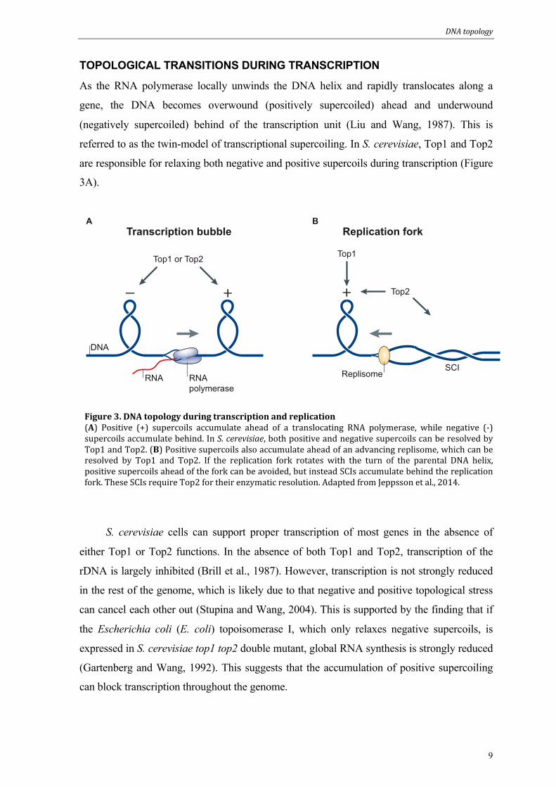
TOPOLOGICAL TRANSITIONS DURING TRANSCRIPTION
As the RNA polymerase locally unwinds the DNA helix and rapidly translocates along a
gene, the DNA becomes overwound (positively supercoiled) ahead and underwound
(negatively supercoiled) behind of the transcription unit (Liu and Wang, 1987). This is
referred to as the twin-model of transcriptional supercoiling. In S. cerevisiae, Top1 and Top2
are responsible for relaxing both negative and positive supercoils during transcription (Figure
3A).
Figure 3. DNA topology during transcription and replication (A) Positive (+) supercoils accumulate ahead of a translocating RNA polymerase, while negative (-‐) supercoils accumulate behind. In S. cerevisiae, both positive and negative supercoils can be resolved by Top1 and Top2. (B) Positive supercoils also accumulate ahead of an advancing replisome, which can be resolved by Top1 and Top2. If the replication fork rotates with the turn of the parental DNA helix, positive supercoils ahead of the fork can be avoided, but instead SCIs accumulate behind the replication fork. These SCIs require Top2 for their enzymatic resolution. Adapted from Jeppsson et al., 2014.
S. cerevisiae cells can support proper transcription of most genes in the absence of
either Top1 or Top2 functions. In the absence of both Top1 and Top2, transcription of the
rDNA is largely inhibited (Brill et al., 1987). However, transcription is not strongly reduced
in the rest of the genome, which is likely due to that negative and positive topological stress
can cancel each other out (Stupina and Wang, 2004). This is supported by the finding that if
the Escherichia coli (E. coli) topoisomerase I, which only relaxes negative supercoils, is
expressed in S. cerevisiae top1 top2 double mutant, global RNA synthesis is strongly reduced
(Gartenberg and Wang, 1992). This suggests that the accumulation of positive supercoiling
can block transcription throughout the genome.
A BTranscription bubble Replication fork
Top2
Top1 or Top2 Top1
DNA
RNA RNApolymerase
ReplisomeSCI

DNA topology
10
Using more sensitive techniques, differences of removing Top1 or Top2 functions in S.
cerevisiae could be detected. In the transcription of the rDNA, top2 mutant cells displayed
slower transcription elongation, indicative of that Top2 is the main topoisomerase that
removes positive supercoils in this region (French et al., 2011). top1 mutant cells on the other
hand accumulated negative supercoils, highlighting the importance of this topoisomerase in
the resolution of supercoils behind the transcription machineries in the rDNA. Top2 was also
recently shown to have a specific role for the proper transcription of long (>3 kilobase pairs
(kb)) S. cerevisiae genes throughout the genome (Joshi et al., 2012). The authors speculated
that in long genes, topological stress ahead of the transcription unit was more often converted
into positive supercoils, which Top2’s double strand passing mechanism is more efficient in
resolving than Top1’s nicking mechanism. In short genes on the other hand, the topological
stress ahead of the transcription machinery more often might remain as increased twist or
overwound DNA, which Top1 is fully capable of resolving.
Related to this thesis, a study showed that genes situated within 100 kb of a telomere
gradually escaped from the transcription stalling caused by expressing E. coli topoisomerase I
in top1 top2 mutant cells (Joshi et al., 2010). These results strongly indicated that topological
stress in the form of positive supercoils or overwound DNA can dissipate over S. cerevisiae
chromosome ends. Another important point concerning topology during transcription related
to this thesis, is that re-orienting a pair of highly expressed RNAPII genes from a tandem to a
convergent orientation, did not reduce their transcription levels (Prescott and Proudfoot,
2002). However, such convergently oriented RNAPII genes are highly dependent on both
Top1 and Top2 for their proper transcription (Garcia-Rubio and Aguilera, 2012). This is true
also if the transcript lengths of the convergently oriented genes is shorter than 3 kb. These
findings indicate that high levels of topological stress accumulate at closely situated
convergently oriented highly expressed genes.
TOPOLOGICAL TRANSITIONS DURING REPLICATION
The unwinding of the parental DNA helix by the replicative helicase during replication
causes the region ahead of the replication fork to become positively supercoiled (Figure 3B).
In S. cerevisiae cells, Top1 or Top2 can resolve this topological stress in order to allow
replication fork progression. In the absence of both Top1 and Top2 functions, replication
stalls a few kb from origins (Brill et al., 1987; Kim and Wang, 1989). The fact that top1 top2
double mutants cannot replicate their chromosomes shows that Top3 is unable to support
DNA topology
11
proper replication progression. Top3 role during unchallenged replication remains unknown,
but it has been suggested to resolve structures formed between two converging replication
forks (Mankouri and Hickson, 2007).
Another way to diminish positive supercoils ahead of the fork, and promote fork
progression, is if the replication fork rotates with the turn of the parental helix. This would
then channel positive supercoils ahead of the replication fork into SCIs behind the fork. Fork
rotation was suggested to occur mainly during replication termination when the length of the
region between the two converging forks becomes to short for topoisomerases to act on
(Champoux, 2001; Sundin and Varshavsky, 1980). The SCIs formed during replication need
to be resolved by Top2 to allow proper chromosome segregation in anaphase (Spell and
Holm, 1994).

Structural Maintenance of Chromosomes
12
STRUCTURAL MAINTENANCE OF CHROMOSOMES In eukaryotes, the well-conserved SMC complexes, cohesin, condensin and Smc5/6 perform
fundamental processes to organize chromosomes and maintain genome stability. Cohesin
holds newly replicated sister chromatids together to ensure bipolar attachment of
chromosomes and accurate segregation. Condensin is required for chromosome condensation,
which is important to allow complete chromosome separation during mitosis. Smc5/6 is less
well characterized than the other two complexes, but has been shown to promote DNA repair
by homologous recombination, timely DNA replication, and segregation of the rDNA.
Condensin will not be discussed in detail below, since it lies outside the scope of thesis.
Figure 4. Structure and composition of SMC complexes (A) Domains of an unfolded SMC protein. The SMC protein then folds back on itself at the hinge domain, which brings the Walker A and Walker B motives together to form the head domain. (B) Structure and composition of the three SMC complexes in S. cerevisiae. Adapted from Jeppsson et al., 2014.
STRUCTURE AND COMPOSITION OF SMC COMPLEXES
The eukaryotic SMC complexes are built around a core of a unique heterodimer of SMC
proteins. SMC proteins are 1000-1500 amino acids in length and have a characteristic
structure. In the center of an SMC protein there is a hinge domain, and at both the N- and C-
termini there are globular domains containing Walker A and Walker B motifs, respectively
A
B
HingeWalker A
N
Walker B
C
Cohesin Condensin Smc5/6Smc1 Smc3 Smc2 Smc4 Smc5 Smc6
Scc1
Scc3
Wpl1
Pds5
Brn1
Ycs4 Ycg1
Mms21
Nse1Nse3
Nse4
Nse5 Nse6

Structural Maintenance of Chromosomes
13
(Figure 4A). The protein folds back on itself at the hinge domain, which brings the N- and C-
termini together to form a functional ATPase domain. The two regions between the hinge
domain and each terminus interact with each other to form a long anti-parallel coiled-coil
structure. The SMC proteins then dimerize in specific pairs for each SMC complex, by
interacting at the hinge domains to form V-shaped heterodimers. In addition to the SMC
proteins, each complex contain a number of non-SMC subunits. One of these subunits is a
member of the kleisin protein family, which bridges the two ATPase-containing head
domains of the SMC heterodimer, and thereby transforms the V-shaped dimer into the
characteristic ring-shaped SMC complex structure (Figure 4B) (Jeppsson et al., 2014). The
subunits of cohesin and Smc5/6 are presented in more detail below.
Cohesin composition
The four canonical subunits of cohesin are Smc1 and Smc3 that make up the core
heterodimer, the kleisin subunit Scc1, and Scc3 (Guacci et al., 1997; Michaelis et al., 1997;
Toth et al., 1999). The head domains of the Smc1-Smc3 heterodimer are bridged by Scc1,
which creates a well-characterized tripartite ring structure (Haering et al., 2008; Haering et
al., 2002). In addition to these four subunits there are cohesin-interacting proteins important
for its function. One of them is Pds5, which binds to cohesin through Scc1 (Hartman et al.,
2000; Panizza et al., 2000). Another cohesin-interacting protein is Wapl (Kueng et al., 2006),
which binds to Pds5. However unlike Pds5, Wapl was shown to interact with cohesin in in a
substochiometric manner, showing that cohesin complexes do not always contain Wapl
(Chan et al., 2012). In human cells there is also a protein called sororin, which interacts with
cohesin and is needed for its function (Schmitz et al., 2007). However, sororin does not
associate with cohesin throughout the cell cycle, instead it has been suggested to interact with
cohesin only when the complex holds sister chromatids together (Nishiyama et al., 2010).
Smc5/6 composition
The core heterodimer of Smc5/6 is, as the name implies, composed of the SMC proteins
Smc5 and Smc6 (Fousteri and Lehmann, 2000). In addition, the complex contains six other
subunits, out of which two have only been found in yeast. Nse1, Nse3, and the kleisin-like
protein Nse4 form a subcomplex, which bridges the head domains of Smc5 and Smc6
(Palecek et al., 2006). Mms21 is a small ubiquitin-like modifier (SUMO) E3 ligase that binds
Structural Maintenance of Chromosomes
14
to the coiled-coil arm of Smc5 (Zhao and Blobel, 2005). Both S. cerevisiae and
Schizosaccharomyces pombe (S. pombe) have the additional subunits Nse5 and Nse6
(Pebernard et al., 2006; Zhao and Blobel, 2005). However, although they share the same
names, they are not conserved on the sequence level between the two yeast species. They also
associate with different parts of the remaining complex, since Nse5 and Nse6 in S. cerevisiae
associate with the hinge domains of Smc5 and Smc6 (Duan et al., 2009), whereas S. pombe
Nse5 and Nse6 associate with the head domains of the heterodimer (Palecek et al., 2006).
FUNCTIONS OF SMC COMPLEXES
The SMC complexes act as functional units, with little evidence that individual subunits can
perform individual tasks. In vitro studies have shown that cohesin and condensin can link two
DNA duplexes together in an ATP-dependent manner. Cohesin was shown to promote
intermolecular DNA linking, while condensin promoted intramolecular DNA linking
(Kimura et al., 1999; Losada and Hirano, 2001). In addition, unpublished data from the
Sjögren lab have shown that Smc5/6, similarly to cohesin can link two different DNA
molecules together in an ATP-dependent manner (Kanno and Sjögren, unpublished).
Potentially, the basal mechanism of the in vivo functions of SMC complexes is to bridge two
DNA loci. This could account for cohesin’s and condensin’s functions in sister chromatid
cohesion and condensation. However, since SMC complexes affect a wide variety of
chromosomal processes, the regulation or downstream effects of such bridging functions
most likely are extensive. Smc5/6 also includes a SUMO-ligase, whose targets can be
involved in many processes, which are unrelated to DNA bridging. In the two sections below,
the in vivo functions of cohesin and Smc5/6, which relate to this thesis, are introduced.
Cohesin functions
The main function of cohesin is sister chromatid cohesion (Guacci et al., 1997; Michaelis et
al., 1997). Through the action of holding sister chromatids together, cohesin counteracts the
pulling forces of the spindle apparatus and thereby promotes chromosome biorientation
(Tanaka et al., 2000). The mechanism by which cohesin holds sister chromatids together has
been well studied. Cohesin forms a ring structure in vivo (Gruber et al., 2003) and is capable
of topologically entrap DNA molecules within its ring (Haering et al., 2008; Murayama and
Uhlmann, 2014). Artificial cleavage of the ring structure abolishes cohesion between sister

Structural Maintenance of Chromosomes
15
chromatids (Gruber et al., 2003; Haering et al., 2008). Since cohesin assembles as a complex
before associating with chromatin (Ciosk et al., 2000), the ring structure requires opening to
allow topological entrapment of DNA. This has been proposed to occur by the transient
opening of the Smc1-Smc3 hinge interface (Gruber et al., 2006). The Smc1-Smc3 interface
was therefore termed cohesin’s “entry gate”. Conversely, to allow the dynamic interaction of
cohesin with chromosomes, and the cleavage-independent removal of cohesin from
chromosome arms in prophase (see below), DNA molecules should also be able to exit from
the cohesin ring. This was proposed to occur, not through the Smc1-Smc3 interface, but
instead through the Smc3-Scc1 interface (Buheitel and Stemmann, 2013; Chan et al., 2012;
Huis in 't Veld et al., 2014). Together these findings support a ring model where cohesin’s
topological entrapment of sister chromatids is how the spindle force is counteracted. The
simplest form of a ring model is the “one-ring” or “embracement” model in which a single
cohesin complex encircles the two sister chromatids (Haering et al., 2002). However, if two
DNA molecules can actually be entrapped within a single ring remains unknown. There are
also alternative ring models, such as the “handcuff”-model (Huang et al., 2005; Zhang et al.,
2008), in which two cohesin complexes interact, each with its own entrapped sister
chromatid.
Cohesin also protects SCIs from resolution by Top2 on long (26 kb) plasmids in G2/M-
phase (Farcas et al., 2011). This is however not the case on shorter (14 kb) plasmids
(Koshland and Hartwell, 1987). Importantly, results from the study of longer plasmids
suggested that cohesin was able to hold plasmids together even if they were not intertwined.
The fact that Top2 is essential in mitosis (Holm et al., 1985), and that chromosomes
missegregate when Top2 is inactivated solely during mitosis (Uemura et al., 1987), suggests
that SCIs are also protected from resolution on linear chromosomes until cohesin is removed.
In addition to sister chromatid cohesion, cohesin promotes condensation of the rDNA
in S. cerevisiae (Guacci et al., 1997). In mouse embryonic fibroblasts, depletion of Wapl,
which counteracts cohesin’s stable association with chromosomes, causes condensation of
interphase chromosomes (Tedeschi et al., 2013). Similarly, deletion of Wapl in S. cerevisiae
cause increased condensation the right arm of chromosomes 12, where the rDNA array is
located (Lopez-Serra et al., 2013). This suggests that a balanced and dynamic association of
cohesin with chromosomes is required to properly organize chromosomes.

Maintenance of genome stability
16
Cohesin also affects transcription in human cells, and has been suggested to perform its
gene regulatory function by mediating long-rang chromosomal interactions in cis between
enhancers and promoters (Hadjur et al., 2009; Kagey et al., 2010).
Smc5/6 functions
Mutations in Smc5/6 subunits cause cells to be hypersensitive to DNA damaging agents such
as MMS, ultraviolet light (UV) and ionizing irradiation, and also to nucleotide-depletion by
HU (Andrews et al., 2005; Lehmann et al., 1995; Pebernard et al., 2006; Verkade et al.,
1999). Epistasis analyses have shown that Smc5/6 functions in DNA repair by homologous
recombination (Andrews et al., 2005; Torres-Rosell et al., 2005; Verkade et al., 1999). A
function in homologous recombination is supported by the observation that homologous
recombination-dependent structures accumulate at damaged replication forks in Smc5/6
mutants (Ampatzidou et al., 2006; Branzei et al., 2006). Smc5/6 has been suggested both to
recruit recombination proteins, and later to promote the resolution of recombination
intermediates at the damaged forks (Irmisch et al., 2009). Smc5/6 also function in
homologous recombination during meiosis, by preventing and resolving aberrant
recombination intermediates (Copsey et al., 2013; Lilienthal et al., 2013; Xaver et al., 2013).
Unlike most other proteins involved in homologous recombination in yeast, Smc5/6 is
also essential in unchallenged cells (Lehmann et al., 1995). This essential function remains
largely elusive. Unchallenged S. cerevisiae cells fail to properly segregate the rDNA in
Smc5/6 mutants (Torres-Rosell et al., 2005). This was suggested to be due to that Smc5/6
mutants failed to complete the replication of the rDNA before entering anaphase (Torres-
Rosell et al., 2007). However, deleting the endogenous rDNA array and instead placing a
single rDNA unit on a multicopy plasmid, which simplifies its segregation, did not improve
the growth of Smc5/6 mutants (Torres-Rosell et al., 2005). This shows that there is another
essential function performed by Smc5/6, other than promoting segregation of the rDNA.
In human cells, depletion of both Smc5 and Smc6 resulted in chromosomes displaying
an abnormal structure in metaphase, and aberrant linkages between sister chromatids during
anaphase (Gallego-Paez et al., 2014). Transiently arresting the cells in G2-phase reduced the
structural defects, which indicated that cell with lower levels of Smc5 and Smc6 required
more time to complete replication.
Maintenance of genome stability
17
In the projects presented in this thesis, we have discovered new functions of Smc5/6 in
DNA replication, chromosome segregation and the maintenance of stalled forks caused by
HU. These functions are summarized in the Results and Discussion chapter, and detailed
descriptions are found in Paper I, Paper II, and Paper IV.
CHROMOSOMAL ASSOCIATION OF SMC COMPLEXES
The SMC complexes perform their functions through the association with chromosomes.
Therefore, knowledge about when and where they associate with chromosomes can lead to
better understanding of the functions of SMC complexes. Their association with
chromosomes is highly regulated during the cell cycle. The complexes do not associate with
specific recognition sequences. Instead chromosomal features, such as centromeres and gene
orientation, are important factors in the localization of SMC complexes. The chromosomal
association of cohesin and Smc5/6 are introduced in the sections below.
The chromosomal association of cohesin
Cohesin is loaded onto chromosomes before replication by the Scc2-Scc4 complex (Ciosk et
al., 2000). At this stage, cohesin’s association with chromosomes is dynamic, since Wapl
promotes cohesin dissociation and Scc2-Scc4 continuously loads new complexes (Chan et al.,
2012; Gerlich et al., 2006; Lopez-Serra et al., 2013). During replication, when sister
chromatid cohesion is established, a subset of cohesin complexes become stably associated
with chromosomes. This is achieved by the acetylation of Smc3 by Eco1, which counteracts
Wapl’s destabilizing activity against cohesin (Rolef Ben-Shahar et al., 2008; Sutani et al.,
2009; Unal et al., 2008). In human cells, Smc3 acetylation leads to the recruitment of sororin,
which is also required to counteract Wapl (Nishiyama et al., 2010; Schmitz et al., 2007).
Detailed chromatin immunoprecipitation (ChIP) analyses in yeast have shown that
cohesin localizes at core centromeres and along chromosome arms in between convergently
oriented genes (Lengronne et al., 2004; Tanaka et al., 1999). The loading complex, Scc2-
Scc4, is however not found at the cohesin sites on chromosome arm. Instead it is found at
core centromeres and highly transcribed genes (Hu et al., 2011; Lengronne et al., 2004).
These findings have led to a model in which cohesin is loaded at Scc2-Scc4 sites, and then
relocates by being pushed by transcription machineries to finally reside in between

Maintenance of genome stability
18
convergently oriented genes (Lengronne et al., 2004). The relocation from the initial loading
sites was later suggested to dependent on ATP hydrolysis of the complex (Hu et al., 2011).
Cohesive cohesin complexes need to be removed from chromosomes to allow
chromosome segregation in anaphase. In human cells, this is completed through a two-step
mechanism. First Wapl promotes dissociation of cohesin from chromosome arms, in a
pathway called the prophase pathway (Kueng et al., 2006; Waizenegger et al., 2000). The
remaining cohesin around centromeres is then cleaved by separase at anaphase onset (Hauf et
al., 2001). In S. cerevisiae, a prophase pathway does not exist, instead all cohesin complexes
are cleaved at anaphase onset (Uhlmann et al., 1999).
The reloading of cohesin after anaphase, starts already in telophase in human cells
(Gerlich et al., 2006), whereas it occurs in late G1-phase in S. cerevisiae (Michaelis et al.,
1997; Uhlmann and Nasmyth, 1998). This is explained by that Scc1 is not present in S.
cerevisiae cells until late G1, since all Scc1 was cleaved in anaphase. The fact that the
chromosomal binding pattern of S. cerevisiae cohesin in late G1 is indistinguishable from the
pattern seen after DNA replication (Lopez-Serra et al., 2013), shows that no new binding sites
are created during cohesion establishment. To date, it is also unknown if cohesion
establishment occurs at all cohesin sites. However, a study in human cells showed that
acetylated Smc3 was only present at a small subset of cohesin sites (Deardorff et al., 2012).
This indicates that cohesion establishment does not occur at all cohesin sites on chromosomes
during replication.
Lastly, cohesin is also enriched around an induced DNA double strand break, and
establishes new cohesion throughout the genome in response to DNA damage (Strom et al.,
2007; Unal et al., 2007).
The chromosomal association of Smc5/6
Using ChIP-on-chip in S. cerevisiae, Smc5/6 was shown to associate around centromeres and
at various positions along chromosome arms. Unlike cohesin, this association occurred
specifically after replication (Lindroos et al., 2006). An interesting finding in this study was
that Smc5/6 displayed a chromosome-length dependent binding pattern, with a higher density
of binding sites on longer chromosomes, compared to short chromosomes. Smc5/6 was also
found to be enriched at or around the rDNA (Lindroos et al., 2006; Torres-Rosell et al.,
2005). In addition, the complex was found to accumulate around an induced DNA double

Maintenance of genome stability
19
strand break (De Piccoli et al., 2006; Lindroos et al., 2006). The accumulation at DNA
breaks, unlike the association to the rest of the undamaged genome required Mre11, a
member of the MRX complex that is involved in the initial processing of DNA breaks
(Lindroos et al., 2006). In this study, Smc5/6 was also shown to accumulate around
replication forks stalled by HU in the absence of a functional checkpoint (Lindroos et al.,
2006). In human cells, the chromosomal association of Smc5/6 has been analyzed by
chromatin fractionation and microscopy. These assays showed that Smc5/6 associated with
chromatin in interphase, but largely dissociated in mitosis when chromosomes were
condensing (Gallego-Paez et al., 2014).
Analysis of the chromosomal association of Smc5/6 has been one of the main focuses
of this thesis. Our findings are summarized in the Results and Discussion chapter, and
described in detail in Paper I, Paper II, Paper III and Paper IV).
Methodology
20
METHODOLOGY In this chapter the model organism and the principal methods used in the thesis are described.
MODEL ORGANISM
In all four papers presented in this thesis, the budding yeast S. cerevisiae was used as model
organism. This unicellular eukaryote represents an excellent experimental system since it has
a short life cycle (of around 90 minutes in ideal conditions), is easy to cultivate, and to
genetically manipulate.
The S. cerevisiae genome was the first eukaryotic genome to be completely sequenced
(Dujon, 1996; Goffeau et al., 1996). S. cerevisiae has 16 linear chromosomes with a total of
just over 12 million bp, excluding the multiple rDNA repeats on chromosome 12 which can
vary in number. The shortest chromosome, chromosome 1, has a length of 230 kb, and the
longest, chromosome 12, has a length of approximately 2350 kb including the rDNA array.
The S. cerevisiae genome contains around 6000 genes with an average ORF length of 1450
bp. This means that the genome is highly gene dense, with more than 70 % of the genome
consisting of ORFs. The intergenic regions are therefore very short, for example with an
average of only 326 bp in between convergently oriented ORFs. In addition, only 4 % of
genes have introns (Dujon, 1996).
This small and gene dense genome, consisting of many relatively short chromosomes,
has obvious distinctions from genomes of many multicellular eukaryotes, which are larger
and more complex. However, research using S. cerevisiae, in which many processes and
proteins are conserved to human cells, has proven to be an excellent approach by which a less
complicated system is used to ask complex questions and discover basic mechanism, which
later can be addressed in human cells. One example related to this thesis, is the discovery of
how sister chromatids are held together by the cohesin complex, which was first discovered
in S. cerevisiae and later proven to be well-conserved in human cells. In addition, working in
a system that allows great detail and highly controlled experiments has strong potential to
lead to unexpected findings. One examples of this is found in Paper I, where we could
analyze the replication timing of specific chromosomes, which is considerably more difficult
in multicellular eukaryotes.

Methodology
21
CHROMATIN IMMUNOPRECIPITATION
ChIP is a technique that allows the analysis of where a specific target protein associates to
chromosomes. This type of information adds important details to the understanding of
chromosome-related functions of target proteins. The first step of the ChIP method is to grow
cells under desired conditions. Cells are then harvested and treated with formaldehyde, which
crosslinks proteins bound to chromosomes. After cell lysis, chromatin is sheared by
sonication into fragments of around 300-500 bp in length. Using a specific antibody, the
target protein is then immunoprecipitated, which also brings down the DNA fragments that
are crosslinked to the protein. Thereafter, crosslinks are reversed and DNA is purified for
downstream analysis (Figure 5) (Katou et al., 2006).
Crosslink proteins to DNA
Lyze cells and shear chromatin by sonication
Immunoprecipitate target protein with antibody coupled to magnetic beads
Reverse crosslinks and purify DNA
DNA
Epitope tag
Target protein
Analyze DNA by sequencing, qPCR or hybridization to microarrays
Figure 5. Chromatin immunoprecipitation workflow Overview of chromatin immunoprecipitation. In this schematic, the target protein is labeled with an epitope tag (green rectangle), which is recognized by antibodies coupled to magnetic beads.

Methodology
22
The amount of DNA for specific loci in the ChIP fraction relative to the amount in the
input fraction is then analyzed using tiling microarrays (ChIP-on-chip), massive parallel
sequencing (ChIP-seq) or quantitative PCR (ChIP-qPCR). Both ChIP-on-chip and ChIP-seq
allow genome-wide analysis of the chromosomal association of a protein of interest in a
single experiment, however ChIP-seq provides higher spatial resolution and signal-to-noise
ratio than ChIP-on-chip (Ho et al., 2011). ChIP-qPCR on the other hand provides fully
quantitative data for specific loci.
ChIP is a population-based assay and the obtained results represent an average of a
target proteins binding profile in the population. Therefore it is not possible from a single
experiment to know if weak signal at a particular locus, relative to another locus, is due to
that fewer cells in the population have the target protein bound to that site, or if the target
protein has a more dynamic association to that specific site. To exclude false positive binding
sites, it is important to use the proper controls. For ChIP-on-chip and ChIP-seq, one way to
do so is to analyze the input fraction, and not only the ChIP fraction (Ho et al., 2011; Nakato
et al., 2013). In S. cerevisiae, experiments are often performed using epitope-tagged target
proteins, and an antibody recognizing the epitope tag. This allows for a more detailed control
to exclude false positive binding sites, by performing experiments on cells lacking the epitope
tag on the protein of interest. In addition, the same epitope tag and high quality antibody can
be used to study any target protein of choice, which minimizes experimental differences
between studies of different proteins. Although there are advantages of using epitope tagged
target proteins, it is important to always control that the small epitope tag does not interfere
with the target protein’s function.
TWO-DIMENSIONAL GEL ELECTROPHORESIS
Two-dimensional gel electrophoresis analysis is a powerful technique that visualizes DNA
structures present at specific sites in the genome. This allows for analysis of replication fork
progression through, or homologous recombination within, any chromosomal locus of
interest. The technique is based on the finding that branched and linear DNA molecules of the
same molecular mass can be separated by gel electrophoresis (Bell and Byers, 1983). Briefly,
genomic DNA is carefully purified and then digested using restriction enzymes. The
restriction enzymes should be chosen so that the locus of interest resides close to the center of
a 3-6 kb restriction fragment. The digested DNA is separated by gel electrophoresis in the
first dimension, using low agarose concentration and voltage. This results in a separation

Methodology
23
largely based on the mass of the DNA molecules. The sample lanes are then excised and the
second dimension gel electrophoresis is performed in a 90 degrees angle to the direction of
the first dimension. In the second dimension the higher voltage and agarose concentration,
and the fact that the intercalating agent ethidium bromide is added, contribute to that the
separation of DNA molecules is not only based on mass but also on their structure. Using the
standard Southern blot technique, the DNA is then transferred to a membrane and detection
of the locus of interest is achieved using a specific radiolabeled probe. The results show the
characteristic pattern of replication or recombination intermediates present within the locus of
interest (Figure 6) (Friedman and Brewer, 1995).
The technique was first developed to detect replication origins on plasmids (Brewer
and Fangman, 1987), but has been extended to monitor replication progression on linear
chromosomes (developed in (Brewer and Fangman, 1988) and used in Paper II and Paper
III), replication termination (Fachinetti et al., 2010), recombination intermediates at stalled
replication forks (developed in (Branzei et al., 2006) and used in Paper IV), hemicatenane
formation (developed in (Lopes et
al., 2003) and used in Paper II).
Related to Paper II and Paper III,
SCIs are fully replicated sister
chromatids that are wrapped around
each other. Therefore, restriction
digestion of the genome into smaller
fragments will dissolve them, which
prevents the use of the two-
dimensional gel electrophoresis
technique to detect SCIs.
Hemicatenanes or recombination
intermediates on the other hand, have
more stable junctions between the
sister chromatids that allows their
detection.
Figure 6. Replication intermediates visualized by two-‐dimensional gel electrophoresis Schematic representation of replication intermediates detected by two-‐dimensional gel electrophoresis. The 1N spot represents linear DNA molecules, in which a replication fork is not present in the investigated fragment.
1st
1N
2nd

Methodology
24
ADDITIONAL TECHNIQUES IMPORTANT FOR THE THESIS
In Paper I, replication completion of specific chromosomes was monitored by pulse-field gel
electrophoresis (PFGE). Since S. cerevisiae chromosomes are relatively short, they can
penetrate and be separated on agarose gels in their intact form, as long as they are purified
from non-replicating cells. However, the branched structures of replicating chromosomes
prevent entry into the gel (Hennessy et al., 1991). Therefore, only chromosomes that have
completed, or not commenced, replication can be resolved by PFGE. To prevent the
visualization of chromosomes that have not commenced replication, cells were made to
replicate in the presence of bromodeoxyuridine (BrdU) during a single S-phase. When bulk
replication appeared complete by the standard fluorescence-activated cell sorting analysis
(FACS) method, cells were harvested and chromosomes were separated by PFGE. The
chromosomes were then transferred to a membrane and chromosomes were detected using an
anti-BrdU antibody. The signal for each specific chromosome was then quantified relative to
the total signal of all chromosomes, including those in the well that did not penetrate the gel.
This assay allows the quantitative detection of the timing of replication completion of specific
chromosomes.
In Paper I and Paper II, plasmid assays were used to monitor SCIs dynamics. Since
SCIs cannot be directly visualized on linear chromosomes, direct analysis of SCIs has to be
performed on circular plasmids, which remain intertwined during DNA preparation as long as
Top2 is kept inactive. In the plasmid assays used for the two papers in this thesis, reporter
plasmids were introduced into S. cerevisiae cells and their topology was analyzed using
standard gel electrophoresis and Southern blotting.
In Paper II, live-cell imaging was used to monitor chromosome segregation. As part of
this technique, individual cells are followed through mitosis, carrying a fluorescently labeled
chromosomal locus. The technique allows detailed studies of when and at what positions
sister chromatids separate from each other, and when the two chromatids successfully
segregate into the mother and daughter cells. To visualize chromosomes, tetracycline
operators were integrated at a specific locus. The cells also express tetracycline repressors,
tagged with the fluorescent protein td-Tomato, which bind to the operators with high
specificity. Tubulin was also tagged with another fluorescent protein, GFP, which allowed
spindle elongation to be monitored. By measuring spindle length using a computer software,
the definition of a specific time point in anaphase could be established, i.e. when the spindle
had reached a defined length. The results from this assay were presented as when the sister
chromatids separated from each other (at the locus of interest) relative to spindle elongation,

Methodology
25
and when the sister chromatids (again at the locus of interest) segregated successfully into the
mother and daughter cell relative to the time of their separation.

Results and discussion
26
RESULTS AND DISCUSSION The four papers presented in this thesis are centered on Smc5/6, its association to
chromosomes and its functions during DNA replication and chromosome segregation. The
findings of each paper are presented and discussed below.
PAPER I
This investigation was started based on the previous intriguing finding that Smc5/6 associates
to chromosomes in a chromosome-length dependent manner, with more binding sites per kb
on longer chromosomes as compared to shorter ones (Lindroos et al., 2006). Since Smc5/6
did not associate with specific recognition sequences or established chromosomal features,
we hypothesized that the level of topological stress on chromosomes was an important factor
for Smc5/6 chromosomal recruitment. Transcription-induced topological stress has been
suggested to dissipate over chromosome ends in S. cerevisiae (Joshi et al., 2010). Longer
chromosomes might therefore suffer from increased topological stress, as the distance to an
“open” chromosome end is longer for a larger part of the chromosome. Supporting our
hypothesis, the absence of Top1 or Top3 specifically delayed the replication of long S.
cerevisiae chromosomes. A similar phenotype was found for Smc5/6 mutants, which created
the possibility that the complex helps to reduce topological stress during replication of long
budding yeast chromosomes.
Using ChIP-on-chip, Smc5/6 association with chromosomes was then shown to require
DNA replication. Since Smc5/6 associates with higher density to longer chromosomes, this
could also be a reflection of the topological status of the chromosome during S-phase. To
further investigate this, Smc5/6 chromosomal association was assayed in top2 mutants and in
cells treated with camptothecin (CPT) to inactivate Top1. The presence of CPT did not alter
Smc5/6 chromosomal association. However, after an S-phase in the absence of Top2
function, the number of Smc5/6 chromosomal binding sites was significantly increased.
Inactivation of Top2 still allows the completion of replication (Bermejo et al., 2007), but
increases the number of SCIs, as judged by chromosome breakage during segregation (Spell
and Holm, 1994) and plasmid assays (DiNardo et al., 1984; Koshland and Hartwell, 1987).
Therefore, Smc5/6 chromosomal association could be triggered by the presence of SCIs. The
increase in Smc5/6 chromosomal binding sites in top2 mutant cells was strongest on
chromosomes of intermediate length. We speculated that this reflected the potential for SCIs
to move along chromosomes and resolve passively over chromosome ends. On long
Results and discussion
27
chromosomes, SCIs might be stable even in wild-type cells, due to low level of passive
resolution over chromosome ends. However on chromosomes of intermediate length, SCIs
might be less stable in wild-type cells and an increased number of SCIs could be more easily
detected. On the shortest chromosomes, any potential increase of SCIs due to the absence of
Top2 during replication might be reduced since SCIs more easily can dissipate over
chromosome ends.
To further challenge the idea that Smc5/6 chromosomal association was triggered by
SCIs, ChIP-on-chip of Smc5/6 was performed in cells carrying either the short chromosome 3
in a circularized form, or the long chromosome 4 divided into two fragments. The results
showed that Smc5/6 is strongly enriched on the circular version of chromosome 3, as
compared to the linear version. This result could indicate that in the absence of chromosome
ends, SCIs are stabilized and can no longer be passively resolved by rotation of “open”
chromosome ends. This is supported by the observation that short circular chromosomes
break, unlike linear ones, during chromosome segregation in the absence of Top2 function
(Spell and Holm, 1994). When the long chromosome 4 is present as two fragments, each with
their own centromere, Smc5/6 peak density is reduced, as compared to on the endogenous
chromosome 4. The lower levels of peak density on the new shorter chromosomes are similar
to those on natural chromosomes of the same length. These results further strengthen the
finding that Smc5/6 association to chromosomes is more influenced by chromosome length
than sequence or any other established chromosomal feature.
Smc5/6 was then shown to promote the intertwining of a reporter plasmid in the
absence of Top2 function. The experiments were performed in the absence of Top2 function,
since at that time intertwined plasmids were not detectable in wild-type cells due to technical
limitations. In top2 smc6 double mutant, less intertwined dimers and more supercoiled
monomeric form of the reporter plasmids were found, as compared to a top2 single mutant.
This result suggested that Smc5/6 promotes the formation of intertwinings by assisting
replication fork rotation. Altogether the results allowed us to propose a speculative model in
which Smc5/6 promotes fork rotation by sequestering SCIs behind the fork. This would
reduce positive supercoils ahead of the replication fork to allow replication of regions with
high topological stress.
The finding that Smc5/6 promote replication fork rotation and therefore the
intertwining of plasmids was later challenged by Farcas and colleagues (Farcas et al., 2011).
They developed a technique that for the first time allowed the detection of intertwined
Results and discussion
28
plasmids in wild-type cells. Their results show that Smc6 function had no obvious effect on
the formation of intertwined plasmids. Several possibilities can account for the differences in
the two studies. We used small plasmid (4.6 kb) and detected how Smc5/6 affected its
topological status in the absence of Top2 function, while Farcas and colleagues used a 26 kb
plasmid to investigate the role of Smc5/6 in wild-type background. In addition, the
temperature-sensitive alleles used to inactivate Smc5/6 were different in the two studies.
Additional experiments are required to understand the difference in results by the two studies.
PAPER II
This investigation was started to challenge the hypothesis that Smc5/6 chromosomal
association is triggered by SCIs, as presented in Paper I. After the publication of Paper I,
cohesin was shown to protect SCIs from resolution by Top2 on long plasmids (Farcas et al.,
2011). If Smc5/6 chromosomal association is indeed triggered by SCIs, it would be expected
to be dependent on cohesin function. However, a previous investigation showed, using ChIP-
on-chip, that Smc5/6 chromosomal binding pattern in a cohesin mutant was altered into more
numerous, jagged and narrow peaks, which indicated that cohesin controlled the positioning
of Smc5/6 on, but not the association to, chromosomes (Lindroos et al., 2006). If, as this
result suggested, Smc5/6 remained associated with chromosomes although the sister
chromatids prematurely separated due to nonfunctional cohesin, it would be difficult to
reconcile with an SCI-dependent association.
To explore this in more detail, we first decided to revisit cohesin’s role in Smc5/6
chromosomal association using ChIP-seq and ChIP-qPCR, which provide higher resolution
and quantitative data, respectively. Our new results clearly showed that Smc5/6 required the
function of cohesin and the cohesin loader Scc2 to associate with chromosomes. The reason
why the previous study (Lindroos et al., 2006) failed to detect that Smc5/6 chromosomal
association is dependent on cohesin remains unknown. However, it does not depend on the
lower resolution of the ChIP-on-chip assay compared to ChIP-seq. This is because the
previous study could detect that Smc5/6 chromosomal association is dependent on the Scc2,
and our new quantitative results showed that the absence of cohesin or its loader reduce
Smc5/6 chromosomal association to the same low levels. In addition, the similarly low levels
of Smc5/6 in Scc2 and cohesin mutants also suggest that the role of Scc2 in the chromosomal
association is mediated through cohesin loading and not through direct loading of Smc5/6.

Results and discussion
29
Our finding that cohesin and its loader Scc2 are required for Smc5/6 chromosomal
association was strengthen by experiments using Eco1 and Pds5 mutants. Both mutants fail to
establish sister chromatid cohesion (Chan et al., 2013; Skibbens et al., 1999; Toth et al.,
1999). In the absence of Eco1 function, cohesin is loaded onto chromosomes but cohesion is
not established (Skibbens et al., 1999; Toth et al., 1999). The fact that Smc5/6 did not
associate with chromosomes in this mutant shows that cohesin loading onto chromosomes is
not sufficient to promote Smc5/6 association. Instead the chromosomal association of Smc5/6
also requires cohesion establishment. In the absence of the cohesion antiestablishment factor
Wapl (Wpl1 in S. cerevisiae), Eco1 becomes largely dispensable for cohesion establishment,
and the eco1 wpl1 double mutant is viable (Rolef Ben-Shahar et al., 2008; Sutani et al., 2009;
Unal et al., 2008). In this double mutant Smc5/6 also associated with chromosomes at wild-
type levels. Altogether these results show that chromatids need to be held together for Smc5/6
to associate to the replicated genome.
The use of ChIP-seq provided a higher resolution of the specific binding sites, and the
use of no tag-control experiments allowed us to exclude several false positive binding sites
from the previous investigation. In wild-type background, strong Smc5/6 binding sites were
found at core centromeres and in between centromere-proximal convergently oriented genes.
At these sites Smc5/6 co-localized with cohesin. Cell-cycle experiments also showed that
Smc5/6 enrichment at these sites was low or absent in G1-phase, reached its peak in G2/M-
phase, and disappeared in anaphase. By focusing on the Smc5/6 enrichment in
pericentromeric regions, where strong signals were detected, and correlating it to
chromosome length we found a clear positive correlation (Figure 7A). In agreement with
previous investigations, (Lindroos et al., 2006) and Paper I, this showed that more Smc5/6
was found in pericentomeric regions on longer chromosomes as compared to short ones.
Since S. cerevisiae chromosome ends had been suggested to be topologically “open” (Joshi et
al., 2010), we decided to see if the proximity to a chromosome end correlated with the
Smc5/6 enrichment in pericentromeric regions. Interestingly, the correlation to the length of
the shorter chromosome arm was even stronger than the correlation to whole chromosome
length (Figure 7B). These two different correlation analyses are in part overlapping, since
longer S. cerevisiae chromosomes more often have longer shorter arms, than shorter
chromosomes. Nevertheless, especially for the longer chromosomes the correlation was
improved. Our previous interpretation on the length-dependency of Smc5/6 chromosomal
association, presented in Paper I (Figure 7C), was based on that SCIs would be able to
swivel off chromosome ends more easily on shorter chromosomes as compared to long ones.

Results and discussion
30
Based on these new findings, we instead speculated that the topological stress during
replication in the pericentromeric region is dependent on the distance to an “open”
chromosome end. Pericentromeric regions found further away from a telomere might
therefore suffer from higher levels of topological stress, which causes more replication fork
rotation and thereby leaves more SCIs behind, bound by Smc5/6.
0
1000
2000
3000
4000
5000
6000
0 100 200 300 400 500 600Shorter arm length (kb)
Enrichment
(100 kb region spanning CEN) R = 0.86672
BA
0
1000
2000
3000
4000
5000
6000
Enrichment
(100 kb region spanning CEN)
0 500 1000 1500 2000Chromosome length (kb)
R = 0.75172
Chromosome length (kb)
0.01
0.02
0.03
0.04
0.05
0.06
peaks/kb per chromosome
0.07
0200 600 1000 1400 1800
cc=0,879
C
Figure 7. Chromosome length, and the length of the shorter chromosome arm, correlates with Smc5/6 chromosomal enrichment around centromeres. (A) Smc5/6 enrichment in pericentromeric regions, as determined by ChIP-‐seq, correlates with chromosome length (Paper II). (B) The correlation of Smc5/6 enrichment in pericentromeric regions with the length of the shorter chromosome arm is enhanced, as compared to whole chromosome length (Paper II). (C) The peak density (binding sites per kb) of Smc5/6, as determined by ChIP-‐on-‐chip, correlates with chromosome length (Paper I).
In Paper I, we found that Smc5/6 accumulates on chromosomes in a top2 mutant. In
this study using ChIP-seq and ChIP-qPCR, we confirmed this finding and could add new
details due to the higher resolution and more quantitative data obtained by these assays. First,
the accumulation of Smc5/6 in top2 mutant cells occurred only on chromosome arms and not
around centromeres. Secondly, the new Smc5/6 binding sites were found in between
convergently oriented genes and co-localizing with cohesin. The chromosomal association
pattern of cohesin was shown not to be affected by Top2 inactivation, ruling out the
possibility that the Smc5/6 accumulation was caused by a change in cohesin binding pattern.
The Smc5/6 chromosomal association in top2 mutant cells was shown to be dependent on
cohesin by ChIP analyzes, although some remaining chromatin association was detected
using chromosome spread analysis. Inactivation of Top2 during a G1- or G2/M-arrest did not
change the chromosomal association pattern of Smc5/6. These findings show that Top2 needs
to be inactive during replication to cause accumulation of Smc5/6 on chromosome arms. This
also suggests that it is not the absence of Top2’s function in transcription that results in
additional Smc5/6 chromosome binding.

Results and discussion
31
Smc5/6 had previously been shown to accumulate at DNA double strand breaks and to
function in homologous recombination (De Piccoli et al., 2006; Lindroos et al., 2006; Torres-
Rosell et al., 2005), so the possibility existed that DNA breaks occurred at cohesin sites in
top2 mutant cells, which then triggered the recruitment of Smc5/6. However, we found that
the DNA damage checkpoint, as indicated by Rad53 phosphorylation, was not activated after
cells had replicated in the absence of Top2 function. In addition, in the absence of Mre11, a
factor required for Smc5/6 to accumulate at DNA breaks (Lindroos et al., 2006), Smc5/6
accumulated on chromosome arms when Top2 function was inactivated. In the absence of
Rad52, cells cannot perform canonical homologous recombination (Krogh and Symington,
2004; Paques and Haber, 1999). Since Smc5/6 accumulated on chromosome arms when Top2
function was impaired also in rad52Δ mutant background, this shows that it is not Rad52-
mediated homologous recombination at cohesin sites in top2 mutant cells that recruits
Smc5/6. Taken together, these results indicate that Smc5/6 chromosomal binding in the
absence of Top2 function is not caused by DNA double strand breaks or recombination
intermediates.
Top2 had previously been shown to promote replication termination (Fachinetti et al.,
2010). In top2 mutant cells, termination was slightly delayed as compared to wild-type cells.
This created the possibility that Smc5/6 association in top2 mutant cells in G2/M-arrest, was
due to incomplete replication, and that Smc5/6 associated to the remaining replication forks.
However, ChIP-seq of a polymerase epsilon subunit after an S-phase in a top2 mutant did not
reveal any enrichment of the polymerase on chromosomes. Furthermore, the Smc5/6
chromosomal binding pattern was not altered in the absence of the Rrm3 helicase, as detected
by ChIP-on-chip. In Rrm3 mutant cells, replication fork pausing occurs at specific sites in the
genome (Ivessa et al., 2003; Ivessa et al., 2000). Our logic for this experiment was that if
Smc5/6 binding was due to fork pausing, we expected to see more Smc5/6 on chromosomes
in the absence of Rrm3. This finding was later challenged by quantitative ChIP-qPCR
analysis in Paper III and is discussed in detail below. More importantly, however, Smc5/6
sites were investigated by two-dimensional gel electrophoresis, which showed that Smc5/6
chromosomal association in top2 mutant cells is not due to the persistence of replication forks
in G2/M-arrest.
We then decided to analyze if restored Top2 activity after replication was able to
remove Smc5/6 that had accumulated on chromosomes due to the absence of Top2 during
replication. It had previously been showed that Top2 is required only at the time of mitosis to
ensure cell survival (Holm et al., 1985). This suggests that Top2 can resolve any accumulated

Results and discussion
32
entanglements if it is reactivated just before, or during, chromosome segregation. We
confirmed this using live-cell imaging to study chromosome segregation. In addition, using a
small reporter plasmid, we showed that Top2 reactivation led to resolution of an intertwined
plasmid during a G2/M-arrest. After these control experiments, Smc5/6 was shown, using
ChIP-seq and ChIP-qPCR, to dissociate from chromosomes when Top2 activity was restored
in G2/M-arrest. Smc5/6 enrichment was reduced to levels seen in wild-type background,
showing that the excess of Smc5/6 on chromosomes was removed. If Top2 was kept
inactivated during a prolonged G2/M-arrest, there was no reduction in Smc5/6 enrichment
levels, further strengthening the conclusion that the accumulation of Smc5/6 in top2 mutant
cells is not due to a delay in replication termination. These results indicate that Smc5/6
associates to a chromosomal structure that Top2 can resolve after DNA replication.
Since Top2 inactivation after S-phase did not affect Smc5/6 chromosomal association,
this created the possibility to analyze segregation of chromosomes in the absence of Top2
function during mitosis with high or low levels of Smc5/6 binding, resulting from the
inactivation of Top2 before or after replication, respectively. Chromosome segregation was
again investigated by live-cell imaging, and the results showed that chromosomes with higher
levels of Smc5/6 enrichment missegregated and had more difficulty in separating its sister
chromatids, than the same chromosomes with lower level of Smc5/6 enrichment. These
results show that Smc5/6 enrichment on chromosomes correlate with the segregation-
inhibiting structures present on chromosomes in top2 mutants. These structures are most
likely SCIs, but the development of assays capable of directly visualizing these structures is
needed to confirm this hypothesis.
Lastly, we posed the question if Smc5/6 had a function in the segregation of entangled
chromosomes. Thus, we decided to analyze chromosome segregation in the absence of both
Top2 and Smc5/6 functions. The experiment was performed by investigating the shortest S.
cerevisiae chromosome, since this chromosome is able to segregate correctly in the majority
of top2 mutant cells, likely due to the passive resolution of SCIs over chromosome ends.
Longer chromosomes missegregate at high rates in top2 mutant cells, which makes it difficult
to address if Smc5/6 also has a segregation-promoting function on these chromosomes. The
results showed that the shortest chromosome missegregates three-fold as often in top2 smc6
double mutants, as compared to top2 single mutants. The increased missegregation was not
due to a failure in the removal of cohesin, which was proposed to occur in S. pombe top2
smc6 double mutants (Outwin et al., 2009). The authors of that study suggested that top2
smc6 mutants failed to remove a subset of cohesin complexes, which are normally removed

Results and discussion
33
by a separase-independent pathway in S. pombe. In S. cerevisiae this pathway does not exist
(Schmidt et al., 2009), instead all cohesin complexes are cleaved during anaphase (Ciosk et
al., 2000; Uhlmann et al., 1999). Our results show that Smc5/6 either promotes Top2-
independent resolution of SCIs, or prevents aberrant linkages on short entangled
chromosomes that cannot be passively resolved. Future investigations will be needed to
analyze if these aberrant sister chromatid linkages require recombination to be formed. It
would also be interesting to see if restored Top2 activity can resolve them, after an S-phase in
the absence of both Top2 and Smc5/6 functions.
Altogether, our results suggest that Smc5/6 indicates the positions where chromosomes
are entangled. If so, the results are in favor of that cohesin directly protects SCIs also on
linear chromosomes, at least in pericentromeric regions. Smc5/6 is also shown to promote the
segregation of entangled sister chromatids. Furthermore, the results strengthen the possibility
that topological stress during S-phase is important for the formation of SCIs.
PAPER III
This investigation was started to further explore the hypothesis, presented in Paper II, that
topological stress during S-phase is one of the factors that determines if Smc5/6 associates to
chromosomes, potentially by promoting SCI formation. Topological stress in the form of
positive supercoils accumulates ahead of translocating enzymatic machineries, such as the
replisome and RNA polymerases, when they unwind the DNA helix (Brill et al., 1987; Liu
and Wang, 1987). Consistently, strong convergent transcription results in the accumulation of
high levels of topological stress (Garcia-Rubio and Aguilera, 2012). To locally increase the
topological stress in the S. cerevisiae genome, we therefore artificially increased the
transcription levels of two closely situated convergently oriented genes, MCR1 and DBR1.
Thereafter, ChIP-on-chip and ChIP-qPCR were used to assay the chromosomal association of
Smc5/6. The increase in transcription levels was achieved by replacing the endogenous
promoters of the two genes with strong constitutively active promoters. The results showed
that Smc6 specifically accumulated to a high level in between the two genes when they were
overexpressed.
In Paper II, we found that Smc5/6 co-localizes with cohesin in both wild-type and top2
mutant cells. In addition, the enrichment levels of the two complexes at individual binding
sites correlated. Cohesin was previously suggested to be pushed by the transcription
machinery to finally reside in between convergently oriented genes (Lengronne et al., 2004).

Results and discussion
34
Therefore, the overexpression of the two convergently oriented genes, MCR1 and DBR1,
could lead to the positioning of more cohesin, which in turn could allow more Smc5/6
binding. However, this possibility was excluded since ChIP-qPCR of Scc1 (a cohesin
subunit) showed that there was no increase in cohesin enrichment by high MCR1 and DBR1
transcription.
We then used two-dimensional gel electrophoresis to monitor replication fork
progression through the two genes, and found that the increased transcription also resulted in
distinct replication fork pausing. The pausing signal was seen strongest in mid-S-phase, and
was almost completely absent when bulk replication was finished. The fork pausing was
largely dependent on the increased transcription of the DBR1 gene, which orientation opposes
the incoming fork. Overexpression of MCR1 alone, which is oriented co-directionally with
replication fork progression, did not cause detectable fork pausing. Hence, the pausing is due
to head-on-collision of the replication fork and the transcription machinery. Our data is in
agreement with the previous finding that highly expressed RNAPII genes on plasmids pause
the replication fork only when they are oriented against the incoming replication fork (Prado
and Aguilera, 2005). This is also consistent with that fork pausing seen at RNAPIII-
transcribed genes is limited to head-on-collisions (Deshpande and Newlon, 1996). However,
a ChIP-on-chip-based study by Azvolinsky and colleagues suggested that highly expressed
RNAPII-genes pause the replication fork independently of their orientation (Azvolinsky et
al., 2009). The experimental rationale of this study was to detect replication fork pausing by
ChIP-on-chip of the DNA polymerase epsilon subunit, Pol2, in asynchronously growing
cells. Potentially, the ChIP-on-chip-based study suffers from detecting false-positive “pausing
sites”, since a later study revealed that ChIP has the potential to detect erroneous signals
within highly expressed ORFs (Teytelman et al., 2013). This is further supported by that the
authors were unable to confirm distinct fork pausing in genes oriented co-directionally with
the replication fork using two-dimensional gel electrophoresis. The finding that high
transcription can produce artifactual signals within ORFs highlights the importance of
validating ChIP-on-chip data by the use of ChIP-qPCR and “no tag”-controls, when possible.
In addition, to further ascertain that the investigated ChIP-signal at a highly transcribed locus
is not due to a false positive signal, it can be useful to, if possible, separate the high
transcription from the accumulation of the target protein. This can be achieved by, for
example, looking at different cell cycle stages or in mutants in which one of the events
(binding of the target protein or transcription levels) is altered, but not the other.
Results and discussion
35
In our study, high convergent transcription resulted in both local Smc5/6 chromosomal
association and replication fork pausing. Therefore, the possibility existed that the
transcription-induced Smc5/6 enrichment in between the two genes was due to direct
association with the paused fork. However, more detailed analysis of the location of fork
pausing showed that it occurs within the DBR1 ORF, whereas Smc5/6 enrichment peaks in
between the two overexpressed genes. This spatial discrepancy argued against that Smc5/6
directly associated to the paused fork.
To further explore the correlation between the two events, we sought for factors
regulating fork pausing with the idea to test if they also affected Smc5/6 enrichment. We
found that in the absence of the helicase Rrm3, fork pausing was strongly enhanced in mid-S-
phase. However, in the end of S-phase no paused replication forks were detected, similar to
wild-type background, indicating that the forks had resumed, or replication termination had
occurred at the paused fork. Rrm3 moves with the replication fork (Azvolinsky et al., 2006),
and assists it past obstacles consisting of non-histone proteins bound tightly to DNA, for
example at centromeres, inactive origins, RNAPIII-genes and in the rDNA (Ivessa et al.,
2003; Ivessa et al., 2000). However, Rrm3’s role in assisting replication forks past highly
expressed RNAPII-transcribed genes is controversial. The plasmid-based study mentioned
above found that Rrm3 counteracts fork pausing when a replication fork meets an opposing
highly expressed RNAPII gene (Prado and Aguilera, 2005), whereas the ChIP-on-chip-based
study, also mentioned above, suggested that Rrm3 had no role in assisting the fork past highly
expressed RNAPII genes in the unaltered genome (Azvolinsky et al., 2009). Our data is in
agreement with the plasmid-based study. The finding of Azvolinsky and colleagues that
Rrm3 has no role in assisting the fork past highly expressed RNAPII genes was based in part
on ChIP-on-chip experiments of Pol2 in asynchronously growing cells of wild-type and
rrm3Δ background (Azvolinsky et al., 2009). As discussed above, false positive signals
within highly expressed genes could have complicated such experimental approach. They did
however detect distinct fork pausing at two loci using two-dimensional gel electrophoresis, at
which the absence of Rrm3 did not affect the pausing signal. The difference in the results
from our and Azvolinsky and colleagues’s studies could be explained that we used cells
passing through S-phase synchronously and took samples at two time points, whereas
Azvolinsky and colleagues only used asynchronously growing cells. Our results show that the
enhanced pausing seen in rrm3Δ background is transient and that the forks eventually pass
the obstacle or replication termination occurs at the pausing site. Hence, it could be difficult
to detect Rrm3’s role in assisting the replication fork past highly expressed RNAPII genes

Results and discussion
36
using asynchronously growing cells. Alternatively, the dissimilarity could be explained by the
difference between the two endogenous loci investigated by two-dimensional gel
electrophoresis by Azvolinsky and colleagues, and our artificial experimental setup. The two
loci displaying distinct fork pausing in the study of Azvolinsky and colleagues contained a
single highly expressed RNAPII gene opposing the replication fork (Azvolinsky et al., 2009),
whereas in our study Rrm3’s function was only assessed at a locus with two highly expressed
convergently oriented RNAPII genes. Based on these experimental differences, future time
course experiments on synchronized cells at highly expressed RNAPII genes in the unaltered
genome, and at our MCR1-DBR1 locus when only DBR1 is overexpressed, have the potential
to clarify the role Rrm3 role in assisting replication forks past these obstacles.
Having found that the absence of Rrm3 strongly enhanced fork pausing in our
experimental setup, we turned to see if Rrm3 also affected Smc5/6 enrichment. ChIP-qPCR
was performed in a time course experiment in wild-type and rrm3Δ background. In wild-type
background Smc5/6 enrichment levels were low in G1-phase, increased during S-phase,
peaked after bulk replication was completed, and remained high in a prolonged G2/M-arrest.
This showed that Smc5/6 enrichment peaks after the fork pausing is strongest, and that
Smc5/6 remains bound to the MCR1-DBR1 locus when replication intermediates are barely
detectable. Similarly, Smc5/6 enrichment levels were low in G1-phase and increased during
S-phase in rrm3Δ background. However, when bulk replication was completed there was a
drastic increase in Smc5/6 enrichment as compared to wild-type background. This increase
was transient and the enrichment levels of Smc5/6 returned to the levels seen in wild-type
background after extending the G2/M-arrest. The transient increase of Smc5/6 enrichment at
the altered MCR1-DBR1 locus in the absence of Rrm3 is also seen at the Smc5/6 binding sites
in the unaltered genome. Since Rrm3 only assists replication fork progression at distinct sites
and not all throughout the genome (Azvolinsky et al., 2006), this may indicate that all Smc5/6
binding sites are Rrm3-regulated sites. This interesting possibility warrants closer in
investigation in the future.
The results show that Smc5/6 enrichment after MCR1-DBR1 overexpression is not due
to the direct association to the paused fork. In Paper II, we proposed that Smc5/6 associates
with SCIs in the unaltered genome. This might be the case also at the MCR1-DBR1 locus.
Potentially, high topological stress in this region results in fork rotation, which leaves the
newly formed sister chromatids intertwined and bound by Smc5/6 in its wake, before the
replication fork comes to a complete pause. However, if the Smc5/6 accumulation would
represent SCIs, this would be surprising since Top2 was shown in Paper II, to counteract

Results and discussion
37
Smc5/6 accumulation on chromosome arms. An important future experiment to challenge
this idea would be to test if transcription-induced Smc5/6 accumulation requires sister
chromatids to be held together by cohesin. This also has the potential to separate the pausing
event from Smc5/6 enrichment, since fork pausing was not affected by the absence of cohesin
function.
The transient increase in Smc5/6 enrichment seen in rrm3Δ background could be
connected to break repair by homologous recombination, since broken forks are detected by
two-dimensional gel electrophoresis. This can be addressed by testing if factors required for
Smc5/6 accumulation at DNA double strand breaks, such as Mre11 (Lindroos et al., 2006),
are needed for the transient increase of Smc5/6 enrichment in rrm3Δ background.
In summary, our data show that Smc5/6 accumulates in between MCR1 and DBR1
when they are overexpressed. The increased transcription also results in replication fork
pausing, due to head-on-collision of the fork and the RNAPII transcription machinery.
However, the Smc5/6 enrichment and fork pausing are temporally and spatially distinct.
Whereas Smc5/6 enrichment peaks in late S-phase and remains high in a prolonged G2/M-
arrest, the fork pausing is strongest in mid-S-phase. Smc5/6 also accumulates in between the
two genes and the fork pauses within the DBR1 ORF. Enhanced fork pausing, due to the
absence of the Rrm3 helicase, leads to transiently increased Smc5/6 levels, but again Smc5/6
enrichment peaks after bulk replication completion. This indicates a complex relationship
between replication fork pausing and Smc5/6 enrichment. To further correlate Smc5/6
enrichment and replication fork pausing, it could be of interest to see if Smc5/6 enrichment
occurs when only DBR1 is overexpressed. Potentially, strong Smc5/6 enrichment requires
high convergent transcription, unlike distinct fork pausing that only requires high
transcription opposing the replication fork. Regardless, our results strengthen the hypothesis
that the levels of topological stress and/or replication obstacles are important determinants in
Smc5/6 chromosomal association after S-phase. In addition, our data supports the view that
opposing, but not co-directional, highly expressed RNAPII transcription pauses the
replication fork, and that Rrm3 assists the fork past highly expressed RNAPII-transcribed
genes.
In the future, it will be important to assess the functional importance of transcription-
induced Smc5/6 accumulation. One alternative could be to test if creating additional Smc5/6
binding sites on a short chromosome by the use of high convergent transcription results in
that the short chromosome now needs Smc5/6 for its proper replication, as shown for longer

Results and discussion
38
chromosomes with more Smc5/6 binding sites in Paper I. However, we did not detect any
replication defects at MCR1-DBR1 by two-dimensional gel electrophoresis in smc6-56
mutant cells, which argues against this possibility. Instead analyzing chromosome
segregation distal to the site of transcription-induced Smc5/6 accumulation in top2-4 could be
used to see if the Smc5/6 enrichment represents a structure that inhibits chromosome
segregation in the absence of Top2 function. Segregation experiments could also be
performed in top2-4 smc6-56 double mutant to see if Smc5/6 function is important to
segregate this locus, similar to chromosomes with many Smc5/6 sites as shown in Paper II.
PAPER IV
This study was started to analyze the function of the Nse5 subunit of Smc5/6. It was a
collaborative study led by Jennifer Cobb’s laboratory, to which Dana Branzei’s laboratory
contributed with two-dimensional gel electrophoresis analysis and we, in Camilla Sjögren’s
laboratory, contributed with ChIP-on-chip and BrdU-IP analysis. The study was built around
two hypomorphic Nse5 alleles, nse5-ts1 and nse5-ts2. These alleles rendered cells
temperature-sensitive, with nse5-ts1 cells being inviable at 37°C, whereas nse5-ts2 cells
showed very slow growth under these conditions. However, the majority of the experiments
were performed at their permissive temperature, 25°C, at which these mutants, under
unchallenged conditions, grow as wild-type cells.
Nse5 was first shown to interact with SUMO (Smt3 in S. cerevisiae) by yeast-two-
hybrid analysis. SUMO is a small protein that can be conjugated to target proteins and
modify their functions. Nse5 also interacted with a mutated form of SUMO, which cannot be
conjugated to target proteins. This result indicated that Nse5 interacts with SUMO through
non-covalent interactions. Consistently, sumoylated forms of Nse5 could not be detected by
SUMO pull-down experiments. Yeast-two-hybrid analysis of the two nse5-ts mutants showed
that they did not interact with SUMO. Based on these results and the proximity of the Nse5-
Nse6 subcomplex to the SUMO-ligase Mms21 within Smc5/6, the question was asked if
Nse5 affected sumoylation of Smc5, which is Mms21-dependent in the presence of MMS
(Zhao and Blobel, 2005). The results showed that Smc5 sumoylation was strongly reduced in
both nse5-ts mutants.
Hypomorphic alleles of the Smc5/6 complex, including SUMO-ligase deficient alleles
of Mms21, cause HU-sensitivity (Ampatzidou et al., 2006; Chen et al., 2009; Cost and
Cozzarelli, 2006; Hu et al., 2005; Pebernard et al., 2006). Therefore, the two nse5-ts alleles

Results and discussion
39
were tested for HU-sensitivity. The results showed that nse5-ts1, but not nse5-ts2, cells were
HU-sensitive. Together with the previous results, these findings separate Smc5 sumoylation
from the functions of Smc5/6 during HU-induced replication stress. ChIP-qPCR was then
used to examine replisome stability at early firing origins in HU-treated cells. ChIP-qPCR of
the polymerase epsilon subunit Pol2 was performed in a time-course experiment in cells
released from G1-arrest into HU-containing medium. In wild-type and nse5-ts2 cells, Pol2
could be detected at early, but not late, firing origins when cells commenced S-phase.
However in nse5-ts1 cells, barely any Pol2 enrichment was detected at early origins
throughout the time course experiment. These results are in line with that nse5-ts1, but not
nse5-ts2, are sensitive to HU-induced replication stress and shows that sumoylation of Smc5
is not required for replisome stability at stalled forks.
To further characterize the nse5-ts1 allele, it was combined and compared with the
SUMO-ligase deficient allele mms21-11. nse5-ts1 mutants were shown to be more sensitive
to HU than mms21-11 mutants. Combining the two mutant alleles resulted in an increased
sensitivity to HU, as compared to either single mutant. Similarly, Smc5 sumoylation levels
were lower in nse5-ts1 cells as compared to mms21-11 cells, and barely detectable in the
double mutant. ChIP-qPCR was then again used to analyze replisome stability at forks stalled
by HU. In mms21-11 cells, polymerase subunits were detected at early firing origins, similar
to wild-type cells. However, nse5-ts1 mms21-11 double mutants exhibited the same reduction
of polymerase subunits detected at early firing origins as seen in nse5-ts1 cells. This showed
that although mms21-11 cells displayed HU-sensitivity, replisome components remained at
the stalled forks at wild-type levels.
In an attempt to correlate the HU-sensitivity with events taking place at the stalled
forks, two-dimensional gel electrophoresis analysis of an early origin was performed. In these
experiments psoralen crosslinking was used to enhance the detection of X-shaped molecules.
The results showed that in the presence of HU, X-shaped molecules accumulated in nse5-ts1,
mms21-11 and nse5-ts1 mms21-11 mutants at an early origin. The accumulation of X-shaped
molecules was strongest in the double mutant. These X-shaped structures were dependent on
Rad51, a protein that promotes the strand invasion step during homologous recombination
(Krogh and Symington, 2004; Paques and Haber, 1999). This indicated that the X-shaped
molecules that accumulate at replication origins in the absence of proper Nse5 and/or Mms21
functions are formed by homologous recombination.

Results and discussion
40
To further characterize why nse5-ts1 mutants displayed strong HU-sensitivity, we
performed BrdU-IP-chip in the presence of HU, in the mutant and in wild-type cells. The
results showed that early origins had fired in nse5-ts1 cells similar to wild-type cells. The
replication forks had also progressed similar distances away from the origins in the two
strains. The fact that late origins had not fired in the mutant was a strong indication that the
intra-S-phase checkpoint was intact in the mutant. However, the signal of incorporated BrdU
was lower in nse5-ts1, than in wild-type cells. These results indicated that DNA synthesis
was lower in nse5-ts1 mutant, potentially because replisomes occasionally collapse and
dissociate from DNA.
We then turned to see if Smc5/6 was recruited to the stalled forks. Smc5/6 had
previously been shown to associate to HU-induced stalled forks in checkpoint mutants
(Lindroos et al., 2006). We now showed, using ChIP-on-chip, that Smc5/6 was also clearly
enriched around the early firing origins in HU-treated wild-type cells. However, in nse5-ts1
cells, Smc5/6 was not detected. This result was confirmed at a few early origins using ChIP-
qPCR. Using this technique, nse5-ts2 and mms21-11 mutants were shown not to impair the
recruitment of Smc5/6 to stalled forks. Lastly, co-immunoprecipitation analysis of Nse6-
Smc5 interaction, as a measure of the stability of the complex, showed that this interaction
was weakened in nse5-ts1 cells. These results indicated that Nse5 is important for the stability
of Smc5/6.
In summary, the results showed that Nse5 is important for Smc5 sumoylation.
However, the functional consequence of Smc5 sumoylation remains unknown since the
results separated the function of Smc5/6 in HU-induced replication stress from Smc5
sumoylation. Smc5/6 was also shown to localize around replication forks stalled by HU. This
localization required a stable complex and proper Nse5 function. Since HU causes fork
stalling that is not site-specific, and ChIP-assays are performed on populations of cells, it is
not possible, in this case, to say if Smc5/6 associated directly to the stalled forks or behind the
forks, as was seen in Paper III where Smc5/6 accumulated behind transcription-induced
paused forks. Our data suggested that Smc5/6 stabilizes stalled forks during HU-induced
replication stress. This required a functional Nse5 subunit, but not the SUMO-ligase activity
of Mms21. In addition, Smc5/6 prevented the accumulation of homologous recombination
intermediates. This function required a functional Mms21 subunit, whose critical sumoylation
target remains to be determined since Smc5 sumoylation was shown to be dispensable.

Results and discussion
41
Unlike the other well-conserved subunits of Smc5/6, Nse5 and Nse6 have so far only
been found in yeast. In addition, they are not conserved on the sequence level between S.
cerevisiae and S. pombe (Duan et al., 2009). Nse5 and Nse6 are also essential in S. cerevisiae
(Duan et al., 2009; Zhao and Blobel, 2005), but not in S. pombe (Pebernard et al., 2006).
Furthermore, the Nse5-Nse6 subcomplexes bind to different places to the Smc5-Smc6
heterodimer in the two yeast species. Whereas Nse5-Nse6 in S. cerevisiae was found to
associate to the hinge domains of Smc5-Smc6 (Duan et al., 2009), Nse5-Nse6 in S. pombe
was found to associate to the head domains and the adjacent coil-coiled regions of the Smc5-
Smc6 heterodimer (Palecek et al., 2006). Criticism could be raised against studying Nse5 in
S. cerevisiae, since it potentially will lead to S. cerevisiae-specific findings. However, using
these two well-established model organisms to understand the species-specific differences
will potentially lead to new insights into Smc5/6 functions, applicable to other species as
well. It would be interesting to see if also S. pombe Nse5 is important to allow the recruitment
of Smc5/6 under replication stress conditions. In addition, it will be interesting to characterize
the two nse5 alleles at restrictive temperatures in unchallenged S. cerevisiae cells, to see if
they under these conditions also affect the stability of the complex and association with
chromatin. Potentially, investigations of why Smc5/6 in S. cerevisiae, but not S. pombe,
requires Nse5 to promote survival will add important clues to the elusive essential function of
the otherwise well-conserved complex.

Perspectives and concluding remarks
42
PERSPECTIVES AND CONCLUDING REMARKS The overall aim of this thesis was to investigate Smc5/6 function and chromosomal
association, and how the topological status of chromosomes affects the two. We have reached
this aim by revealing new functions of Smc5/6 and topoisomerases during replication (Paper
I). Our data also suggest that Smc5/6 associates to entangled chromosomal loci, and
promotes the segregation of short entangled chromosomes (Paper II). In the absence of
cohesin function, Smc5/6 does not associate with chromosomes after replication (Paper II).
Therefore, phenotypes found in cohesin mutants could also stem from the lack of chromatin-
bound Smc5/6. In addition, we speculate that the level of topological stress on chromosomes
during S-phase affects their level of entanglement (Paper I, Paper II and Paper III). Lastly,
investigations of Nse5 during replication stress suggest that Smc5/6 maintains replication
forks that have been stalled by HU treatment, and prevents aberrant homologous
recombination at these forks (Paper IV). Our studies have revealed an intricate interplay
between chromosome structure, topoisomerases and Smc5/6, in which topological structures
affect SMC complexes and SMC complexes affect topological structures. Our findings allow
us to speculate that topological structures are more than problems that cells need to
overcome, and instead are important to link the processes of DNA replication and
chromosome segregation, and thereby promote genome stability. In the sections below, this
hypothesis is extended, and future challenges related to our main findings are discussed.
Potential benefits of topological structures
Topological structures, such as SCIs and positive supercoils, have mainly been considered as
problems that cells need to overcome. On a speculative note, I would like to propose that
these structures play important roles in the faithful segregation of chromosomes. Recent data
have showed that SCIs are tightly regulated by SMC complexes. The most telling example is
that cohesin maintains SCIs on plasmids until anaphase, by preventing Top2 to resolve them
prematurely (Farcas et al., 2011). This revived the hypothesis of that SCIs contribute to
proper sister chromatid cohesion (see below). In addition, condensin has been shown to
promote Top2-dependent resolution of SCIs (Baxter et al., 2011; Charbin et al., 2014), and
our data suggest that Smc5/6 helps to form them, and prevents them from becoming aberrant
linkages, in the absence of Top2 (Paper I and Paper II). Our results also indicate that
topological stress during replication promotes SCI formation, possibly by promoting
replication fork rotation (Paper I, Paper II and Paper III). These results open for that

Perspectives and concluding remarks
43
topological stress during replication is not just a problem that replication forks need to
overcome, but also has the potential to contribute to proper chromosomes segregation,
through SCI formation and/or sister chromatid cohesion (see below). There are a few
indications in the literature of these ideas. Reducing the length of an S. cerevisiae
chromosome below 150 kb results in that it is no longer stably transmitted (Murray et al.,
1986). The authors hypothesized at the time that SCIs held chromosomes together and that
such structures were not stable on chromosomes shorter than 150 kb. Subsequent analyses
found that plasmids were not intertwined in metaphase cells, which showed that something
other than SCIs holds counteracts the pulling forces of the spindle (Koshland and Hartwell,
1987). This was then discovered to be the cohesin complex (Guacci et al., 1997; Michaelis et
al., 1997). Based on the finding of cohesin, SCIs were considered to be an unwanted
byproduct of DNA replication, which could cause problems in chromosome segregation. In
the light of that cohesin maintains SCIs on longer plasmids (Farcas et al., 2011), it would be
interesting to revisit the former hypothesis that SCIs are needed for proper chromosome
segregation of much longer linear chromosomes. Smc5/6 associate with chromosomes in a
length-dependent manner (Paper I) and potentially marks entangled loci (Paper II).
Therefore, it would be interesting to investigate Smc5/6 chromosomal association on
chromosomes of shorter than natural length to see if it correlates with the lack of proper
segregation also in Top2-proficient cells. Lastly, if SCIs have a function for proper cohesion
on linear chromosomes, our data would then indicate that also the topological stress during
replication (Paper I and Paper II) have positive functions in promoting fork rotation (and
SCI formation) around centromeres.
FUTURE SMC CHALLENGES
Although research of SMC complexes has advanced our knowledge of chromosome
dynamics substantially over the years, there are still many remaining mysteries of how the
SMC complexes function. One of the most obvious ones is that the essential function of
Smc5/6 still remains elusive. Hypomorphic Smc5/6 mutants showed some improved growth
when homologous recombination was inhibited (Torres-Rosell et al., 2005). However,
Smc5/6’s function in recombination cannot alone account for Smc5/6’s essential function,
since homologous recombination is not required for growth in unchallenged S. cerevisiae
cells (Krogh and Symington, 2004; Paques and Haber, 1999). Nor can Smc5/6’s function in
rDNA maintenance account for that unchallenged cells require the complex for viability. As
mentioned before, this is due to the observation that relocalization of the rDNA to a

Perspectives and concluding remarks
44
multicopy plasmid, from its endogenous position on chromosome 12, did not improve the
growth of Smc5/6 mutants (Torres-Rosell et al., 2005). Therefore, the essential function of
Smc5/6 must lie outside the maintenance of the rDNA. Similarly, the replication delay of
long chromosomes seen in mutants of Smc5/6 (Paper I) is unlikely to be the essential
function since mms21-CH and top1Δ mutants, which are viable, display a similar phenotype.
Smc5/6 was recently shown to restrain Mph1 helicase function in promoting fork regression
in vitro (Xue et al., 2014). In addition, deletion of Mph1 has been shown to restore growth,
although extremely poorly, to cells lacking Smc5/6 (Chen et al., 2009). Potentially, one
reason that Smc5/6 mutants fail to proliferate is that Mph1 promotes aberrant fork reversal,
which causes toxic replication intermediates. It would be interesting to combine factors that
potentially can alleviate the requirement of Smc5/6, such as deletion of Mph1 and inhibition
of recombination. It would also be possible to engineer S. cerevisiae cells that do not have
long chromosomes, which require Smc5/6 for their proper replication. Although Smc5/6 is
essential in yeast (Lehmann et al., 1995) and mouse (Ju et al., 2013), surprisingly, this well-
conserved complex is not essential in Drosophila melanogaster (Li et al., 2013) and
Caenorhaditis elegans (Bickel et al., 2010). Future analyses of the reason for this difference
have the potential to add important clues to the essential function of Smc5/6.
Another SMC mystery that relates to this thesis is the mechanism of sister chromatid
cohesion. The dominant model is that a single cohesin ring embraces two DNA molecules
from different sister chromatids (Gruber et al., 2003; Haering et al., 2002; Nasmyth and
Haering, 2005). An elegant study recently succeeded with the reconstitution of cohesin’s
topological embracement of DNA in vitro (Murayama and Uhlmann, 2014). Potentially such
experiments may in the future lead to answering the question if a single cohesin ring can
encircle two DNA molecules. In addition, knowledge is also lacking if all, or just a subset of,
cohesin complexes on chromosomes become cohesive during S-phase. Since sister chromatid
cohesion is required for Smc5/6 chromosomal association, uncovering this mechanism will
shed light on the regulation of Smc5/6 as well. Ideas on how cohesion establishment can be
further explored are discussed in the sections below.
Distinguishing between SCIs and cohesin-mediated cohesion
Cohesin associates with chromosomes in a dynamic manner before DNA replication (Bernard
et al., 2008; Gerlich et al., 2006; Lopez-Serra et al., 2013), showing that cohesin can associate
with chromosomes in a non-cohesive state. Through a poorly understood mechanism

Perspectives and concluding remarks
45
occurring during replication, sister chromatid cohesion is established, and cohesin is
stabilized on chromosomes (Lopez-Serra et al., 2013). In the absence of cohesion
establishment, e.g. in S. cerevisiae eco1 mutants, sister chromatids separate as soon as they
are formed, but cohesin association with these chromosomes remains unaltered (Lengronne et
al., 2006). Altogether this shows that the presence of cohesin on chromosomes does not
indicate whether or not sister chromatids are cohesed. Smc5/6, on the other hand,
accumulates on chromosomes during replication (Paper I) and dissociates when sister
chromatid cohesion is dissolved in anaphase (Paper II). In addition, Smc5/6 localizes to a
subset of cohesin sites on chromosomes, and requires sister chromatid cohesion to associate
with chromosomes (Paper II). Apart from the localization at core centromeres, Smc5/6
chromosomal association could therefore mark sites where cohesin is cohesive, i.e. has
established cohesion. This would potentially stand as an alternative model to that Smc5/6
associates to SCIs. This alternative model would lead to the unexpected conclusion that
longer chromosomes have more cohesive cohesin than shorter ones in wild-type cells. If so,
the reduction in transmission stability of chromosomes shorter than 150 kb in otherwise wild-
type cells (as discussed above) could be due to a reduction in cohesive cohesin, as opposed to
a reduction of SCIs. Interestingly, a study suggested that enhanced replication fork pausing
promote cohesion establishment (Fernius and Marston, 2009). Potentially replication forks
need to slow down at specific loci (e.g. cohesin sites) to promote cohesion establishment.
Incorporating our model proposed in Paper II to this idea, creates the possibility that lower
topological stress on chromosomes shorter than normal length, does not slow down
replication forks enough to promote proper cohesin-dependent sister chromatid cohesion
establishment.
This alternative model of Smc5/6 association would also suggest that Top2 has a
function in preventing excessive cohesion establishment, since Smc5/6 accumulates on
chromosomes in the absence of Top2. top2 mutants would then have more cohesive cohesin
along chromosome arms, which recruits more Smc5/6. The hypothesis that replication fork
slowdown promotes cohesion establishment could also be used to explain this unanticipated
hypothesis, since replication termination is slowed down in top2 mutants (Fachinetti et al.,
2010). This could allow for more time for cells to establish cohesion, which would results in
more cohesive cohesin (and Smc5/6) along chromosome arms.
The hallmark of cohesion establishment is the acetylation of Smc3 by Eco1 during
replication (Rolef Ben-Shahar et al., 2008; Unal et al., 2008). This post-translational
modification is then reversed by the histone deacetylase Hos1, after cohesin-cleavage in

Perspectives and concluding remarks
46
anaphase (Beckouet et al., 2010). Therefore the chromosomal association of acetylated Smc3
follows that of Smc5/6. In human cells, the cohesin-associated protein Sororin is another
protein that associates with chromosomes in a manner that coincides precisely with the timing
of cohesion (Nishiyama et al., 2010). Sororin’s chromosomal association depends on the
acetylation of Smc3 (Nishiyama et al., 2010). However, this is not the case for Smc5/6 in
yeast, since the complex also associates with chromosomes in eco1 wpl1 double mutant. In
this double mutant the cohesion anti-establishment function of Wpl1 is deleted, which leaves
Eco1 dispensable and cohesion partially rescued, without any Smc3 acetylation (Rolef Ben-
Shahar et al., 2008; Rowland et al., 2009; Sutani et al., 2009). This shows that Smc3-
acetylation does not recruit Smc5/6 to cohesin sites. A future experiment that would explore
if Smc5/6 association correlates with cohesive cohesin would be to perform ChIP against the
acetylated form of Smc3 in wild-type and top2 mutant cells. ChIP-seq of acetylated Smc3 has
been performed successfully in human cells, which showed that acetylated Smc3 overlaps
with a subset of Rad21 (the human homolog of Scc1) peaks (Deardorff et al., 2012).
A strong argument against that cohesive cohesin complexes, and not SCIs, attracts
Smc5/6 to chromosomes stems from the fact that Top2 restoration causes dissociation of
Smc5/6 from chromosome arms. This would then argue for the unlikely scenario that Top2
activity can remove cohesive cohesin in G2/M-arrested cells. Top2’s well-established role in
the resolution of SCIs therefore strongly argues that it Smc5/6 is more likely to associate with
SCIs than cohesive cohesins. In addition, unpublished experiments from the Sjögren lab have
failed to detect any interactions between of Smc5/6 and cohesin by mass spectrophotometry
(T. Kanno and K. Jeppsson). However, these two alternative models could be united if SCIs
were exclusively present at cohesive cohesin complexes. To distinguish between the
hypotheses that SCI or cohesive cohesin are attracting Smc5/6 to linear chromosomes, new
techniques would be needed that either has solved the formidable task of detecting SCIs on
linear chromosomes, and/or allowed the creation of cohesed linear chromosomes that
completely lack intertwinings between its sister chromatids.
Correlating chromosomal binding sites with function
SMC complexes have been extensively studied by ChIP analyses under various conditions.
The knowledge of when and where the complexes associate to chromosomes correlates, has
not only led to more detailed understanding of already known functions of SMC complexes
(for example as in Paper IV), but also spurred investigations that found new unexpected

Perspectives and concluding remarks
47
functions of SMC complexes (for example as in Paper I). In future studies attention will be
turned towards understanding which specific functions the different SMC complexes perform
at particular binding sites. Moving towards this goal, it is important to remember that
although ChIP, especially in combination with massive parallel sequencing, is a powerful
technique that allows the interrogation of a target proteins association to every position in the
genome with high resolution in a single experiment, the experiments are based on populations
of cells. The fact that ChIP data is a population-average of a target protein’s chromatin
enrichment at a particular time point opens for several scenarios of why high enrichment
signal is detected at some loci, and low signal other loci. This could be due to that the protein
interacts with “strong” loci in all of the cells in the population. Alternatively, it could also
depend on that the target protein associates in a more stable manner at the “strong” loci, and
more dynamically at “weak” loci. One step towards understanding if “weak” sites result from
a more dynamic interaction of SMC complexes would be to use a recently developed ChIP
assay, which allows measurements of how dynamic interactions are between the target
protein and chromosomes (Poorey et al., 2013). In this assay, the experiment is repeated with
different durations of formaldehyde crosslinking, and then uses mathematical models to
calculate on- and off-rates of the target protein, based on the relationship between cross-
linking time and ChIP-signal.
To distinguish what function an SMC complex performs at a particular site, a site-
specific functional assay along with the ability to create or remove binding of the SMC
complex to that locus is an ideal experimental setup. Although not a trivial task, an
experiment of this kind has been performed investigating cohesin (Chang et al., 2005). In
G2/M-arrested cells, a specific locus with or without cohesin binding was looped-out from
the rest of the chromosome by highly efficient site-specific recombination to form circles. If
this locus was cohesed, the two excised circles (one from each sister chromatid) were held
together at a high rate, as monitored by GFP-targeted to this locus. Using this assay, the
authors could draw conclusions of the regulation of cohesin and cohesion by silencing factors
associating to this locus (Chang et al., 2005; Wu et al., 2011).
Similarly, site-specific functional analyses at a locus with or without Smc5/6
enrichment are possible based on the findings presented in Paper III. As mentioned before,
since high convergent transcription site-specifically recruits Smc5/6, chromosome
segregation of this locus could be monitored in the absence or presence of Smc5/6
enrichment, in various mutants.

Perspectives and concluding remarks
48
A method that could be used to differentiate between binding sites of an SMC complex
is to perform ChIP against specific post-translational modifications carried by subunits of the
complex. This would require that the post-translational modification has been characterized
and highly specific antibodies have been produced that specifically recognizes the modified
form of the subunit. As mentioned above, this has been performed for the acetylated version
of Smc3 in human cells (Deardorff et al., 2012). Their results showed that acetylated Smc3
overlapped with a small subset of cohesin sites. However, it remains unknown if sister
chromatid cohesion is uniquely established at sites where acetylated Smc3 is present.
In addition, our understanding of how SMC complexes associate with chromosomes
needs to move from a two-dimensional to a three-dimensional view. Since cohesin and
condensin function to promote chromosome condensation, and Smc5/6 associates with
cohesin sites, the possibility exists that two different bindings sites on the same chromosome,
detected by ChIP, are created because a single SMC complex links two distant chromosomal
loci intramolecularly (Figure 8).
Figure 8. Potential three-‐dimensional conformations of SMC bound chromatin The upper panel shows a ChIP-‐map of an SMC complex. Two examples of the three-‐dimensional chromosome structure, which the ChIP-‐map could represent, are displayed below. Adapted from Jeppsson et al., 2014.
ChIP map of an SMC complex
Chromosome structure
SMC complex
or
2D
3D

Perspectives and concluding remarks
49
A solution can be to combine ChIP data with data from chromosome conformation
capture techniques (for example Hi-C). Although Hi-C techniques do not yet provide good
resolution in cis, a recent study showed that cohesin promotes the local interactions within
short regions of S. pombe chromosomes, to form what the authors named as “globules”
(Mizuguchi et al., 2014). Not only were these “globules” cohesin-dependent, but cohesin was
also found localizing at their borders. The finding that particular cohesin sites are in close
proximity to each other, leads to a three-dimensional understanding of cohesin’s
chromosomal association and creates the possibility that cohesin at these loci promotes
condensation. An important future step towards a better three-dimensional view of genomes
is to improve the resolution of Hi-C techniques, especially in cis (i.e. between loci on the
same chromosome).
Analysis of replication termination
Our results indicate that SCI formation and sister chromatid cohesion promote the
chromosomal association of Smc5/6 (Paper II). Both of these processes are also linked to
replication termination. SCI formation has been suggested to occur mainly at termination
sites, when the region between the two forks becomes gradually shorter. The short length of
the region between the two forks might prevent topoisomerases to resolve the few remaining
supercoils ahead of the forks. This would leave fork rotation as a means to reduce the
topological stress ahead of the fork, in order to complete replication (Wang, 2002). Cohesion
is formed during replication and is not theoretically needed before replication termination,
since sister chromatids are not able to separate before (Guacci and Koshland, 2012). In
addition, Smc5/6 is found specifically at cohesin sites (Paper II), which opens for that SCIs
might be present at cohesive cohesin sites after replication has completed. These connections
suggest that studies of replication termination will increase our understanding of Smc5/6.
However, replication termination is difficult to analyze since it does not occur at
specific sites but instead in wider regions, i.e. termination regions or fork merger zones
(Fachinetti et al., 2010; McGuffee et al., 2013). In a population of cells it is also difficult to
temporally synchronize these events. In addition, replication termination intermediates are
short-lived due to their rapid resolution, which results in that converging forks are difficult to
analyze by two-dimensional gel electrophoresis analysis. One idea to allow more detailed
studies of replication termination is to create termination “hot spots” at which all cells in a
population terminate replication in temporally synchronous manner. Pausing replication forks

Perspectives and concluding remarks
50
long enough should create site-specific replication termination, when the converging fork
arrives. Potentially, further development of the experimental setup described in Paper III can
achieve this task.
The recent finding that a specific post-translational modification of terminated
replisomes promotes their disassembly (Maric et al., 2014; Moreno et al., 2014), enables the
use of specific antibodies against the post-translationally modified version of the protein in
ChIP-assays to further investigate replication termination.
Visualization of topological structures on linear chromosomes
The visualization of topological structures, such as supercoils and SCIs, present on linear
chromosomes in vivo poses a significant challenge for the future. An initial attempt have been
made to take on the daunting task of measuring levels of topological stress on linear
chromosomes in vivo, using a assay based on psoralen crosslinking (Bermudez et al., 2010).
Psoralen intercalates DNA, and the more unwound the DNA helix is the more psoralen will
intercalate. Specifically purifying psoralen crosslinked DNA therefore allows analysis of
which regions have a higher degree of negative supercoiling. The results suggested that
domains with different degrees of topological stress exist on S. cerevisiae chromosomes, and
that regions close to chromosome ends contain less topological stress than the rest of the
genome. The authors speculated that coupling this assay to massive parallel sequencing,
instead of microarray hybridization would improve the resolution, and potentially lead to
more detailed findings (Bermudez et al., 2010).
To date there is no technique available to directly detect SCIs present on linear
chromosomes in vivo. Since SCIs are just two DNA double helices that are wrapped around
each other, these structures have the potential to be highly mobile, especially when
chromosomes are extracted from cells for analysis. Conclusions of SCI dynamics on linear
chromosomes, including ours, are therefore based on indirect observations. For example,
chromosome length-dependent missegregation in the absence of Top2 activity ((Spell and
Holm, 1994) and Paper II) is concluded to depend on the failure of resolving SCIs, without
directly visualizing SCIs. An attempt has been made to analyze SCIs on human metaphase
chromosomes ex vivo (Bauer et al., 2012). In this study chromosomes were deproteinized and
then Top2-sensitive DNA linkages could be visualized between the sister chromatids.
However, the results from this assay does not provide evidence if the DNA linkages are truly
SCIs, or potentially due to unfinished replication, nor does it provide information of which
Perspectives and concluding remarks
51
chromosomal loci are entangled. This is also true for assays looking at DNA-linkages, e.g.
ultra-fine bridges, between segregating chromosomes in vivo (Liu et al., 2014; Oliveira et al.,
2010; Wang et al., 2010).
Our data showed site-specific alterations (Smc5/6 accumulation) on chromosomes in
the absence of Top2, conditions which cause reporter plasmids to become highly intertwined
and linear chromosomes to missegregate in a length-dependent manner (Paper II). The
accumulation of Smc5/6 follows in a precise manner what would be expected for SCIs. Based
on this, we proposed that Smc5/6 associates to SCIs (Paper II). These findings highlight
specific loci and conditions that should be focused on when assays are being developed to
directly visualize SCIs.
First, a step could be taken to investigate if Top2 uses its well-characterized catalytic
function at these sites. Our experimental setup of restoring Top2 activity after replication,
which causes Smc5/6 dissociation from chromosome arms (Paper II), can be extended to
directly map the sites of Top2 catalytic activity. This can be achieved by treating cells with
the Top2 poison etoposide before Top2 activity is restored. Etoposide prevents Top2 to
religate the created double strand break (Pommier et al., 2010), resulting in that Top2 remains
covalently bound to the DNA cut site. Thereafter Top2-DNA complexes can be purified and
Top2 cut sites can be mapped with nucleotide resolution (Haffner et al., 2010; Mirault et al.,
2006). This experiment would pinpoint the loci of Top2 catalytic action, and if they coincide
with Smc5/6 chromosomal binding would further strengthen the idea that SCIs are present at
those sites. However, the exact structure that Top2 resolves at these loci would still remain
unknown. Instead, applying an assay to site-specifically recombine out circles from
chromosomal regions bound by Smc5/6 has the potential to confine whatever topological
structures that were present at Smc5/6 binding sites in vivo. Identification of which structures
they are could be performed by subsequent gel electrophoresis analysis. The visualization of
SCIs on linear chromosomes will not be straightforward, but the successful accomplishment
of this task will be highly rewarding. It will not only provide valuable knowledge of
chromosome structure, it will also help to answer if SCIs contribute to sister chromatid
cohesion, and add important knowledge to the regulation of Smc5/6.

Perspectives and concluding remarks
52
FINAL REMARKS
The discovery of topoisomerases solved the conundrum about how the DNA helix could be
replicated and segregated, without the problems of DNA tangling (Wang, 2002). Later,
studies of SMC complexes provided a fundamentally new understanding of how
chromosomes are organized to promote correct segregation. Since cancerous cells are often
aneuploid, investigating the processes by which chromosomes are replicated and segregated
can lead to more detailed knowledge of how cells become aneuploid. This emphasizes the
importance of further studies of SMC complexes and topoisomerases. In addition, mutations
in subunits of SMC complexes and associated proteins have been found in several genetic
disorders (Deardorff et al., 2012; Deardorff et al., 2007; Gordillo et al., 2008; Krantz et al.,
2004; Musio et al., 2006; Payne et al., 2014; Tonkin et al., 2004; Vega et al., 2005). This
highlights the importance of elucidating the mechanisms of their functions to learn more of
the etiology of these diseases. The mechanisms of topoisomerases are already well
characterized, and they are also the targets of several anti-cancer agents (Pommier et al.,
2010). However, these drugs suffer from the targeting of healthy cells. Potentially, further
studies of the regulation of topoisomerases can lead to improving the specificity of anti-
cancer agents.
The studies presented in this thesis reveal new functions of Smc5/6 and topoisomerases
during DNA replication, and of Smc5/6 in chromosome segregation. For example, our results
add more specific knowledge about the outcomes of Top2 inhibition during different stages
of the cell cycle (Paper II). Another important conclusion of this thesis is that Smc5/6
associates with chromosomes only at times and positions where sister chromatids are in close
proximity to each other, e.g. at replication forks (Paper III and Paper IV) and at cohesin
sites where chromosomes might be entangled (Paper II). Since our results (Paper I, Paper
II and Paper IV) and previous studies (Copsey et al., 2013; Gallego-Paez et al., 2014;
Lilienthal et al., 2013; Torres-Rosell et al., 2005; Xaver et al., 2013) have shown that aberrant
linkages between sister chromatids arise in Smc5/6 mutants under a variety of conditions,
further investigations of which DNA transactions that occur at the Smc5/6 binding sites have
the potential to shed light on the mechanism of Smc5/6 and its elusive, yet essential function.
Speculatively, our results might also enable future detailed studies of entanglements on linear
chromosomes. Lastly, our findings suggest that topological stress is dealt with on a
chromosomal scale (Paper I and Paper II), and that such stress during replication can
influence chromosome segregation.

Acknowledgements
53
ACKNOWLEDGEMENTS I would like to take this opportunity to thank past and present colleagues at the Department of
Cell and Molecular Biology at Karolinska Institutet for being part of creating such a
stimulating work environment. I would also like to thank and acknowledge the following
people for their contribution to the thesis and for their support during my time as a graduate
student.
My supervisor Camilla, thank you for opening the world of chromosome biology to me.
Your drive, enthusiasm and joy of science have been very inspiring. I have enjoyed every
moment of my PhD studies, and I am looking forward to our future discoveries.
Shirahige-sensei, thank you for hosting me so many times in your lab in Tokyo. You have
empowered me and been a mentor to me, which I really appreciate. Working in your lab has
been very inspiring. I am looking forward to continue to work with you and I hope can repay
some of the knowledge I have been given, with discoveries in the future.
Tomoyuki, if I ever reach halfway to becoming a scientist of your caliber, I would be
delighted. Thank you for everything you taught me, and our many inspiring discussions! I am
looking forward to follow your findings in your new research field and hope we can find a
way to collaborate in the future. My former supervisor Peter, thank you for introducing me to
science. The training I received in your lab taught me about precision, accuracy and critical
scientific thinking. My co-supervisors Christer and Lena, thank you for creating a great
work environment in our corridor.
Kristian C, it has been a pleasure to work together with you. Your composure and
organizational skills has taught me a lot. Abrahan, thank you for being a great friend and
colleague. I am looking forward to visit you in Mexico. Davide, Andreas, Takaharu,
Sidney, Martin and Ingrid, thank you for being great colleagues and for help through the
years. Fosco, Chris, Ana, Maartje, Pei-Shang, Emma and Elin, thank you, current and
former office mates. I appreciate our discussions of science and not science. A special thanks
to Chris for proofreading my manuscript and thesis. Elisabeth, Veronica, Janet, Rosa-
Amanda and Zdravko, thank you for all the help with practical things. You have always met
me with a smile and a quick word that has brightened up my day. I have appreciated it a lot.
Yuki, Makiko, Nakato-san, Sutani-san, Sujiraporn, Minamino-san, Bando-san and all
other members of the Shirahige lab, thank you for welcoming me into your lab! I am very
impressed by your hard work and dedication to science.

Acknowledgements
54
Conferences and research travels during my time as a student were supported by Helge
Ax:son Johnsons Stiftelse, Eva & Oscar Ahréns Stiftelse and the Japan Society for Promoting
Science.
Tack till mamma, pappa, farmor, Andy, Hanna och Hugo för ert stöd.
Till sist, Fanny, tack för allt.

References
55
REFERENCES Ampatzidou, E., Irmisch, A., O'Connell, M.J., and Murray, J.M. (2006). Smc5/6 is required for repair at collapsed replication forks. Mol Cell Biol 26, 9387-9401.
Andrews, E.A., Palecek, J., Sergeant, J., Taylor, E., Lehmann, A.R., and Watts, F.Z. (2005). Nse2, a component of the Smc5-6 complex, is a SUMO ligase required for the response to DNA damage. Mol Cell Biol 25, 185-196.
Ansari, A., and Hampsey, M. (2005). A role for the CPF 3'-end processing machinery in RNAP II-dependent gene looping. Genes & development 19, 2969-2978.
Azvolinsky, A., Dunaway, S., Torres, J.Z., Bessler, J.B., and Zakian, V.A. (2006). The S. cerevisiae Rrm3p DNA helicase moves with the replication fork and affects replication of all yeast chromosomes. Genes & development 20, 3104-3116.
Azvolinsky, A., Giresi, P.G., Lieb, J.D., and Zakian, V.A. (2009). Highly transcribed RNA polymerase II genes are impediments to replication fork progression in Saccharomyces cerevisiae. Mol Cell 34, 722-734.
Bates, A.D., and Maxwell, A. (2005). DNA topology, 2nd edn (Oxford ; New York: Oxford University Press).
Bauer, D.L., Marie, R., Rasmussen, K.H., Kristensen, A., and Mir, K.U. (2012). DNA catenation maintains structure of human metaphase chromosomes. Nucleic acids research 40, 11428-11434.
Baxter, J., Sen, N., Martinez, V.L., De Carandini, M.E., Schvartzman, J.B., Diffley, J.F., and Aragon, L. (2011). Positive supercoiling of mitotic DNA drives decatenation by topoisomerase II in eukaryotes. Science 331, 1328-1332.
Beckouet, F., Hu, B., Roig, M.B., Sutani, T., Komata, M., Uluocak, P., Katis, V.L., Shirahige, K., and Nasmyth, K. (2010). An Smc3 acetylation cycle is essential for establishment of sister chromatid cohesion. Mol Cell 39, 689-699.
Bell, L., and Byers, B. (1983). Separation of branched from linear DNA by two-dimensional gel electrophoresis. Analytical biochemistry 130, 527-535.
Bell, S.P., and Stillman, B. (1992). ATP-dependent recognition of eukaryotic origins of DNA replication by a multiprotein complex. Nature 357, 128-134.
Bermejo, R., Capra, T., Gonzalez-Huici, V., Fachinetti, D., Cocito, A., Natoli, G., Katou, Y., Mori, H., Kurokawa, K., Shirahige, K., et al. (2009). Genome-organizing factors Top2 and Hmo1 prevent chromosome fragility at sites of S phase transcription. Cell 138, 870-884.
Bermejo, R., Capra, T., Jossen, R., Colosio, A., Frattini, C., Carotenuto, W., Cocito, A., Doksani, Y., Klein, H., Gomez-Gonzalez, B., et al. (2011). The replication checkpoint protects fork stability by releasing transcribed genes from nuclear pores. Cell 146, 233-246.
Bermejo, R., Doksani, Y., Capra, T., Katou, Y.M., Tanaka, H., Shirahige, K., and Foiani, M. (2007). Top1- and Top2-mediated topological transitions at replication forks ensure fork progression and stability and prevent DNA damage checkpoint activation. Genes & development 21, 1921-1936.
Bermudez, I., Garcia-Martinez, J., Perez-Ortin, J.E., and Roca, J. (2010). A method for genome-wide analysis of DNA helical tension by means of psoralen-DNA photobinding. Nucleic acids research 38, e182.

References
56
Bernard, P., Schmidt, C.K., Vaur, S., Dheur, S., Drogat, J., Genier, S., Ekwall, K., Uhlmann, F., and Javerzat, J.P. (2008). Cell-cycle regulation of cohesin stability along fission yeast chromosomes. The EMBO journal 27, 111-121.
Bickel, J.S., Chen, L., Hayward, J., Yeap, S.L., Alkers, A.E., and Chan, R.C. (2010). Structural maintenance of chromosomes (SMC) proteins promote homolog-independent recombination repair in meiosis crucial for germ cell genomic stability. PLoS genetics 6, e1001028.
Branzei, D., Sollier, J., Liberi, G., Zhao, X., Maeda, D., Seki, M., Enomoto, T., Ohta, K., and Foiani, M. (2006). Ubc9- and mms21-mediated sumoylation counteracts recombinogenic events at damaged replication forks. Cell 127, 509-522.
Brewer, B.J., and Fangman, W.L. (1987). The localization of replication origins on ARS plasmids in S. cerevisiae. Cell 51, 463-471.
Brewer, B.J., and Fangman, W.L. (1988). A replication fork barrier at the 3' end of yeast ribosomal RNA genes. Cell 55, 637-643.
Brill, S.J., DiNardo, S., Voelkel-Meiman, K., and Sternglanz, R. (1987). Need for DNA topoisomerase activity as a swivel for DNA replication for transcription of ribosomal RNA. Nature 326, 414-416.
Buheitel, J., and Stemmann, O. (2013). Prophase pathway-dependent removal of cohesin from human chromosomes requires opening of the Smc3-Scc1 gate. The EMBO journal 32, 666-676.
Chabes, A., Georgieva, B., Domkin, V., Zhao, X., Rothstein, R., and Thelander, L. (2003). Survival of DNA damage in yeast directly depends on increased dNTP levels allowed by relaxed feedback inhibition of ribonucleotide reductase. Cell 112, 391-401.
Champoux, J.J. (2001). DNA topoisomerases: structure, function, and mechanism. Annual review of biochemistry 70, 369-413.
Chan, K.L., Gligoris, T., Upcher, W., Kato, Y., Shirahige, K., Nasmyth, K., and Beckouet, F. (2013). Pds5 promotes and protects cohesin acetylation. Proceedings of the National Academy of Sciences of the United States of America 110, 13020-13025.
Chan, K.L., Roig, M.B., Hu, B., Beckouet, F., Metson, J., and Nasmyth, K. (2012). Cohesin's DNA exit gate is distinct from its entrance gate and is regulated by acetylation. Cell 150, 961-974.
Chang, C.R., Wu, C.S., Hom, Y., and Gartenberg, M.R. (2005). Targeting of cohesin by transcriptionally silent chromatin. Genes & development 19, 3031-3042.
Charbin, A., Bouchoux, C., and Uhlmann, F. (2014). Condensin aids sister chromatid decatenation by topoisomerase II. Nucleic acids research 42, 340-348.
Chen, Y.H., Choi, K., Szakal, B., Arenz, J., Duan, X., Ye, H., Branzei, D., and Zhao, X. (2009). Interplay between the Smc5/6 complex and the Mph1 helicase in recombinational repair. Proceedings of the National Academy of Sciences of the United States of America 106, 21252-21257.
Ciosk, R., Shirayama, M., Shevchenko, A., Tanaka, T., Toth, A., Shevchenko, A., and Nasmyth, K. (2000). Cohesin's binding to chromosomes depends on a separate complex consisting of Scc2 and Scc4 proteins. Mol Cell 5, 243-254.

References
57
Ciosk, R., Zachariae, W., Michaelis, C., Shevchenko, A., Mann, M., and Nasmyth, K. (1998). An ESP1/PDS1 complex regulates loss of sister chromatid cohesion at the metaphase to anaphase transition in yeast. Cell 93, 1067-1076.
Copsey, A., Tang, S., Jordan, P.W., Blitzblau, H.G., Newcombe, S., Chan, A.C., Newnham, L., Li, Z., Gray, S., Herbert, A.D., et al. (2013). Smc5/6 coordinates formation and resolution of joint molecules with chromosome morphology to ensure meiotic divisions. PLoS genetics 9, e1004071.
Cost, G.J., and Cozzarelli, N.R. (2006). Smc5p promotes faithful chromosome transmission and DNA repair in Saccharomyces cerevisiae. Genetics 172, 2185-2200.
De Piccoli, G., Cortes-Ledesma, F., Ira, G., Torres-Rosell, J., Uhle, S., Farmer, S., Hwang, J.Y., Machin, F., Ceschia, A., McAleenan, A., et al. (2006). Smc5-Smc6 mediate DNA double-strand-break repair by promoting sister-chromatid recombination. Nat Cell Biol 8, 1032-1034.
Deardorff, M.A., Bando, M., Nakato, R., Watrin, E., Itoh, T., Minamino, M., Saitoh, K., Komata, M., Katou, Y., Clark, D., et al. (2012). HDAC8 mutations in Cornelia de Lange syndrome affect the cohesin acetylation cycle. Nature 489, 313-317.
Deardorff, M.A., Kaur, M., Yaeger, D., Rampuria, A., Korolev, S., Pie, J., Gil-Rodriguez, C., Arnedo, M., Loeys, B., Kline, A.D., et al. (2007). Mutations in cohesin complex members SMC3 and SMC1A cause a mild variant of cornelia de Lange syndrome with predominant mental retardation. American journal of human genetics 80, 485-494.
Deshpande, A.M., and Newlon, C.S. (1996). DNA replication fork pause sites dependent on transcription. Science 272, 1030-1033.
DiNardo, S., Voelkel, K., and Sternglanz, R. (1984). DNA topoisomerase II mutant of Saccharomyces cerevisiae: topoisomerase II is required for segregation of daughter molecules at the termination of DNA replication. Proceedings of the National Academy of Sciences of the United States of America 81, 2616-2620.
Duan, X., Yang, Y., Chen, Y.H., Arenz, J., Rangi, G.K., Zhao, X., and Ye, H. (2009). Architecture of the Smc5/6 Complex of Saccharomyces cerevisiae Reveals a Unique Interaction between the Nse5-6 Subcomplex and the Hinge Regions of Smc5 and Smc6. The Journal of biological chemistry 284, 8507-8515.
Dujon, B. (1996). The yeast genome project: what did we learn? Trends in genetics : TIG 12, 263-270.
Evrin, C., Clarke, P., Zech, J., Lurz, R., Sun, J., Uhle, S., Li, H., Stillman, B., and Speck, C. (2009). A double-hexameric MCM2-7 complex is loaded onto origin DNA during licensing of eukaryotic DNA replication. Proceedings of the National Academy of Sciences of the United States of America 106, 20240-20245.
Fachinetti, D., Bermejo, R., Cocito, A., Minardi, S., Katou, Y., Kanoh, Y., Shirahige, K., Azvolinsky, A., Zakian, V.A., and Foiani, M. (2010). Replication termination at eukaryotic chromosomes is mediated by Top2 and occurs at genomic loci containing pausing elements. Mol Cell 39, 595-605.
Farcas, A.M., Uluocak, P., Helmhart, W., and Nasmyth, K. (2011). Cohesin's concatenation of sister DNAs maintains their intertwining. Mol Cell 44, 97-107.

References
58
Fernius, J., and Marston, A.L. (2009). Establishment of cohesion at the pericentromere by the Ctf19 kinetochore subcomplex and the replication fork-associated factor, Csm3. PLoS genetics 5, e1000629.
Fousteri, M.I., and Lehmann, A.R. (2000). A novel SMC protein complex in Schizosaccharomyces pombe contains the Rad18 DNA repair protein. The EMBO journal 19, 1691-1702.
French, S.L., Sikes, M.L., Hontz, R.D., Osheim, Y.N., Lambert, T.E., El Hage, A., Smith, M.M., Tollervey, D., Smith, J.S., and Beyer, A.L. (2011). Distinguishing the roles of Topoisomerases I and II in relief of transcription-induced torsional stress in yeast rRNA genes. Mol Cell Biol 31, 482-494.
Friedman, K.L., and Brewer, B.J. (1995). Analysis of replication intermediates by two-dimensional agarose gel electrophoresis. Methods in enzymology 262, 613-627.
Gallego-Paez, L.M., Tanaka, H., Bando, M., Takahashi, M., Nozaki, N., Nakato, R., Shirahige, K., and Hirota, T. (2014). Smc5/6-mediated regulation of replication progression contributes to chromosome assembly during mitosis in human cells. Molecular biology of the cell 25, 302-317.
Gangloff, S., McDonald, J.P., Bendixen, C., Arthur, L., and Rothstein, R. (1994). The yeast type I topoisomerase Top3 interacts with Sgs1, a DNA helicase homolog: a potential eukaryotic reverse gyrase. Mol Cell Biol 14, 8391-8398.
Garcia-Rubio, M.L., and Aguilera, A. (2012). Topological constraints impair RNA polymerase II transcription and causes instability of plasmid-borne convergent genes. Nucleic acids research 40, 1050-1064.
Gartenberg, M.R., and Wang, J.C. (1992). Positive supercoiling of DNA greatly diminishes mRNA synthesis in yeast. Proceedings of the National Academy of Sciences of the United States of America 89, 11461-11465.
Gerlich, D., Koch, B., Dupeux, F., Peters, J.M., and Ellenberg, J. (2006). Live-cell imaging reveals a stable cohesin-chromatin interaction after but not before DNA replication. Current biology : CB 16, 1571-1578.
Goffeau, A., Barrell, B.G., Bussey, H., Davis, R.W., Dujon, B., Feldmann, H., Galibert, F., Hoheisel, J.D., Jacq, C., Johnston, M., et al. (1996). Life with 6000 genes. Science 274, 546, 563-547.
Gordillo, M., Vega, H., Trainer, A.H., Hou, F., Sakai, N., Luque, R., Kayserili, H., Basaran, S., Skovby, F., Hennekam, R.C., et al. (2008). The molecular mechanism underlying Roberts syndrome involves loss of ESCO2 acetyltransferase activity. Human molecular genetics 17, 2172-2180.
Goto, T., and Wang, J.C. (1985). Cloning of yeast TOP1, the gene encoding DNA topoisomerase I, and construction of mutants defective in both DNA topoisomerase I and DNA topoisomerase II. Proceedings of the National Academy of Sciences of the United States of America 82, 7178-7182.
Greenfeder, S.A., and Newlon, C.S. (1992a). Replication forks pause at yeast centromeres. Mol Cell Biol 12, 4056-4066.
Greenfeder, S.A., and Newlon, C.S. (1992b). A replication map of a 61-kb circular derivative of Saccharomyces cerevisiae chromosome III. Molecular biology of the cell 3, 999-1013.

References
59
Gruber, S., Arumugam, P., Katou, Y., Kuglitsch, D., Helmhart, W., Shirahige, K., and Nasmyth, K. (2006). Evidence that loading of cohesin onto chromosomes involves opening of its SMC hinge. Cell 127, 523-537.
Gruber, S., Haering, C.H., and Nasmyth, K. (2003). Chromosomal cohesin forms a ring. Cell 112, 765-777.
Guacci, V., and Koshland, D. (2012). Cohesin-independent segregation of sister chromatids in budding yeast. Molecular biology of the cell 23, 729-739.
Guacci, V., Koshland, D., and Strunnikov, A. (1997). A direct link between sister chromatid cohesion and chromosome condensation revealed through the analysis of MCD1 in S. cerevisiae. Cell 91, 47-57.
Hadjur, S., Williams, L.M., Ryan, N.K., Cobb, B.S., Sexton, T., Fraser, P., Fisher, A.G., and Merkenschlager, M. (2009). Cohesins form chromosomal cis-interactions at the developmentally regulated IFNG locus. Nature 460, 410-413.
Haering, C.H., Farcas, A.M., Arumugam, P., Metson, J., and Nasmyth, K. (2008). The cohesin ring concatenates sister DNA molecules. Nature 454, 297-301.
Haering, C.H., Lowe, J., Hochwagen, A., and Nasmyth, K. (2002). Molecular architecture of SMC proteins and the yeast cohesin complex. Mol Cell 9, 773-788.
Haffner, M.C., Aryee, M.J., Toubaji, A., Esopi, D.M., Albadine, R., Gurel, B., Isaacs, W.B., Bova, G.S., Liu, W., Xu, J., et al. (2010). Androgen-induced TOP2B-mediated double-strand breaks and prostate cancer gene rearrangements. Nature genetics 42, 668-675.
Hartman, T., Stead, K., Koshland, D., and Guacci, V. (2000). Pds5p is an essential chromosomal protein required for both sister chromatid cohesion and condensation in Saccharomyces cerevisiae. The Journal of cell biology 151, 613-626.
Hauf, S., Waizenegger, I.C., and Peters, J.M. (2001). Cohesin cleavage by separase required for anaphase and cytokinesis in human cells. Science 293, 1320-1323.
Hennessy, K.M., Lee, A., Chen, E., and Botstein, D. (1991). A group of interacting yeast DNA replication genes. Genes & development 5, 958-969.
Ho, J.W., Bishop, E., Karchenko, P.V., Negre, N., White, K.P., and Park, P.J. (2011). ChIP-chip versus ChIP-seq: lessons for experimental design and data analysis. BMC genomics 12, 134.
Holm, C., Goto, T., Wang, J.C., and Botstein, D. (1985). DNA topoisomerase II is required at the time of mitosis in yeast. Cell 41, 553-563.
Hu, B., Itoh, T., Mishra, A., Katoh, Y., Chan, K.L., Upcher, W., Godlee, C., Roig, M.B., Shirahige, K., and Nasmyth, K. (2011). ATP hydrolysis is required for relocating cohesin from sites occupied by its Scc2/4 loading complex. Current biology : CB 21, 12-24.
Hu, B., Liao, C., Millson, S.H., Mollapour, M., Prodromou, C., Pearl, L.H., Piper, P.W., and Panaretou, B. (2005). Qri2/Nse4, a component of the essential Smc5/6 DNA repair complex. Molecular microbiology 55, 1735-1750.
Huang, C.E., Milutinovich, M., and Koshland, D. (2005). Rings, bracelet or snaps: fashionable alternatives for Smc complexes. Philosophical transactions of the Royal Society of London Series B, Biological sciences 360, 537-542.

References
60
Huis in 't Veld, P.J., Herzog, F., Ladurner, R., Davidson, I.F., Piric, S., Kreidl, E., Bhaskara, V., Aebersold, R., and Peters, J.M. (2014). Characterization of a DNA exit gate in the human cohesin ring. Science 346, 968-972.
Irmisch, A., Ampatzidou, E., Mizuno, K., O'Connell, M.J., and Murray, J.M. (2009). Smc5/6 maintains stalled replication forks in a recombination-competent conformation. The EMBO journal 28, 144-155.
Ivessa, A.S., Lenzmeier, B.A., Bessler, J.B., Goudsouzian, L.K., Schnakenberg, S.L., and Zakian, V.A. (2003). The Saccharomyces cerevisiae helicase Rrm3p facilitates replication past nonhistone protein-DNA complexes. Mol Cell 12, 1525-1536.
Ivessa, A.S., Zhou, J.Q., and Zakian, V.A. (2000). The Saccharomyces Pif1p DNA helicase and the highly related Rrm3p have opposite effects on replication fork progression in ribosomal DNA. Cell 100, 479-489.
Jeppsson, K., Kanno, T., Shirahige, K., and Sjogren, C. (2014). The maintenance of chromosome structure: positioning and functioning of SMC complexes. Nature reviews Molecular cell biology 15, 601-614.
Joshi, R.S., Pina, B., and Roca, J. (2010). Positional dependence of transcriptional inhibition by DNA torsional stress in yeast chromosomes. The EMBO journal 29, 740-748.
Joshi, R.S., Pina, B., and Roca, J. (2012). Topoisomerase II is required for the production of long Pol II gene transcripts in yeast. Nucleic acids research 40, 7907-7915.
Ju, L., Wing, J., Taylor, E., Brandt, R., Slijepcevic, P., Horsch, M., Rathkolb, B., Racz, I., Becker, L., Hans, W., et al. (2013). SMC6 is an essential gene in mice, but a hypomorphic mutant in the ATPase domain has a mild phenotype with a range of subtle abnormalities. DNA repair 12, 356-366.
Kagey, M.H., Newman, J.J., Bilodeau, S., Zhan, Y., Orlando, D.A., van Berkum, N.L., Ebmeier, C.C., Goossens, J., Rahl, P.B., Levine, S.S., et al. (2010). Mediator and cohesin connect gene expression and chromatin architecture. Nature 467, 430-435.
Katou, Y., Kaneshiro, K., Aburatani, H., and Shirahige, K. (2006). Genomic approach for the understanding of dynamic aspect of chromosome behavior. Methods in enzymology 409, 389-410.
Kim, R.A., and Wang, J.C. (1989). Function of DNA topoisomerases as replication swivels in Saccharomyces cerevisiae. Journal of molecular biology 208, 257-267.
Kimura, K., Rybenkov, V.V., Crisona, N.J., Hirano, T., and Cozzarelli, N.R. (1999). 13S condensin actively reconfigures DNA by introducing global positive writhe: implications for chromosome condensation. Cell 98, 239-248.
Kitamura, E., Tanaka, K., Kitamura, Y., and Tanaka, T.U. (2007). Kinetochore microtubule interaction during S phase in Saccharomyces cerevisiae. Genes & development 21, 3319-3330.
Kobayashi, T. (2003). The replication fork barrier site forms a unique structure with Fob1p and inhibits the replication fork. Mol Cell Biol 23, 9178-9188.
Kobayashi, T., and Horiuchi, T. (1996). A yeast gene product, Fob1 protein, required for both replication fork blocking and recombinational hotspot activities. Genes to cells : devoted to molecular & cellular mechanisms 1, 465-474.
References
61
Koc, A., Wheeler, L.J., Mathews, C.K., and Merrill, G.F. (2004). Hydroxyurea arrests DNA replication by a mechanism that preserves basal dNTP pools. The Journal of biological chemistry 279, 223-230.
Koshland, D., and Hartwell, L.H. (1987). The structure of sister minichromosome DNA before anaphase in Saccharomyces cerevisiae. Science 238, 1713-1716.
Krantz, I.D., McCallum, J., DeScipio, C., Kaur, M., Gillis, L.A., Yaeger, D., Jukofsky, L., Wasserman, N., Bottani, A., Morris, C.A., et al. (2004). Cornelia de Lange syndrome is caused by mutations in NIPBL, the human homolog of Drosophila melanogaster Nipped-B. Nature genetics 36, 631-635.
Krogh, B.O., and Symington, L.S. (2004). Recombination proteins in yeast. Annual review of genetics 38, 233-271.
Kueng, S., Hegemann, B., Peters, B.H., Lipp, J.J., Schleiffer, A., Mechtler, K., and Peters, J.M. (2006). Wapl controls the dynamic association of cohesin with chromatin. Cell 127, 955-967.
Lee, M.P., Brown, S.D., Chen, A., and Hsieh, T.S. (1993). DNA topoisomerase I is essential in Drosophila melanogaster. Proceedings of the National Academy of Sciences of the United States of America 90, 6656-6660.
Lehmann, A.R., Walicka, M., Griffiths, D.J., Murray, J.M., Watts, F.Z., McCready, S., and Carr, A.M. (1995). The rad18 gene of Schizosaccharomyces pombe defines a new subgroup of the SMC superfamily involved in DNA repair. Mol Cell Biol 15, 7067-7080.
Lengronne, A., Katou, Y., Mori, S., Yokobayashi, S., Kelly, G.P., Itoh, T., Watanabe, Y., Shirahige, K., and Uhlmann, F. (2004). Cohesin relocation from sites of chromosomal loading to places of convergent transcription. Nature 430, 573-578.
Lengronne, A., McIntyre, J., Katou, Y., Kanoh, Y., Hopfner, K.P., Shirahige, K., and Uhlmann, F. (2006). Establishment of sister chromatid cohesion at the S. cerevisiae replication fork. Mol Cell 23, 787-799.
Li, X., Zhuo, R., Tiong, S., Di Cara, F., King-Jones, K., Hughes, S.C., Campbell, S.D., and Wevrick, R. (2013). The Smc5/Smc6/MAGE complex confers resistance to caffeine and genotoxic stress in Drosophila melanogaster. PloS one 8, e59866.
Lilienthal, I., Kanno, T., and Sjogren, C. (2013). Inhibition of the Smc5/6 complex during meiosis perturbs joint molecule formation and resolution without significantly changing crossover or non-crossover levels. PLoS genetics 9, e1003898.
Lindroos, H.B., Strom, L., Itoh, T., Katou, Y., Shirahige, K., and Sjogren, C. (2006). Chromosomal association of the Smc5/6 complex reveals that it functions in differently regulated pathways. Mol Cell 22, 755-767.
Liu, L.F., and Wang, J.C. (1987). Supercoiling of the DNA template during transcription. Proceedings of the National Academy of Sciences of the United States of America 84, 7024-7027.
Liu, Y., Nielsen, C.F., Yao, Q., and Hickson, I.D. (2014). The origins and processing of ultra fine anaphase DNA bridges. Current opinion in genetics & development 26, 1-5.
Lopes, M., Cotta-Ramusino, C., Liberi, G., and Foiani, M. (2003). Branch migrating sister chromatid junctions form at replication origins through Rad51/Rad52-independent mechanisms. Mol Cell 12, 1499-1510.

References
62
Lopes, M., Cotta-Ramusino, C., Pellicioli, A., Liberi, G., Plevani, P., Muzi-Falconi, M., Newlon, C.S., and Foiani, M. (2001). The DNA replication checkpoint response stabilizes stalled replication forks. Nature 412, 557-561.
Lopez-Serra, L., Lengronne, A., Borges, V., Kelly, G., and Uhlmann, F. (2013). Budding yeast Wapl controls sister chromatid cohesion maintenance and chromosome condensation. Current biology : CB 23, 64-69.
Losada, A., and Hirano, T. (2001). Intermolecular DNA interactions stimulated by the cohesin complex in vitro: implications for sister chromatid cohesion. Current biology : CB 11, 268-272.
Mankouri, H.W., and Hickson, I.D. (2007). The RecQ helicase-topoisomerase III-Rmi1 complex: a DNA structure-specific 'dissolvasome'? Trends in biochemical sciences 32, 538-546.
Maric, M., Maculins, T., De Piccoli, G., and Labib, K. (2014). Cdc48 and a ubiquitin ligase drive disassembly of the CMG helicase at the end of DNA replication. Science 346, 1253596.
McGuffee, S.R., Smith, D.J., and Whitehouse, I. (2013). Quantitative, genome-wide analysis of eukaryotic replication initiation and termination. Mol Cell 50, 123-135.
Michaelis, C., Ciosk, R., and Nasmyth, K. (1997). Cohesins: chromosomal proteins that prevent premature separation of sister chromatids. Cell 91, 35-45.
Mirault, M.E., Boucher, P., and Tremblay, A. (2006). Nucleotide-resolution mapping of topoisomerase-mediated and apoptotic DNA strand scissions at or near an MLL translocation hotspot. American journal of human genetics 79, 779-791.
Mizuguchi, T., Fudenberg, G., Mehta, S., Belton, J.M., Taneja, N., Folco, H.D., FitzGerald, P., Dekker, J., Mirny, L., Barrowman, J., et al. (2014). Cohesin-dependent globules and heterochromatin shape 3D genome architecture in S. pombe. Nature 516, 432-435.
Moreno, S.P., Bailey, R., Campion, N., Herron, S., and Gambus, A. (2014). Polyubiquitylation drives replisome disassembly at the termination of DNA replication. Science 346, 477-481.
Morham, S.G., Kluckman, K.D., Voulomanos, N., and Smithies, O. (1996). Targeted disruption of the mouse topoisomerase I gene by camptothecin selection. Mol Cell Biol 16, 6804-6809.
Moyer, S.E., Lewis, P.W., and Botchan, M.R. (2006). Isolation of the Cdc45/Mcm2-7/GINS (CMG) complex, a candidate for the eukaryotic DNA replication fork helicase. Proceedings of the National Academy of Sciences of the United States of America 103, 10236-10241.
Murayama, Y., and Uhlmann, F. (2014). Biochemical reconstitution of topological DNA binding by the cohesin ring. Nature 505, 367-371.
Murray, A.W., Schultes, N.P., and Szostak, J.W. (1986). Chromosome length controls mitotic chromosome segregation in yeast. Cell 45, 529-536.
Musio, A., Selicorni, A., Focarelli, M.L., Gervasini, C., Milani, D., Russo, S., Vezzoni, P., and Larizza, L. (2006). X-linked Cornelia de Lange syndrome owing to SMC1L1 mutations. Nature genetics 38, 528-530.
Nakato, R., Itoh, T., and Shirahige, K. (2013). DROMPA: easy-to-handle peak calling and visualization software for the computational analysis and validation of ChIP-seq data. Genes to cells : devoted to molecular & cellular mechanisms 18, 589-601.

References
63
Nasmyth, K., and Haering, C.H. (2005). The structure and function of SMC and kleisin complexes. Annual review of biochemistry 74, 595-648.
Newlon, C.S. (1988). Yeast chromosome replication and segregation. Microbiological reviews 52, 568-601.
Nishiyama, T., Ladurner, R., Schmitz, J., Kreidl, E., Schleiffer, A., Bhaskara, V., Bando, M., Shirahige, K., Hyman, A.A., Mechtler, K., et al. (2010). Sororin mediates sister chromatid cohesion by antagonizing Wapl. Cell 143, 737-749.
Oliveira, R.A., Hamilton, R.S., Pauli, A., Davis, I., and Nasmyth, K. (2010). Cohesin cleavage and Cdk inhibition trigger formation of daughter nuclei. Nat Cell Biol 12, 185-192.
Outwin, E.A., Irmisch, A., Murray, J.M., and O'Connell, M.J. (2009). Smc5-Smc6-dependent removal of cohesin from mitotic chromosomes. Molecular and cellular biology 29, 4363-4375.
Palecek, J., Vidot, S., Feng, M., Doherty, A.J., and Lehmann, A.R. (2006). The Smc5-Smc6 DNA repair complex. bridging of the Smc5-Smc6 heads by the KLEISIN, Nse4, and non-Kleisin subunits. The Journal of biological chemistry 281, 36952-36959.
Panizza, S., Tanaka, T., Hochwagen, A., Eisenhaber, F., and Nasmyth, K. (2000). Pds5 cooperates with cohesin in maintaining sister chromatid cohesion. Current biology : CB 10, 1557-1564.
Paques, F., and Haber, J.E. (1999). Multiple pathways of recombination induced by double-strand breaks in Saccharomyces cerevisiae. Microbiology and molecular biology reviews : MMBR 63, 349-404.
Payne, F., Colnaghi, R., Rocha, N., Seth, A., Harris, J., Carpenter, G., Bottomley, W.E., Wheeler, E., Wong, S., Saudek, V., et al. (2014). Hypomorphism in human NSMCE2 linked to primordial dwarfism and insulin resistance. The Journal of clinical investigation 124, 4028-4038.
Pebernard, S., Wohlschlegel, J., McDonald, W.H., Yates, J.R., 3rd, and Boddy, M.N. (2006). The Nse5-Nse6 dimer mediates DNA repair roles of the Smc5-Smc6 complex. Mol Cell Biol 26, 1617-1630.
Pommier, Y., Leo, E., Zhang, H., and Marchand, C. (2010). DNA topoisomerases and their poisoning by anticancer and antibacterial drugs. Chemistry & biology 17, 421-433.
Poorey, K., Viswanathan, R., Carver, M.N., Karpova, T.S., Cirimotich, S.M., McNally, J.G., Bekiranov, S., and Auble, D.T. (2013). Measuring chromatin interaction dynamics on the second time scale at single-copy genes. Science 342, 369-372.
Prado, F., and Aguilera, A. (2005). Impairment of replication fork progression mediates RNA polII transcription-associated recombination. The EMBO journal 24, 1267-1276.
Prescott, E.M., and Proudfoot, N.J. (2002). Transcriptional collision between convergent genes in budding yeast. Proceedings of the National Academy of Sciences of the United States of America 99, 8796-8801.
Remus, D., Beuron, F., Tolun, G., Griffith, J.D., Morris, E.P., and Diffley, J.F. (2009). Concerted loading of Mcm2-7 double hexamers around DNA during DNA replication origin licensing. Cell 139, 719-730.

References
64
Rolef Ben-Shahar, T., Heeger, S., Lehane, C., East, P., Flynn, H., Skehel, M., and Uhlmann, F. (2008). Eco1-dependent cohesin acetylation during establishment of sister chromatid cohesion. Science 321, 563-566.
Rowland, B.D., Roig, M.B., Nishino, T., Kurze, A., Uluocak, P., Mishra, A., Beckouet, F., Underwood, P., Metson, J., Imre, R., et al. (2009). Building sister chromatid cohesion: smc3 acetylation counteracts an antiestablishment activity. Mol Cell 33, 763-774.
Schmidt, C.K., Brookes, N., and Uhlmann, F. (2009). Conserved features of cohesin binding along fission yeast chromosomes. Genome biology 10, R52.
Schmitz, J., Watrin, E., Lenart, P., Mechtler, K., and Peters, J.M. (2007). Sororin is required for stable binding of cohesin to chromatin and for sister chromatid cohesion in interphase. Current biology : CB 17, 630-636.
Skibbens, R.V., Corson, L.B., Koshland, D., and Hieter, P. (1999). Ctf7p is essential for sister chromatid cohesion and links mitotic chromosome structure to the DNA replication machinery. Genes & development 13, 307-319.
Sogo, J.M., Lopes, M., and Foiani, M. (2002). Fork reversal and ssDNA accumulation at stalled replication forks owing to checkpoint defects. Science 297, 599-602.
Spell, R.M., and Holm, C. (1994). Nature and distribution of chromosomal intertwinings in Saccharomyces cerevisiae. Mol Cell Biol 14, 1465-1476.
Strom, L., Karlsson, C., Lindroos, H.B., Wedahl, S., Katou, Y., Shirahige, K., and Sjogren, C. (2007). Postreplicative formation of cohesion is required for repair and induced by a single DNA break. Science 317, 242-245.
Stupina, V.A., and Wang, J.C. (2004). DNA axial rotation and the merge of oppositely supercoiled DNA domains in Escherichia coli: effects of DNA bends. Proceedings of the National Academy of Sciences of the United States of America 101, 8608-8613.
Sundin, O., and Varshavsky, A. (1980). Terminal stages of SV40 DNA replication proceed via multiply intertwined catenated dimers. Cell 21, 103-114.
Sutani, T., Kawaguchi, T., Kanno, R., Itoh, T., and Shirahige, K. (2009). Budding yeast Wpl1(Rad61)-Pds5 complex counteracts sister chromatid cohesion-establishing reaction. Current biology : CB 19, 492-497.
Tanaka, T., Cosma, M.P., Wirth, K., and Nasmyth, K. (1999). Identification of cohesin association sites at centromeres and along chromosome arms. Cell 98, 847-858.
Tanaka, T., Fuchs, J., Loidl, J., and Nasmyth, K. (2000). Cohesin ensures bipolar attachment of microtubules to sister centromeres and resists their precocious separation. Nat Cell Biol 2, 492-499.
Tedeschi, A., Wutz, G., Huet, S., Jaritz, M., Wuensche, A., Schirghuber, E., Davidson, I.F., Tang, W., Cisneros, D.A., Bhaskara, V., et al. (2013). Wapl is an essential regulator of chromatin structure and chromosome segregation. Nature 501, 564-568.
Tercero, J.A., Labib, K., and Diffley, J.F. (2000). DNA synthesis at individual replication forks requires the essential initiation factor Cdc45p. The EMBO journal 19, 2082-2093.
Teytelman, L., Thurtle, D.M., Rine, J., and van Oudenaarden, A. (2013). Highly expressed loci are vulnerable to misleading ChIP localization of multiple unrelated proteins. Proceedings of the National Academy of Sciences of the United States of America 110, 18602-18607.

References
65
Tonkin, E.T., Wang, T.J., Lisgo, S., Bamshad, M.J., and Strachan, T. (2004). NIPBL, encoding a homolog of fungal Scc2-type sister chromatid cohesion proteins and fly Nipped-B, is mutated in Cornelia de Lange syndrome. Nature genetics 36, 636-641.
Torres-Rosell, J., De Piccoli, G., Cordon-Preciado, V., Farmer, S., Jarmuz, A., Machin, F., Pasero, P., Lisby, M., Haber, J.E., and Aragon, L. (2007). Anaphase onset before complete DNA replication with intact checkpoint responses. Science 315, 1411-1415.
Torres-Rosell, J., Machin, F., Farmer, S., Jarmuz, A., Eydmann, T., Dalgaard, J.Z., and Aragon, L. (2005). SMC5 and SMC6 genes are required for the segregation of repetitive chromosome regions. Nat Cell Biol 7, 412-419.
Toth, A., Ciosk, R., Uhlmann, F., Galova, M., Schleiffer, A., and Nasmyth, K. (1999). Yeast cohesin complex requires a conserved protein, Eco1p(Ctf7), to establish cohesion between sister chromatids during DNA replication. Genes & development 13, 320-333.
Uemura, T., Ohkura, H., Adachi, Y., Morino, K., Shiozaki, K., and Yanagida, M. (1987). DNA topoisomerase II is required for condensation and separation of mitotic chromosomes in S. pombe. Cell 50, 917-925.
Uhlmann, F., Lottspeich, F., and Nasmyth, K. (1999). Sister-chromatid separation at anaphase onset is promoted by cleavage of the cohesin subunit Scc1. Nature 400, 37-42.
Uhlmann, F., and Nasmyth, K. (1998). Cohesion between sister chromatids must be established during DNA replication. Current biology : CB 8, 1095-1101.
Unal, E., Heidinger-Pauli, J.M., Kim, W., Guacci, V., Onn, I., Gygi, S.P., and Koshland, D.E. (2008). A molecular determinant for the establishment of sister chromatid cohesion. Science 321, 566-569.
Unal, E., Heidinger-Pauli, J.M., and Koshland, D. (2007). DNA double-strand breaks trigger genome-wide sister-chromatid cohesion through Eco1 (Ctf7). Science 317, 245-248.
Vega, H., Waisfisz, Q., Gordillo, M., Sakai, N., Yanagihara, I., Yamada, M., van Gosliga, D., Kayserili, H., Xu, C., Ozono, K., et al. (2005). Roberts syndrome is caused by mutations in ESCO2, a human homolog of yeast ECO1 that is essential for the establishment of sister chromatid cohesion. Nature genetics 37, 468-470.
Verkade, H.M., Bugg, S.J., Lindsay, H.D., Carr, A.M., and O'Connell, M.J. (1999). Rad18 is required for DNA repair and checkpoint responses in fission yeast. Molecular biology of the cell 10, 2905-2918.
Waizenegger, I.C., Hauf, S., Meinke, A., and Peters, J.M. (2000). Two distinct pathways remove mammalian cohesin from chromosome arms in prophase and from centromeres in anaphase. Cell 103, 399-410.
Wallis, J.W., Chrebet, G., Brodsky, G., Rolfe, M., and Rothstein, R. (1989). A hyper-recombination mutation in S. cerevisiae identifies a novel eukaryotic topoisomerase. Cell 58, 409-419.
Wang, J.C. (2002). Cellular roles of DNA topoisomerases: a molecular perspective. Nature reviews Molecular cell biology 3, 430-440.
Wang, L.H., Mayer, B., Stemmann, O., and Nigg, E.A. (2010). Centromere DNA decatenation depends on cohesin removal and is required for mammalian cell division. Journal of cell science 123, 806-813.
References
66
Wu, C.S., Chen, Y.F., and Gartenberg, M.R. (2011). Targeted sister chromatid cohesion by Sir2. PLoS genetics 7, e1002000.
Xaver, M., Huang, L., Chen, D., and Klein, F. (2013). Smc5/6-Mms21 prevents and eliminates inappropriate recombination intermediates in meiosis. PLoS genetics 9, e1004067.
Xue, X., Choi, K., Bonner, J., Chiba, T., Kwon, Y., Xu, Y., Sanchez, H., Wyman, C., Niu, H., Zhao, X., et al. (2014). Restriction of replication fork regression activities by a conserved SMC complex. Mol Cell 56, 436-445.
Yeeles, J.T., Deegan, T.D., Janska, A., Early, A., and Diffley, J.F. (2015). Regulated eukaryotic DNA replication origin firing with purified proteins. Nature 519, 431-435.
Zhang, N., Kuznetsov, S.G., Sharan, S.K., Li, K., Rao, P.H., and Pati, D. (2008). A handcuff model for the cohesin complex. The Journal of cell biology 183, 1019-1031.
Zhao, X., and Blobel, G. (2005). A SUMO ligase is part of a nuclear multiprotein complex that affects DNA repair and chromosomal organization. Proceedings of the National Academy of Sciences of the United States of America 102, 4777-4782.