lever and freiman,5 and in the heart by johnson and jason.6 little
TRANSCRIPT

THE NATURAL HISTORY OF THE SARCOID GRANULOMA*H. J. BARRIE, B.M., and A. BOGOCH, M.D.
(From the Department of Pathology, University of Toronto, Toronto, Ont.)
In the many papers on sarcoidosis, little attention has been paid tothe earliest phase of development of the characteristic lesions of thisdisease. Most authors, like Pautrierl and Ricker and Clark,2 believedthat the aggregate of epithelioid cells is the earliest recognizable lesion.An earlier phase characterized by perivascular inflammatory reactionhas been claimed by Kissmeyer,3 described in the skin by Kyrle4 andLever and Freiman,5 and in the heart by Johnson and Jason.6 Littlehas been written about the significance of the granuloma in the naturalhistory of sarcoidosis or the fundamental question whether the aggre-gates represent clumping of cells or the results of cell division. Further-more, do the epitheioid cells represent a slow progressive reaction tosome foreign body or are they sequelae of an acute exudative process?To investigate these points, a study was made of the cases of sar-
coidosis in the necropsy files of the Banting Institute. Several caseswere rejected because the lesions were open to question or because thepicture was confused by coincident disease such as disseminated lupusand widespread deposits of a seminoma. There remained 24 cases suit-able for this study. A full description of the distribution of the lesionsin this series of cases is being published separately. Use was made alsoof biopsy material obtained through Dr. W. L. Robinson from the De-partment of Surgical Pathology.
AGE CHANGES IN THE GRANULOMA OF SARCOIDOSISIt was easier to find changes indicative of increasing age in the
granulomata than to determine the duration of the disease in any onecase; therefore, the life cycle of the granuloma as a histologic struc-ture will first be described.
Mature Granulomatous LesionThe characteristic lesion of sarcoidosis has been described so often
that we need mention only those features which relate to its genesis.The lesions are roughly spherical and the plump or fusiform epithe-lioid cells are often cupped into one another as if by pressure. Thegranuloma contains reticulin fibers and it often has been assumed thatthese represent "inclusions" of the original stromal fibers of the tissue
* Presented at the Forty-ninth Annual Meeting of the American Association ofPathologists and Bacteriologists, New York City, April lo, 1952.
Received for publication, October 23, 1952.
45'

BARRIE AND BOGOCH
involved. We believe, however, that these fibers are produced by theepithelioid cells at an early stage of development because "inclusions"of collagen fibers are absent and reticulin may be found readily aroundnew-formed epithelioid cells. Furthermore, the very early lesions maycontain no reticulin at all (Fig. i). The original stroma of the tissueinvolved is displaced to the periphery of the granuloma as an even,concentric, compression capsule, which later becomes thickened andhyaline when eccentric peripheral fibrosis occurs in the retrogressivestage of the lesion. A mature sarcoid lesion may be considered wronglyto contain collagen fibers if it is not appreciated that a large aggregateis commonly composed of two to four semi-confluent miliary granu-lomata which have formed along the course of a small vessel. Theresultant structure resembles a shrub with several nests of sarcoidgranuloma in its branches as shown in Figures i and 2, in which onemiliary granuloma has taken shape and others are developing. Whena band of collagen appears to be in a central position, it is actually partof the capsules of aggregated miliary granulomata.The cupping of the cells and the formation of a compression capsule
are strongly in favor of the granuloma being formed locally, not byaggregation of cells, but by cell proliferation. However, though thefully formed epithelioid cells sometimes stain intensely with eosin andhematoxylin, we have never seen indisputable mitotic figures in themand it is doubtful if they are capable of further complete division.Amitotic nuclear division in a mature epithelioid cell seems to resultin the formation of multinucleated cells. Therefore the aggregate mustbe formed initially by division of a parent cell of the epithelioid cellor of an intermediate form.
The Early PhaseIn the study of the early phase of sarcoidosis it soon became ap-
parent that there was evidence to support the belief that the granu-loma develops by cell proliferation. Immature granulomata werecommon in the cases listed in Table I and had the following charac-teristics:
Cell Pleomorphism and Hyperchromatism. Besides a few fullyformed epitheioid cells with their pale nuclei, there were numeroussmaller, usually fusiform cells with more angular, darkly stainingnuclei and relatively scanty, angulated eosinophilic cytoplasm (Fig.2); and accompanying them were abundant mononuclear cells andtransitional forms. These young cells had the appearance one associ-ates with cells in active division, but the mitotic figures were rare and
452

SARCOID GRANULOMA
only occasionally seen in the transitional forms. It was of course diffi-cult to identify from a "still" picture the exact parent cell of theepitheioid cell and we have no means of determining if lymphocytesas well as monocytes played an important r8le. Multinucleate cellswere already forming and had not the abundance of nuclei found laterbut contained two or three darkly staining nuclei (Fig. 3).
Disorder in CeUular Arrangement. The early fusiform hyperchro-matic cells were not cupped into one other but were grouped in a hap-hazard manner or were arranged in bands of parallel cells resemblinga sarcoma. These ill defined cellular masses lacked a compression cap-sule but the latter was beginning to form around small aggregatesbecause of local hypertrophy of the epithelioid cells (Figs. I, 2, and 3).
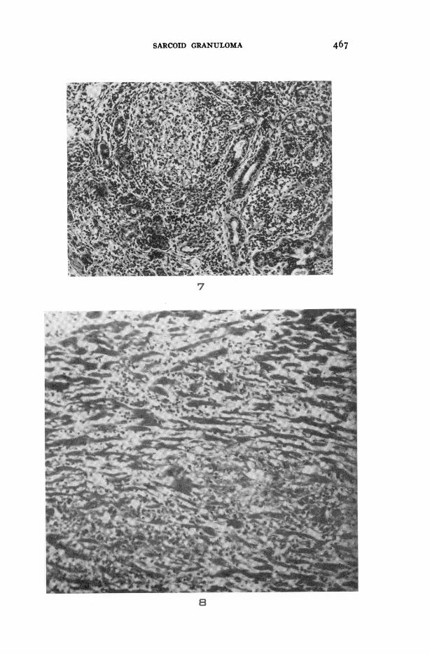
The Presence of a Mononuclear Cell Infiltration in a Wider AreaThan the Field of the Granuloma. Mononuclear cell infiltration in awider area than the field of the granuloma was most striking in thelungs (Figs. 4 and 5) where the early granuloma, sometimes inter-stitial but often intra-alveolar, was surrounded by a nebula of lympho-cytes and monocytes, gradually fading out into the surroundingparenchyma. The less mature the granulomata, the wider the cellularinfiltration; and, conversely, once the sarcoid lesions had become ma-ture, the cloud of infiltration had largely disappeared. This mono-nuclear cell infiltration, usually accompanied by destruction ofparenchyma, was also found in the liver (Fig. 6), parotid gland (Fig.7), heart (Fig. 8), and central nervous system. This feature was somarked that it is strange so few observers have mentioned it. As willbe indicated in the paragraph on retrogressive lesions, this diffuseinflammation leaves behind it a variable amount of fibrosis. Whilegranulomata in a proliferative phase were found in the spleen, nodes,and bone marrow, the mononuclear cell infiltration in these organswas either absent or difficult to recognize.
Sarcoidosis, as it affects certain organs, appears to progress throughthree stages: (I) diffuse inflammation; (2) cell proliferation leadingto the formation of epithelioid cells; (3) hypertrophy of epithelioidcells to form localized cell aggregates. We have not made a specialstudy of sarcoidosis in the skin but from the papers of Kyrle4 andLever and Freiman" we may judge that a similar process occurs there.
Retrogressive LesionsThe sarcoid granuloma is an avascular structure dependent for its
life on the diffusion of fluids from nearby vessels. If, as we believe,the granuloma is a response to some foreign agent, the stability or
453
BARRIE AND BOGOCH
otherwise of the granuloma may throw light on the nature of thecausative agent, besides reflecting the influences of natural ageingand of changes in the surrounding circulation.The histologic pattern in a group of nodes may appear astonishingly
stable, as has been shown by biopsy after a long interval from the siteof a previous excision. The tendency for sarcoidosis to persist appar-ently unaltered for long periods is also evident clinically in some cases.This does not enable us to assert that the foreign agent has no dele-terious effects on its host, the epithelioid cell, for death of anepithelioid cell might be followed by local replacement, and mostgranulomata contain occasional mononuclear cells from which suchreplacement could be derived.
Retrogressive changes may be divided into active degeneration,simple atrophy, and fibrosis.
Active Degeneration. Many forms of active degeneration other thansimple atrophy occurred in the cells composing the granulomata. Thuslysis, fragmentation, and coagulative necrosis were encountered fre-quently. Lysis was most apparent in the nuclei, especially those ofgiant cells which were often reduced to shadowy outlines (Fig. 9).This aroused interesting speculation regarding the field of cytoplasmrelated to these nuclei. It is possible that some of the calcified Schau-mann bodies might have had their origin in micro-infarcts. Fragmen-tation of the cytoplasm was common, although it was always difficultto exclude the possibility of artefact. Coagulative necrosis occurredin the centers of the granulomata and has often been described. Thereis no justification for calling it fibrinoid necrosis. It has a slightlyrefringent quality and is more eosinophilic than the caseous necrosisin tuberculosis.
In the majority of instances, when active degenerative changes wereoccurring, the cells in the immediate neighborhood showed signs ofincreased activity, made evident by irregularity of outline, hyperchro-matism and irregularity of the nucleus, and the presence of smallmultinucleated cells containing only two or three nuclei (Fig. io).
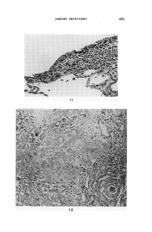
Simple Atrophy. Many of the cases showed simple atrophy of thegranuloma. Here the epithelioid cells showed indistinct outlines. Thechromatin stained poorly and was limited to the periphery of thenuclei. They were surrounded by little clusters of lymphocytes whichwere sharply limited to the lymphatic pathways in contrast to thediffuse exudate found in the early stage (Fig. ii). The multinucleatedcells often survived the longest, as was also the case when the lesionunderwent fibrosis.The difference between the slow atrophy and the more active degen-
454

SARCOID GRANULOMA
erative lesions apparently was not related to disturbances in thesurrounding vascular system and we believed it more probably repre-sented differences in the degree of activity of the causative agent.
Fibrosis. There is ample clinical evidence that lesions in the eye andskin, and therefore probably elsewhere, may undergo atrophy andabsorption without fibrosis. In 7 of the cases, however, the latter wasa marked feature. This process may also provide clues to the signifi-cance of the granuloma.
Fibrosis occurred in two forms. In one, the fibrous tissue wasarranged in parallel strands and its relation to granuloma formationwas not always obvious. It is probable that some of it represented asequel of the diffuse mononuclear cell infiltration we have alreadydescribed. In the other form, fibrosis was nummular and obviouslyrelated to the granulomata. It always started in the periphery of thegranuloma though it was often eccentric and, because of the compoundnature of the aggregates, often partially divided a granuloma into theshape of a heart. It was usually hyaline and in 3 cases there was ma-terial present which showed the staining reaction of amyloid. Itusually has been assumed that the epithelioid cells become transformedinto fibroblasts but the evidence for this is very scanty. Hortega'ssilver carbonate stain encrusts fibroblasts well but not epithelioid cells.Using this technique it was seen that the onset of the fibrosis was asso-ciated with the migration of fibroblasts into the periphery of thegranuloma. This occurred only when the epithelioid cells were disap-pearing. As long as epithelioid cells remained, all extracellular fibrilsin the granuloma were slender and argyrophilic and could be classedas reticulin. As the epithelioid cells disappeared, two processes led tofibrosis. One was the migration of fibroblasts into the periphery andthe other was the gradual coarsening of the reticulin fibers of thegranuloma into hyaline collagen. There was no evidence to suggestthat the epithelioid cell ever took on the function of a fibroblast. Wetake the opposite standpoint and assert that the granuloma, whileviable, is remarkably resistant to fibrosis. The tendency for the giantcells to persist longest in a hyaline lesion is illustrated in Figure I2.We may remark here, too, that besides being resistant to fibrosis,
the small complex of epithelioid cells and reticulin fibers was found tobe relatively impenetrable to polymorphonuclear infiltration and, inpneumonia, could be found like a little island in a sea of polymorpho-nuclear cells. The little granuloma has therefore a great capacity forisolation and this may be the clue to its function, as discussed in alater paragraph.
In some of the cases showing the most fibrosis, doubly refractile
455

BARRIE AND BOGOCH
particles were present in the granuloma. That some of this was silicondioxide was made likely by the presence of silicotic lesions elsewhere.The epithelioid cells and giant cells often contained carbon particlesalso and it is obvious that their load may not be confined to any oneforeign substance. Coincident phagocytosis of silicon dioxide wouldbe expected to modify the natural history of the sarcoid lesion.
Age of the DiseaseSo far, we have been dealing with histologic features only. To what
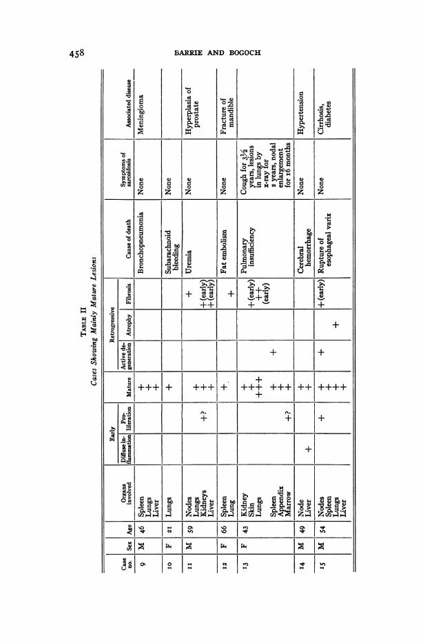
extent does our division of the lesions of sarcoidosis into early, mature,and retrogressive types agree with the clinical course of the disease?A major difficulty here was the common absence of symptoms refer-able to sarcoidosis and in i8 of our cases the disease was an accidentalfinding, death being due to other causes. Even when death was due topulmonary insufficiency as in case I2 there was no means of deter-mining how long sarcoidosis had been present in the lungs beforesymptoms occurred. A further but lesser difficulty lay in the fact thatin many cases the various organs contained lesions of different ages.This corresponds to the well known clinical experience that sarcoidosishas recurrent phases of activity. Nevertheless, by taking into accountthe age of the majority of the lesions, it was possible to divide thecases into those in which the lesions were predominantly early, mature,or retrogressive, respectively, and the resulting correlations with theclinical findings are shown in Tables I, II, and III. It can be seen thatall 4 cases with cardiac involvement fall into the early group, as sar-coidosis of the heart shows all the features which we have describedas the early phase.That this probably did represent an early phase and was not merely
due to some special reaction of the myocardium was shown by the factthat in 3 of the 4 cases with involvement of the heart, two other organsalso contained early lesions. In the group with retrogressive lesions,there were no scars in the heart to suggest that sarcoidosis had beenpresent at one time and had healed. The average age for the cases inTable I was 43, in Table II, 48, and in Table III, 6o years. The signifi-cance of this would be hard to define on statistical grounds. Clinicaldata can be seen to be of little help except in case 4 which has beenpublished by Oille, Ritchie, and Barrie7 and must be mentioned againhere. This was the case of a 43-year-old woman who, because ofsymptoms of an iron-deficiency anemia, had an electrocardiogram andfluoroscopic examination of the lungs in May, I948. Both were nor-mal. In August she was febrile for a week and suffered pain in the
456

SARCOID GRANULOMA
c
do~~~~~ocis
ce
co0(4
+++++
+ +
cd
bCo 4)E
(4)1
.41 -4*1
3 C)i~
(0
U
4L)0
z
0
(1204L)
Ca;(44L)
0 t
+ +
It -bm
4'.
ICi2
U~4
502laI0 0
0
o
*s 0
Cd
>0ZO
I:
It4) M.t,
E:R4)
w
cd
C.P
4.) 4)
d~4)C1$,0d
-O'
j3.Cd,4
A .:
Cd
.40
C)
O.
4)
*gyX
O0 cd cd
+
+ ++
+
+ +
0
Zj(
Iux
I oS
12a-Io
1..
4c)o.u
Cd
12C)-
Cd
I++
++
+
++
u](0
+
boI
0
'I
Cd
-4
IaqCa0
P*4
0
0
'4
(2Cd
.4cd
U
+++ +1
+ ++ +1
Z A4 W4 M
00
1o4, 2}:
IX4~~~~~~~~~l
457
1*4)
0
IL)0
z
4)
:3 0(4-1 cd
d
4.)(1(4
c4)>
Z44
o
u
co
cn
4)4
00
Ca
0
C)
a)-
co
0
.o
cn%4)j
ll
0cocis9tsIcis
.I
(4 oo
BARRIE AND BOGOCH
a)00z
0
fA0
4)00
eq
0
0
ei
000z
a)
54 I..8 d4
++
.-
"Ao
tn0)4. w
:. >
'4-0 a)
0 --
4 054"0
a)90
z
0
a)
+
+.0
o
I
I>
cn d
o o
eq o
rA0
C'_s
+
+
+
a)O CO >,,-0
o C
0. $u
cf
458
V
;8
.uq)co
:
0 o)
'0
.5Ca
0
o
100
4%
,A
2
bC
q
e'-4
0
To._._-0
a)
0r1
z
00a)
0000540
pq
I
I+
I;
10
I)
Cs.X4
q
Uk)
U1
00
.C0aL)
a.)00z
a)bo,0I "
++
a)4
)-0
'0 )
00 0
cOa0,0.0cwqm
.1-1u
a)
0zz
NU)
I r.
1-.ce
o bOD
I++
I+++
I+
COOCO
,I
in

0
cis
C-)C
o =r-boU o
a
00
z
.0
Ei=ir_
0
0
+tt
U
o t0
's =c-Do
00.4Cd )
00
zcoO
8
a0Dsm
+
a CQlae105
~
I-
I4
0
10
C:0z
.0
1O-5.
I+
I++
I...
0
.4)
I IX
I$
I
00
40I..
9:Z0z
I-
10
IS0U
1-aV
54
$:'+ C++
_
I++++I
._
.4>= 4 )
WI)
0u
1 0
I=
0
z
0
l:3
0.),0
u0
ImI+
I++
I+
; Z='0
.4Z
0011)
.~~
SARCOID GRANULOMA 459
U
z_0
z
U
U
+
4-2cso
4-
I;IEi0
IQco, Q
I
I
0r.0
z
.M
u
0
5..
ri4
I
+++
4L d0
10 0
1v =1
0
0
U
Io d-10
00C)s
I Ez
l u2
.190
z
I
;++
c
0o>,
o-4
C.)"04
-4
E- s
the
lala
0
0
*0
0
r'0
v afilu.0 5..
U00z
u5.4
I0d
1.5U
u
0-
:30
.4z
+1.
Io
0 C#ljeq
1*

BARRIE AND BOGOCH
ears and swelling of the lymph nodes. She then developed a slightcough. An electrocardiogram was again normal on October 26 but 2weeks later she suddenly became so tired that she could not get out ofbed. Two weeks after this she was unexpectedly found dead in bed.At necropsy, the heart was widely infiltrated by sarcoidosis and therewere also lesions in the liver, lungs, and lymph nodes. Invasion of theheart may be assumed to have occurred after the last normal electro-cardiogram and the cardiac lesions, therefore, were less than 4 weeksold. This remarkable case gives us a time standard with which to judgethe development of the cardiac lesions of sarcoidosis, and the patient'sfebrile episode 3 months before death strongly suggests that that wasthe time of onset of the diffuse inflammatory phase.We had no clues from our material which would reveal how long
the mature and retrogressive lesions had been present in the body.Case 2 was difficult to interpret. Proliferative lesions were present inlungs, spleen, and hypothalamus in spite of the fact that it was prob-able that diabetes insipidus of 6 years' standing was due to sarcoidosisof the mid-brain.The tables emphasize the benign nature of the disease. Death was
caused by sarcoidosis in only 6 cases and in 5 of these there was in-volvement of either the heart or the central nervous system.
DIsCUSSIONIt would seem that certain deductions can reasonably be made from
a study of the natural history of sarcoidosis as it has been outlinedand from the facts already available in the literature. For purposes ofcomparison, a brief survey of granulomas of some other types mightbe helpful.The granulomatous reaction is a response to a foreign agent either
introduced from without or produced by modification of some normalstructure in the body. Exogenous agents may be divided into thosewhich are not capable of self-propagation and those which are. Of thefirst, the best example is silicon dioxide. This slowly soluble but highlyactive substance has a portal of entry (the lungs), and the granuloma-tous response is found along the migratory routes of the monocyteswhich phagocytize the particles. A certain concentration of the agentis necessary for granuloma formation and therefore the lungs and thesites nearest to them contain the more numerous granulomata. It isprobable that individual monocytes containing silicon dioxide may bepresent anywhere in the body for they can migrate through the wallsof pulmonary veins and enter the blood stream, but no granuloma
460

SARCOID GRANULOMA
develops because of the small amount of silica present. In the liver,spleen, and marrow, where blood-borne silica is filtered out and con-centrated, it is well known that silicotic nodules may develop.Of exogenous agents capable of proliferation, an important example
is Mycobacterium tuberculosis. With this agent the localization oflesions is not controlled by the initial concentration alone, but also bythe ability of the migrating bacterium to proliferate in its immediatesurroundings. The portal of entry in the majority of cases is the sameas in silicosis. Thus tubercles develop at the same sites as silicoticnodules as wel as in certain organs which seem to favor the prolifera-tion of tubercle bacteria, such as the genito-urinary tract and the cen-tral nervous system.
Finally, granulomas may develop from a modification of somenormal component of the body. Sometimes this is a localized phenome-non such as the formation of giant cells around altered elastica in alocally destructive lesion in the lung, but here the cause is usuallyobvious. When generalized and due to abnormalities of antigen-anti-body reaction, the primary lesion occurs in components of the vascularsystem rather than in individual organs and the distribution of lesionsis very wide, including skin, serous surfaces, joints, muscles, and vis-cera. Sometimes, however, disease believed to be due to hypersensi-tivity, such as rheumatic fever, may show a remarkble organspecifidty which may affect the heart alone; arsenical reactions mayaffect the heart and skin, and "malignant granuloma" affects the mid-line of the face.
Against this background the main features of the lesions in sar-coidosis may be reviewed. On such limited material, no dogmaticstatement is possible about the significance of the diffuse mononuclearcell infiltration that was found in the lungs, liver, heart, salivary glands,and central nervous system, and which probably occurs also in theskin. It remains, however, an interesting finding and opens the possi-bility that it represents one of the missing links in sarcoidosis-thestage of active organ invasion.The first stage that we can definitely recognize as being part of the
disease is an early proliferative phase which is recognizable in all sitesin which the granuloma may be found, that is, in the lymphatics, in themigratory routes of the phagocytes in the organs mentioned, and in thefilter systems of the body, the lymph nodes, the spleen, and the mar-row. A reasonable interpretation of this sequence is that some foreinagent is present that is capable of damaging parenchymal cells andproducing some fibrosis. This foreign agent is removed from contact
46I

BARRIE AND BOGOCH
with the tissues by phagocytosis. The phagocytes, however, may them-selves be damaged by the (foreign) agent, react by proliferation andthen by hypertrophy, and become transformed into epithelioid cells.
Proliferation of phagocytes and enlargement of the daughter cellscontinue to a point at which some stability occurs. It would seem mostreasonable to suggest that this point of stability is reached when theforeign agent is diluted sufficiently by successive division of epithelioidcells to impair its capacity to induce further cell division. The granu-loma thus represents merely a stabilization of the phagocytic process,the purpose of which is to isolate the foreign agent. That the lattermay finally be dissolved, neutralized, or removed is suggested by thefrequent disappearance of the granuloma. That it may damage theepithelioid cells themselves has been considered in the discussion onretrogressive changes.When the epithelioid cell dies as a result of this injury three things
may occur: Firstly, if the epithelioid cell is placed centrally it mayremain in situ and give the picture of coagulative necrosis. Secondly,if placed peripherally, the epithelioid cell may be absorbed. The for-eign agent may then be liberated and stimulate the peripheral fibrosisthat is common in old sarcoid lesions. Thirdly, the liberated foreignagent may be picked up once more by phagocytes and the granulomathus may extend and may coalesce with other lesions.When the behavior of sarcoid is compared to the three types of
granuloma that have been discussed, it can be seen at once that it ismore complicated than the behavior of the body to silica. The distri-bution of the earliest lesions in lung, liver, skin, central nervous sys-tem, and salivary glands makes it tempting to suggest a virus as theetiologic agent. Search for a virus in sarcoidosis has so far been un-productive but it is possible that these observations have been madeat an unsuitable phase in the development of the lesion. By the timethe granulomas of sarcoid have become established they may not beharboring an active virus but only some changed component of thevirus or of the body's tissues.
In the precise differentiation between the role of hypersensitivityand organismal invasion the threads of the clues run out, but it is be-lieved that the interpretation of the disease up to this point may havevalue in understanding its natural history.
SUMMARY
Formation of the sarcoid granuloma is often preceded by a phase ofdiffuse mononuclear cell exudation.
462
SARCOMD GRANULOMA 463
The granuloma is formed first by proliferation and then by hyper-trophy of monocytes.
Early sarcoid lesions may therefore be recognized by three features:pleomorphism and hyperchromatism of the cells; disorder in cellulararrangement; the presence of an outlying infiltration of mononuclearcells.Of 24 necropsied cases of sarcoidosis, the predominant lesions were
early in 8, mature in 7, and retrogressive in 9.The granuloma does not undergo fibrosis by a change of the epithe-
ioid cells into fibroblasts. Fibrosis occurs from the periphery andfollows atrophy or necrosis of the epithelioid cells.The granuloma is probably a protective device whose function is to
isolate some substance from the body tissues. Proliferation of mono-cytes and hypertrophy into epithelioid cells may dilute this substanceto a point where it has little further action on the phagocytes. Thegranuloma then becomes relatively stable.Breakdown of the epithelioid cells may liberate this substance,
which then causes fibrosis or is re-phagocytosed.
REFERENCESI. Pautrier, L. M. La maladie de Besnier-Boeck-Schaumnrm. Masson et Cie,
Pans, I940, 344 PP.2. Ricker, W., and Clak. M. Sarcoidosis. A clinicopathologic review of three
hundred cases, including twenty-two autopsies. Am. J. Clin. Path., i949, ig,725-749-
3. Kissneyer, A. La maladie de Boeck: Sarcoldes cutanes b6ignes multiples.Levin & Munksgaard, CopenhageI, 1932, 147 PP.
4. Kyrle, J. Die Anfangsstadien des Boeckschen Lupoids; Beitrag zur Frage dertuberkul6sen Atiologie dieser Dermatose. Arch. f. Dermat. u. Syph., 1921,131, 33-68.
5. Lever, W. F., and Freiman, D. G. Sarcoidosis. Report of an atypical case witherythrodermic lesions, subcutaneous nodes and asteroid inclusion bodies ingiant cells. Arch. Dermat. & Syph., 948, 57, 639-654.
6. Johnson, J. B., and Jason, R-S. Sarcoidosis of the heart Report of a case andreview of the literature. Am. Heart J., 1944, 27, 246-258.
7. Oille, W. A, Ritchie, R. C., and Barrie, E J. The age of the lesions in a caseof cardiac sarcoidosis. Canad. M. A. J., 1953, 68, 277-278.
[ IUustrations foUow ]
BARRIE AND BOGOCH
LEGENDS FOR FIGURES
FIG. I. Early sarcoid nodule. One miliary granuloma is already formed but as yetcontains no reticulin. Surgical specimen for biopsy of retro-orbital tumor.Laidlaw's stain. X 256.
FIG. 2. Stage of cell division in early sarcoid. Same section as Figure i. Hema-toxylin and eosin stain. X 256.
FIG. 3. Case 6. Early sarcoid granuloma in heart. Stage of cell division. Hema-toxylin and eosin stain. X 538.
FIG. 4. Case 3. Diffuse mononuclear cell exudate surrounding an early granulomain the lungs. Hematoxylin and eosin stain. X I28.
464

SARCOID GRANULOMA 465
2
3
1
4

BARRIE AND BOGOCH
FIG. 5. Case 4. Diffuse mononuclear cell exudate and very early sarcoid aggregatein lungs. Hematoxylin and eosin stain. X 256.
FIG. 6. Case 4. Diffuse mononuclear cell exudate and loss of parenchyma in theliver. A sarcoid giant cell is present in the upper portion of the field. Hema-toxylin and eosin stain. X I28.
FIG. 7. Diffuse mononuclear cell exudate in parotid gland. An early sarcoid granu-loma is visible in the upper portion of the field. Surgical biopsy. Hematoxylinand eosin stain. X I28.
FIG. 8. Case 4. Diffuse mononuclear cell infiltration and atrophy of fibers at pe-riphery of sarcoid granuloma in heart. Hematoxylin and eosin stain. X 54.
S ~~~~~~6
466
5
SARCOID GRANULOMA 467
7
8

BARRIE AND BOGOCH
FIG. 9. Case 4. Lysis of nuclei in one giant cell and a Schaumann body in the other.Hematoxylin and eosin stain. X 538.
FIG. IO. Coagulative necrosis in the center of a sarcoid aggregate. The neighboringepithelioid cells show signs of activity. Surgical specimen of lymph node.Hematoxylin and eosin stain. X 256.
FIG. II. Case i6. Atrophic sarcoid lesion. A few residual epithelioid cells are ac-companied by lymphocytes sharply limited to the lymphatic pathways. Hema-toxylin and eosin stain. X 256.
FIG. I2. Case I7. Hyaline fibrosis of lymph nodes with persistence of giant cells.Hematoxylin and eosin stain. X I50.
10
468
9
SARCOID GRANULOMA
11
12
469