leukocyte cultures - journal of bacteriology - american society for
TRANSCRIPT

JOURNAL OF BACTERIOLOGY, July, 1966Copyright © 1966 American Society for Microbiology
Vol. 92, No. 1Printed in U.S.A.
Replication of Mumps Virus in HumanLeukocyte Cultures
HUU DUC-NGUYEN1 AND WERNER HENLE2Virus Laboratories, The Children's Hospital of Philadelphia, and School ofMedicine, University of Pennsylvania,
Philadelphia, Pennsylvania
Received for publication 3 March 1966
ABSTRACr
Duc-NGuYEN, Huu (The Children's Hospital of Philadelphia, Philadelphia,Pa.), AND WERNER HENLE. Replication of mumps virus in human leukocyte cul-tures. J. Bacteriol. 92:258-265. 1966.-Human peripheral leukocyte cultures main-tained in the presence of phytohemagglutinin (PHA) were found to support tosome extent the replication of mumps virus. When such cultures were exposed,within 24 hr after their initiation, to a high input multiplicity of virus, successfulinfection, as determined by immunofluorescence and plaque assays, did not be-come evident before the 3rd or 4th day. On exposure of cultures 4 to 5 days old,viral replication was detectable within 2 days. In both instances, peak immuno-fluorescence and virus titers were reached when the cultures were 7 to 9 days oldand composed mainly of blast forms. With decreasing input multiplicities of infec-tion, cells containing viral antigen and production of infectious viral progenybecame detectable with increasing delay. No significant viral replication was notedin surviving cells maintained in the absence of PHA. These results indicate thatmainly, if not solely, the PHA-stimulated cells of the lymphocytic series supportviral multiplication. The extent of the infectious process was limited, however,because the life span of the cultures was not significantly shortened, the yields ofinfectious virus per immunofluorescent cell were at all times low, and most infectedcells contained only a few well-delineated small masses of antigen, suggestive of anabortive infection. Only fresh cultures were capable of synthesizing interferon onstimulation by mumps, Newcastle disease, or Sendai viruses. When the cultureswere set up in the presence of PHA, this capacity was lost within 24 hr. PHA per sefailed to induce detectable production of an interferon under the conditions used.The implications of these findings are discussed.
Several viruses were found to replicate to someextent in suspensions of human peripheral leuko-cytes which were prepared or cultured in the pre-sence of phytohemagglutinin (PHA). This sub-stance, originally used to separate erythrocytesfrom leukocytes, was shown subsequently to stim-ulate mytosis of lymphocytic elements and tochange many into blast forms (8, 10). Berg andRosenthal (2) were able to maintain measles virusthrough serial passages at 3- to 4-day intervals infresh human leukocyte cultures. Maximal virustiters were obtained, however, only between the4th and 7th days. Measles virus also grew in
1 Present address: Section of Viral Oncology, Na-tional Cancer Institute, National Institutes of Health,U.S. Public Health Service, Bethesda, Md.
2 Career Investigator, 1-K6-AI-22,683, NationalInstitutes of Health, U.S. Public Health Service.
leukocyte suspensions consisting mainly of blastforms, which were derived from leukemic patients.Berg (1) reported similar results on infection offresh leukocyte suspensions with echovirus 9. Sub-sequently, Nahmias and co-workers (9) and Gres-ser and Chany (5) infected not only fresh but also4-day-old leukocyte cultures with herpes simplexvirus and poliovirus type 1, respectively. Whenfresh cultures were used, evidence of viral replica-tion was not noted before the 4th day, and peaktiters were observed by the 6th day or later. Oninfection of 4-day-old cultures, significant viralreplication became evident and maximal titerswere obtained sooner than in fresh suspensions.These results indicated that polymorphonuclearcells, which degenerate rapidly, contributed rela-tively little, if at all, to the maintenance of thevarious viruses, and that mainly the PHA.
258
Dow
nloa
ded
from
http
s://j
ourn
als.
asm
.org
/jour
nal/j
b on
23
Nov
embe
r 20
21 b
y 21
1.21
0.20
7.64
.

MUMPS VIRUS IN HUMAN LEUKOCYTE CULTURES
stimulated lymphocytic elements were responsiblefor supporting viral replication. Indeed, in theabsence of PHA, the cultures failed to supportherpes simplex virus. The yields of virus in all in-stances were low in relation to the number of avail-able cells.The delay in viral replication and the low yields
of progeny conceivably might be related to thefact that fresh leukocyte suspensions are capableof synthesizing interferon on appropriate viralstimulation (4), with both lymphocytes and poly-morphonuclear cells being involved (7). Further-more, PHA per se was reported to induce inter-feron synthesis in human leukocytes (14). Thus,interferon could well reduce, delay, or abort viralreplication in fresh leukocyte cultures. WhetherPHA-stimulated leukocytes which have beenmaintained in culture for some time retain thecapacity of interferon synthesis has not as yet beenclearly demonstrated.The present studies, concerned with infection
of leukocyte cultures by mumps virus, were under-taken to clarify further the cultural conditions re-quired for viral replication, the extent of cellularsusceptibility, and the roles played by PHA andinterferon in the infectious process.
MATERIALS AND METHODS
Leukocyte cultures. The technique described byMoorhead et al. (8) was used with some modifications.Blood treated with heparin was allowed to settleat 37 C within a syringe kept in an upright position.When about one-third of the volume had been clearedof red cells, the leukocyte-rich plasma was expelledupward through a bent needle into a centrifuge tube,and the cells were sedimented at 600 X g for 10 min.The cells were washed three times in medium 199supplemented with 10% inactivated fetal calf serum(30 min at 56 C), and transferred to plastic or glassflasks. After being gassed with a mixture of air and5% CO2, the flasks were incubated in a prone posi-tion at 37 C for 1 hr to permit attachment of poly-morphonuclear cells and monocytes to the surface ofthe vessels. The suspension of unattached cells wasdecanted and counted. Differential counts revealedthat the granulocytes were reduced by this procedureby, at most, 85%. The desired numbers of cells weresedimented and resuspended in medium 199 contain-ing 20% fetal calf serum, 1% phytohemagglutinin-P,and penicillin (100 units per ml) and streptomycin(100 jug/ml). The final suspensions, containing be-tween 106 and 2 X 10⁶ cells per milliliter, were dis-pensed into Erlenmeyer flasks, prescription bottles,or screw-capped vials, leaving an air space of about80% of the total volume. Half of the medium wasreplaced on the second day, care being taken not todisturb the settled cells. Further feedings did not seemto prolong the survival of the cultures and, therefore,were usually omitted. Cell counts were made at in-tervals, by use of a 0.1% solution of trypan blue as
diluent to differentiate between viable and dead(staining) cells.
Viruses. The Ricki strain of mumps virus, isolatedin chick embryos from a case of parotitis, was usedin its 7th to 9th amniotic passage. The amniotic fluidseeds, stored at -70 C, contained approximately108 plaque-forming units (PFU) and 102.6 hemaggluti-nating (HA) units per ml. The virus was added toleukocyte cultures in a concentration sufficient toattain desired input multiplicities. Nonadsorbed viruswas usually, but not always, removed after adsorptionperiods of 1 to 3 hr at 37 C by washing of the cellstwice with medium.
For production of interferon, Sendai or Newcastledisease (Victoria strain) virus was used in the form ofinfected chick embryo allantoic fluids. For assay ofinterferon activity, the Indiana strain of vesicularstomatitis virus (VSV) was used as challenge. Stockvirus was prepared from infected chick embryo fibro-blast cultures.
Hemadsorption plaque assay ofmumps virus. Mono-layers of chick embryo fibroblasts were prepared inplastic flasks (Falcon Plastic Co.) according to routineprocedures (6). Enough cells were seeded to obtainconfluent sheets within 18 hr. After washing withHanks solution, 0.4-ml amounts of the test materialswere introduced, and the virus was permitted toadsorb for 1 hr at 37 C. The cell sheets were thenoverlaid with the following mixture: methyl cellulose(Methocel; viscosity, 1,500 centipoises; Dow Chemi-cal Co., Midland, Mich.), 1.5%; medium 199 (lOX),3%; Scherer's maintenance medium (lOX), 5%; in-activated horse serum, 20%; NaHCO3 solution(7.5%), 5%; and antibiotics. After incubation at 37C for 10 days, the flasks were placed at 4 C for 1 hr,the overlay was poured off, and the cell sheets werewashed with cold phosphate-buffered saline (PBS)solution until all cellulose was removed. After additionof 3 ml of a 0.6% suspension of chicken red cells inPBS, the flasks were kept at 4 C for 30 min, drained,washed, and examined for foci of hemadsorption.These were readily identifiable (Fig. 1) and countableunder illumination by a 60-w light source.
Immunofluorescence assays. Cells were sedimentedfrom culture samples and, after decanting of thesupernatant fluids, were resuspended in the remainingmedium. Drops of this suspension were spreadevenly over cover slips (6 by 30 mm), and allowed todry at 37 C. The preparations were fixed in fresh ace-tone at room temperature for 10 min, and then ex-posed for 1 hr at 37 C to fluorescein isothiocyanate-conjugated human y-globulin obtained from donorswho had been hyperimmunized against mumps. Thecover slips were thoroughly washed in two changes ofPBS, dipped in distilled water, dried, fixed on slidesby means of a semipermanent mounting medium (12),and examined under ultraviolet illumination (OsramHBO 200-w lamp) with a Zeiss microscope. The per-centage of cells containing viral antigen was deter-mined by counting from 200 to 1,000 cells, dependingupon the extent of the infection.
Interferon production and assay. Suspensions con-taining up to 107 viable cells per milliliter of medium(2% fetal calf serum) were exposed to mumps, Sendai,
259VOL. 92, 1966
Dow
nloa
ded
from
http
s://j
ourn
als.
asm
.org
/jour
nal/j
b on
23
Nov
embe
r 20
21 b
y 21
1.21
0.20
7.64
.

DUC-NGUYEN AND HENLE
FIG. 1. Hemadsorption plaques induced by mumps virus on monolayers of chick embryo fibroblasts. The flaskshad been inoculated with a 1O-2, 10-3, and 10-35 dilution of virus.
or Newcastle disease virus (NDV) at input multiplicitiesin excess of 1, and incubated at 37 C for 24 to 36 hr.The clarified medium was concentrated twofold withCarbowax polyethylene glycol compound 20-M(Union Carbide Chemical Co.). This material wasusually dialyzed at 4 C successively against 100 vol-umes of KCl buffer (pH 2), phosphate buffer (pH 7.2),and Hanks solution, each for 24 hr. For some tests,the medium was used merely after clarification at100,000 X g for 1 hr. For assay of interferon activity,serial twofold dilutions were added to human amnionor human embryonic kidney cultures (purchased fromFlow Laboratories, Rockville, Md.). These werechallenged 24 hr later with about 104 TCID5c of VSV.Virus controls showed marked cytopathic effects in24 hr and complete destruction of the cell sheets in48 hr. The results were read at this time, and thehighest dilution of an interferon preparation protect-ing at least 75% of the cells was taken as the endpoint. In one test, HeLa cells were used according tothe method developed by Paucker (11), based onchanges in pH of the cultures.
RESULTS
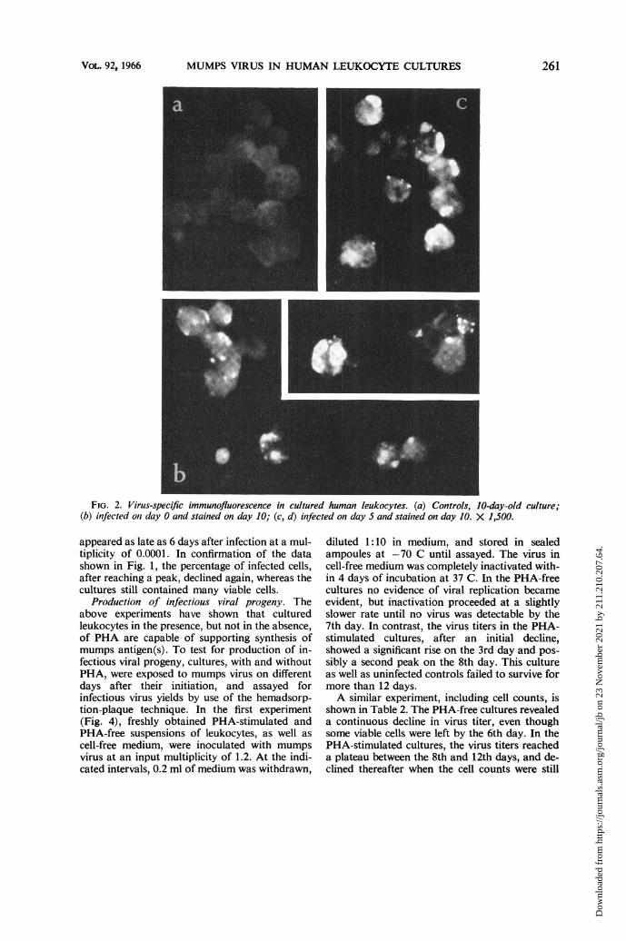
Development of virus-specific immunofluores-cence. In initial experiments, leukocyte cultureswere set up with or without PHA and exposedimmediately to mumps virus at input multiplicitiesranging from 0.1 to >5. After incubation forvarying periods of time, cover-slip preparations ofcells were made and tested for virus-specificimmunofluorescence. In the PHA-stimulated cul-tures, only rare cells, if any, were found to containviral antigen within the first 48 hr. Stainable cellsappeared thereafter in gradually increasing num-bers ranging from 30 to 80% of the total popula-tion. The appearance of the stained cells is illus-
trated in Fig. 2. The infected cells generally werelarger than the lymphocytes present at start ofthe cultures, and contained viral antigen in well-delineated round or oval masses of varying sizes.Cultures set up without PHA and infected withmumps virus failed to reveal detectable numbersof antigen-containing cells at any time.The gradual spread of the infection in PHA-
stimulated cultures is shown in Fig. 3. In thisexperiment, leukocytes of one donor were exposedto mumps virus at an input multiplicity of 1.1, andreplicate cultures were set up for assay of viablecells and virus-specific immunofluorescence at theindicated intervals. The cell counts, after the usualinitial decline, revealed evidence of cellular repli-cation, and the cultures, consisting at that timelargely of blast forms, survived for 3 weeks. Thepercentage of infected cells reached its peak by the13th day and declined thereafter, well in advanceof the death of the culture.
These results indicated that mainly the PHA-stimulated lymphocytic elements of the culturesupported the synthesis of viral antigen(s). Thisis further evident from the fact that, upon inocula-tion of mumps virus into 3- to 5-day-old cultures,many cells became infected within a few days,their number depending, of course, upon themultiplicity of infection. In the experiment pre-sented in Table 1, PHA-stimulated leukocyteswere infected on the 3rd day at varying multiplici-ties. With the larger doses of virus, more than50% of the cells contained antigen 48 hr later, butas the dose was decreased antigen became detect-able successively later. In other experiments ex-tending over longer periods, immunofluorescence
260 J. BACTERIOL.
Dow
nloa
ded
from
http
s://j
ourn
als.
asm
.org
/jour
nal/j
b on
23
Nov
embe
r 20
21 b
y 21
1.21
0.20
7.64
.
MUMPS VIRUS IN HUMAN LEUKOCYTE CULTURES
FIG. 2. Virus-specific immunofluorescence in cultured human leukocytes. (a) Controls, 10-day-old culture;(b) infected oni day 0 and stained on day 10; (c, d) infected on day 5 and stainied on day 10. X 1,500.
appeared as late as 6 days after infection at a mul-tiplicity of 0.0001. In confirmation of the datashown in Fig. 1, the percentage of infected cells,after reaching a peak, declined again, whereas thecultures still contained many viable cells.
Production of infectious viral progeny. Theabove experiments have shown that culturedleukocytes in the presence, but not in the absence,of PHA are capable of supporting synthesis ofmumps antigen(s). To test for production of in-fectious viral progeny, cultures, with and withoutPHA, were exposed to mumps virus on differentdays after their initiation, and assayed forinfectious virus yields by use of the hemadsorp-tion-plaque technique. In the first experiment(Fig. 4), freshly obtained PHA-stimulated andPHA-free suspensions of leukocytes, as well ascell-free medium, were inoculated with mumpsvirus at an input multiplicity of 1.2. At the indi-cated intervals, 0.2 ml of medium was withdrawn,
diluted 1:10 in medium, and stored in sealedampoules at -70 C until assayed. The virus incell-free medium was completely inactivated with-in 4 days of incubation at 37 C. In the PHA-freecultures no evidence of viral replication becameevident, but inactivation proceeded at a slightlyslower rate until no virus was detectable by the7th day. In contrast, the virus titers in the PHA-stimulated cultures, after an initial decline,showed a significant rise on the 3rd day and pos-sibly a second peak on the 8th day. This cultureas well as uninfected controls failed to survive formore than 12 days.A similar experiment, including cell counts, is
shown in Table 2. The PHA-free cultures revealeda continuous decline in virus titer, even thoughsome viable cells were left by the 6th day. In thePHA-stimulated cultures, the virus titers reacheda plateau between the 8th and 12th days, and de-clined thereafter when the cell counts were still
VOL. 92, 1966 261
Dow
nloa
ded
from
http
s://j
ourn
als.
asm
.org
/jour
nal/j
b on
23
Nov
embe
r 20
21 b
y 21
1.21
0.20
7.64
.

DUC-NGUYEN AND HENLE
N
-
zJ0
30
11 I2 4 6
100- Z
14 U)5)JI- -J50
I8 10 12 14 16 18 20 22 24
AGE OF CULTURE IN DAYS
FIG. 3. Viable-cell counts and percentage of im-munofluorescent cells in leukocyte cultures infected onthe day ofpreparation.
TABLE 1. Relation between multiplicity ofinfection and development of virus-
specific immunofluorescence
Input multiplicityDay afterinfectiona
8 4 0.4 0.04 0.004
1 Ob 0 0 0 02 3 2 0 0 03 1 3 3 0 04 1 1 2 1 0
a The leukocyte cultures were inoculated 3 daysafter their initiation in the presence of PHA.
b Symbols: 0, no fluorescence; 1, 1 to 20%;2, 21 to 50%; 3, 51 to 80% of the cells showed posi-tive fluorescence.
relatively high. A comparison of the cell countsand plaque-forming units revealed that the yieldsof infectious virus per cell were very low.The two suggestive peaks in virus titers seen in
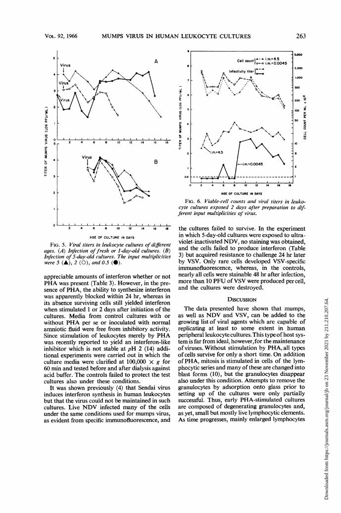
Fig. 4 were evident also when other cultures wereinfected at multiplicities of 0.5 to 5 on the day ofpreparation or 1 day later (Fig. SA). Dependingon the general viability of leukocyte cultures fromindividual donors, the final decline in virus titersoccurred between the 8th and 15th days. The lossof the cultures could not be ascribed to viral ac-tion, because control, uninfected cultures, as arule, failed to survive longer. When cultures wereinfected at similar multiplicities on the 5th dayafter their initiation, peak virus titers were reachedwithin 2 to 3 days thereafter, and only small,secondary peaks were noted after another 4 to 6days (Fig. 5B).When low multiplicities of infection were em-
ployed, evidence of viral replication was consider-
ably delayed as was observed also in the immuno-fluorescence tests. In the experiment shown in Fig.6, leukocyte cultures were exposed 24 hr aftertheir initiation to virus at input multiplicities of4.5 and 0.0045. The results obtained with thelarger dose conformed to those just described.With the smaller dose, virus became temporatilydetectable 6 days after infection of the culture, andwas in significant titers only after the 8th day. Thecell counts for the two cultures failed to showstriking differences during the observation periodin spite of the fact that the yields of virus differedon the average by a factor of 10. This showedagain that the mumps infection does not markedlyreduce the survival time of the leukocyte cultures.
Interferon synthesis. Leukocyte cultures weretested for their capacity to produce interferonafter exposure to mumps, Sendai, or Newcastledisease (NDV) virus. Fresh cultures produced
Medium control
Culture without PHACulture with PHA
..*-*- .-Wz- -.
0 2 4 6 8 10 12 14 16 is
AGE OF CULTURE IN DAYS
FIG. 4. Viral titers in leukocyte cultures with orwithout phytohemagglutinin and infected on the dayofpreparation.
TABLE 2. Viable-cell counts and mumps virus titers inPHA-stimulated and PHA-free leukocyte cultures
No PHA PHA-stimulatedDayafter .-infec- Viable Virus Viable Virustiona cells per titer cells per titer
ml (PFU/ml) ml (PFU/ml)
1 3.04 X 10⁵ 5.82 X 10² 1.62 X 10⁵ 5.59 X 10²² 2.03 X 10⁵ 1.25 X 10² 1.12 X 10⁵ 1.33 X 1026 8.00 X 10³ 2.50 X 101 5.40 X 10⁴ 1.20 X 1038 0 0 8.80 X 10⁴ 1.70 X 1049 0 0 9.60 X 10⁴ 1.20 X 10412 0 0 3.40 X 10⁴ 9.75 X 10314 0 0 1.80 X 10⁴ 4.60 X 10²
a The leukocyte cultures were infected on the day of theirpreparation.
n
>L
IL
coa:cr
1.0
0.4
262 J. BACTERIOL.
Dow
nloa
ded
from
http
s://j
ourn
als.
asm
.org
/jour
nal/j
b on
23
Nov
embe
r 20
21 b
y 21
1.21
0.20
7.64
.
MUMPS VIRUS IN HUMAN LEUKOCYTE CULTURES
a
nCL
a.
:)
U-
5
0
9
a
7
-i
aI
D
LL
Virus
A.... %.
'.
0% % o
%\6i '14 £0r
2 4 6 8 10 12 14 16
2
B
AGE OF CULTURE IN DAYS
FIG. 5. Viral titers in leukocyte cultures of differentages. (A) Infection offresh or 1-day-old cultures. (B)Infection of 5-day-old cultures. The input multiplicitieswere 5 (A), 2 (0), and 0.5 (0).
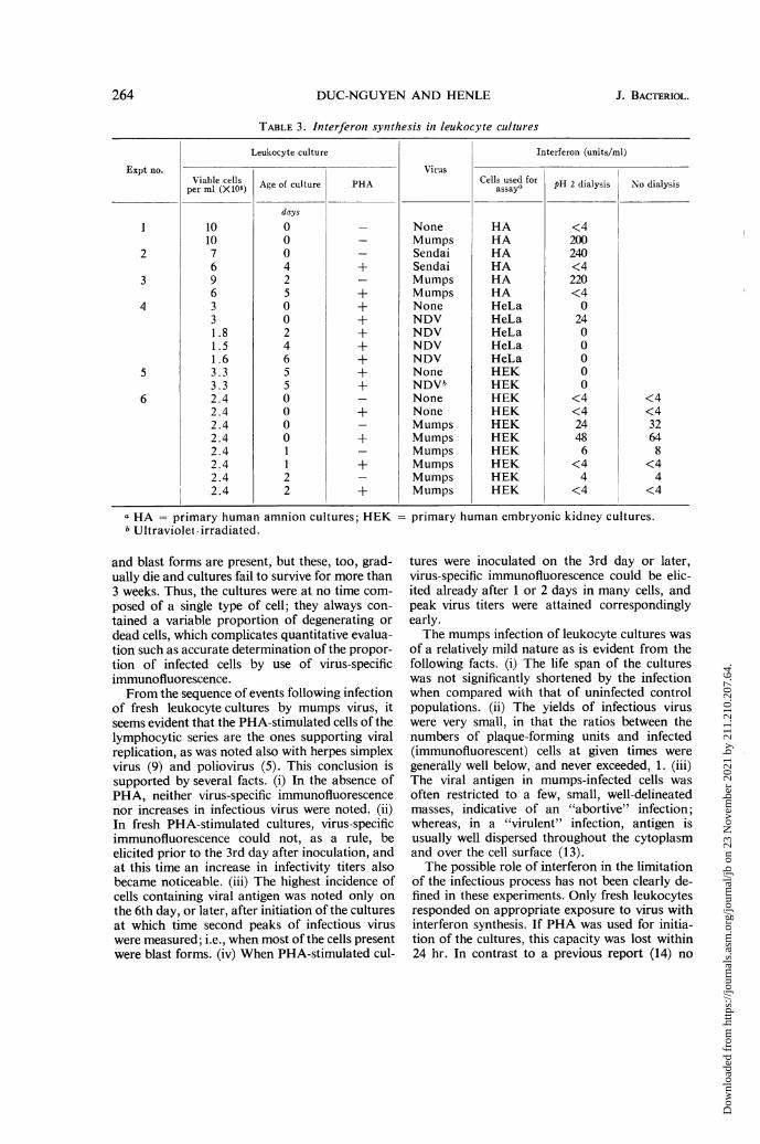
appreciable amounts of interferon whether or notPHA was present (Table 3). However, in the pre-sence of PHA, the ability to synthesize interferonwas apparently blocked within 24 hr, whereas inits absence surviving cells still yielded interferonwhen stimulated 1 or 2 days after initiation of thecultures. Media from control cultures with orwithout PHA per se or inoculated with normalamniotic fluid were free from inhibitory activity.Since stimulation of leukocytes merely by PHAwas recently reported to yield an interferon-likeinhibitor which is not stable at pH 2 (14) addi-tional experiments were carried out in which theculture media were clarified at 100,000 x g for60 min and tested before and after dialysis againstacid buffer. The controls failed to protect the testcultures also under these conditions.
It was shown previously (4) that Sendai virusinduces interferon synthesis in human leukocytesbut that the virus could not be maintained in suchcultures. Live NDV infected many of the cellsunder the same conditions used for mumps virus,as evident from specific immunofluorescence, and
2 4 6 10 12 14 16 1i
500
2,000
1,000
500
200 -
100 a
50 F-5
50
AGE OF CULTURE IN DAYS
FiG. 6. Viable-cell counts and viral titers in leuko-cyte cultures exposed 2 days after preparation to dif-ferent input multiplicities of virus.
the cultures failed to survive. In the experimentin which 5-day-old cultures were exposed to ultra-violet-inactivated NDV, no staining was obtained,and the cells failed to produce interferon (Table3) but acquired resistance to challenge 24 hr laterby VSV. Only rare cells developed VSV-specificimmunofluorescence, whereas, in the controls,nearly all cells were stainable 48 hr after infection,more than 10 PFU of VSV were produced per cell,and the cultures were destroyed.
DISCUSSION
The data presented have shown that mumps,as well as NDV and VSV, can be added to thegrowing list of viral agents which are capable ofreplicating at least to some extent in humanperipheral leukocyte cultures. This type of host sys-tem is far from ideal, however, for the maintenanceof viruses. Without stimulation by PHA, all typesof cells survive for only a short time. On additionof PHA, mitosis is stimulated in cells of the lym-phocytic series and many of these are changed intoblast forms (10), but the granulocytes disappearalso under this condition. Attempts to remove thegranulocytes by adsorption onto glass prior tosetting up of the cultures were only partiallysuccessful. Thus, early PHA-stimulated culturesare composed of degenerating granulocytes and,as yet, small but mostly live lymphocytic elements.As time progresses, mainly enlarged lymphocytes
VOL. 92, 1966 263
Cell countIl i i.m.s4.5C o-o i.m.z0.0045
Infectivity titer
* X' 1 , . '
ia\-/ S A *
A \*4
i'./ w~~~VS
2
Dow
nloa
ded
from
http
s://j
ourn
als.
asm
.org
/jour
nal/j
b on
23
Nov
embe
r 20
21 b
y 21
1.21
0.20
7.64
.
DUC-NGUYEN AND HENLE
TABLE 3. Interferon synthesis in leukocyte cultures
Leukocyte culture Interferon (units/ml)
Expt no. VirusViable cells Age of culture PHA Cells used for pH 2 dialysis No dialysis
days
1 10 0 _ None HA <410 0 _ Mumps HA 200
2 7 0 _ Sendai HA 2406 4 + Sendai HA <4
3 9 2 _ Mumps HA 2206 5 + Mumps HA <4
4 3 0 + None HeLa 03 0 + NDV HeLa 24I.8 2 + NDV HeLa 01.5 4 + NDV HeLa 01.6 6 + NDV HeLa 0
5 3.3 5 + None HEK 03.3 5 + NDVb HEK 0
6 2.4 0 _ None HEK <4 <42.4 0 + None HEK <4 <42.4 0 _ Mumps HEK 24 322.4 0 + Mumps HEK 48 642.4 1 _ Mumps HEK 6 82.4 1 + Mumps HEK <4 <42.4 2 _ Mumps HEK 4 42.4 2 + Mumps HEK <4 <4
a HA = primary human amnion cultures; HEKb Ultraviolet-irradiated.
and blast forms are present, but these, too, grad-ually die and cultures fail to survive for more than3 weeks. Thus, the cultures were at no time com-posed of a single type of cell; they always con-tained a variable proportion of degenerating ordead cells, which complicates quantitative evalua-tion such as accurate determination of the propor-tion of infected cells by use of virus-specificimmunofluorescence.From the sequence of events following infection
of fresh leukocyte cultures by mumps virus, itseems evident that the PHA-stimulated cells of thelymphocytic series are the ones supporting viralreplication, as was noted also with herpes simplexvirus (9) and poliovirus (5). This conclusion issupported by several facts. (i) In the absence ofPHA, neither virus-specific immunofluorescencenor increases in infectious virus were noted. (ii)In fresh PHA-stimulated cultures, virus-specificimmunofluorescence could not, as a rule, beelicited prior to the 3rd day after inoculation, andat this time an increase in infectivity titers alsobecame noticeable. (iii) The highest incidence ofcells containing viral antigen was noted only onthe 6th day, or later, after initiation of the culturesat which time second peaks of infectious viruswere measured; i.e., when most of the cells presentwere blast forms. (iv) When PHA-stimulated cul-
= primary human embryonic kidney cultures.
tures were inoculated on the 3rd day or later,virus-specific immunofluorescence could be elic-ited already after 1 or 2 days in many cells, andpeak virus titers were attained correspondinglyearly.The mumps infection of leukocyte cultures was
of a relatively mild nature as is evident from thefollowing facts. (i) The life span of the cultureswas not significantly shortened by the infectionwhen compared with that of uninfected controlpopulations. (ii) The yields of infectious viruswere very small, in that the ratios between thenumbers of plaque-forming units and infected(immunofluorescent) cells at given times weregenerally well below, and never exceeded, 1. (iii)The viral antigen in mumps-infected cells wasoften restricted to a few, small, well-delineatedmasses, indicative of an "abortive" infection;whereas, in a "virulent" infection, antigen isusually well dispersed throughout the cytoplasmand over the cell surface (13).The possible role of interferon in the limitation
of the infectious process has not been clearly de-fined in these experiments. Only fresh leukocytesresponded on appropriate exposure to virus withinterferon synthesis. If PHA was used for initia-tion of the cultures, this capacity was lost within24 hr. In contrast to a previous report (14) no
264 J. BACTERIOL.
Dow
nloa
ded
from
http
s://j
ourn
als.
asm
.org
/jour
nal/j
b on
23
Nov
embe
r 20
21 b
y 21
1.21
0.20
7.64
.

MUMPS VIRUS IN HUMAN LEUKOCYTE CULTURES
interferon-like inhibitor was found in the media ofPHA-stimulated control cultures, even when di-alysis against pH 2 buffer was omitted. The con-ditions used for induction or assay of thisinhibitor may have been less than optimal. It isconceivable that subdetectable amounts of inter-feron were produced by a proportion of the cellsin response to PHA, and that these cells becamerefractory within 24 hr to restimulation of inter-feron synthesis by the various viruses. This sug-gestion is based upon the observation (3) that,after completion of virus-induced interferon pro-duction, cells not only resist viral infection butalso cannot be immediately restimulated to pro-duce more interferon. Thus, although PHA isneeded to produce virus-susceptible cells, at thesame time it may render them partially resistant.To resolve this problem, it would be essential tofind potent inducers of mitosis and blast formswhich neither stimulate per se nor block virus-in-duced interferon synthesis.
ACKNOWLEDGMENT
This investigation was conducted under the spon-sorship of the Commission on Viral Infections, ArmedForces Epidemiological Board, and supported by theOffice of the Surgeon General, United States ArmyMedical Research and Development Command.
LITERATURE CITED
1. BERG, R. B. 1961. Multiplication of ECHO 9virus in suspensions of human leukocytes. Proc.Soc. Exptl. Biol. Med. 108:742-744.
2. BERG, R. B., AND M. S. ROSENTHAL. 1961. Propa-gation of measles virus in suspensions of humanand monkey leukocytes. Proc. Soc. Exptl. Biol.Med. 106:581-585.
3. CANTELL, K., AND K. PAUCKER. 1963. Quantita-tive studies on viral interference in suspended
L cells. IV. Production and assay of interferon.Virology 21:11-21.
4. GRESSER, I. 1961. Production of interferon bysuspensions of human leukocytes. Proc. Soc.Exptl. Biol. Med. 108:799-803.
5. GRESSER, I., AND C. CHANY. 1964. Multiplicationof poliovirus type I in preparations of humanleukocytes and its inhibition by interferon. J.Immunol. 92:889-895.
6. HENLE, G., F. DEINHARDT, V. V. BERGS, AND W.HENLE. 1958. Studies on persistent infection oftissue cultures. I. General aspects of the sys-tem. J. Exptl. Med. 108:537-560.
7. LEE, S. H. S., AND R. L. OZERE. 1965. Productionof interferon by human mononuclear leuko-cytes. Proc. Soc. Exptl. Biol. Med. 118:190-195.
8. MOORHEAD, P. S., P. C. NOWELL, W. J. MELL-MAN, D. M. BATTIPS, AND D. A. HUNGERFORD.1960. Chromosome preparation of leukocytescultured from human peripheral blood. Exptl.Cell Res. 20:613-616.
9. NAHMIAS, A. J., S. KIBRICK, AND R. C. ROSAN.1964. Viral leukocyte interrelationships. I.Multiplication of a DNA virus-Herpessimplex-in human leukocyte cultures. J.Immunol. 93:69-74.
10. NOWELL, P. C. 1960. Phytohemagglutinin: aninitiator of mitosis in cultures of normal humanleukocytes. Cancer Res. 20:462-466.
11. PAUCKER, K. 1965. The serologic specificity ofinterferon. J. Immunol. 94:371-378.
12. RODRIGUEZ, J., AND F. DEINHARDT. 1960. Prepa-ration of a semipermanent mounting mediumfor fluorescent antibody studies. Virology12:316-317.
13. WALKER, D. L., AND H. C. HINZE. 1962. A carrierstate of mumps virus in human conjunctivacells. I. General characteristics. J. Exptl. Med.116:739-750.
14. WHEELOCK, E. F. 1965. Interferon-like virus-inhibitor induced in human leukocytes byphytohemagglutinin. Science 149:310-311.
265V'OL. 92, 1966
Dow
nloa
ded
from
http
s://j
ourn
als.
asm
.org
/jour
nal/j
b on
23
Nov
embe
r 20
21 b
y 21
1.21
0.20
7.64
.