lesson 1.5 workbook would a patient with broca’s...
TRANSCRIPT
W o r k b o o kLesson 1.5 32
LESSON 1.5 WORKBOOK
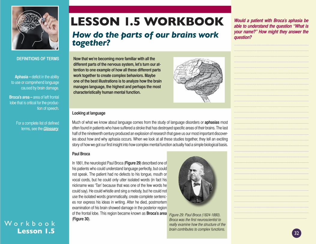
Now that we’re becoming more familiar with all the different parts of the nervous system, let’s turn our at-tention to one example of how all these different parts work together to create complex behaviors. Maybe one of the best illustrations is to analyze how the brain manages language, the highest and perhaps the most characteristically human mental function.
Looking at language
Much of what we know about language comes from the study of language disorders or aphasias most often found in patients who have suffered a stroke that has destroyed specific areas of their brains. The last half of the nineteenth century produced an explosion of research that gave us our most important discover-ies about how and why aphasia occurs. When we look at all these studies together, they tell an exciting story of how we got our first insight into how complex mental function actually had a simple biological basis.
Paul Broca
In 1861, the neurologist Paul Broca (Figure 29) described one of his patients who could understand language perfectly, but could not speak. The patient had no defects to his tongue, mouth or vocal cords, but he could only utter isolated words (in fact his nickname was ‘Tan’ because that was one of the few words he could say). He could whistle and sing a melody, but he could not use the isolated words grammatically, create complete sentenc-es nor express his ideas in writing. After he died, postmortem examination of his brain showed damage in the posterior region of the frontal lobe. This region became known as Broca’s area (Figure 30).
DEFINITIONS OF TERMS
Aphasia – deficit in the ability to use or comprehend language
caused by brain damage.
Broca’s area – area of left frontal lobe that is critical for the produc-
tion of speech.
For a complete list of defined terms, see the Glossary.
Would a patient with Broca’s aphasia be able to understand the question “What is your name?” How might they answer the question?________________________________________________________________________________________________________________________________________________________________________________________________________________________________________________________________________________________________________________________________________________________________________________________________________________________________________________________________________________________________________________________________________________________________________________________________________________________________________________________________________________________________________________________________________________________________________________________________________________________________________________________________________________________________________________________________________________________________________________________________________________________________________________________________________________________________
How do the parts of our brains work together?
Figure 29: Paul Broca (1824-1880). Broca was the first neuroscientist to really examine how the structure of the brain contributes to complex functions.

W o r k b o o kLesson 1.5 33
LESSON READINGBroca studied a further 8 patients, all of whom had the same symptoms, and all of whom had damage in the same area of the left cerebral hemisphere near the frontal cortex when their brains were autopsied. This discovery led Broca to state: “We speak with the left hemisphere!”
You can watch a teenage girl, Srarah, with Broca’s aphasia online - see this unit on the student website or click below:
■ Video: Broca’s aphasia - Sarah Scott - teenage stroke
Carl Wernicke
The next step was taken in 1876 by Carl Wernicke (Figure 31). He described another kind of aphasia. Wernicke’s aphasia involved a fail-ure to understand what was being said or written, rather than speak. While Broca’s patients could understand language but not speak, Wernicke’s patients could speak, but could not understand the lan-guage they heard. Their conversations were fluent, but unintelligible.
In this case the damage was located in the posterior part of the temporal lobe where it joins the parietal and oc-cipital lobes. This region became known as Wernicke’s area (Figure 32), and Wernicke might have said (but he didn’t) “We understand language at the back of the left hemisphere!”
DEFINITIONS OF TERMS
Wernicke’s area – area of the temporal lobe that is critical for
understanding language
For a complete list of defined terms, see the Glossary.
Would a patient with Wernicke’s aphasia be able to understand the question “What is your name?” How might they answer the question?_____________________________________________________________________________________________________________________________________________________________________________________________________________________________________________________________________________________________________________________________________________________________________________________________________________________________________________
What does the part of the brain that Bro-ca’s area is found in control? What about Wernicke’s area?__________________________________________________________________________________________________________________________________________________________________________________________________________________________________________________________________________________________________________________________________________________________________________________________________________________________________________________________________________________________________________________________________________________________________________________________________________________
Figure 30: Broca’s area. Broca’s area is situated next to the part of the frontal lobe that controls movement.
Figure 31: Carl Wernicke (1848-1905). Wernicke no-ticed that not all language deficits were the result of damage to Broca’s area.
Figure 32: Wernicke’s area. Wernicke’s area is located where the parietal and tem-poral lobes meet. It encircles the auditory cortex.

W o r k b o o kLesson 1.5 34
LESSON READINGYou can watch a patient with Wernicke’s aphasia online - see this unit on the student website or click below:
■ Video: Wernicke’s aphasia
Wernicke was the first to appreciate that different components of a single behavior are processed in different regions in the brain. His theory proposes that language involves separate motor and sensory programs, each governed by several regions of the cortex (Figure 33).
• A motor program that controls the movements of the mouth, tongue, palate and vocal cords is located in front of the motor area in Broca’s area.
• A sensory program that controls word per-ception is located in the temporal lobe area in Wernicke’s Area. This area is surrounded by the auditory cortex, as well as the areas that integrate auditory, visual, and touch sensation into complex perceptions and that are therefore known as as-sociation cortex.
Wernicke’s model for the organization of language has been elaborated on over the years but is still in use today. According to the most up-to-date model, language is processed in a specialized pathway that involves several areas of the brain, as follows:
• The initial perceptions of language are formed in separate sensory areas of the cortex specialized for hearing words (auditory cortex) or reading words (visual cortex).
• These perceptions are then conveyed to an area of the association cortex called the angular gyrus that is able to transform auditory and visual information into a single code that is shared by both speech and the written word.
• From the angular gyrus this code is conveyed to Wernicke’s area, where it is recognized as language and associated with meaning. Without that association, the ability to comprehend language is lost.
• The common neural code is then relayed from Wernicke’s area to Broca’s area, where it is trans-formed into a motor representation that can lead to either to spoken or written language.
When this last stage, where the single code is transformed to a spoken or written motor representation cannot take place, the ability to express language is lost.
What pathways through the brain are activat-ed when someone says ‘Hi!’ to you and you reply. Draw them out.__________________________________________________________________________________________________________________________________________________________________________________________________________________________________________________________________________________________________________________________________________________________________________________________________________________________________________________________________________________________________________
What pathways through the brain are activat-ed when you read this message: “Say Hi out loud”.___________________________________________________________________________________________________________________________________________________________________________________________________________________________________________________________________________________________________________________________________________________________________________________________________________________________________________________________________________________________________________________________________________________________________________________________________________________
Wernicke’s area Broca’s area
Motor cortex
Figure 33: Language processing. Both Wer-nicke’s and Broca’s areas contribute when we hear a spoken word and then repeat it.

W o r k b o o kLesson 1.5 35
LESSON READINGUsing this reasoning and proposed neural pathway, Wernicke correctly deduced that a third type of aphasia must exist, that would result if the connection between Broca’s and Wernicke’s area is damaged. He predicted that patients with conduction aphasia would be able to understand language and would also be able to speak, but that they would not be able to use words correctly. Indeed patients with conduction aphasia can understand words they hear and read perfectly, and can also speak and write quite fluently, yet, they cannot speak coherently. They omit parts of words or substitute incor-rect sounds. They are painfully aware of their errors, but unable to put them right.
Our current understanding of language processing
Until recently, everything we knew about language came from studies of patients who had suffered brain damage. Now PET and fMRI imaging let us look at language in behaving uninjured, healthy people. PET scans show where individual words are coded in the brain of healthy subjects when words are read or heard (Figure 34). The data shows that not only are reading and listening processed separately, but the mere act of thinking about a word’s meaning activates still a different area in the frontal cortex. Thus, lan-guage processing occurs in parallel in a number of different areas as well as in serial via Wernicke’s and Broca’s areas.
Wernicke’s discoveries also provided the first evidence for distributed processing, which is the important concept that different types of information are routed to a number of different areas in the brain in order to organize a response. This evidence and studies using PET scans and fMRI has led to the current thinking that our brains organize language using a modular format that consists of processing centers each having more or less independent functions, that are connected together in serial and in parallel.
We now appreciate that all cognitive abilities, not just language, are constructed using a similar format. This means that while specific brain regions are concerned with simple processing operations, complex functions like perception, movement, thought, and memory are all made possible because several brain regions, each with their own specific function, are linked together in serial and in parallel. As a result, dam-age to a single area need not result in the loss of an entire behavior. Even if a behavior initially disappears, it may partially return as undamaged parts of the same functional module reorganize their linkages.
DEFINITIONS OF TERMS
Distributed processing – theory suggesting that information is
processed in several different parts of the brain.
Conduction aphasia – language disorder in which patients can
understand language, and speak without any problem, but they omit parts of words or substitute incor-
rect words.
For a complete list of defined terms, see the Glossary.
Would a patient with conduction aphasia be able to understand the question “What is your name?” How might they answer the question?____________________________________________________________________________________________________________________________________________________________________________________________________________________________________________________________________________________________________________________________________________________________________________________________________________________________________
If Wernicke’s area wasn’t activated when we read words, does that mean we need to hear words in order to understand them?_________________________________________________________________________________________________________________________________________________________________________________________________________________________________________________________________________________________________________________________________________________________________________________________________________________________________________________________________________________________________________________________________________________________________________________________________________________________________________________________________________________________
Figure 34: PET scans of language areas. PET scans taken while subjects were hearing, seeing, speaking and thinking about words. Only when patients were listening to words, did their Wernicke’s areas show activity. The PET scans taken while the subject was seeing, speak-ing and thinking show activation of the occipital cortex, Broca’s area and frontal lobes.

W o r k b o o kLesson 1.5 36
Remember to identify your sources
STUDENT RESPONSES
We have leearned that many areas of our brain are involved in complex behaviors. Do you consider that this is a good or a bad thing when you are thinking about cases of brain injury or disease? What could it mean in terms of patient recovery?
_____________________________________________________________________________________________________
____________________________________________________________________________________________________
_____________________________________________________________________________________________________
_____________________________________________________________________________________________________
_____________________________________________________________________________________________________
_____________________________________________________________________________________________________
_____________________________________________________________________________________________________
_____________________________________________________________________________________________________
_____________________________________________________________________________________________________
_____________________________________________________________________________________________________
_____________________________________________________________________________________________________
_____________________________________________________________________________________________________
_____________________________________________________________________________________________________
____________________________________________________________________________________________________
_____________________________________________________________________________________________________
_____________________________________________________________________________________________________
_____________________________________________________________________________________________________
_____________________________________________________________________________________________________
_____________________________________________________________________________________________________
_____________________________________________________________________________________________________
_____________________________________________________________________________________________________
_____________________________________________________________________________________________________
_____________________________________________________________________________________________________
_____________________________________________________________________________________________________