leith et al 2010
TRANSCRIPT

Leith, J. L., Koutsikou, S., Lumb, B. M., & Apps, R. (2010). Spinalprocessing of noxious and innocuous cold information: differentialmodulation by the periaqueductal gray: differential modulation by theperiaqueductal gray. Journal of Neuroscience, 30(14), 4933 - 4942. DOI:10.1523/JNEUROSCI.0122-10.2010
Publisher's PDF, also known as Version of record
Link to published version (if available):10.1523/JNEUROSCI.0122-10.2010
Link to publication record in Explore Bristol ResearchPDF-document
University of Bristol - Explore Bristol ResearchGeneral rights
This document is made available in accordance with publisher policies. Please cite only the publishedversion using the reference above. Full terms of use are available:http://www.bristol.ac.uk/pure/about/ebr-terms

Behavioral/Systems/Cognitive
Spinal Processing of Noxious and Innocuous ColdInformation: Differential Modulation by thePeriaqueductal Gray
J. Lianne Leith, Stella Koutsikou, Bridget M. Lumb, and Richard AppsDepartment of Physiology and Pharmacology, University of Bristol, Bristol BS8 1TD, United Kingdom
In addition to cold being an important behavioral drive, altered cold sensation frequently accompanies pathological pain states. However,in contrast to peripheral mechanisms, central processing of cold sensory input has received relatively little attention. The present studycharacterized spinal responses to noxious and innocuous intensities of cold stimulation in vivo and established the extent to which theyare modulated by descending control originating from the periaqueductal gray (PAG), a major determinant of acute and chronic pain. Inlightly anesthetized rats, hindpaw cooling with ethyl chloride, but not acetone, was sufficiently noxious to evoke withdrawal reflexes,which were powerfully inhibited by ventrolateral (VL)-PAG stimulation. In a second series of experiments, subsets of spinal dorsal hornneurons were found to respond to innocuous and/or noxious cold. Descending control from the VL-PAG distinguished between activityin nociceptive versus non-nociceptive spinal circuits in that innocuous cold information transmitted by non-nociceptive class 1 andwide-dynamic-range class 2 neurons remained unaltered. In contrast, noxious cold information transmitted by class 2 neurons and allcold-evoked activity in nociceptive-specific class 3 neurons was significantly depressed. We therefore demonstrate that spinal responsesto cold can be powerfully modulated by descending control systems originating in the PAG, and that this control selectively modulatestransmission of noxious versus innocuous information. This has important implications for central processing of cold somatosensationand, given that chronic pain states are dependent on dynamic alterations in descending control, will help elucidate mechanisms under-lying aberrant cold sensations that accompany pathological pain states.
IntroductionUnderstanding the neural mechanisms underlying cold sensationis important behaviorally, given the vital role of temperature per-ception in survival, but also clinically, given the aberrant coldresponses frequently observed in neuropathic pain (Ochoa andYarnitsky, 1994; Jorum et al., 2003). Peripheral mechanisms ofcold somatosensation in relation to both acute and chronic painstates have received much recent interest. However, there is asurprising lack of knowledge regarding central processing of coldinformation, in particular whether cold-evoked responses at thespinal level are modulated by descending control systems, whichare known to have powerful modulatory effects on other sensorymodalities (Lovick and Bandler, 2005; Heinricher et al., 2009).
Innocuous cooling activates subsets of A�- and C-fiber low-threshold afferents (Bessou and Perl, 1969; Leem et al., 1993;Campero et al., 1996). More intense cold stimuli additionallyactivate populations of nociceptive afferents (both A�- andC-fiber units), which display a range of activation thresholds and
encode changes in stimulus intensity (LaMotte and Thalhammer,1982; Lynn and Carpenter, 1982; Leem et al., 1993; Simone andKajander, 1996, 1997; Cain et al., 2001). At the spinal level, elec-trophysiological studies have demonstrated that wide-dynamic-range and nociceptive-specific neurons in the dorsal horn areexcited by cold stimuli and encode intensity to noxious cold overa wide range of temperatures (Christensen and Perl, 1970; Khasabovet al., 2001; Brignell et al., 2008). Additionally, cold stimulation ofthe hindpaw or face evokes intensity-dependent Fos expressionin spinal and medullary dorsal horn neurons, respectively(Strassman et al., 1993; Abbadie et al., 1994; Doyle and Hunt,1999; Todd et al., 2005).
Spinal processing of sensory information is subject to dynamicdescending modulation from supraspinal structures, which is a ma-jor determinant of the acute pain experience evoked by noxiousmechanical and heat stimuli (Millan, 2002; Heinricher et al.,2009). In particular, the periaqueductal gray (PAG) is a keysource of descending control that operates in different behavioralstates (Keay and Bandler, 2001; Lovick and Bandler, 2005), and itis now recognized that chronic pain states are dependent on de-scending control from brainstem centers, including the PAG(Pertovaara et al., 1996; Urban and Gebhart, 1999; Pertovaara,2000; Monhemius et al., 2001; Pertovaara and Wei, 2003; Vanegasand Schaible, 2004; Carlson et al., 2007). Descending controlfrom the PAG profoundly modulates processing of mechanicaland heat information (Heinricher et al., 2009). However, it re-
Received Jan. 8, 2010; revised Feb. 2, 2010; accepted Feb. 17, 2010.We gratefully acknowledge the financial support of the Biotechnology and Biological Sciences Research Council,
the technical assistance of Barbara Carruthers and Rachel Bissett, and Dr. Lucy Donaldson and Dr. James Dunham forcomments on the manuscript.
Correspondence should be addressed to Dr. Bridget M. Lumb, Department of Physiology and Pharmacol-ogy, University of Bristol, School of Medical Sciences, University Walk, Bristol BS8 1TD, UK. E-mail: [email protected].
DOI:10.1523/JNEUROSCI.0122-10.2010Copyright © 2010 the authors 0270-6474/10/304933-10$15.00/0
The Journal of Neuroscience, April 7, 2010 • 30(14):4933– 4942 • 4933

mains unknown to what extent processing of cold information issubject to descending control.
By recording noxious cold-evoked withdrawal reflexes anddorsal horn neuronal activity in response to both innocuous andnoxious cold stimuli, the present study provides direct evidencethat descending control from the ventrolateral (VL)-PAG selec-tively modulates transmission of noxious cold information.Dynamic alterations in descending control underlie central sen-sitization and chronic pain states; therefore, changes in descend-ing modulation of cold inputs during the transition from acute tochronic pain may contribute to aberrant responses to cold thataccompany neuropathic pain.
Materials and MethodsAnimal preparationAll experiments were performed in accordance with the UK Animals(Scientific Procedures) Act 1986 and associated guidelines. Male adultWistar rats (280 –320 g; n � 38; Harlan) were housed in standard condi-tions and handled frequently to minimize stress on the day of theexperiment.
Anesthesia was induced using 4% halothane in O2, and the jugularvein was cannulated for anesthetic maintenance using a constant intra-venous infusion of alphaxalone (�25 mg � kg �1 � h �1 Alfaxan; Jurox).The carotid artery was exposed and cannulated for recording of bloodpressure, and the trachea was intubated. Body temperature was main-tained within physiological limits by means of a feedback-controlledheating blanket and rectal probe. In animals in which dorsal horn neu-ronal activity was to be recorded, a laminectomy was performed betweenT11 and T13 to expose the lumbar spinal cord. Animals were then posi-tioned in a stereotaxic frame and a craniotomy was performed to allowaccess to the PAG with glass micropipettes.
In neuronal recording experiments, anesthesia was maintained at alevel at which there were no precipitous changes in blood pressure inresponse to minor noxious stimuli, and in electromyographic (EMG)recording experiments it was reduced to a level at which animals weremoderately responsive to firm pinch of the contralateral forepaw. Ani-mals were allowed to stabilize at these levels for a minimum of 30 minbefore recording of neuronal or EMG activity.
Recording of skin temperatureSurface skin temperature on the hindpaw was recorded using a K-typethermocouple (Physitemp) held in place with a small dab of cyanoacry-late glue (distant from the site of stimulation; Loctite; Henkel). The ther-mocouple was connected to a digital thermometer (BAT-12; Physitemp),and the output signal was digitized using a 1401plus data acquisitionsystem (Cambridge Electronic Design). Subcutaneous skin temperaturewas recorded using a T-type thermocouple (made in-house) connectedto a digital thermometer, and then the output signal was digitized via a1401plus.
Recording of EMG activityAn intramuscular bipolar electrode, custom made from two shortlengths of Teflon-coated, 0.075-mm-diameter, stainless steel wire (Ad-vent Research Materials), was inserted into the biceps femoris of the lefthind leg to record EMG activity during the withdrawal reflex. The EMGsignal was amplified (�5000) and filtered (50 Hz to 5 kHz; NeuroLogSystem; Digitimer), before being captured for subsequent analysis via a1401plus (Cambridge Electronic Design) onto a PC running Spike2 ver-sion 5 software (Cambridge Electronic Design). The magnitude of thewithdrawal reflex evoked by thermal stimuli was quantified by measuringthe modulus of the EMG using Spike2 software. This value was thencorrected for background noise by subtracting noise over the same lengthof time as the response (measured before application of the thermalstimulus). In some experiments, surface and subsurface skin tempera-tures were recorded simultaneously, and it was therefore possible to mea-sure the threshold temperature at which the withdrawal reflex occurredin addition to response magnitude. If no EMG was observed following
PAG stimulation, withdrawal threshold was assigned as 0°C for dataanalysis.
Recording of dorsal horn neuronal activityThe vertebral column was clamped at each end of the laminectomy tomaximize stability during neuronal recordings. The dura was removed, apool was made with skin flaps, and the whole area was filled with agar tofurther stabilize the preparation. Once set, a small window was cut out ofthe agar over the desired recording site and filled with warm paraffin oil.A glass-coated tungsten microelectrode (�5 M�; Melanie Ainsworth;Northamptonshire, UK) was lowered into the cord. Extracellular single-unit neuronal activity was amplified (�5000) and filtered (500 Hz to 10kHz; NeuroLog System; Digitimer) before being captured at 10 kHz forsubsequent analysis via a 1401plus (Cambridge Electronic Design) onto aPC running Spike2 version5 software (Cambridge Electronic Design).
Functional classification of spinal neuronsSingle units were isolated using gentle tapping, stroking, or firm pinch ofthe hindlimb, delivered manually, as a search stimulus. It was not feasibleto use cooling as a search stimulus; therefore, it must be borne in mindthat the search stimuli used biased the population to mechanically sen-sitive neurons.
Once a unit had been identified, the peripheral receptive field wascharacterized using low-threshold (brush, tap) and high-threshold(pinch) mechanical stimuli. According to their mechanical responseproperties, units were classified as class 1 (low threshold, non-noxious),class 2 (low and high threshold, wide dynamic range), or class 3 (highthreshold, nociceptive specific) (Menetrey et al., 1977; 1979). Neuronswere then tested for responsiveness to the following thermal stimuli:noxious heat (50°C water; 1 ml), innocuous cooling (acetone; 1 ml), ornoxious cold [ethyl chloride (EC); 1 ml].
Responses to acetone and EC were quantified by counting the totalnumber of spikes evoked until activity returned to the prestimulus level;this value was then corrected for spontaneous activity of the neuron overthe same length of time as the response (measured before application ofthe thermal stimulus). In some neurons, afferent input was further char-acterized by monitoring responses to percutaneous electrical stimuli (1ms square pulse) delivered to the center of the receptive field via needleelectrodes. Thresholds for A- and C-fiber activation were established andrepeated sweeps were made at both 1.5 and 3 times C-fiber thresholdvoltage (a train of 20 1 ms square pulses delivered at 0.1 Hz). All neuronstested showed responses at latencies consistent with input from both A-and C-fiber afferents and were therefore classified as C positive (Watersand Lumb, 2008).
Antidromic testing of spinal neurons for a supraspinal projectionIn some experiments, dorsal horn neurons were tested for a supraspinalprojection to the caudal brainstem. Supraspinal projection neurons wereidentified by their antidromic responses to electrical stimulation in thevicinity of the contralateral inferior olivary complex [�12.5 mm caudalto bregma, 1.2–1.5 mm lateral to the midline, and 8.5–9.0 mm deep to thecortical surface according to the brain atlas of Paxinos and Watson(2005)] using a bipolar stimulating electrode (interpolar distance of 0.5mm; SNE-100X; Harvard Apparatus). Single pulses (20 –100 �A, 0.1 msduration, at a rate of 0.1 Hz) were delivered via the stimulating electrode,and dorsal horn neurons were classified as projection neurons if they metthe following standard criteria for antidromic activation: (1) an all-or-none response to stimulation, (2) constant latency responses, (3) fre-quency following to three stimuli delivered at a rate of 100 Hz, and (4)collision of the antidromic spike with a spontaneous or evoked ortho-dromic spike (Lipski, 1981).
Cold stimulation of the skinTwo different cooling stimuli were used. One milliliter of either 100% EC(Acorus Therapeutics) or 100% acetone (Fisher Scientific) was appliedtopically to the hindpaw (in withdrawal reflex experiments) or the hind-limb receptive field (in dorsal horn neuronal recording experiments)using a pipette. Care was taken not to touch the skin with the pipette tip.Because of the position of the animal in the stereotaxic frame, the pipette
4934 • J. Neurosci., April 7, 2010 • 30(14):4933– 4942 Leith et al. • Midbrain Control of Spinal Cold Somatosensation
was aimed at the lateral paw, but the coolant very rapidly (�1 s) spreadover the whole paw, including both dorsal and plantar surfaces.
Neuronal activation in the PAGGlass micropipettes (Harvard Apparatus) were driven stereotaxicallyinto the VL-PAG at �7.6 –7.8 mm caudal to bregma, 0.8 –1.0 mm lateralto midline, and 5.3–5.5 mm deep to the cortical surface according to thebrain atlas of Paxinos and Watson (2005). Micropipettes were filled witha 50 mM solution of the excitatory amino acid DL-homocysteic acid(DLH; Sigma) containing pontamine sky blue dye (Gurr) to mark injec-tion sites. Microinjections of DLH (60 – 80 nl) were given under micro-scopic guidance using a custom-made, paraffin-filled pressure injectionsystem attached to a 1 �l syringe (SGE Analytical Science). The concen-tration of DLH was the same as that used in previous studies of descend-ing control from the PAG (Waters and Lumb, 1997; McMullan andLumb, 2006a, 2006b; Koutsikou et al., 2007; Waters and Lumb, 2008),and since only very small amounts of DLH were injected, it is likely thatits administration results in excitation of PAG neuronal cell bodies ratherthan depolarizing block (Lipski et al., 1988). Consistent with previousstudies (see above), microinjections into the VL-PAG evoked transientdecreases in mean arterial pressure (data not shown).
Experimental protocolDescending modulation of cold-evoked withdrawal reflexes. Following pre-paratory surgery, a glass micropipette containing DLH solution was low-ered vertically into the left VL-PAG (ipsilateral to the stimulatedhindpaw). Flexion withdrawal reflexes were recorded in response to ECdelivered to the left hindpaw at 5 min intervals. After three “baseline”withdrawal responses to EC were recorded, an injection of DLH wasmade into the VL-PAG. EC was applied again to the paw 10 s after DLHinjection (“PAG” in bar charts) and responses to three further applica-tions were conducted at 5 min intervals (at 5, 10, and 15 min post-DLHmicroinjection, termed “PAG�5 min,” “PAG�10 min,” and “PAG�15min,” respectively, in bar charts).
Descending modulation of cold-evoked neuronal activity. Following iso-lation of a neuron and characterization of its receptive field and responseproperties, a glass micropipette containing DLH solution was loweredvertically into the left VL-PAG (ipsilateral to the stimulated hindlimb).Neuronal activity was recorded in response to acetone or EC delivered tothe receptive field of the neuron at 5 min intervals. After three baselineneuronal responses to acetone or EC were recorded, an injection of DLHwas made into the VL-PAG. Acetone or EC was applied again to thereceptive field 10 s after DLH injection (PAG in bar charts), and re-sponses to three further applications were conducted at 5 min intervals(at 5, 10, and 15 min post-DLH microinjection; termed PAG�5 min,PAG�10 min, and PAG�15 min, respectively, in bar charts).
HistologyAt the end of experiments, animals were killed by overdose of sodiumpentobarbital (30 mg i.v. bolus; Sigma). The brain was removed, fixed for24 h in paraformaldehyde solution (4% in 0.1 M phosphate buffer), andthen cryoprotected in 30% sucrose solution for 24 h. Brains were sec-tioned at 60 �m and PAG injection sites marked with pontamine sky bluedye were localized with reference to the rat brain atlas of Paxinos andWatson (2005) and plotted onto standard transverse diagrams of themidbrain.
Data analysisAll EMG and neuronal activity data are displayed as mean � SEM. Allstatistical analysis was performed using Prism 4 (GraphPad). Post-DLH(PAG on bar charts) and recovery responses (PAG�5 min, PAG�10min, and PAG�15 min on bar charts) to acetone or EC were comparedwith baseline responses using Kruskal–Wallis test followed by Dunn’smultiple comparison test. Significance was taken at the 5% level.
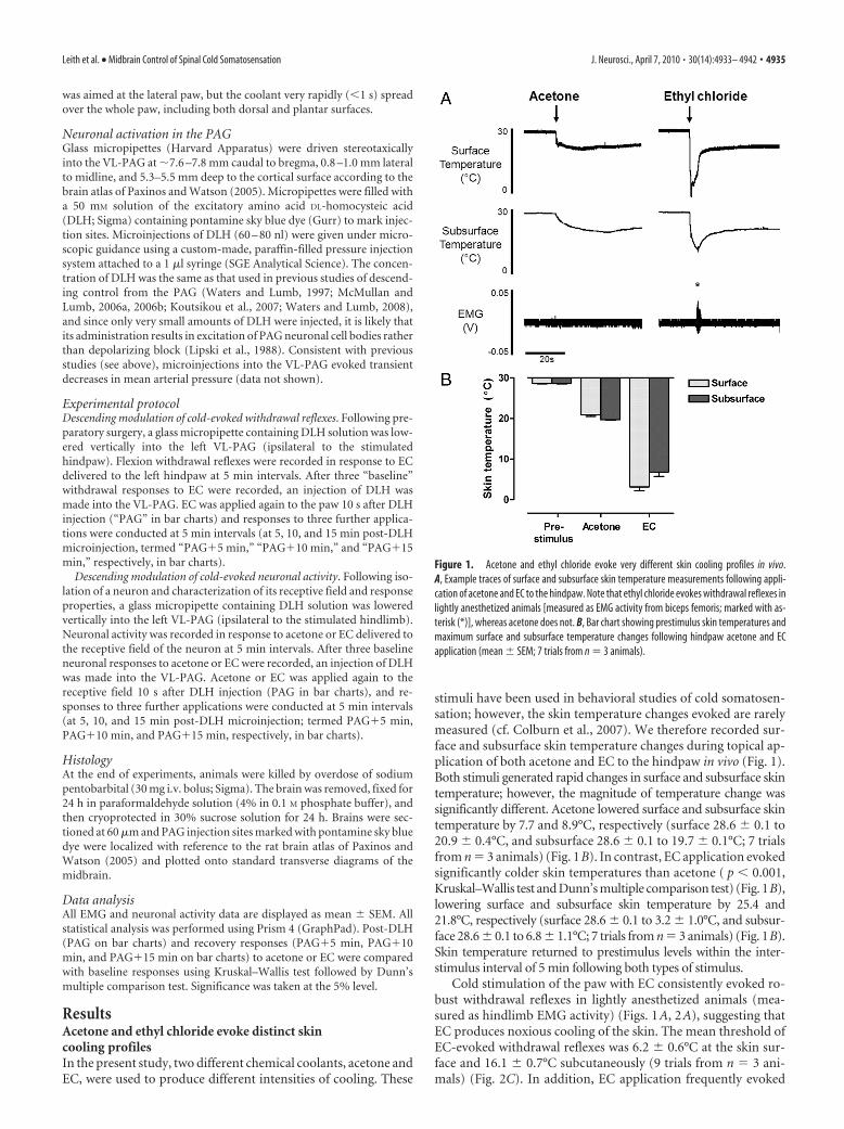
ResultsAcetone and ethyl chloride evoke distinct skincooling profilesIn the present study, two different chemical coolants, acetone andEC, were used to produce different intensities of cooling. These
stimuli have been used in behavioral studies of cold somatosen-sation; however, the skin temperature changes evoked are rarelymeasured (cf. Colburn et al., 2007). We therefore recorded sur-face and subsurface skin temperature changes during topical ap-plication of both acetone and EC to the hindpaw in vivo (Fig. 1).Both stimuli generated rapid changes in surface and subsurface skintemperature; however, the magnitude of temperature change wassignificantly different. Acetone lowered surface and subsurface skintemperature by 7.7 and 8.9°C, respectively (surface 28.6 � 0.1 to20.9 � 0.4°C, and subsurface 28.6 � 0.1 to 19.7 � 0.1°C; 7 trialsfrom n � 3 animals) (Fig. 1B). In contrast, EC application evokedsignificantly colder skin temperatures than acetone ( p � 0.001,Kruskal–Wallis test and Dunn’s multiple comparison test) (Fig. 1B),lowering surface and subsurface skin temperature by 25.4 and21.8°C, respectively (surface 28.6 � 0.1 to 3.2 � 1.0°C, and subsur-face 28.6 � 0.1 to 6.8 � 1.1°C; 7 trials from n � 3 animals) (Fig. 1B).Skin temperature returned to prestimulus levels within the inter-stimulus interval of 5 min following both types of stimulus.
Cold stimulation of the paw with EC consistently evoked ro-bust withdrawal reflexes in lightly anesthetized animals (mea-sured as hindlimb EMG activity) (Figs. 1A, 2A), suggesting thatEC produces noxious cooling of the skin. The mean threshold ofEC-evoked withdrawal reflexes was 6.2 � 0.6°C at the skin sur-face and 16.1 � 0.7°C subcutaneously (9 trials from n � 3 ani-mals) (Fig. 2C). In addition, EC application frequently evoked
Figure 1. Acetone and ethyl chloride evoke very different skin cooling profiles in vivo.A, Example traces of surface and subsurface skin temperature measurements following appli-cation of acetone and EC to the hindpaw. Note that ethyl chloride evokes withdrawal reflexes inlightly anesthetized animals [measured as EMG activity from biceps femoris; marked with as-terisk (*)], whereas acetone does not. B, Bar chart showing prestimulus skin temperatures andmaximum surface and subsurface temperature changes following hindpaw acetone and ECapplication (mean � SEM; 7 trials from n � 3 animals).
Leith et al. • Midbrain Control of Spinal Cold Somatosensation J. Neurosci., April 7, 2010 • 30(14):4933– 4942 • 4935

increases in mean arterial blood pressure, indicative of a noxiousintensity of stimulation (data not shown). In the preparationused here, noxious intensities of stimulation are required toevoke withdrawal reflexes (McMullan et al., 2004), which indi-cates that EC is largely a noxious cold stimulus. In contrast, in thesame animals withdrawal reflexes were never observed followingacetone application, nor did acetone evoke any changes in bloodpressure, suggesting it produces innocuous cooling of the skin.Nonetheless, it is likely that the temperature changes produced byacetone application will activate a small proportion of nocicep-tive afferents, since some cold-responsive nociceptors are re-ported to have activation thresholds of 20°C (although mosthave thresholds at much colder temperatures) (Cain et al., 2001;Campero et al., 1996; Simone and Kajander, 1996). Clearly, how-ever, activation of such afferents is not generally sufficient toproduce overt behavioral responses to acetone, either in lightlyanesthetized rats (this study) or in awake normal rats, whichshow little or no response to acetone (Choi et al., 1994; Decosterdand Woolf, 2000; Kim et al., 2009; Hulse et al., 2010); therefore,we consider acetone to be predominantly (though not exclu-sively) an innocuous cooling stimulus.
Cold-evoked withdrawal reflexes are significantly depressedby VL-PAG stimulationDescending control originating in the PAG has been shown topowerfully modulate withdrawal reflexes evoked by noxious me-chanical and thermal (heat) stimuli (e.g., Mayer et al., 1971; Fardin etal., 1984; Carstens et al., 1990; McMullan and Lumb, 2006a; Leithet al., 2007). However whether this control extends to noxiouscold stimulation remains unclear; therefore, the effect of VL-PAGstimulation on cold-evoked withdrawal thresholds and responsemagnitudes was investigated.
PAG stimulation strongly depressed cold-evoked withdrawalreflexes in vivo to the extent that the responses were frequentlyabolished (Fig. 2). Cold-evoked withdrawal reflex magnitude wassignificantly reduced to 0.1 � 0.1% of baseline ( p � 0.01, n � 5;Kruskal–Wallis test and Dunn’s multiple comparison test) (Fig.2B), and withdrawal threshold temperature was significantlycolder (from 6.2 � 0.6°C at the skin surface and 16.1 � 0.7°C s.c.to 0°C at both the surface and the subsurface; both p � 0.05, n �3; Kruskal–Wallis test and Dunn’s multiple comparison test)(Fig. 2C). Reflex magnitude and threshold recovered partiallyduring the 15 min period following PAG stimulation (Fig. 2B,C).
Although stimulation of the PAG has been shown to altercardiovascular parameters (Carrive, 1993; Bandler et al., 2000)that might alter the rate and extent of cooling produced by thestimulus, the profile of skin cooling evoked by EC was not alteredfollowing PAG stimulation (Fig. 2A). This suggests that the de-pression of withdrawal reflexes is caused mainly by direct centralmodulatory effects on neuronal excitability rather than being sec-ondary to changes in peripheral blood flow.
The data therefore provide evidence that cold-evoked with-drawal reflexes can be strongly modulated by descending controlfrom the VL-PAG. However, given that acetone does not evokewithdrawal reflexes in lightly anesthetized preparations, this ap-proach does not allow us to test whether the PAG also modulatesresponses to innocuous cold. We therefore examined the activityof dorsal horn neurons in the spinal cord in response to bothacetone and EC and whether this activity could be altered follow-ing stimulation of the PAG.
Figure 2. Noxious cold-evoked withdrawal reflexes are depressed by VL-PAG stimulation. A, Typ-ical examples of EC-evoked withdrawal reflexes (measured as EMG activity) with corresponding sur-face and subsurface skin temperature measurements before and after stimulation of the VL-PAG; thewithdrawal reflex is abolished by PAG stimulation, however the profile of skin cooling remains unal-tered. B, C, The effects of VL-PAG stimulation on withdrawal reflex magnitude (n � 5) (B) andthreshold (surface and subsurface; both n � 3) (C). Data are expressed as mean � SEM. Statisticalanalysis compared post-DLH (PAG) and recovery (PAG�5 min, PAG�10 min, PAG�15 min) groupswith baseline responses using Kruskal–Wallis test followed by Dunn’s multiple comparison test;*p � 0.05; **p � 0.01. D, Injection sites in the PAG from which the effects of PAG stimulationon EC-evoked responses were tested; coordinates are relative to bregma (Paxinos and Watson,2005). DM, Dorsomedial; DL, dorsolateral; L, lateral; VL, ventrolateral.
4936 • J. Neurosci., April 7, 2010 • 30(14):4933– 4942 Leith et al. • Midbrain Control of Spinal Cold Somatosensation

Subsets of class 1, 2, and 3 spinal neurons respond to acetoneand ethyl chlorideA total of 47 lumbar dorsal horn neurons (mean depth in cord729 � 40 �m; range 280 –1430 �m) were studied and classifiedby responses to low- and high-threshold mechanical stimula-tion of the receptive field according to the scheme defined byMenetrey et al. (1977); class 1, n � 6; class 2, n � 27; and class 3,n � 14. Subsets of neurons in each class (class 1, n � 2; class 2, n �8; class 3, n � 7) were antidromically identified as supraspinallyprojecting, at least to the level of the caudal brainstem. There wasno distinction between a neuron’s response to cold stimuli andwhether or not a supraspinal projection could be detected.
Response characteristics of all spinalneurons to acetone and EC are summa-rized in Figure 3. The response of eachneuron to each type of cold stimulus wasconsistent from trial to trial. The majority(66%; 4/6) of non-nociceptive class 1 neu-rons responded to cold [acetone only, n �0 (0%); acetone and EC, n � 3 (50%); EConly, n � 1 (17%); neither n � 2 (33%)](Fig. 3A). Class 1 neurons tended to showprolonged, high-frequency responses toboth acetone and EC. In the three neuronsthat responded to both stimuli, responsesto EC were always similar to or less thanthe responses to acetone (Fig. 3B,C, toppanels) suggesting, as expected, that theseneurons are not able to encode stimulusintensity into the noxious range. This in-dicates that the EC response in class 1 neu-rons represents the innocuous coolingcomponent of the stimulus, since EC willactivate low-threshold, cooling-sensitiveafferents in addition to those in the noci-ceptive range.
The majority (85%; 23/27) of wide-dynamic-range class 2 neurons respondedto cold [acetone only, n � 2 (7%); acetoneand EC, n � 17 (63%); EC only, n � 4(15%); neither, n � 4 (15%)] (Fig. 3A).Class 2 neurons showed a range of re-sponse magnitudes to both acetone andEC that appeared to be related to receptivefield location; neurons with receptivefields on proximal regions of the hindlimb(e.g., thigh) tended to show greater re-sponses to cold than those with distal re-ceptive fields (e.g., paw) (Fig. 3C, middlepanel). This is unlikely to be caused by adifferent skin-cooling profile, since tem-perature measurements at the thigh werenot significantly different compared withthose at the paw (data not shown). In-stead, the greater responses of neuronswith proximal receptive fields are likely tobe due to the higher degree of conver-gence of input in the spinal cord from pe-ripheral afferents (Willis and Coggeshall,1991). All class 2 neurons that were re-sponsive to both cold stimuli showedgreater responses to EC than acetone (Fig.3B,C, middle panels), indicative of theirability to encode stimulus intensity into
the noxious range (Khasabov et al., 2001).Only half of class 3 neurons (50%; 7/14) responded to cold
stimuli [acetone only, n � 0 (0%); acetone and EC, n � 4 (29%);EC only, n � 3 (21%); neither, n � 7 (50%)] (Fig. 3A). In class 3neurons, unlike class 2, there was no clear relationship betweenreceptive field location and response magnitude, and they did notencode stimulus intensity to the same extent, although it shouldbe noted that the sample size of class 3 neurons is small (Fig. 3C,bottom panel). A large proportion of class 3 neurons did notrespond to either acetone or EC. However, it remains possiblethat these neurons are cold responsive because the stimulus used
Figure 3. Subpopulations of class 1, 2, and 3 dorsal horn neurons respond to acetone and ethyl chloride. A, Summary of theproportions of class 1, 2, and 3 neurons that responded to acetone and/or EC. B, Typical examples of responses to acetone and ECin cold-responsive class 1, 2, and 3 neurons (in each example the two sets of responses are recorded from the same neuron; rawdorsal horn (DH) recordings and corresponding peristimulus time histograms (spikes per 1 s bin) are shown). Note, however, thatthe majority of class 3 neurons did not respond to acetone. Arrows indicate onset of cold stimulus. C, For each neuron class,responses (mean spikes � SEM) to the two cold stimuli are grouped in relation to receptive field location; numbers above each barrepresent sample size.
Leith et al. • Midbrain Control of Spinal Cold Somatosensation J. Neurosci., April 7, 2010 • 30(14):4933– 4942 • 4937

in this study may not have achievedtemperatures adequate for their activa-tion. Somewhat unexpectedly, a smallproportion (4/14; 29%) of nociceptive-specific class 3 neurons (as classified bymechanical stimulation) also respondedto acetone. However, as stated above, theskin temperatures produced by acetoneapplication (�20°C) are likely to be suffi-cient to activate a small proportion of no-ciceptive afferents that could, in turn,activate nociceptive-specific class 3 neu-rons in the spinal cord. Therefore, acetoneresponses observed in class 3 neurons mayindeed represent nociceptive activity (seeDiscussion).
An important additional considerationwhen interpreting these data is that boththe cooling stimuli used here involve alow-threshold mechanical component(i.e., application of liquid to the skin). It istherefore possible that part of the re-sponse observed was due to stimulationof mechanically sensitive afferents. Todetermine the extent of firing evokedsolely by this mechanical stimulus, a con-trol stimulus of the same volume of roomtemperature water was applied to the re-ceptive field of some neurons (n � 9;data not shown). In class 1 and 2 neu-rons, room temperature water evokeda brief (�1 s), modest increase (�10spikes) in firing, but no response was ob-served in class 3 neurons, which indi-cates that the response to acetone andEC (typically of long duration and highfiring frequency) (Fig. 3) was predomi-nantly due to skin cooling rather thanany mechanical stimulation.
Descending control from the PAGmodulates spinal neuronal responses tonoxious but not innocuous coldResponses of non-nociceptive class 1 neu-rons to both acetone and EC remainedunaltered after stimulation of the VL-PAG, as shown in Figure 4 [117 � 63 and122 � 16% of baseline responses; n � 3and n � 4 (including one projection neu-ron), respectively; both p 0.05, Kruskal–Wallis test and Dunn’s multiple comparison test], indicating thatdescending control from the PAG does not significantly alterresponses to innocuous cold in these neurons.
Similarly, stimulation of VL-PAG did not significantly alterresponses of wide-dynamic-range class 2 neurons to acetone asshown in Figure 5 [123 � 45% of baseline responses; n � 6 (nonepositively identified as projection neurons); p 0.05, Kruskal–Wallis test and Dunn’s multiple comparison test]. However, re-sponses of class 2 neurons to EC were significantly reduced byVL-PAG stimulation [to 17 � 7% of baseline; n � 7 (includingtwo projection neurons); p � 0.01, Kruskal–Wallis test andDunn’s multiple comparison test] (Fig. 5), indicating that de-scending control from the PAG selectively depresses responses to
noxious versus innocuous cold in these neurons. EC responsesrecovered to baseline levels within 5 min of VL-PAG stimulation(PAG�5 min, PAG�10 min, and PAG�15 min, all p 0.05compared with baseline, Kruskal–Wallis test and Dunn’s multi-ple comparison test) (Fig. 5).
Stimulation of VL-PAG strongly depressed cold-evoked activityin all nociceptive-specific class 3 neurons tested; responses to ECwere significantly depressed as shown in Figure 6 [to 14 � 11% ofbaseline responses; n � 4 (including two projection neurons);p � 0.01, Kruskal–Wallis Test and Dunn’s multiple comparisontest]. These responses recovered only partially during the 15 minperiod following VL-PAG stimulation and remained significantlylower than baseline levels 15 min after VL-PAG stimulation
Figure 4. Cold-evoked activity remains unaltered in non-nociceptive class 1 neurons following PAG stimulation. A, Typicalexamples of a class 1 neuron response to acetone and EC [recorded from the same neuron; peristimulus time histograms (spikes per1 s bin) are shown] before and after PAG stimulation. B, The effect of VL-PAG stimulation on class 1 neuronal responses to acetone(n � 3) and EC (n � 4). Data are expressed as mean � SEM of normalized spike counts in response to acetone or EC. Statisticalanalysis compared post-DLH (PAG) and recovery (PAG�5 min, PAG�10 min, PAG�15 min) groups with baseline responses usingKruskal–Wallis test followed by Dunn’s multiple comparison test; ns, p 0.05. C, Injection sites in the PAG from which the effectsof PAG stimulation on acetone- and/or EC-evoked responses were tested; coordinates are relative to bregma (Paxinos and Watson,2005). DM, Dorsomedial; DL, dorsolateral; L, lateral; VL, ventrolateral.
4938 • J. Neurosci., April 7, 2010 • 30(14):4933– 4942 Leith et al. • Midbrain Control of Spinal Cold Somatosensation

(37 � 8%, p � 0.05 compared with baseline, Kruskal–Wallis test,and Dunn’s multiple comparison test) (Fig. 5). This suggests thatdifferent mechanisms may be responsible for descending modu-lation of class 3 neuronal responses (that remain depressed overthe time period studied) compared with class 2 neuronal re-sponses (that recover more rapidly). A total of 4/14 class 3 neu-rons (29%) also responded to acetone (in addition to EC), and inone of these neurons (not positively identified as a projectionneuron) the effects of descending control from the PAG weretested. VL-PAG stimulation abolished the response to acetone inthis neuron (Fig. 6A,B). Noxious pinch-evoked activity was alsotested in this neuron, which was similarly abolished by PAG stim-ulation (data not shown).
DiscussionAn understanding of the spinal processingof cold sensory input, including its de-scending control, is essential not only indetermining how forward transmission ofcold signals may be altered in different be-havioral states, but ultimately in the de-velopment of novel strategies for thetreatment of aberrant cold sensations thataccompany pathological pain states. Here,we show that spinal responses to cold canbe powerfully modulated by descendingcontrol systems originating in the PAGand that this control selectively modulatestransmission of noxious versus innocuousinformation.
Neural mechanisms ofcold somatosensationTwo different intensities of cold stimuliwere used to investigate central process-ing of both innocuous and noxious cold,providing novel information regardingmechanisms underlying behavioral re-sponses to cold. Acetone produced onlymild cooling of the skin and never evokedwithdrawal reflexes in lightly anesthetizedrats, consistent with observations thatnormal rats show little or no response toacetone, and with the use of acetone as atest for cold allodynia following neuropa-thy (Choi et al., 1994; Decosterd andWoolf, 2000; Kim et al., 2009; Hulse et al.,2010). Together with reports from humanstudies that temperatures between 15 and20°C are perceived as cool (Greenspan etal., 1993), this suggests that acetone pro-duces predominantly innocuous cooling.In contrast, EC produced a more intensecold stimulus, generating skin surfacetemperatures approaching 0°C in thepresent study, and its application consis-tently evoked withdrawal reflexes accom-panied by increases in blood pressure.Together, this indicates that EC is a nox-ious cold stimulus. EC has been used pre-viously in behavioral studies, and similartemperature changes have been reported(Hao et al., 1996, 1999; Sjolund et al.,1998). These temperature changes are suf-
ficiently cold to activate populations of nociceptive afferents(LaMotte and Thalhammer, 1982; Lynn and Carpenter, 1982;Leem et al., 1993; Simone and Kajander, 1996, 1997). However,temperatures reported to produce cold pain sensation in humansand nociceptive behaviors in animals are highly variable, likelydue to the wide variety of stimuli used and responses measured,making comparison between studies difficult. In humans, tem-peratures of �10�15°C and below are reported to evoke noxioussensations (Chery-Croze, 1983; Yarnitsky and Ochoa, 1990;Davis, 1998; Gottrup et al., 1998; Harrison and Davis, 1999). Weobserved cold-evoked withdrawal in lightly anesthetized rats atskin surface temperatures of �6°C, which is consistent with
Figure 5. Noxious cold-evoked activity is selectively inhibited in wide-dynamic-range class 2 neurons by PAG stimulation.A, Typical examples of a class 2 neuron response to acetone and EC [recorded from the same neuron; peristimulus time histograms(spikes per 1 s bin) are shown] before and after PAG stimulation. B, The effect of VL-PAG stimulation on class 2 neuronal responsesto acetone (n � 6) and EC (n � 7). Data are expressed as mean � SEM of normalized spike counts in response to acetone or EC.Statistical analysis compared post-DLH (PAG) and recovery (PAG�5 min, PAG�10 min, PAG�15 min) groups with baselineresponses using Kruskal–Wallis test followed by Dunn’s multiple comparison test; ns, p0.05; **p�0.01. C, Injection sites in thePAG from which the effects of PAG stimulation on acetone- and/or EC-evoked responses were tested; coordinates are relative tobregma (Paxinos and Watson, 2005). DM, Dorsomedial; DL, dorsolateral; L, lateral; VL, ventrolateral.
Leith et al. • Midbrain Control of Spinal Cold Somatosensation J. Neurosci., April 7, 2010 • 30(14):4933– 4942 • 4939

Allchorne et al. (2005), who defined coldthreshold in normal rats at 5°C, and Jasminet al. (1998), who reported nocifensive be-haviors �3°C. Using operant assays, Vi-erck et al. (2004) reported thresholds for“lick/guard/jump” behaviors at �4°C;however, escape behaviors were observedat much warmer temperatures (�16°C).
The present study provides novel in-formation regarding the population ofspinal neurons excited by acetone and EC,both of which are used in behavioral painstudies. We found that the majority ofclass 1 and 2 neurons are cold responsive,while half of class 3 neurons were not.Consistent with our data, Khasabov et al.(2001) found that an almost identical pro-portion of wide-dynamic-range (class 2)neurons responded to cold delivered via aPeltier thermode, which encoded intensityto noxious cold over a broad range of tem-peratures. Furthermore, functional ana-tomical studies have shown that hindpawand facial cold stimulation evoke intensity-dependent Fos expression in dorsal hornneurons, including NK-1-positive lamina Iprojection neurons, which are believed toform spinal-brainstem feedback loops withimportant roles in the development ofchronic pain states (Strassman et al., 1993;Abbadie et al., 1994; Doyle and Hunt, 1999;Suzuki et al., 2002; Todd et al., 2005). How-ever, in contrast to our findings, Khasabovet al. (2001) found a higher proportion ofcold-responsive class 3 neurons, but aroundhalf (42%) only responded to cooling�0°C.Many cold-sensitive nociceptors have thre-sholds of �0°C (Simone and Kajander,1996; 1997) (although this could be in re-sponse to tissue damage). It is possible,therefore, that the cold “insensitive” class 3neurons described here are cold responsivebut with thresholds at very low tempera-tures; therefore, the stimulus we used wouldnot have produced cooling sufficient to ac-tivate them.
A seemingly unexpected finding wasthat some nociceptive-specific neurons(classified by standard mechanical meth-ods) responded to acetone application,which is generally considered to be innoc-uous. However, this activity may be due tonociceptive input, since the temperatures produced by acetoneapplication are sufficient to activate nociceptive afferents withcold thresholds at relatively warm temperatures (20°C) (Cainet al., 2001; Campero et al., 1996; Simone and Kajander, 1996).Any class 3 neurons receiving afferent input from these lower-threshold nociceptors could therefore be acetone responsive. Ifthis is the case, and given the small proportion of nociceptorswith thresholds of 20°C, it is not surprising that a small pro-portion of class 3 neurons were found to be activated by acetoneapplication. An additional possibility is that information trans-mitted by nociceptive-specific class 3 neurons is interpreted by
higher centers as nociceptive, regardless of the intensity of theinput.
Descending modulation of spinal cold processingStimulation of descending pathways from the PAG has beenshown to powerfully modulate the responses of spinal neurons toperipheral mechanical and thermal (heat) stimulation in vivo(Mayer et al., 1971; Jones and Gebhart, 1988; Sandkuhler et al.,1991; Waters and Lumb, 1997, 2008; McMullan and Lumb,2006b). This modulatory control is selective for responses to nox-ious versus innocuous stimulus intensities (Bennett and Mayer,
Figure 6. Cold-evoked activity in class 3 nociceptive-specific neurons is inhibited by PAG stimulation. A, Examples of a class 3neuron response to acetone and EC [recorded from the same neuron; peristimulus time histograms (spikes per 1 s bin) are shown]before and after PAG stimulation. B, The effect of VL-PAG stimulation on mean class 3 neuronal responses to acetone (n � 1) andEC (n � 4). Data are expressed as mean � SEM of normalized spike counts in response to acetone or EC. Statistical analysiscompared post-DLH (PAG) and recovery (PAG�5 min, PAG�10 min, PAG�15 min) groups with baseline responses usingKruskal–Wallis test followed by Dunn’s multiple comparison test; *p � 0.05; **p � 0.01. C, Injection sites in the PAG from whichthe effects of PAG stimulation on acetone- and/or EC-evoked responses were tested; coordinates are relative to bregma (Paxinosand Watson, 2005); DM, Dorsomedial; DL, dorsolateral; L, lateral; VL, ventrolateral.
4940 • J. Neurosci., April 7, 2010 • 30(14):4933– 4942 Leith et al. • Midbrain Control of Spinal Cold Somatosensation
1979; Gebhart et al., 1983; Sandkuhler et al., 1991; Waters andLumb, 1997). We therefore hypothesized that this selective con-trol might also extend to the modality of cold and tested thisdirectly by examining whether acetone- and EC-evoked re-sponses of spinal dorsal horn neurons were altered by PAG stim-ulation. We found that spinal processing of cold input can also bepowerfully modulated by the PAG, and that this control differ-entiates between activity in nociceptive versus non-nociceptivecircuits. This pattern of descending control therefore resemblesthat for other modalities and suggests that stimulus intensity (i.e.,nociceptive versus non-nociceptive) rather than modality is thedetermining factor in whether spinal activity is subject to de-scending control.
Our findings do not, however, include cold-specific neuronsthat are not driven by mechanical stimulation (Han et al., 1998),since these would not have been encountered in our experiments.Given that these neurons will also contribute to cold-evoked be-haviors, it will be important to investigate whether these cells arealso modulated by descending pathways.
Functional significanceTemperature perception is vital for survival, providing environ-mental information that drives appropriate behavioral responsesand, if necessary, escape from potentially harmful conditions.Knowledge of the central processing and modulation of cold sen-sory input is therefore important from a behavioral perspective,yet has received little attention. The present study demonstratesthat descending control originating in the PAG can dramaticallyalter spinal responses to noxious cold input, leaving informationregarding innocuous cold unaltered. In normal animals, modu-lation of spinal nociception by the VL-PAG is hypothesized to actas part of a coordinated passive coping strategy triggered by in-escapable stressors (Keay and Bandler, 2001; Lumb, 2002; Lovickand Bandler, 2005). Selective inhibition of noxious informationwould allow an animal to respond appropriately to threatening orstressful situations without the distraction of nociceptive input,therefore enabling the generation of adaptive behaviors that arebeneficial to survival.
Recent evidence suggests that the PAG retains the capacity tomodulate cold responses in pathophysiological states, since elec-trical stimulation of the PAG attenuates enhanced behavioralresponses to cold in neuropathic rats (Lee et al., 2000). It is nowrecognized that alterations in descending controls from brain-stem centers, including the PAG, contribute to central sensitiza-tion and chronic pain states (Pertovaara et al., 1996; Urban andGebhart, 1999; Pertovaara, 2000; Monhemius et al., 2001;Pertovaara and Wei, 2003; Vanegas and Schaible, 2004). Indeed,cold allodynia is dependent on descending control systems, sincelidocaine block of the rostroventromedial medulla (RVM), a ma-jor relay of outflow from the PAG, attenuates cold allodynia inmodels of neuropathic injury (Taylor et al., 2007). Therefore,knowledge of descending influences on spinal processing of coldinformation is clinically important, given the heightened coldsensitivity frequently reported by patients with neuropathic in-jury (Ochoa and Yarnitsky, 1994; Jorum et al., 2003). We hypoth-esize that dysfunction of descending control from the PAG(decreased descending inhibition and/or increased facilitation),likely acting through the RVM and other medullary structures,contributes to cold allodynia, driving the exaggerated behavioralresponses (Choi et al., 1994) and increased spinal neuronal excit-ability to cold (Brignell et al., 2008) observed in neuropathic painmodels, in addition to peripheral mechanisms. Future studiesinvestigating dynamic modulation of cold responsiveness in the
transition from acute to chronic pain will provide important in-formation regarding underlying neural mechanisms responsiblefor altered cold responses in pathological pain states.
ReferencesAbbadie C, Honore P, Besson JM (1994) Intense cold noxious stimulation
of the rat hindpaw induces c-fos expression in lumbar spinal cord neu-rons. Neuroscience 59:457– 468.
Allchorne AJ, Broom DC, Woolf CJ (2005) Detection of cold pain, coldallodynia and cold hyperalgesia in freely behaving rats. Mol Pain 1:36.
Bandler R, Keay KA, Floyd N, Price J (2000) Central circuits mediating pat-terned autonomic activity during active vs passive emotional coping.Brain Res Bull 53:95–104.
Bennett GJ, Mayer DJ (1979) Inhibition of spinal cord interneurons by nar-cotic microinjection and focal electrical stimulation in the periaqueductalcentral gray matter. Brain Res 172:243–257.
Bessou P, Perl ER (1969) Response of cutaneous sensory units with unmy-elinated fibers to noxious stimuli. J Neurophysiol 32:1025–1043.
Brignell JL, Chapman V, Kendall DA (2008) Comparison of icilin- andcold-evoked responses of spinal neurones, and their modulation of me-chanical activity, in a model of neuropathic pain. Brain Res 1215:87–96.
Cain DM, Khasabov SG, Simone DA (2001) Response properties of mech-anoreceptors and nociceptors in mouse glabrous skin: an in vivo study.J Neurophysiol 85:1561–1574.
Campero M, Serra J, Ochoa JL (1996) C-polymodal nociceptors activatedby noxious low temperature in human skin. J Physiol 497:565–572.
Carlson JD, Maire JJ, Martenson ME, Heinricher MM (2007) Sensitizationof pain-modulating neurons in the rostral ventromedial medulla afterperipheral nerve injury. J Neurosci 27:13222–13231.
Carrive P (1993) The periaqueductal gray and defensive behavior: func-tional representation and neuronal organization. Behav Brain Res58:27– 47.
Carstens E, Hartung M, Stelzer B, Zimmermann M (1990) Suppression of ahind limb flexion withdrawal reflex by microinjection of glutamate ormorphine into the periaqueductal gray in the rat. Pain 43:105–112.
Chery-Croze S (1983) Relationship between noxious cold stimuli and themagnitude of pain sensation in man. Pain 15:265–269.
Choi Y, Yoon YW, Na HS, Kim SH, Chung JM (1994) Behavioral signs ofongoing pain and cold allodynia in a rat model of neuropathic pain. Pain59:369 –376.
Christensen BN, Perl ER (1970) Spinal neurons specifically excited by nox-ious or thermal stimuli: marginal zone of the dorsal horn. J Neurophysiol33:293–307.
Colburn RW, Lubin ML, Stone DJ Jr, Wang Y, Lawrence D, D’Andrea MR,Brandt MR, Liu Y, Flores CM, Qin N (2007) Attenuated cold sensitivityin TRPM8 null mice. Neuron 54:379 –386.
Davis KD (1998) Cold-induced pain and prickle in the glabrous and hairyskin. Pain 75:47–57.
Decosterd I, Woolf CJ (2000) Spared nerve injury: an animal model of per-sistent peripheral neuropathic pain. Pain 87:149 –158.
Doyle CA, Hunt SP (1999) A role for spinal lamina I neurokinin-1-positiveneurons in cold thermoreception in the rat. Neuroscience 91:723–732.
Fardin V, Oliveras JL, Besson JM (1984) A reinvestigation of the analgesiceffects induced by stimulation of the periaqueductal gray matter in the rat.I. The production of behavioral side effects together with analgesia. BrainRes 306:105–123.
Gebhart GF, Sandkuhler J, Thalhammer JG, Zimmermann M (1983) Quan-titative comparison of inhibition in spinal cord of nociceptive informa-tion by stimulation in periaqueductal gray or nucleus raphe magnus of thecat. J Neurophysiol 50:1433–1445.
Gottrup H, Nielsen J, Arendt-Nielsen L, Jensen TS (1998) The relationshipbetween sensory thresholds and mechanical hyperalgesia in nerve injury.Pain 75:321–329.
Greenspan JD, Taylor DJ, McGillis SL (1993) Body site variation of coolperception thresholds, with observations on paradoxical heat. Somato-sens Mot Res 10:467– 474.
Han ZS, Zhang ET, Craig AD (1998) Nociceptive and thermoreceptive lam-ina I neurons are anatomically distinct. Nat Neurosci 1:218 –225.
Hao JX, Yu W, Xu XJ, Wiesenfeld-Hallin Z (1996) Capsaicin-sensitive af-ferents mediate chronic cold, but not mechanical, allodynia-like behaviorin spinally injured rats. Brain Res 722:177–180.
Hao JX, Xu IS, Xu XJ, Wiesenfeld-Hallin Z (1999) Effects of intrathecal
Leith et al. • Midbrain Control of Spinal Cold Somatosensation J. Neurosci., April 7, 2010 • 30(14):4933– 4942 • 4941
morphine, clonidine and baclofen on allodynia after partial sciatic nerveinjury in the rat. Acta Anaesthesiol Scand 43:1027–1034.
Harrison JL, Davis KD (1999) Cold-evoked pain varies with skin type andcooling rate: a psychophysical study in humans. Pain 83:123–135.
Heinricher MM, Tavares I, Leith JL, Lumb BM (2009) Descending controlof nociception: specificity, recruitment and plasticity. Brain Res Rev60:214 –225.
Hulse R, Wynick D, Donaldson LF (2010) Intact cutaneous C fibre afferentproperties in mechanical and cold neuropathic allodynia. Eur J Pain.Advance online publication. Retrieved February 24, 2010. doi:10.1016/j.ejpain.2009.10.001.
Jasmin L, Kohan L, Franssen M, Janni G, Goff JR (1998) The cold plate as atest of nociceptive behaviors: description and application to the study ofchronic neuropathic and inflammatory pain models. Pain 75:367–382.
Jones SL, Gebhart GF (1988) Inhibition of spinal nociceptive transmissionfrom the midbrain, pons and medulla in the rat: activation of descendinginhibition by morphine, glutamate and electrical stimulation. Brain Res460:281–296.
Jorum E, Warncke T, Stubhaug A (2003) Cold allodynia and hyperalgesia inneuropathic pain: the effect of N-methyl-D-aspartate (NMDA) receptorantagonist ketamine–a double-blind, cross-over comparison with alfen-tanil and placebo. Pain 101:229 –235.
Keay KA, Bandler R (2001) Parallel circuits mediating distinct emotionalcoping reactions to different types of stress. Neurosci Biobehav Rev25:669 – 678.
Khasabov SG, Cain DM, Thong D, Mantyh PW, Simone DA (2001) En-hanced responses of spinal dorsal horn neurons to heat and cold stimulifollowing mild freeze injury to the skin. J Neurophysiol 86:986 –996.
Kim HY, Gwak YS, Shim I (2009) An electrophysiological method for quan-tifying neuropathic pain behaviors in rats: measurement of hindlimbwithdrawal EMG magnitude. J Physiol Sci 59:473– 476.
Koutsikou S, Parry DM, MacMillan FM, Lumb BM (2007) Laminar organi-zation of spinal dorsal horn neurones activated by C- vs A-heat nocicep-tors and their descending control from the periaqueductal grey in the rat.Eur J Neurosci 26:943–952.
LaMotte RH, Thalhammer JG (1982) Response properties of high-thresholdcutaneous cold receptors in the primate. Brain Res 244:279–287.
Lee BH, Park SH, Won R, Park YG, Sohn JH (2000) Antiallodynic effectsproduced by stimulation of the periaqueductal gray matter in a rat modelof neuropathic pain. Neurosci Lett 291:29 –32.
Leem JW, Willis WD, Chung JM (1993) Cutaneous sensory receptors in therat foot. J Neurophysiol 69:1684 –1699.
Leith JL, Wilson AW, Donaldson LF, Lumb BM (2007) Cyclooxygenase-1-derived prostaglandins in the periaqueductal gray differentially controlC- versus A-fiber-evoked spinal nociception. J Neurosci 27:11296 –11305.
Lipski J (1981) Antidromic activation of neurones as an analytic tool in thestudy of the central nervous system. J Neurosci Methods 4:1–32.
Lipski J, Bellingham MC, West MJ, Pilowsky P (1988) Limitations of thetechnique of pressure microinjection of excitatory amino acids for evok-ing responses from localized regions of the CNS. J Neurosci Methods26:169 –179.
Lovick TA, Bandler R (2005) The organisation of the midbrain periaque-ductal grey and the integration of pain behaviours. In: The neurobiologyof pain (Hunt SP, Koltzenburg M, eds), pp 267–287. Oxford: Oxford UP.
Lumb BM (2002) Inescapable and escapable pain is represented in distincthypothalamic-midbrain circuits: specific roles for Adelta- and C-nociceptors.Exp Physiol 87:281–286.
Lynn B, Carpenter SE (1982) Primary afferent units from the hairy skin ofthe rat hind limb. Brain Res 238:29 – 43.
Mayer DJ, Wolfle TL, Akil H, Carder B, Liebeskind JC (1971) Analgesiafrom electrical stimulation in the brainstem of the rat. Science 174:1351–1354.
McMullan S, Lumb BM (2006a) Midbrain control of spinal nociceptiondiscriminates between responses evoked by myelinated and unmyelinatedheat nociceptors in the rat. Pain 124:59 – 68.
McMullan S, Lumb BM (2006b) Spinal dorsal horn neuronal responses tomyelinated versus unmyelinated heat nociceptors and their modulationby activation of the periaqueductal grey in the rat. J Physiol 576:547–556.
McMullan S, Simpson DA, Lumb BM (2004) A reliable method for the pref-erential activation of C- or A-fibre heat nociceptors. J Neurosci Methods138:133–139.
Menetrey D, Giesler GJ Jr, Besson JM (1977) An analysis of response prop-erties of spinal cord dorsal horn neurones to nonnoxious and noxiousstimuli in the spinal rat. Exp Brain Res 27:15–33.
Menetrey D, Chaouch A, Besson JM (1979) Responses of spinal cord dorsalhorn neurones to non-noxious and noxious cutaneous temperaturechanges in the spinal rat. Pain 6:265–282.
Millan MJ (2002) Descending control of pain. Prog Neurobiol 66:355– 474.Monhemius R, Green DL, Roberts MH, Azami J (2001) Periaqueductal grey
mediated inhibition of responses to noxious stimulation is dynamicallyactivated in a rat model of neuropathic pain. Neurosci Lett 298:70 –74.
Ochoa JL, Yarnitsky D (1994) The triple cold syndrome: cold hyperalgesia,cold hypoaesthesia and cold skin in peripheral nerve disease. Brain117:185–197.
Paxinos G, Watson C (2005) The rat brain in stereotaxic coordinates. Syd-ney: Academic.
Pertovaara A (2000) Plasticity in descending pain modulatory systems. ProgBrain Res 129:231–242.
Pertovaara A, Wei H (2003) A dissociative change in the efficacy of supraspinalversus spinal morphine in the neuropathic rat. Pain 101:237–250.
Pertovaara A, Wei H, Hamalainen MM (1996) Lidocaine in the rostroven-tromedial medulla and the periaqueductal gray attenuates allodynia inneuropathic rats. Neurosci Lett 218:127–130.
Sandkuhler J, Willmann E, Fu QG (1991) Characteristics of midbrain con-trol of spinal nociceptive neurons and nonsomatosensory parameters inthe pentobarbital-anesthetized rat. J Neurophysiol 65:33– 48.
Simone DA, Kajander KC (1996) Excitation of rat cutaneous nociceptors bynoxious cold. Neurosci Lett 213:53–56.
Simone DA, Kajander KC (1997) Responses of cutaneous A-fiber nocicep-tors to noxious cold. J Neurophysiol 77:2049 –2060.
Sjolund KF, von Heijne M, Hao JX, Xu XJ, Sollevi A, Wiesenfeld-Hallin Z(1998) Intrathecal administration of the adenosine A1 receptor agonistR-phenylisopropyl adenosine reduces presumed pain behaviour in a ratmodel of central pain. Neurosci Lett 243:89 –92.
Strassman AM, Vos BP, Mineta Y, Naderi S, Borsook D, Burstein R (1993)Fos-like immunoreactivity in the superficial medullary dorsal horn in-duced by noxious and innocuous thermal stimulation of facial skin in therat. J Neurophysiol 70:1811–1821.
Suzuki R, Morcuende S, Webber M, Hunt SP, Dickenson AH (2002) Super-ficial NK1-expressing neurons control spinal excitability through activa-tion of descending pathways. Nat Neurosci 5:1319 –1326.
Taylor BK, Abhyankar SS, Vo NT, Kriedt CL, Churi SB, Urban JH (2007)Neuropeptide Y acts at Y1 receptors in the rostral ventral medulla toinhibit neuropathic pain. Pain 131:83–95.
Todd AJ, Spike RC, Young S, Puskar Z (2005) Fos induction in lamina Iprojection neurons in response to noxious thermal stimuli. Neuroscience131:209 –217.
Urban MO, Gebhart GF (1999) Supraspinal contributions to hyperalgesia.Proc Natl Acad Sci U S A 96:7687–7692.
Vanegas H, Schaible HG (2004) Descending control of persistent pain: in-hibitory or facilitatory? Brain Res Brain Res Rev 46:295–309.
Vierck CJ Jr, Kline Rt, Wiley RG (2004) Comparison of operant escape andinnate reflex responses to nociceptive skin temperatures produced by heatand cold stimulation of rats. Behav Neurosci 118:627– 635.
Waters AJ, Lumb BM (1997) Inhibitory effects evoked from both the lateraland ventrolateral periaqueductal grey are selective for the nociceptiveresponses of rat dorsal horn neurones. Brain Res 752:239 –249.
Waters AJ, Lumb BM (2008) Descending control of spinal nociception fromthe periaqueductal grey distinguishes between neurons with and withoutC-fibre inputs. Pain 134:32– 40.
Willis WD, Coggeshall RE (1991) Sensory mechanisms of the spinal cord,2nd Ed. New York: Plenum.
Yarnitsky D, Ochoa JL (1990) Release of cold-induced burning pain byblock of cold-specific afferent input. Brain 113:893–902.
4942 • J. Neurosci., April 7, 2010 • 30(14):4933– 4942 Leith et al. • Midbrain Control of Spinal Cold Somatosensation