leishmania - mymensingh medical college file/leishmania akram.pdf · leishmania is a...
TRANSCRIPT
Leishmania
Prof. Dr. Md. Akram Hossain
March 09

Introduction
� Leishmania is a hemoflagellate, different species of causes the diseaseleishmaniasis.
� Clinically leishmaniasis are manifested as cutaneous, mucocutaneous andvisceral lesions. Of its three clinical forms, Visceral leishmaniasis is the mostsevereone.severeone.
� The different forms of Leishmaniasis constitute severe public health problems.Above 12 millions peoples are infected and some 350 million people are atrisk throughout the world.
� All of these parasitic diseases are transmitted by biting ofdifferent species ofsand flies.– Visceral Leishmaniasis (VL) is fatal while left untreated.– Mucocutaneous Leishmaniasis (MCL) is a mutilating disease.– Diffuse cutaneous Leishmaniasis (DCL) is a disabling disease.
1/7/2014 2Prof. Muhammad Akram

Epidemiology
� Leishmaniasis is prevalent through out the tropical and sub-tropical regions of Africa, Asia, the Mediterranean, SouthernEurope (old world) as well as South and Central America(newworld).(newworld).
� Annual incidence of leishmaniasis is estimated to be 1 to 1.5million cases of Cutaneous leishmaniasis and 500000 casesof Visceral leishmaniasis.
� About 350 million people are at risk with an overallprevalence of over 12 million people worldwide.
1/7/2014 3Prof. Muhammad Akram

Host factors
� Age and sex :In East Africa and India the peak age group is5-9 years. In Southern Europe and China, people of all agegroups are affected. Males are affected twice as often asfemale.
� Socioeconomic status: VL is usually more prevalent in theunder-developed countries than developed countries. Poorpeople are affected most by VL.
� Population movement :Recently, the population movementis regarded as the important factor in epidemiology.Movement of the population fromendemic to non-endemicareas results in the spread of infection.
1/7/2014 4Prof. Muhammad Akram
Environmental Factors
� Season:– There is a seasonal variation of VL incidence in the endemicand
hyper-endemic regions. Usually there is high prevalence during andafter rainy season. There are two peaks of VL incidence in endemicregions of Bangladesh. One takes place in June and the other inregions of Bangladesh. One takes place in June and the other inNovember-December, which coincides, respectively to usual pre andpost-monsoon seasons.
� Rural area:– The disease is usually confined to rural areas where conditions for
breeding of sand flies is favourable compared to urban areas.
� Altitude:– Plains are the homeland of VL. It does not occur in altitude over 2000
feet as the sand flies are not found above this altitude due tounfavorableclimatic conditions.
1/7/2014 5Prof. Muhammad Akram

Bangladesh situation
� It affects 88 countries of the world of which 72 are developing and 13 ofthem are among the least developed countries.
� In Bangladesh, India, Nepal and Sudan, 90% cases of VisceralLeishmaniasis are reported. Conversely, 90% cases of Cutaneousleishmaniasis occur in Afghanistan, Brazil, Iran, Peru, Saudi Arabia andSyria.Syria.
� n India, VL has been continuing as endemic in the states of Bihar, UttarPradesh and West Bengal. More than 1 lakh new cases occur in Indiaevery year and state of Bihar accounts for more than 90% of those.
� In Bangladesh, about 18% of the total population (20 million) live in areaswith active transmission of VL.– Highest number of VL cases were recorded in the district of Mymensingh
followed by Pabna, Tangail and Gazipur. The incidence was assumed to be inthe range of 15000-30000 per year.
1/7/2014 6Prof. Muhammad Akram

Historical Background
LeishmanandDonovan first described VL in 1903.
Both of these physicians separately but simultaneouslydemonstrated parasites in stained smears fromthe spleenof patients suffering from a malaria-like illness, whichof patients suffering from a malaria-like illness, whichbecame known as Visceral Leishmaniasis and its causativeagent was namedLeishmania donovani as per the names oftwo discoverers.
Swaminath et al.,1942, proved using human volunteers thatthe Leishmania parasite could be transmitted by thePhlebotomine sand flies.
1/7/2014 7Prof. Muhammad Akram

Taxonomy
�Phylum - Euglenozoa,
�Class - Kinetoplastea, – Order - Kinetoplastida,
– Family - Tyopanosomatidae, – Family - Tyopanosomatidae,
– Genus -Leishmania, Subgenus - Leishmania sensu stricto & Viannia,
• Species - L.donovani, L.infantum, L.tropica, Complexes: L.donovani, L.tropica, L.major, L.ethiopica, L.maxicana, L. braziliensis, L.guyanensis
1/7/2014 8Prof. Muhammad Akram

Clinical Classification
1. Visceral leishmaniasis or Kala-azaris caused primarily byL. donovani,
L. chagasi, or L. infantum. Dogs (feral or domesticated) are a reservoir for
L. chagasi.
2. Cutaneous leishmaniasisusually divided into
1. Old World leishmaniasiscaused primarily by L. tropica, L. major, L.1. Old World leishmaniasiscaused primarily by L. tropica, L. major, L.
aethiopica, and
2. New World leishmaniasis caused primarily byL. Mexicana or L. braziliensis.
3. Diffuse cutaneous leishmaniasis is caused primarily byL. aethiopica or L.
mexicana.
3. Mucocutaneous leishmaniasis(espundia) is caused primarily byL.
(viannia) braziliensis.
Altogether, there are about 21 leishmanial species that aretransmitted by about
30 species of sand flies.1/7/2014 9Prof. Muhammad Akram
Clinical classification…
� Visceral leishmaniasis or kala-azaron the basis of biological reservoir as follows :– African Kala-Azar - Rodent reservoir.
– Mediterrarean Kala -Azar or infantile kala -azar -Canine reservoir.– Mediterrarean Kala -Azar or infantile kala -azar -Canine reservoir.
– Indian Kala-Azar - Human reservoir.
� Cutaneous leishmaniasis can also be divided further asfollows :– Oriental sore - caused by L.tropica.
– Espundia or Mucocutaneous leishmaniasis-caused by L.braziliensis.
– Dermal leishmanoid or PKDL, a late sequel to visceral infection.
1/7/2014 10Prof. Muhammad Akram

Morphology
� Exist in both amastigote and promastigote forms.� Amastigote (Aflagellar stages)form resides in the
reticulo-endothelial systemof the vertebrate hosts (– It is usually short oval or pear shaped or slender spindle
shaped bodies, about 5-10 um in length. Cell membraneis delicate and can be demonstrated in fresh specimensonly. Nucleus lies centrally. Kinetoplast (DNAonly. Nucleus lies centrally. Kinetoplast (DNAcontaining rod shape body) lies transversely near theanterior end. Flagellum may be of same length as thebody or even longer, projecting from the front. Nucleusis less than 1µm in diameter. It is oval or rounded andsituated in the middle of the cell or side of the cell wall.
� Promastigote (Flagellar stages) : this stage of theparasite is only encountered in cultures and insectvectors (sand flies).– It is usually rounded or oval body, about 2-5 µm in
diameter
1/7/2014 11Prof. Muhammad Akram

life cycle
1/7/2014 12Prof. Muhammad Akram

Life cycle
1/7/2014 13Prof. Muhammad Akram
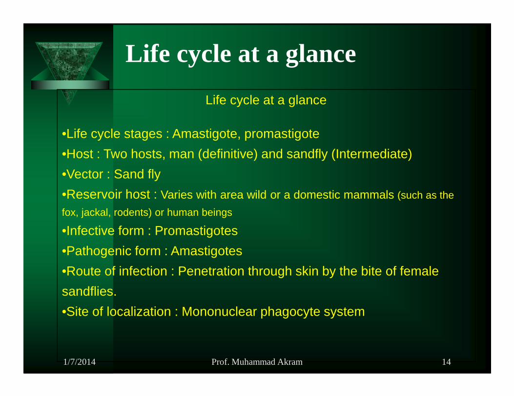
Life cycle at a glance
Life cycle at a glance
•Life cycle stages : Amastigote, promastigote
•Host : Two hosts, man (definitive) and sandfly (Intermediate)
•Vector : Sand fly
•Reservoir host : Varies with area wild or a domestic mammals (such as the •Reservoir host : Varies with area wild or a domestic mammals (such as the
fox, jackal, rodents) or human beings
•Infective form : Promastigotes
•Pathogenic form : Amastigotes
•Route of infection : Penetration through skin by the bite of female
sandflies.
•Site of localization : Mononuclear phagocyte system
1/7/2014 14Prof. Muhammad Akram

Transmission
� Vector borne transmission :VL is transmitted to man by thebite of infectedfemale sand fly. once the vector is infected itremain infective throughout its life time and capable ofinfecting several persons.
� Direct transmission : Due to contamination of the bitewound. Accidental inoculation of parasite during laboratorywork results in leishmaniasis. Transmission by bloodtransfusion is also recorded.
� Congenital transmission : Rarely occurs. Transmission ofinfection to fetus in uterus may occur due to placental defect.
1/7/2014 15Prof. Muhammad Akram

Virulence factors
1. Intracellular parasitism : Most important ***
2. Resistance to serumcomplement components
3. Glycoprotein 63 (gp-63)
4. Acid phosphatases (ACP)
5. Scavanger enzymes: Superoxide dismutase, catalase &
glutathione peroxidase
6. Cysteine proteinases and megasomes
1/7/2014 16Prof. Muhammad Akram
Pathogenesis of VL
� Visceral leishmaniasis is also known as Kala-azar (Hindi: kala=black,azar=sickness). The etiological agents belong to theLeishmania donovanicomplex,L.d donovani, L.d infantum andL.d archibaldi in the old worldandL.d chagasi in the new world.
� Visceral leishmaniasisor Kala azarhasmany featuresin commonwith� Visceral leishmaniasisor Kala azarhasmany featuresin commonwithchronic malaria. Kala-Azar or Dum Dum fever is characterized by a lowdegree of fever with hepatoplenomegaly and severe progressive cachexia(wasting), swollen lymph glands, leucopeania, thrombocytopenia withrelative monocytosis and loss of hair.
� The parasites (Leishmania donovani ) are mainly found in bone marrow,liver and spleen. If left untreated this manifestation of leishmaniasis isfatal in more than 90% of cases.
1/7/2014 17Prof. Muhammad Akram

Pathogenesis of VL
� Leishmaniasis is a slowly progressive disease that can takeup to 7 yearsto become clinically apparent. Even then, signs are frequently nonspecificand a diagnosis ofLeishmania is seldom considered.
� Oneof the mostconsistentfindings (~100%) with Leishmania sp. is the� Oneof the mostconsistentfindings (~100%) with Leishmania sp. is thepresence of hyperproteinemia due to hyperglobulinemia, often inconjunction with hypoalbuminemia (94%). Serum protein electrophoresiscommonly reveals a polyclonal gammopathy consisting primarily of IgGimmunoglobulins and some acute phase proteins.
� Indirectly, increased antibody production and resultant immune-complexdisease are responsible for many of the clinical and laboratoryabnormalities encountered.
1/7/2014 18Prof. Muhammad Akram

Pathogenesis of VL…
� Immune-mediated vasculitis leads to loss of proteins and cells resulting inhypoalbuminemia (94%) and thrombocytopenia (50%), whereas depositionof immune complexes into joints results in polyarthritis and lameness.
� Proteinuria (85%) and azotemia (45%) are associated with� Proteinuria (85%) and azotemia (45%) are associated withglomerulonephritis which is reported to be the leading cause of death inuntreated animals.
� Liver failure occurs less commonly than renal failure, but elevations in liverenzyme activities, such as alanine aminotransferase (61%)and alkalinephosphatase (51%), are not uncommon.
� Hemostatic disorders such as epistaxis (15%), hematuria and disseminatedintravascular coagulation (DIC) are not uncommon.
1/7/2014 19Prof. Muhammad Akram
Cutaneous leishmaniasis or "oriental sore"
� The cutaneous manifestation of leishmaniasis is found mainly in North Africa,and in the Middle East and Central Asia, where it is caused byL. tropica andL. major. In the New World cutaneous leishmaniasis is caused byL. mexicana.
� At the location of the bite of the sandfly a papule develops which enlargesand then starts to necrotize centrally. The ulceration which may be acuteleadingto a moistulcer,or slow in thecaseof a typical dry lesionis invadedleadingto a moistulcer,or slow in thecaseof a typical dry lesionis invadedwith macrophages that serve as the host cells for the parasite and that permitthe amastigotes to multiply.
� The ulceration may last for many months before healing occurs. Eventuallygranulomatous swelling subsides, and the ulceration heals, generally leaving alarge scar and major mutilation, regarded as characteristic in highly endemicareas.
� The lesion is painless and secondary infection is restricted to the dead tissue.� In the case of the Old World disease, when cure has occurred a solid
immunity has been acquired against re-infection by the sameparasite.
1/7/2014 20Prof. Muhammad Akram
Mucocutaneous leishmaniasis or "espundia"
� L. d. braziliensis causes in man a slow healing often very extensiveoriental sore, which self-cures after a variable sometimesextended time.Following the cure of the initial lesion the infection may metastasize,reappearing on the mucosal surfaces of the oronasal region,causing thedisfiguringandeventuallyoftenfataldiseaseof espundia.disfiguringandeventuallyoftenfataldiseaseof espundia.
� The mucocutaneous lesions do not self heal but continue to invade thetissues which become blocked with granulomatous infiltration, eventuallynecrotizing and eroding insidiously.
� An early manifestation is often the end of the nose, which becomesexpanded, so producing the syndrome of 'tapir nose'.
� Death frequently results as an indirect consequence, due tobacterial super-infection or obstruction of airways or the food passage.
1/7/2014 21Prof. Muhammad Akram

Post Kala-azar DermaL Leishmaniasis (PKDL)
� Post Kala - azar dermal leishmaniasis (PKDL) Usually followsrecovery from a Kala -azar infection.
� The disease begins with small measles-like lesions (hypo pigmentedmacules, papules or nodules) appearing on the face and graduallyincrease in size (rarely greater than 1cm in diameter).
� Eventually the lesions spread to the upper trunk, arms, forearms, thighs,legs, abdomen, the neck and the back.
� The multiple lesions can coalesce to form larger lesions andcan lead tothe gross enlargement of facial features such as the nose andlips,giving an appearance similar to leprosy.
� The disease is particularly severe if the lesions spread to the mucosalsurfaces of the nasal septum, hard and soft palate, oropharynx, larynxor the eye lids and the cornea leading to blindness.
1/7/2014 22Prof. Muhammad Akram
Post Kala-azar DermaL Leishmaniasis (PKDL)
� The lesions are usually self-limiting, however those that do not healspontaneously within six months have to be treated.
� Pentavalent antimonials remain the drugs of choice for treating PKDL.
� Sodium stibogluconate at a dose of 20 mg/Kg of body weight administeredintramuscularlyfor 4-5 monthsis recommended. In additionKetoconazoleintramuscularlyfor 4-5 monthsis recommended. In additionKetoconazoleand allopurinol can be given orally to improve response.
� In antimony resistant cases amphotericin B is an effective replacement.Indian PKDL appears any time between 1-7 years after apparent cure ofKala-azar, although longer periods of up to 20-30 years havebeen reported.
� The African form of the disease usually appears within a few months aftercure, in most cases within 6 months, on average within 56 days.
� However it can develop during the treatment of Kala-azar, inwhich casethe termPara Kala azar dermal leishmaniasiswould seem more fitting.
1/7/2014 23Prof. Muhammad Akram

Immunity� Leishmania is an intracellular parasite that escapes from the humural
immune respons by hiding as an amastigote in the lysosomes ofthe hostcell.– This has important implications for the immunological response of the host
towards the parasite, because: the intracellular parasite is not exposed to thehost's humoral response, the circulating antibodies have no effect on theinfectionandmayevenbeharmful. So,theimmunity is entirelycell-mediated.infectionandmayevenbeharmful. So,theimmunity is entirelycell-mediated.
� T cells of the CD8 subtype seem to play an important role in controllingthe infection byLeishmania. Only the cutaneous form of leishmaniasis isself-curing. This indicates that it is possible to develop acertain degree ofimmunity against the parasite, resulting in healing of the ulcers.
� However, the parasites probably never disappear completely from thebody, since in situations where the immune system is compromised, suchas in AIDS, or is suppressed by cancer chemotherapy or in cases of organtransplantation, leishmaniasis may suddenly reappear.
� The fact that the immunological response plays an importantrole incontrolling cutaneous leishmaniasis indicates that vaccination is possible.
1/7/2014 24Prof. Muhammad Akram

Laboratory Diagnosis
�Principle :– Leishmaniasis can be diagnosed by demonstration of the
parasite (LDbodies) fromthe cutaneous, mucosal lesionsand incaseof visceral leishmaniasisfrom Bone marrow,and incaseof visceral leishmaniasisfrom Bone marrow,Spleen or Lymph node aspirate.
– It can also be diagnosed by detection of antigen orantibody by immunological tests and isolation of theorganismby culture if facilities are available. Nucleic acidtechniques are also helpful.
1/7/2014 25Prof. Muhammad Akram

Laboratory Diagnosis
� Microscopy : Demonstration of the parasite (LD bodies) from the Bonemarrow, Spleen or Lymph node aspirate and buffy coat preparation is the hallmark of diagnosis in case of Kala-azar. Leishman stained smear is veryhelpful.
� Immunological tests : Immunological diagnosis is based on the presence ofspecific humoral antibodies in blood or Leishmanial antigen in urine. There isa rangeof serologicalmethodsavailablefor the diagnosisof VL varying ina rangeof serologicalmethodsavailablefor the diagnosisof VL varying inaccuracy and specificity.– Indirect fluorescent antibody tests (IFAT),
– Direct agglutination test (DAT),
– Enzyme linked immunosorbent assay (ELISA),
– Immunochromatography (ICT) is commercially available.
– Aldehyde test is a non-specific serological test, which detects markedincrease inIgG.
� Nucleic acid based techniques :PCR has 90% sensitivity and 100%specificity and can be used where facilities are available.
1/7/2014 26Prof. Muhammad Akram

Treatment :Traditional drugs
� Pentavalent antimonials : Sodium Stibogluconate and meglumineantimoniate were first introduced in the 1940's and have since been used as first-line chemotherapeutic agents against all forms of leishmaniasis including visceralleishmaniasis.Sodium stibogluconateis usually administered at a dose of 20mg per Kg body weight for 20-40 days, however due to wide-spread antimonyresistantcasesof Indian PKDL and kala azar, treatmentover four months isresistantcasesof Indian PKDL and kala azar, treatmentover four months isrecommended.
� Pentamidine : Pentamidine isethionate (Pentamidine) or pentamidinedimethane sulphonate (Lomidine)is used. Both of these compounds are veryeffective in the treatment of Kala-Azar but because of their toxicity and potentialside effects they are used as drugs of second choice.
� Amphotericin B : Amphotericin B is another drug of second choice It is veryactive, in vitro, killing extracellular and intracellular forms of leishmania atconcentrations of 1 mg per millilitre of medium. Amphotericin B was up to 400times as potent as sodium stibogluconate.
1/7/2014 27Prof. Muhammad Akram
Treatment :New drugs
�Lipid associatedAmphotericin B currently
under trial for treatment of leishmaniasis.
�Miltefosine�Miltefosine– First oral drug for visceral leishmaniasis. This new therapy for
“Kala-azar is 95% effective. Miltefosine is likely to cost less andis much easier to deliver than all current therapies.
� Immunotherapy
– Interferon gamma (IFN-gamma)
1/7/2014 28Prof. Muhammad Akram

Prevention
� Early detection by serological diagnosis and treatment of infectedpersons, especially in area where humans are the only or importantreservoirs of infection.
� Personal protection from sandfly bites by :– Using insect repellants.
– Avoiding endemicareasespeciallyat timeswhensandfliesaremostactive.– Avoiding endemicareasespeciallyat timeswhensandfliesaremostactive.
– Use of pyrethroid impregnated bednets and curtains.
� Vector control by the use of insectiside spraying of houses such as DDT,Malathion, Fenitrothion, Propoxur and Diazinon..
� Destruction of stray dogs and infected domestic dogs in areawhere dogsare the main reservoir hosts.
� Elimination and control of rodents in areas where these are the sources ofhuman infection.
1/7/2014 29Prof. Muhammad Akram

Summary1. Leishmania is a hemoflagellate, that causes Leishmaniasisaffecting more
than 12 million people of the world with annual incidence rate of 1 to 1.5million cases of Cutaneous leishmaniasis and 500,000 casesof Visceralleishmaniasis.
2. The genus Leishmania has many species, of which Cutaneous leishmaniasisis caused by– L. tropica, Diffuse leishmaniasis by L. amazonensis,Mucocutaneousleishmaniasisby L. braziliensisandVisceral leishmaniasisMucocutaneousleishmaniasisby L. braziliensisandVisceral leishmaniasisby L. donovani
3. Leishmania has two stages in their life cycle, Amastigote and Promastigoteof which Promastigote is the infective stage found in sand fly and in culture.Amastigote (LD body) stages are found in the tissue macrophages of manand are responsible for pathogenicity. It is also diagnostic form.
4. Leishmaniasis are manifested as cutaneous, mucocutaneousand viscerallesions. Of its three clinical forms, Visceral Leishmaniasis is the mostsevere one. Mucocutaneous leishmaniasis (MCL) is a mutilating disease.Diffuse cutaneous Leishmaniasis (DCL) is a disabling disease.
1/7/2014 30Prof. Muhammad Akram

Summary…
4. Microscopic detection of LD bodies from particular site of lesions is thegold standard for definitive diagnosis. Culture of parasites aid in thedefinitive diagnosis. Antigen and antibody detection alsopracticedreliably through out the world. Antibody detection againstrK 39 antigenby Immunochromatography (ICT) is now popular method of non-invasivediagnosis.invasivediagnosis.
5. Treatment of leishmaniasis is done by 1st line injectable drugpentavalent antimony compounds. In resistant and treatment failurecases, pentamidine and amphotericin B is given. Recently, miltefosinehas shown higher cure rate and safely being used as an oral drug inIndia.
6. Prevention depends on successful control of sand fly vectors byinsecticides, adequate treatment of Kala-azar cases and generating healthconsciousness among illiterate and poor population. Vaccine is not yetavailable.
1/7/2014 31Prof. Muhammad Akram

Study Questions
1. What is leishmaniasis? Name its different forms with causative agents
2. Briefly describe the world situation of leishmaniasis.
3. Name different vectors of leishmaniasis as per geographic location.
4. What are the possible modes of transmission of leishmaniasis?
5. Name the reservoir hosts for leishmaniasis.
6. Outline different virulence factors of leishmania.
7. Briefly describe the pathogenesis of visceral leishmaniasis.
8. What are the immunologic events related in leishmaniasis?
9. How can you diagnose different form of leishmaniasis?
10. What do you know about treatment and new drugs of leishmaniasis?
11. How can we prevent leishmaniasis?
1/7/2014 32Prof. Muhammad Akram