lecture - early vascular development - embryology · image of mouse embryo (19 somite) vascular...
TRANSCRIPT

Image of mouse embryo (19 somite) vasculardistribution (about Human stage 12)
Lecture - Early Vascular DevelopmentFrom Embryology
Contents1 Introduction2 Lecture Objectives3 Lecture Resources4 Development Overview
4.1 Timecourse5 Angiogenesis
5.1 Blood islands5.2 Blood formation5.3 Red blood cells
6 Early vascular systems6.1 Vitelline blood vessels6.2 Embryo blood vessels6.3 Placental blood vessels
7 Blood flow through the embryo8 Blood vessel remodeling
8.1 Vascular Endothelial Growth Factor (VEGF)9 Heart Development
9.1 Early heart development9.2 Heart layers9.3 Heart looping9.4 Heart neural crest9.5 Embryonic heart rate
10 Internet Links11 References
11.1 Online Textbooks12 Terms
IntroductionThis lecture is an introduction to the events in early embryonic developmentthat relate to mesoderm and early cardiovascular development. Textsfrequently separate heart development from vascular development in order tosimplify their descriptions of cardiovascular development, although the twoare functionally and embryonically connected.
Note that later in the course, the late development of the heart and vascularchanges will be further discussed. The complexity of septation, cardiacoutflow separation, remodelling of the peripheral vasculature, and the pre- topost-natal changes may also contribute to the relatively large proportion ofbirth defects associated with this system. These events of vasculardevelopment are covered in a later lecture.
It is important to note also that we are just beginning to understand vasculardevelopment which involves the careful orchestration of a variety ofmoleculular mechanisms. Development does appear to be an independentmechanism preceding both skeletal and smooth muscle development andusing different regulatory mechanisms. In the next few years, there arecertain to be new molecules identified as well as an understanding and appreciation of new roles for known molecules.

[Expand]
[Expand]
[Collapse]
[Expand]
Historic image of early human vasculardevelopment
Lab 3 Assessment
Lecture - Print PDF
Lecture ObjectivesUnderstanding of mesoderm developmentUnderstanding of heart tube formation and early developmentUnderstanding of early blood vessel and blood developmentBrief understanding of vascular growth and regressionBrief understanding of vascular growth factors
Lecture Resources
Movies
ReferencesHill, M.A. (2015).UNSW Embryology (15thed.) Retrieved August24, 2015, from
https://embryology.med.unsw.edu.au
Week 3 | Gastrulation | Implantation | Cardiovascular System Development |Placenta DevelopmentLecture Archive: 2014 Lecture 7 PDF
Moore, K.L., Persaud,T.V.N. & Torchia, M.G.(2011). The developinghuman: clinicallyoriented embryology (9th
ed.). Philadelphia: Saunders.
The following chapter links only work with a UNSW connection.
Chapter 7 - Placenta and Fetal Membranes(http://www.unsw.eblib.com.wwwproxy0.library.unsw.edu.au/patron/Read.aspx?p=1430154&pg=131)Chapter 13 - Cardiovascular System(http://www.unsw.eblib.com.wwwproxy0.library.unsw.edu.au/patron/Read.aspx?p=1430154&pg=311)
Schoenwolf, G.C., Bleyl,S.B., Brauer, P.R. &Francis-West, P.H.(2009). Larsen's humanembryology (4th ed.).
New York; Edinburgh: ChurchillLivingstone.
The following chapter links only work with a UNSW UNSW Library subscription(http://er.library.unsw.edu.au/er/cgi-bin/eraccess.cgi?url=http://www.unsw.eblib.com.wwwproxy0.library.unsw.edu.au/patron/FullRecord.aspx?p=2074524)
Chapter 2 - Second Week: Becoming Bilaminar and Fully ImplantingChapter 12 - Development of the HeartChapter 13 - Development of the Vasculature
ECHO360 Recording
Development Overview

mouse E9.5 heart (stage 10)
The heart develops from cardiogenic mesoderm that originally lies above the cranial end of the developing neural tube.Enlargement of the cranial neural fold brings this region ventrally to its correct anatomical position. The original paired cardiactubes fuse, with the "ventricular" primordia initially lying above the "atria". Growth of the cardiac tube flexes it into an "S-shape" tube, rotating the "ventricles" downward and pushing the "atria" upward.
This is then followed by septation, a complex process which converts this simple tube into a four chambered heart and coveredin a later lecture and lab. A key part of this process is the separation of cardiac outflow (truncus arteriosus) into a separatepulmonary and aortic arch outflow. During embryonic development there is extensive remodelling of the initially right and leftsymmetrical cardiovascular system and a contribution from the neural crest to some vessels.
The Human Heart from day 10 to 25 (scanning electron micrograph)
Timecourse
forms initially in splanchnic mesoderm of prechordal plate region -cardiogenic region
growth and folding of the embryo moves heart ventrally anddownward into anatomical position
heart tube connects to blood vessels forming in splanchnic andextraembryonic mesodermWeek 2-3 pair of thin-walled tubesWeek 3 paired heart tubes fuse, truncus arteriosus outflow, heart

fetal blood
contractingWeek 4 heart tube continues to elongate, curving to form S shapeWeek 5 septation starts, atrial and ventricular
Septation continues, atrial septa remains open until after birth,foramen ovale.
Week 37-38 at birth, pressure difference closes foramen ovale leaving afossa ovals
Human early heart tube (Week 4, Stage 10)
AngiogenesisBegins week 3 in extraembryonic mesoderm and then embryonic splanchnic mesodermBegins as the formation of blood islandsEarliest islands - yolk sac, connecting stalk and chorion (Area vasculosa)Growth factors stimulate growth and development - Vascular Endothelial Growth Factor (VEGF) and PlacentalGrowth Factor (PlGF)
Growing blood vessels follow a gradient generated by target tissues/regions of Vascular Endothelial Growth Factor(VEGF) to establish a vascular bed. Recent findings suggest that Notch signaling acts as an inhibitor for this system,preventing sprouting of blood vessels. Notch is a transmembrane receptor protein involved in regulating celldifferentiation in many developing systems.PIGF is also a VEGF released from the placental trophoblast cells.
angioblasts form clusters called "blood islands"blood islands extend and fuse together to form a primordial vascular network
Blood islands
Blood islands contain cells (haemangioblasts) which are capable ofdifferentiating into 2 populations of cells
Vascular precursors (angioblasts) - form endothelial cellsBlood cell precursors (haemocytoblasts)
These angioblasts migrate, coalesce into cords and form a lumen. Thisprocess of vessel formation is called vasculogenesis and is dominant invery early embryogenesis e.g. formation of the dorsal aortaSprouting from pre-existing vessels is called angiogenesis e.g. brain isan organ which is vascularized by this processNote: the vascular tree undergoes constant remodeling as the embryogrows.
Blood formation
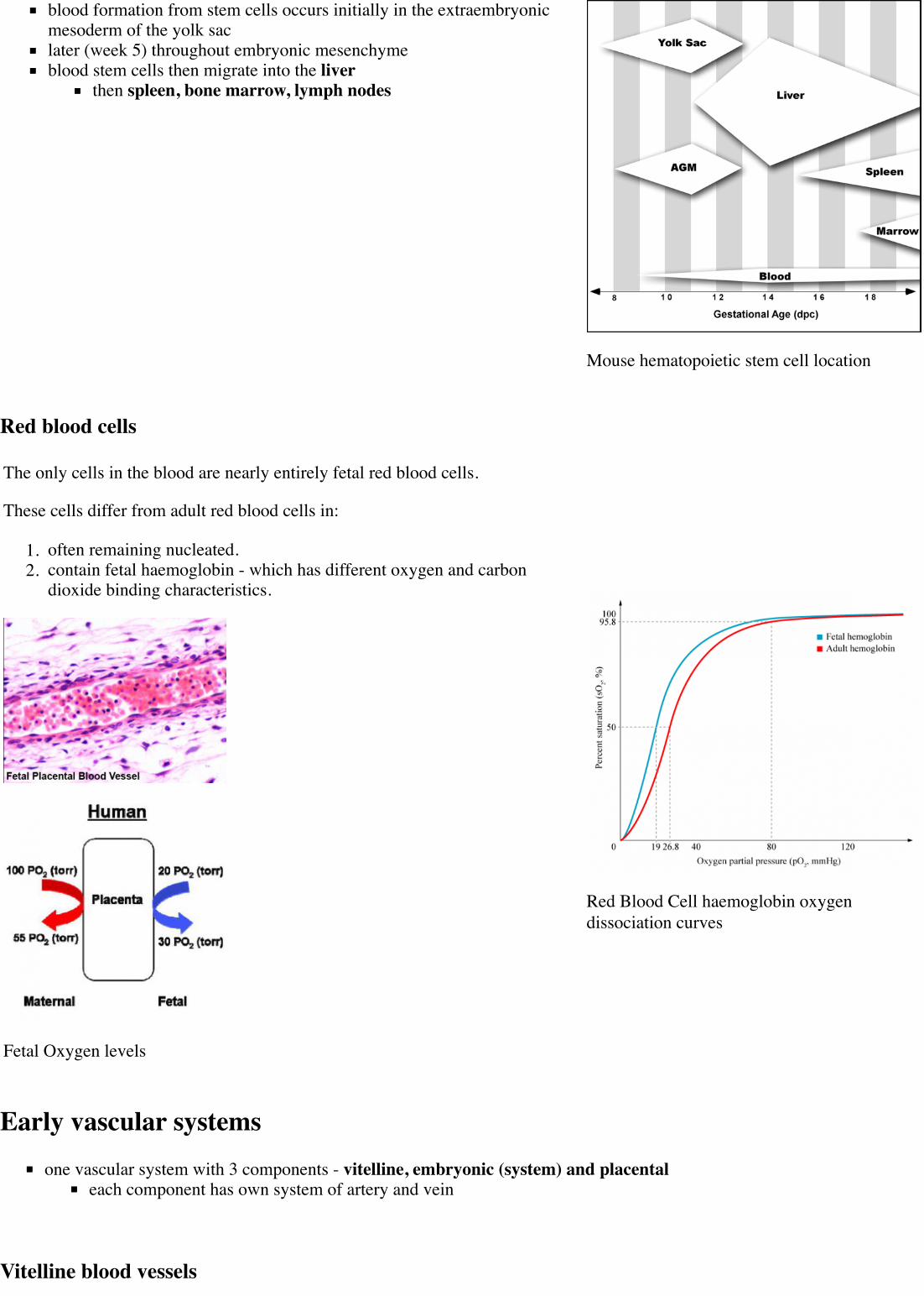
blood formation from stem cells occurs initially in the extraembryonicmesoderm of the yolk saclater (week 5) throughout embryonic mesenchymeblood stem cells then migrate into the liver
then spleen, bone marrow, lymph nodes
Mouse hematopoietic stem cell location
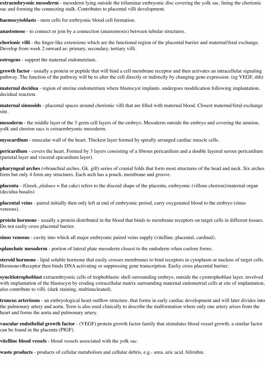
Red blood cells
The only cells in the blood are nearly entirely fetal red blood cells.
These cells differ from adult red blood cells in:
1. often remaining nucleated.2. contain fetal haemoglobin - which has different oxygen and carbon
dioxide binding characteristics.
Fetal Oxygen levels
Red Blood Cell haemoglobin oxygendissociation curves
Early vascular systemsone vascular system with 3 components - vitelline, embryonic (system) and placental
each component has own system of artery and vein
Vitelline blood vessels

Early vascular systems
Week 7 embryo with placenta
Angioblasts form a network of vessels over the yolk sac and connecting stalkJoin into two main vessels, the vitteline veins (omphalomesenteric)Pass through vitello-intestinal duct (yolk sac stalk)Enter caudal end of cardiac tube
Vitelline Arteries - arises from dorsal aorta, contribute to adult GIT arteries (fuse to become superior mesenteric artery(midgut)Vitelline Veins - empties into sinus venosus, contribute to the adultportal system
Embryo blood vessels
(systemic) will form the most of the cardiovascular systemsome vessels have neural crest contributionArterial blood flow - aortic sac → aortic arches → dorsal aorta→umbilical artery
dorsal aorta,paired initially , later fuses from T4 to L4 (gives offsegmental arteries)connect to ventral aorta via pharyngeal arches arteries.caudally, give rise to umbilical arterieslaterally, give rise to intersegmental arteries
Veins - 3 pairs of veins empty into the sinus venosus of the heartvitelline, umbilical (right and left from developing placenta entercaudal cardiac tube; only left persists)cardinal veins - anterior, common, posterior
Vein histology Blood capillary (EM)
Placental blood vessels
(Placenta development covered in next lecture)

form initially in the connecting stalk (then umbilical cord) and anastomose in chorionextend maternally - toward the chorionic villiextend embryonically - toward the sinus venosus and dorsal aorta
Arteries - paired and carry deoxygenated blood (from dorsal aorta) and waste products to the placental villiVeins - paired initially then only left at end of embryonic period and carry oxygenated blood to the embryo (sinus venosus)
Blood flow through the embryoHigh pressure pathway
Maternal Blood | -> umbilical vein -> liver -> anastomosis -> sinus venosus -> atria ventricles-> truncus arteriosus -> aortic sac -> aortic arches-> dorsal aorta-> pair of umbilical arteries | Maternal Blood.
Low pressure pathway
Head - Large veins lateral to dorsal aortae. These are the superior or anterior cardinal veins. Their function is to drain thehead region.Body - Large veins lateral to dorsal aortae. These are the inferior or posterior cardinal veins. Their function is to drain thelower part of the embryo.
Blood vessel remodeling

Early vascular development is laterally symmetrical (paired left and right). With embryo development this scheme is extensivelyremodelled leading to an asymmetric adult system in the body.
Complex balance between Stimulators and Inhibitors of Angiogenisis.
Links: Blood Vessel Development
Vascular Endothelial Growth Factor (VEGF)
required for early stages of blood vessel patterningrequired later for endothelial cell maintenance in tissues.autocrine VEGF loop from endothelial cell secretion involved in vascular growth.
VEGF protein family - VEGF (or VEGF-A), VEGF-B, placental growth factor (PlGF), VEGF-C and VEGF-D.
VEGF receptors - VEGFR-1, -2 and -3.
Cells expressing the receptors are directed in their growth.Note that there are other growth factor families (FGF, Tie, TGF-β, netrins, semaphorins) that can also influence vesselgrowth.Some angiogenic factors also involved in organ development (liver).
Links: Vascular Endothelial Growth Factor
Heart DevelopmentMH - Later development of the heart (septation) will be covered in anotherlecture.
Early heart development
Mesenchymal condensation in splanchnic mesenchyme = cardiogenic plateFollowing bending and folding, the plate comes to lie dorsal to pericardialcoelom
Plate undergoes bilateral canalisation to form 2 cardiac tubes:cranial part of cardiac tubes = ventral aortae which will later join the existing dorsal aortaeCaudally the tubes will join vitelline and umbilical veins
Cardiac tubes fuse to form single heart tube that sinks into coelom (= future pericardial sac)At this stage, the heart is an endothelial tube surrounded by visceral layer of pericardium (epicardium)From the time of the fusion of cardiac tubes, the walls undergo fibrillary movements (forerunner of cardiac contraction)Later, space between epicardium and cardiac endothelium fills with jelly-like material (cardiac jelly), which becomesinvaded by cells of deep layer of epicardium. These are the myoblasts (future cardiac muscle)Combined layer of epicardium and invaded jelly = myoepicardial mantle.Epicardial layer also gives rise to blood islands which form vascular network (future coronary vessels)Heart tube (now within the pericardial coelom) begins to undergo internal and external changes.

mouse E9.5 heart (stage 10)
Heart external
Heart internal
Heart layers
pericardium - covers the heart. Formed by 3 layers consisting of afibrous pericardium and a double layered serous pericardium (parietallayer and visceral epicardium layer).myocardium - muscular wall of the heart. Thickest layer formed byspirally arranged cardiac muscle cells.endocardium - lines the heart. Epithelial tissue lining the inner surfaceof heart chambers and valves.
Heart looping
Week 3Page | Play
Heart LoopingPage | Play

Transverse section- Heart is 2 tubes that fuse in the midline anterior to pharynx.
The pericardial cavity can be imagined as the top of the "horseshoe" of the intraembryonic coelom. (where the arms become thepleural cavity and the ends fuse anteriorly to form a single peritoneal cavity).
This view shows the initial positioning of the ventricles above the atria. The ventricles are rotated into their correct anatomicalposition by the growth of the heart tube, bending into an "S" shape.
Initially...
Heat tube develops a series of constrictions:Truncus arteriosus – ventral aortae meet - OUTFLOWBulbus cordisVentricleAtriumSinus venosus – caudal end of tube, receiving 4 veins - INFLOW
Rapid growth – ‘buckling’ and ‘twisting’Heart tube bends ventrally into pericardial coelomVentricle enlargens, absorbs lower part of bulbus cordis (bulboventricular loop)Ventricle also twists to left- atrium and sinus venosus come to lie dorsal to bulbus cordis and lower part of truncusarteriosusVenous inflow comes to lie directly dorsal to the arterial outflow.Possible abnormality – dextro-rotation, the heart bends or twists to the right. Maybe associated by other abnormalities.Later, sinus venosus becomes absorbed into atrium
Heart neural crest
The mouse model shows that the heart also has contributions from neural crest E8.5 mouse neural crest(http://dev.biologists.org/cgi/content/full/131/14/3367/FIG1)
between the levels of post-otic hindbrain to somite 4, with the most contribution from somite 2 level.
7 somite stage - Migration of cardiac neural crest from the neural tube begins. (level post-otic hindbrain and somite 4)Pathways dorsolateral, medial, and between somites.Then through peri-aortic mesenchyme (lateral to pharynx), through pharyngeal arches (3, 4, 6) into the aortic sac.
32 somite stage: Colonisation of the outflow tract mesenchyme.
Data from: Chan WY, Cheung CS, Yung KM, Copp AJ. Chan WY, Cheung CS, Yung KM, Copp AJ. Cardiac neural crest of themouse embryo: axial level of origin, migratory pathway and cell autonomy of the splotch (Sp2H) mutant effect. Development.2004 Jul;131(14):3367-79. PMID: 15226254 (http://www.ncbi.nlm.nih.gov/pubmed/15226254)
Embryonic heart rate
Ultrasonographic measurement of embryonic heart rate (EHR) shows a steady increase from Stage 9-10 (75 beats/minute)to Stage 18 (130 beats/minute) and on to Stage 20, following which a gradual decrease in EHR occursMaximal EHR is reached when morphological development of the embryonic heart is completed.
Links: Embryonic Heart Rate
Internet LinksEmbryo Images Unit: Embryo Images Online (http://www.med.unc.edu/embryo_images/) Early Cell Populations (cardiogenicsection) | Cardiovascular Development (http://www.med.unc.edu/embryo_images/unit-cardev/cardev_htms/cardevtoc.htm) |Week 3 Development (http://www.med.unc.edu/embryo_images/unit-cardev/cardev_htms/cardev001.htm) | Week 4Development (http://www.med.unc.edu/embryo_images/unit-cardev/cardev_htms/cardev007.htmhttp://www.med.unc.edu/embryo_images/unit-cardev/cardev_htms/cardev007.htm) | HeartChambers and Outflow Tract (http://www.med.unc.edu/embryo_images/unit-cardev/cardev_htms/cardev018.htm) |Atrioventricular Septation (http://www.med.unc.edu/embryo_images/unit-cardev/cardev_htms/cardev022.htm) | Outflow TractSeptation (http://www.med.unc.edu/embryo_images/unit-cardev/cardev_htms/cardev028.htm) | Ventricular Septation

(http://www.med.unc.edu/embryo_images/unit-cardev/cardev_htms/cardev035.htm) | Atrial Septation(http://www.med.unc.edu/embryo_images/unit-cardev/cardev_htms/cardev036.htm) | Atrial Walls(http://www.med.unc.edu/embryo_images/unit-cardev/cardev_htms/cardev040.htm) Aortic Arch Vessels(http://www.med.unc.edu/embryo_images/unit-cardev/cardev_htms/cardev041.htm) | Changes at Birth(http://www.med.unc.edu/embryo_images/unit-cardev/cardev_htms/cardev042.htm)
References
Online Textbooks
Developmental Biology by Gilbert, Scott F. Sunderland (MA): Sinauer Associates, Inc.; c2000 The Heart(http://www.ncbi.nlm.nih.gov/books/bv.fcgi?&rid=dbio.section.3693#3695) | Figure 15.6. Cascade of heart development} |[http://www.ncbi.nlm.nih.gov/books/bv.fcgi?&rid=dbio.figgrp.3698 Figure 15.3. Formation of the chick heart from thesplanchnic lateral plate mesoderm (http://www.ncbi.nlm.nih.gov/books/bv.fcgi?&rid=dbio.figgrp.3702) | Figure 15.4.Fusion of the right and left heart rudiments to form a single cardiac tube (http://www.ncbi.nlm.nih.gov/books/bv.fcgi?&rid=dbio.figgrp.3699) | Figure 15.5. Specification of the atrium and ventricles occurs even before heart looping(http://www.ncbi.nlm.nih.gov/books/bv.fcgi?&rid=dbio.figgrp.3701)Molecular Biology of the Cell 4th ed. Alberts, Bruce; Johnson, Alexander; Lewis, Julian; Raff, Martin; Roberts, Keith;Walter, Peter New York and London: Garland Science; c2002 - Figure 21-35. The vertebrate body plan as a dorsoventralinversion of the insect body plan (http://www.ncbi.nlm.nih.gov/books/bv.fcgi?&rid=mboc4.figgrp.3860) Figure 22-40. Thefour classes of muscle cells of a mammal (http://www.ncbi.nlm.nih.gov/books/bv.fcgi?&rid=mboc4.figgrp.4167)
Stage 12
Stage 13
TermsFor more cardiovascular term definitions and links to related topics use the glossary.
angioblast - the stem cells in blood islands generating endothelial cells which will form the walls of both arteries and veins.(More? Blood Vessel)
angiogenesis - the formation of blood vessels also called vasculogenesis in the embryo.
anlage (German, anlage = primordium) structure or cells which will form a future more developed or differentiated adultstructure.
blood islands - earliest sites of blood vessel and blood cell formation, seen mainly on yolk sac chorion.
cardinal veins - paired main systemic veins of early embryo, anterior, common, posterior.
cardiogenic region - region above prechordal plate in mesoderm where heart tube initially forms.
ectoderm - the layer (of the 3 germ cell layers) which form the nervous system from the neural tube and neural crest and alsogenerates the epithelia covering the embryo.
endoderm - the layer (of the 3 germ cell layers) which form the epithelial lining of the gastrointestinal tract (GIT) and accessoryorgans of GIT in the embryo.
endocardium - lines the heart. Epithelial tissue lining the inner surface of heart chambers and valves.
endothelial cells - single layer of cells closest to lumen that line blood vessels.
extraembryonic mesoderm - mesoderm lying outside the trilaminar embryonic disc covering the yolk sac, lining the chorionicsac and forming the connecting stalk. Contributes to placental villi development.
haemocytoblasts - stem cells for embryonic blood cell formation.
anastomose - to connect or join by a connection (anastomosis) between tubular structures.
chorionic villi - the finger-like extensions which are the functional region of the placental barrier and maternal/fetal exchange.Develop from week 2 onward as: primary, secondary, tertiary villi.
estrogens - support the maternal endometrium.
growth factor - usually a protein or peptide that will bind a cell membrane receptor and then activates an intracellular signalingpathway. The function of the pathway will be to alter the cell directly or indirectly by changing gene expression. (eg VEGF, shh)
maternal decidua - region of uterine endometrium where blastocyst implants. undergoes modification following implantation,decidual reaction.
maternal sinusoids - placental spaces around chorionic villi that are filled with maternal blood. Closest maternal/fetal exchangesite.
mesoderm - the middle layer of the 3 germ cell layers of the embryo. Mesoderm outside the embryo and covering the amnion,yolk and chorion sacs is extraembryonic mesoderm.
myocardium - muscular wall of the heart. Thickest layer formed by spirally arranged cardiac muscle cells.
pericardium - covers the heart. Formed by 3 layers consisting of a fibrous pericardium and a double layered serous pericardium(parietal layer and visceral epicardium layer).
pharyngeal arches (=branchial arches, Gk. gill) series of cranial folds that form most structures of the head and neck. Six archesform but only 4 form any structures. Each arch has a pouch, membrane and groove.
placenta - (Greek, plakuos = flat cake) refers to the discoid shape of the placenta, embryonic (villous chorion)/maternal organ(decidua basalis)
placental veins - paired initially then only left at end of embryonic period, carry oxygenated blood to the embryo (sinusvenosus).
protein hormone - usually a protein distributed in the blood that binds to membrane receptors on target cells in different tissues.Do not easliy cross placental barrier.
sinus venosus - cavity into which all major embryonic paired veins supply (vitelline, placental, cardinal).
splanchnic mesoderm - portion of lateral plate mesoderm closest to the endoderm when coelom forms.
steroid hormone - lipid soluble hormone that easily crosses membranes to bind receptors in cytoplasm or nucleus of target cells.Hormone+Receptor then binds DNA activating or suppressing gene transcription. Easliy cross placental barrier.
syncitiotrophoblast extraembryonic cells of trophoblastic shell surrounding embryo, outside the cytotrophoblast layer, involvedwith implantation of the blastocyst by eroding extracellular matrix surrounding maternal endometrial cells at site of implantation,also contribute to villi. (dark staining, multinucleated).
truncus arteriosus - an embryological heart outflow structure, that forms in early cardiac development and will later divides intothe pulmonary artery and aorta. Term is also used clinically to describe the malformation where only one artery arises from theheart and forms the aorta and pulmonary artery.
vascular endothelial growth factor - (VEGF) protein growth factor family that stimulates blood vessel growth, a similar factorcan be found in the placenta (PIGF).
vitelline blood vessels - blood vessels associated with the yolk sac.
waste products - products of cellular metabolism and cellular debris, e.g.- urea, uric acid, bilirubin.

2015 Course: Week 2 Lecture 1 Lecture 2 Lab 1 | Week 3 Lecture 3 Lecture 4 Lab 2 | Week 4 Lecture 5 Lecture 6 Lab 3 |Week 5 Lecture 7 Lecture 8 Lab 4 | Week 6 Lecture 9 Lecture 10 Lab 5 | Week 7 Lecture 11 Lecture 12 Lab 6 | Week 8Lecture 13 Lecture 14 Lab 7 | Week 9 Lecture 15 Lecture 16 Lab 8 | Week 10 Lecture 17 Lecture 18 Lab 9 | Week 11Lecture 19 Lecture 20 Lab 10 | Week 12 Lecture 21 Lecture 22 Lab 11 | Week 13 Lecture 23 Lecture 24 Lab 12 |Projects: Group 1 | Group 2 | Group 3 | Group 4 | Group 5 | Group 6 | Students | Student Sharing | Moodle page(http://moodle.telt.unsw.edu.au/course/view.php?id=15814)
Retrieved from ‘https://embryology.med.unsw.edu.au/embryology/index.php?title=Lecture_-_Early_Vascular_Development&oldid=194838’
Categories: Heart Cardiovascular 2015 Science-Undergraduate
This page was last modified on 24 August 2015, at 10:23.This page has been accessed 11,896 times.