lecture 4. sample preparation for maldi and lc/ms 4. sample preparation for maldi and lc/ms 1....
TRANSCRIPT
1
1
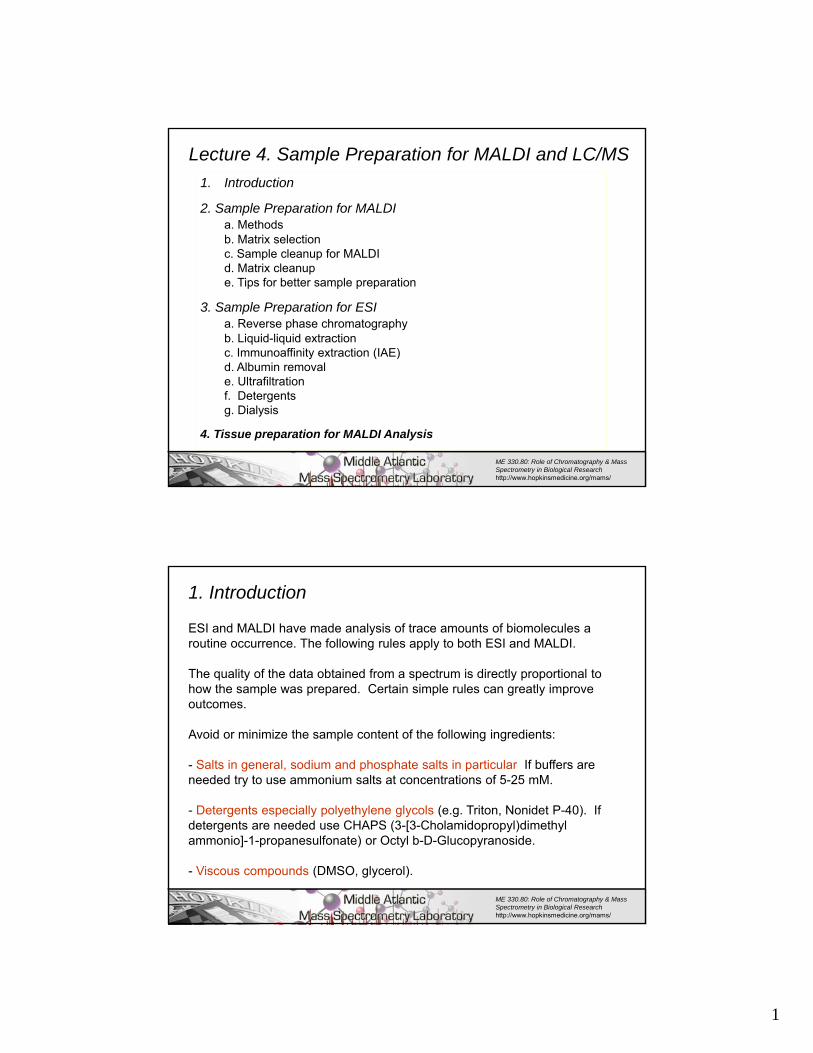
Lecture 4. Sample Preparation for MALDI and LC/MS
1. Introduction
2. Sample Preparation for MALDIa. Methodsb. Matrix selectionc. Sample cleanup for MALDId. Matrix cleanupe. Tips for better sample preparation
3. Sample Preparation for ESIa. Reverse phase chromatographyb. Liquid-liquid extractionc. Immunoaffinity extraction (IAE)d. Albumin removale. Ultrafiltrationf. Detergentsg. Dialysis
4. Tissue preparation for MALDI Analysis
ME 330.80: Role of Chromatography & Mass Spectrometry in Biological Research http://www.hopkinsmedicine.org/mams/
2
1. Introduction
ESI and MALDI have made analysis of trace amounts of biomolecules a routine occurrence. The following rules apply to both ESI and MALDI.
The quality of the data obtained from a spectrum is directly proportional to how the sample was prepared. Certain simple rules can greatly improve outcomes.
Avoid or minimize the sample content of the following ingredients:
- Salts in general, sodium and phosphate salts in particular If buffers are needed try to use ammonium salts at concentrations of 5-25 mM.
- Detergents especially polyethylene glycols (e.g. Triton, Nonidet P-40). If detergents are needed use CHAPS (3-[3-Cholamidopropyl)dimethyl ammonio]-1-propanesulfonate) or Octyl b-D-Glucopyranoside.
- Viscous compounds (DMSO, glycerol).
ME 330.80: Role of Chromatography & Mass Spectrometry in Biological Research http://www.hopkinsmedicine.org/mams/
2
3
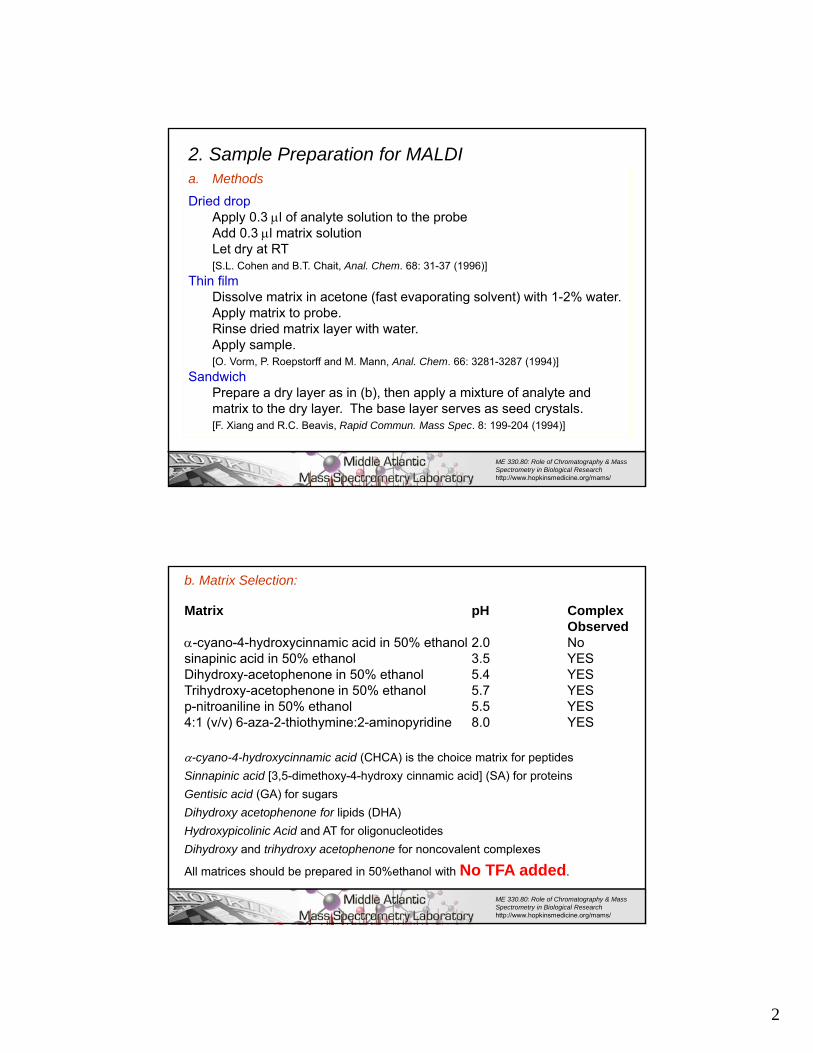
a. Methods
Dried dropApply 0.3 l of analyte solution to the probeAdd 0.3 l matrix solutionLet dry at RT[S.L. Cohen and B.T. Chait, Anal. Chem. 68: 31-37 (1996)]
Thin filmDissolve matrix in acetone (fast evaporating solvent) with 1-2% water.Apply matrix to probe. Rinse dried matrix layer with water.Apply sample.[O. Vorm, P. Roepstorff and M. Mann, Anal. Chem. 66: 3281-3287 (1994)]
SandwichPrepare a dry layer as in (b), then apply a mixture of analyte and matrix to the dry layer. The base layer serves as seed crystals.[F. Xiang and R.C. Beavis, Rapid Commun. Mass Spec. 8: 199-204 (1994)]
2. Sample Preparation for MALDI
ME 330.80: Role of Chromatography & Mass Spectrometry in Biological Research http://www.hopkinsmedicine.org/mams/
4
b. Matrix Selection:
Matrix pH Complex Observed
-cyano-4-hydroxycinnamic acid in 50% ethanol 2.0 Nosinapinic acid in 50% ethanol 3.5 YESDihydroxy-acetophenone in 50% ethanol 5.4 YESTrihydroxy-acetophenone in 50% ethanol 5.7 YESp-nitroaniline in 50% ethanol 5.5 YES4:1 (v/v) 6-aza-2-thiothymine:2-aminopyridine 8.0 YES
-cyano-4-hydroxycinnamic acid (CHCA) is the choice matrix for peptides
Sinnapinic acid [3,5-dimethoxy-4-hydroxy cinnamic acid] (SA) for proteins
Gentisic acid (GA) for sugars
Dihydroxy acetophenone for lipids (DHA)
Hydroxypicolinic Acid and AT for oligonucleotides
Dihydroxy and trihydroxy acetophenone for noncovalent complexes
All matrices should be prepared in 50%ethanol with No TFA added.
ME 330.80: Role of Chromatography & Mass Spectrometry in Biological Research http://www.hopkinsmedicine.org/mams/
3
5
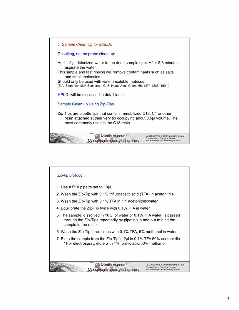
c. Sample Clean Up for MALDI:
Desalting, on the probe clean up:
Add 1-2 l deionized water to the dried sample spot. After 2-3 minutes aspirate the water.
This simple and fast rinsing will remove contaminants such as salts and small molecules.
Should only be used with water insoluble matrices.[E.A. Stemmler, M.V. Buchanan, G. B. Hurst, Anal. Chem. 66: 1274-1285 (1994)]
HPLC: will be discussed in detail later.
Sample Clean up Using Zip-Tips
Zip-Tips are pipette tips that contain immobilized C18, C4 or other resin attached at their very tip occupying about 0.5µl volume. The most commonly used is the C18 resin.
ME 330.80: Role of Chromatography & Mass Spectrometry in Biological Research http://www.hopkinsmedicine.org/mams/
6
Zip-tip protocol:
1. Use a P10 pipette set to 10µl.
2. Wash the Zip-Tip with 0.1% trifluroacetic acid (TFA) in acetonitrile
3. Wash the Zip-Tip with 0.1% TFA in 1:1 acetonitrile:water
4. Equilibrate the Zip-Tip twice with 0.1% TFA in water
5. The sample, dissolved in 10 µl of water or 0.1% TFA water, is passed through the Zip-Tips repeatedly by pipeting in and out to bind the sample to the resin.
6. Wash the Zip-Tip three times with 0.1% TFA, 5% methanol in water
7. Elute the sample from the Zip-Tip in 2µl in 0.1% TFA 50% acetonitrile. * For electrospray, elute with 1% formic acid/50% methanol.
ME 330.80: Role of Chromatography & Mass Spectrometry in Biological Research http://www.hopkinsmedicine.org/mams/

4
7
Purification of α-cyano-4-hydroxycinnamic acid [CHCA]; same protocol could be applied to other matrices
Make a saturated solution in 95% or 100% ethanol (warmed in a flask in a pan of water, but not boiling).
Filter out whatever isn't dissolved. Then add two volumes of water to the filtrate (cold water works best, it gives a better yield). You should have a bright, pale yellow liquid.
Filter it, and keep the retentate. To increase your yield, leave the solution in the refrigerator overnight before filtering, take the second filtrate and leave it in the refrigerator overnight, then refilter to eke out the last.
Dry the powder on the filter, mash it up to get the lumps out.
Repeat the above protocol if the dried matrix is still not bright yellow and fluffy.
ME 330.80: Role of Chromatography & Mass Spectrometry in Biological Research http://www.hopkinsmedicine.org/mams/
8
Zip-tip protocol:
C18 ZipTip pipettes are the tips of choice for peptides and low MW proteins.
C8 Zip Tip pipettes are most suitable for low to intermediate MW proteins.
In many cases, the two devices can be used interchangeably. Because higher molecular weight proteins tend to adsorb tenaciously to hydrophobic surfaces, ZipTipC4 pipette tips are recommended for proteins over 100,000 MW. For more details go to the Millipore web site.
Some other companies have zip Tip like short columns
ME 330.80: Role of Chromatography & Mass Spectrometry in Biological Research http://www.hopkinsmedicine.org/mams/
http://www.millipore.com/lifescience/products.nsf/docs/ziptip
Xu Y, Bruening M.L., and Watson J.T. Mass Spec Rev22, 429-440 (2003)

5
9
d. Matrix Clean up:
Water insoluble matrices:
Fill lower third of an eppendorf tube with matrix, add water, vortex, centrifuge, and discard.
This will remove a significant amount of sodium and potassium ions.
Water soluble matrices:
Can be dissolved and desalted by adding cation exchange beads charged with ammonium ions.
ME 330.80: Role of Chromatography & Mass Spectrometry in Biological Research http://www.hopkinsmedicine.org/mams/
10
e. Tips For better sample preparation
• Vacuum dry sample-matrix spot on the target before acquiring a spectrum. Except for Dihydroxy acetophenone (DHA), as it sublimes in vacuum within an hour from the introduction of the sample plate in the mass spectrometer.
• Raise the pH of sample-matrix mixture by adding ammonium citrate or bicarbonate.
• To decrease suppression, add 0.3 microliter of 150 mM ammonium sulfate, acetate, citrate or bicarbonate (25-100 mM) to the sample deposited on the sample plate before matrix addition.
• To detect intact double stranded DNA using MALDI, prepare the matrix in the following way. Saturated 6-Aza-2-Thiothymine in 150 mM Ammonium Sulfate.
ME 330.80: Role of Chromatography & Mass Spectrometry in Biological Research http://www.hopkinsmedicine.org/mams/
6
11
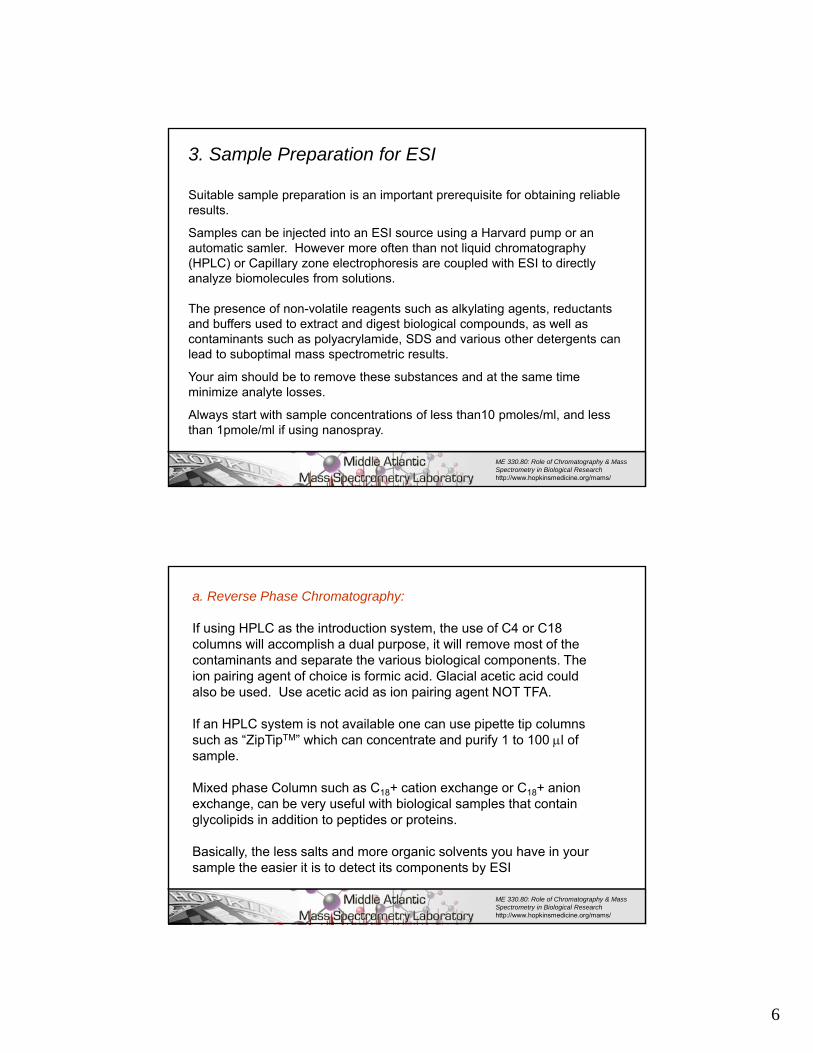
Suitable sample preparation is an important prerequisite for obtaining reliable results.
Samples can be injected into an ESI source using a Harvard pump or an automatic samler. However more often than not liquid chromatography (HPLC) or Capillary zone electrophoresis are coupled with ESI to directly analyze biomolecules from solutions.
The presence of non-volatile reagents such as alkylating agents, reductants and buffers used to extract and digest biological compounds, as well as contaminants such as polyacrylamide, SDS and various other detergents can lead to suboptimal mass spectrometric results.
Your aim should be to remove these substances and at the same time minimize analyte losses.
Always start with sample concentrations of less than10 pmoles/ml, and less than 1pmole/ml if using nanospray.
3. Sample Preparation for ESI
ME 330.80: Role of Chromatography & Mass Spectrometry in Biological Research http://www.hopkinsmedicine.org/mams/
12
a. Reverse Phase Chromatography:
If using HPLC as the introduction system, the use of C4 or C18 columns will accomplish a dual purpose, it will remove most of the contaminants and separate the various biological components. The ion pairing agent of choice is formic acid. Glacial acetic acid could also be used. Use acetic acid as ion pairing agent NOT TFA.
If an HPLC system is not available one can use pipette tip columns such as “ZipTipTM” which can concentrate and purify 1 to 100 l of sample.
Mixed phase Column such as C18+ cation exchange or C18+ anion exchange, can be very useful with biological samples that contain glycolipids in addition to peptides or proteins.
Basically, the less salts and more organic solvents you have in your sample the easier it is to detect its components by ESI
ME 330.80: Role of Chromatography & Mass Spectrometry in Biological Research http://www.hopkinsmedicine.org/mams/

7
13
b. Liquid-Liquid extraction:
Mostly used for drug extraction to isolate drugs after cleavage of the biochemical component to which they are conjugated.
Followed by separation on a reverse phase column.
c. Immunoaffinity extraction (IAE):
IAE can be coupled with HPLC. - The first column is packed with the IEA bound antibodies.- The second column is a trapping column - The third one is an analytical column. - The sample is loaded, - The analyte is bound, then the analyte is eluted off the column, and trapped onto the trapping column, then the analyte is back-flushed onto the appropriate analytical column.
ME 330.80: Role of Chromatography & Mass Spectrometry in Biological Research http://www.hopkinsmedicine.org/mams/
14
d. Removal of serum albumin
Serum proteome mirrors the condition of a patient thus it’s importance for the discovery of biomarkers. A protocol for the removal of albumin should remove the albumin with minimal loss of other proteins.
Depletion of Albumin Component (DAC) Protocol
Colantonio D.A, Dunkinson C., Bovenkamp D.E. and Van Eyk J.E. Effective removal of albumin from serum Proteomics 5, 3831 – 3835 (2005)- To 100–500 uL of serum Sodium chloride is added for a final conc of 0.1 M and incubated with gentle rotation for 60 min at 4oC.-Cold ethanol is then added to the mixture for a final concentration of 42% and incubated for 60 min at 4oC. - The sample is centrifuged at 16 000g for 45 min at 4oC. - The supernatant is transferred to a microtube and its pH adjusted to 5.7 using cold 0.8 M sodium acetate, pH 4.0 and incubated at 4oC for 60 min. The pellet is retained.
ME 330.80: Role of Chromatography & Mass Spectrometry in Biological Research http://www.hopkinsmedicine.org/mams/

8
15ME 330.80: Role of Chromatography & Mass Spectrometry in Biological Research http://www.hopkinsmedicine.org/mams/
- Mixture is centrifuged as described above. The supernatant, containing albumin, is put in a microtube and the second pellet is retained. -Both pellets are combined, resuspended in 10mM Tris buffer, pH 6.8, and 1 M urea. IgG is reduced by adding sepharose conjugated protein G to the resuspended pellets in presence of 0.5% CHAPS and incubating for 60 min. -The sample is centrifuged at 14 000g for 10 min and the albumin-depleted/IgG-reduced supernatant analysed-All protein fractions can be analyzed by 1-D SDS-PAGE.
Other methodsL.F. Steel et al. Molecular & Cellular Proteomics 2, 262-270 (2003).Uses monoclonal antibodies against human serum albumin to develop an immunoaffinity resin effective for removal of both full length albumin and its fragments present in serum,
Enchant™ Life Science Kits Albumin DepletionIt utilizes a dehydrated Cibacron® Blue-based support that processes 10–100 uL of serum or plasma samples. The dehydrated albumin depleting disc, which rapidly hydrates in the presence of water, is equivalent to 200 uL of resin and removes > 2 mg of albumin.
16
e. Ultrafiltration:An ultra filter is a molecular sieve with extremely small pores. Most ultra filtration units are made of a sample cup with a membrane sealed to its bottom. The cup sits inside a centrifuge tube. The membranes have different molecular weight cut-off.
ME 330.80: Role of Chromatography & Mass Spectrometry in Biological Research http://www.hopkinsmedicine.org/mams/
Method:Determine what size particles are to be removed from or concentrated in a solution, and be sure that the filter is chemically compatible with any fluids that it may be exposed to.Determine flow rate of filtration process by taking into consideration the pore size of the filter, the viscosity, volume, the particulate load of the sample, before choosing centrifugation speed and duration.

9
17
Typical MALDI spectrum seen when sample contains polyethylene glycol detergents
ME 330.80: Role of Chromatography & Mass Spectrometry in Biological Research http://www.hopkinsmedicine.org/mams/
f. Detergents
ESI spectrum seen when sample contains polyethylene glycol detergents
18
-
Polyethylene and polypropylene membranes:
Used to cleanup Triton X for a sample of lipid A and a sample of bovine heart cytochrome c.
[T.A. Worrall, R.J. Cotter, A.S. Woods, Anal. Chem. 70, 750-756 (1998)]
ME 330.80: Role of Chromatography & Mass Spectrometry in Biological Research http://www.hopkinsmedicine.org/mams/

10
19
How to eliminate or minimize suppression
Buffer used: 100 mM ammonium citratePpt Seq MW BBI %RI %RI
Ppt+Mtrx Ppt+Mtrx
+Buffer
P1 FERFEIFPKE 1341.5 -3500 59.2 96.3
P2 TYQRTRALV 1107.3 -290 100.0 100.0
P3 SIINFEKL 963.2 -3790 8.9 23.0
P4 IYSTVASSL 940.1 -3120 0.0 14.1
A.S. Woods, A.Y.C. Huang, R.J. Cotter, G.R. Pasternack, D.M. Pardoll and E.M. Jaffee. Simplified High Sensitivity Sequencing of MHC Class I-Associated, Immunoreactive Peptides Using Matrix Assisted Laser Desorption/Ionization Mass Spectrometry, Anal. Biochem. 226 (1995) 15-25.
L. Marzilli, T. Golden, R.J. Cotter and A.S. Woods, The Complementary Use of Pronase and In Source Decay for Peptide Sequencing JASMS 11, 1000-1008 (2000).
ME 330.80: Role of Chromatography & Mass Spectrometry in Biological Research http://www.hopkinsmedicine.org/mams/
20
g. Dialysis:
Frequently it is necessary to remove salts or change the buffer in which a protein is dissolved. This can be achieved by dialysis. The protein solution is placed in a bag made of a semi-permeable membrane and placed in an appropriate buffer. Small molecules pass across the membrane while large ones are retained. The membrane is made of cellulose acetate with pores of 1-20 nm in diameter. The size of the pores determines the molecular weight of the molecules that will be retained.
ME 330.80: Role of Chromatography & Mass Spectrometry in Biological Research http://www.hopkinsmedicine.org/mams/

11
21ME 330.80: Role of Chromatography & Mass Spectrometry in Biological Research http://www.hopkinsmedicine.org/mams/
4. Tissue preparation for MALDI Analysis
Tissue Sectioning, Sample Preparation and Spectra Acquisition: This will be covered in greater details in the imaging lecture.
•Rats are euthanized and the organs quickly removed and frozen in dry ice-chilled isopentane for 15 seconds, prior to storage at –80°C.•Frozen tissue is cut into thin sections (14 m thickness) in a cryostat. Tissue is attached to the cryostat sample stage using ice slush, which should only come in contact with the tissue blocks at the surface opposing the sample stages, and is frozen into a thin layer of ice within 5 seconds. •Water is used instead of optimal cutting temperature compound (OCT) to connect tissue to sample stage, as OCT causes interference and reduces the quality of mass spectra.•Serial brain sections are collected onto MALDI sample targets or poly-L-lysine coated microscopic slide. •After collection, tissue sections are stored at 4 °C until MALDI analysis.
22ME 330.80: Role of Chromatography & Mass Spectrometry in Biological Research http://www.hopkinsmedicine.org/mams/
•Tissue sections on microscopic slides are stained with cresyl violet and a Rat Brain Atlas used for assignment of brain regions analyzed by mass spectrometry.•Matrix solution volumes of 0.1- 0.2, are deposited directly onto the tissue section and allowed to air-dry prior to insertion into the mass spectrometer.•Spectra are acquired. Matrix deposition Can be made with: an artistic air brush or the Chip inkjet printer (Shimadzu, Columbia MD)
MALDI-IM positive ion mode image of sodiated cerebroside 24 : 0 OH at m/z 850.7, using 5.5 nm gold particles for matrix, deposited with an artistic air brushJackson SN, Ugarov M, Egan T, Post JD, Langlais D, Schultz JA and Woods AS JMS 42, 1093–98 (2007). Image of sodiated cerebroside 24:0 OH at m/z 850.7, using 5.5 nm gold particles for matrix, deposited with an artistic airbrush
12
23ME 330.80: Role of Chromatography & Mass Spectrometry in Biological Research http://www.hopkinsmedicine.org/mams/
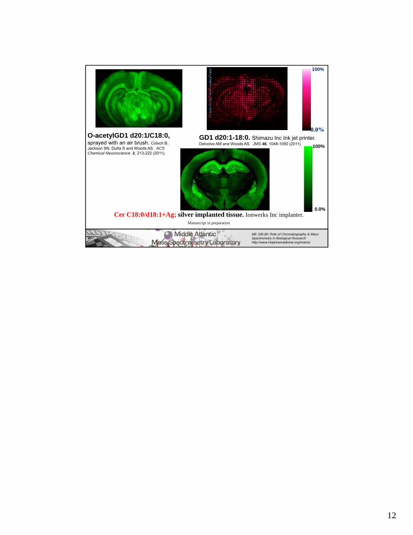
GD1 d20:1-18:0. Shimazu Inc Ink jet printer. Delvolve AM and Woods AS. JMS 46, 1046-1050 (2011).
0.0%
100%
O-acetylGD1 d20:1/C18:0, sprayed with an air brush. Colsch B, Jackson SN, Dutta S and Woods AS. ACS Chemical Neuroscience 2, 213-222 (2011).
Cer C18:0/d18:1+Ag; silver implanted tissue. Ionwerks Inc implanter. Manuscript in preparation
100%
0.0%