lecture 4 muscle physiology(1)
TRANSCRIPT


Overview
• Structure of Muscle and Muscle Fiber• Components of a Muscle Fiber, Myofibril,
and Sarcomere• Structure of Myosin and Actin• Muscle Contraction• Muscle Fiber Types• Fiber Type and Athletic Performance• Muscle Fatigue during Exercise

Three Types of Muscle Tissue
• Smooth muscle: involuntary, hollow organs
• Cardiac muscle: involuntary, heart
• Skeletal muscle: voluntary, skeleton
Figure 1.1

Structure of Muscle
Surrounds entire muscle
Surrounds bundles of muscle fibers (fascicles)
Surrounds individual muscle fibers
Sarcolemma Muscle cell membrane

Structure of a Muscle Fiber (Muscle Cell)
Figure 1.3

• Plasmalemma- plasma membrane– Attach to tendons– Transport in and out of cell– Sarcolemma includes plasmalemma and basement
membrane
MacIntosh, Gardiner, & McComas, Skeletal Muscle, Human Kinetics, 2006
Structure of a Muscle Fiber (Muscle Cell)
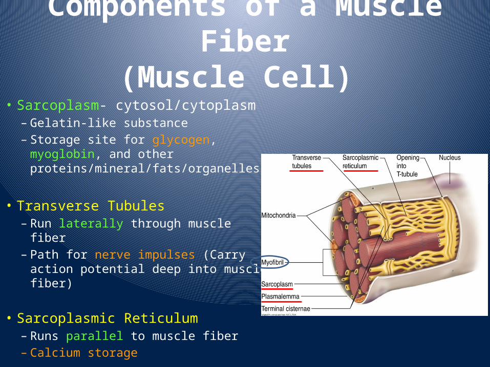
Components of a Muscle Fiber(Muscle Cell)
• Sarcoplasm- cytosol/cytoplasm– Gelatin-like substance– Storage site for glycogen, myoglobin,
and other proteins/mineral/fats/organelles
• Transverse Tubules– Run laterally through muscle fiber– Path for nerve impulses (Carry action
potential deep into muscle fiber)
• Sarcoplasmic Reticulum– Runs parallel to muscle fiber– Calcium storage

Components of a Myofibril
Figure 1.5

Components of a Sarcomere• Sarcomere: *Basic contractile unit of a myofibril
*Composed of interdigitating thick and thin filaments (myosin vs. actin)– I-Band– A-Band– H-Zone– M-Line– Z-Disk (Z-line)
Figure 1.5

Components of a Sarcomere
Figure 1.8

Components of a Sarcomere
(MacIntosh, Gardiner, & McComas, Skeletal Muscle, Human Kinetics, 2006, From Huxley, 1972)

Components of a Sarcomere
4
1
2
3
5
6

Components of a Sarcomere• Sarcomere includes two types of protein
filaments– Thick Filament: Myosin– Thin Filament: Actin
• Alignment of the thick and thin filaments is what give muscle its striations

Myosin
• Comprises 2/3 of skeletal muscle proteins• Two protein strands twisted together• Globular heads (Myosin Cross-bridges)• Titin filaments stabilize myosin

Actin
• Thin filaments are composed of 3 proteins– Actin: globular proteins form strands– Tropomyosin: twists around actin strand– Troponin: bound at intervals to actin
• Anchored to Z-Disk

Actin and Myosin Structure

Muscle Contraction
Sarcomere Actin
Myosin
Sarcomere
Actin Myosin
Muscle Fiber Function - actin and myosin function - whole muscle function & performance
Skeletal Muscle
Muscle Fiber(Myofiber, Muscle Cell)

Muscle Contraction
Muscles are divided into motor units comprised of:
α-motor neuron
Muscle fibers
Figure 1.6

Phases of Muscle Contraction
• Action Potential/Calcium Release• Calcium-Troponin Binding; Tropomyosin Shift• Actin-Myosin Binding• Myosin Power Stroke/ ATP Binding

Resting Membrane Potential(RMP)
• RMP= -70mV• Caused by uneven separation of charged ions
inside (K+) and outside (Na+) the cell• More ions outside the cell than inside• Membrane more permeable to K+• Sodium-Potassium Pumps maintain imbalance
– 3 Na+ out– 2 K+ in

Ions Channels• At rest, almost all the
Na+ channels are closed.
• At rest, few K+
channels are open.– Leaking due to [ ]
gradient

Sodium/Potassium Pump• Resting membrane
potential is maintained by pump– Potassium tends to diffuse
out of cell– Na+/K+ pump moves 3 Na+
out and 2 K+ inside the cell– Use energy from ATP

Action Potential

Action Potential• Occurs when a stimulus of sufficient strength
depolarizes the cell– Opens Na+ channels, and Na+ diffuses into cell
• Inside becomes more positive
• Repolarization– Return to resting membrane potential
• immediately following depolarization• K+ leaves the cell rapidly• Na+ channels close
• All-or-none law– Once a nerve impulse is initiated, it will travel the entire
length of the neuron without losing strength. (gun shot)
Slightly Open
Na+
OpensWide
Na+

Motor Unit

Neuromuscular Junction

Excitation-Contraction Coupling(EC Coupling)
• Action potential travels to Sarcoplasmic Reticulum, causes release of calcium into sarcoplasm
• Calcium binds to troponin on thin filament• Troponin moves tropomyosin, revealing
myosin binding sites on actin• Myosin cross-bridges bind to actin

Muscle Excitation1. Action potential in motor neuron
causes release of acetylcholine (ACh) into synaptic cleft.
2. ACh binds to receptors on motor end plate, leads to depolarization that is conducted down transverse tubules, which causes release of Ca+2 from sarcoplasmic reticulum (SR).

Sliding Filament Theory
• Muscle contraction = muscle fiber shortening
• Myosin power stroke– Myosin bound to actin tilts its head, pulling thin
filament towards the center of the sarcomere– Process is repeated until Z-disk reaches myosin
filaments or until calcium is no longer available

Figure 1.9
Ca++

Energy for Contraction
• ATP binding sites on myosin head• ATPase (on myosin head) splits ATP into ADP
and Pi
• Energy released fuels the tilting of the myosin head (power stroke)
• Additional ATP is required to keep contraction going

Muscle Relaxation
• Calcium pumps return calcium to the SR, stored for future use
• ATP required for calcium pumps
• Troponin and Tropomyosin return to original position
• Thick and thin filaments return to original positions

Muscle Action & Relaxation
• Muscle twitch–Contraction as the result of a
single stimulus–Latent period
• Lasting ~5 ms (immediately after the stimulus)–Contraction
•Tension is developed•40 ms
–Relaxation•50 ms
• It varies among muscle type

Speed of Muscle Twitch
• Speed of shortening is greater in fast fibers– Sarcoplasmic reticulum releases Ca+2 at a faster rate– Higher myosin ATPase activity – quicker ATP release of energy
with ATP hydrolysis
Type IType IIaType IIx

Fiber Type Characteristics Fast Fibers Slow fibers
Characteristic Type 2x Type 2a Type 1
Number of mitochondria Low High/mod High
Resistance to fatigue Low High/mod High
Predominant energy system Anaerobic Combination Aerobic
ATPase Highest High Low
Vmax (speed of shortening) Highest Intermediate Low
Efficiency Low Moderate High
Specific tension High High Moderate


Bergström Muscle Biopsy
http://www.youtube.com/watch?v=Hc4HJj3THuw

Muscle HistochemistryType 2a Type 1Type 2x
Fiber Type and Performance
• Power Athletes– Sprinters– Mostly Fast (70-75%) Twitch (Type 2)
• Endurance Athletes– Distance Runners, Triathletes, Cyclists– Mostly Slow (70-80%) Twitch (Type 1)
• Others– Non-athletes– Equal amount of Fast and Slow Twitch

Other Factors which Influence Muscle Force
• Number of motor units activated• Type of motor units activated (FT or ST)• Muscle size• Initial muscle length• Joint angle• Speed of muscle action (shortening or
lengthening)

Length-Tension Relationship
Figure 1.13
• Length-tension relationship– Optimal sarcomere length = optimal overlap– Too short or too stretched = little or no force develops

Speed-Force Relationship
• Speed-force relationship
– Concentric: maximal force development decreases at higher speeds
– Eccentric: maximal force development increases at higher speeds

Muscle Fatigue during ExerciseM
usc
le F
orc
e
Exercise
<60 sec
Pi & H+
>2 hrCa Release from SR