lecture 3 - columbus labs
TRANSCRIPT
Quiz!

Reminders/Announcements•
Articles for projects are on the Toolkit website –
be sure you pick the project in which you can attend the evening session.
•
I will pass out guidelines for the paper.
•
Start reading “The Double Helix”
•
Wed Office Hours will be in the computer lab – please help each other getting started

Review of Lecture 2
What type of molecule is this? Name the three components.What base is this?
is this a purine
or a pyrimidine?what would we have to change to make adenine?
What sugar is this?what is the difference from the “other sugar”?
Give the full name of this molecule

Nucleic acids•
Polynucleotides
linked 3' to 5' by phosphodiester
bonds
•
Ribonucleic acid (RNA) and deoxyribonucleic acid DNA

5’3’
5’ 3’
Summary of DNA Structure
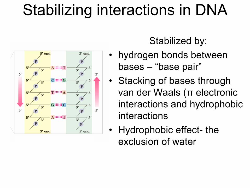
Stabilizing interactions in DNA
Stabilized by:•
hydrogen bonds between bases –
“base pair”
•
Stacking of bases through van der
Waals (π
electronic
interactions and hydrophobic interactions
•
Hydrophobic effect-
the exclusion of water

The “canonical”
base pairs•
The canonical A:T and G:C base pairs have nearly identical overall dimensions –
very important for the stacking in the helix
•
A and T share two H-bonds •
G and C share three H-bonds
•
G:C-rich regions of DNA are more stable •
Polar atoms in the sugar-phosphate backbone also form H-
bonds
Watson –
Crick Base PairsCanonical base pairs

Secondary Structure of DNA•
Sugar-phosphate backbone outside
•
Bases (hydrogen-bonded) inside •
Right-twist closes the gaps between base pairs to 3.4 A (0.34 nm) in B-DNA

Major and minor grooves•
The "tops" of the bases (as we draw them) line the "floor" of the major groove
•
The major groove is large enough to accommodate an alpha helix from a protein
•
Regulatory proteins (transcription factors) can recognize the pattern of bases and H-bonding possibilities in the major groove
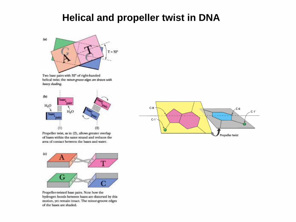
Helical and propeller twist in DNA

B-DNA A-DNA Z-DNA
Right-handedMajor Groove –
wideMinor Groove –
narrowPitch per turn helix –
33.2 Å
Right-handednarrowBroad24.6 Å
Left-handedFlattened out on surfaceNarrow45.6 Å
Comparison of DNA Structures
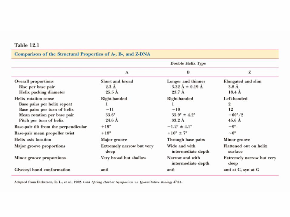
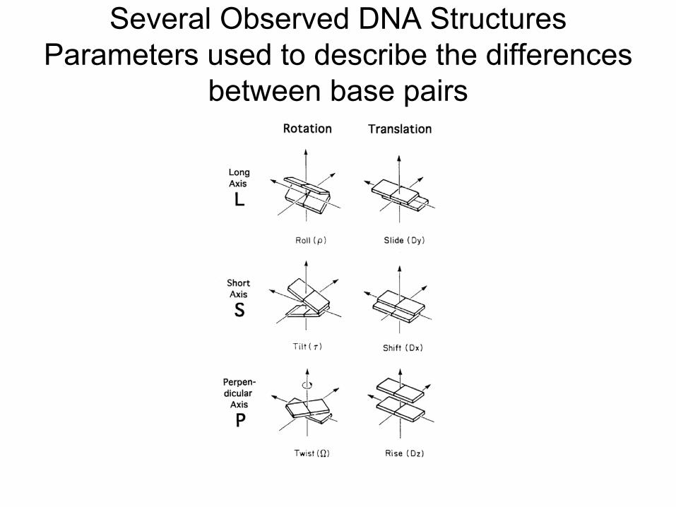
Several Observed DNA Structures Parameters used to describe the differences
between base pairs

Transition of B-DNA to A-DNA

Z-DNADiscovered by Alex Rich
•
Found in G:C-rich regions of DNA •
G goes to syn
conformation
•
C stays anti but whole C nucleoside (base and sugar) flips 180 degrees
•
Result is that G:C H-bonds can be preserved in the transition from B-form to Z-form!

DNA Intercalators
Bis
–
intercalator
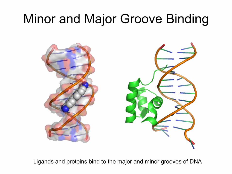
Minor and Major Groove Binding
Ligands and proteins bind to the major and minor grooves of DNA
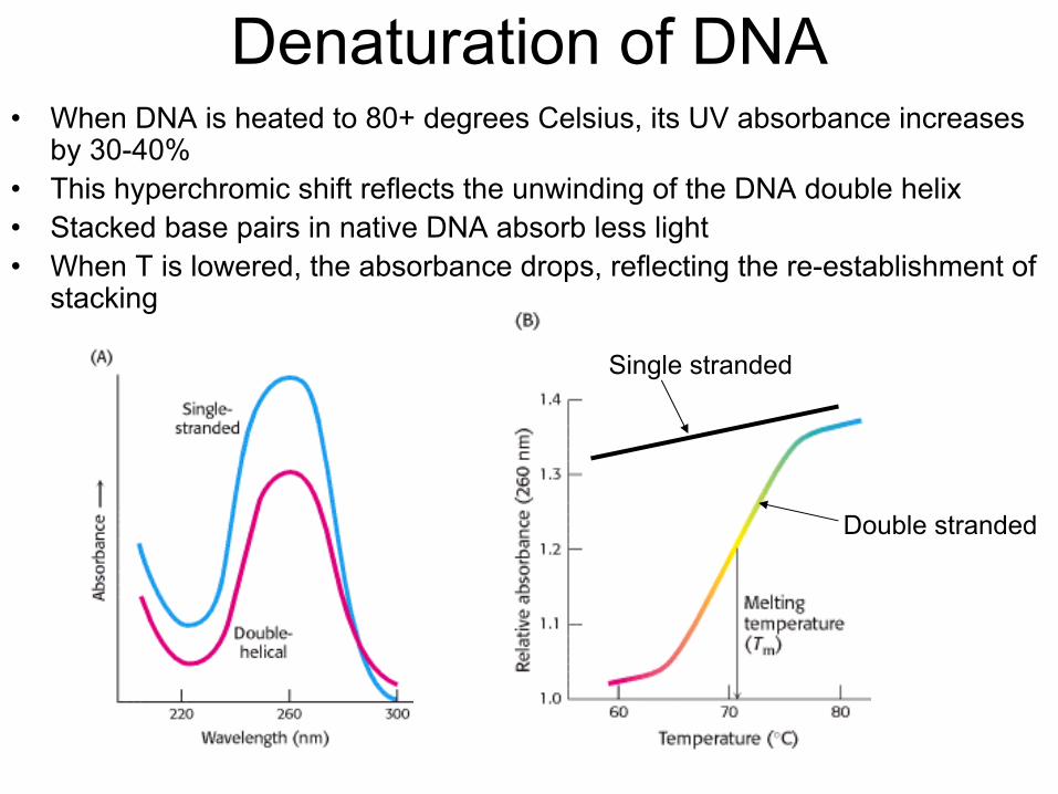
Denaturation
of DNA•
When DNA is heated to 80+ degrees Celsius, its UV absorbance increases by 30-40%
•
This hyperchromic
shift reflects the unwinding of the DNA double helix •
Stacked base pairs in native DNA absorb less light •
When T is lowered, the absorbance drops, reflecting the re-establishment of stacking
Single stranded
Double stranded

reannealing
Denaturation
of DNA

For long strands of DNATm = 69o + 0.41(%G+C)
Tm
Denaturation
of DNA –
GC content
The three H-bonds in a G-C base pair stabilize the DNA compared to the two H- bonds in an A-T base pair

Buoyant density of DNA is and index of GC content
η
= 1.660 + 0.098 (mole fraction GC)

Tertiary Structure of DNA
•
Length of E. coli DNA1.6 million nm, but E. coli cell is only 2000 nm long
Therefore,•
DNA needs to be compacted and folded

Supercoils
–
DNA tertiary structure•
In relaxed duplex DNA, ten bp
per turn of helix •
Circular DNA sometimes has more or less than 10 bp
per turn -
a supercoiled
state
relaxed
supercoiled
L –
linking numberT –
twists W –
writhes L = T + W
Overwinds
DNA helix
Unwinds DNA helix
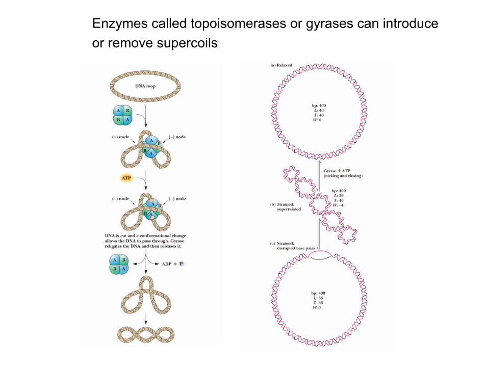
Enzymes called topoisomerases
or gyrases
can introduceor remove supercoils
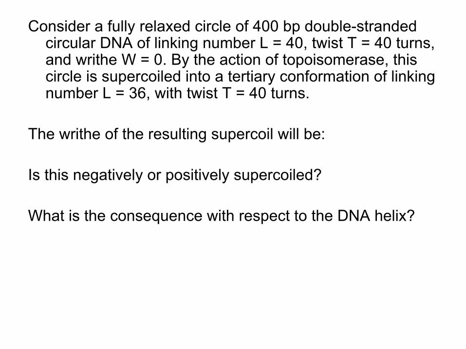
Consider a fully relaxed circle of 400 bp
double-stranded circular DNA of linking number L = 40, twist T = 40 turns, and writhe W = 0. By the action of topoisomerase, this circle is supercoiled
into a tertiary conformation of linking
number L = 36, with twist T = 40 turns.
The writhe of the resulting supercoil
will be:
Is this negatively or positively supercoiled?
What is the consequence with respect to the DNA helix?
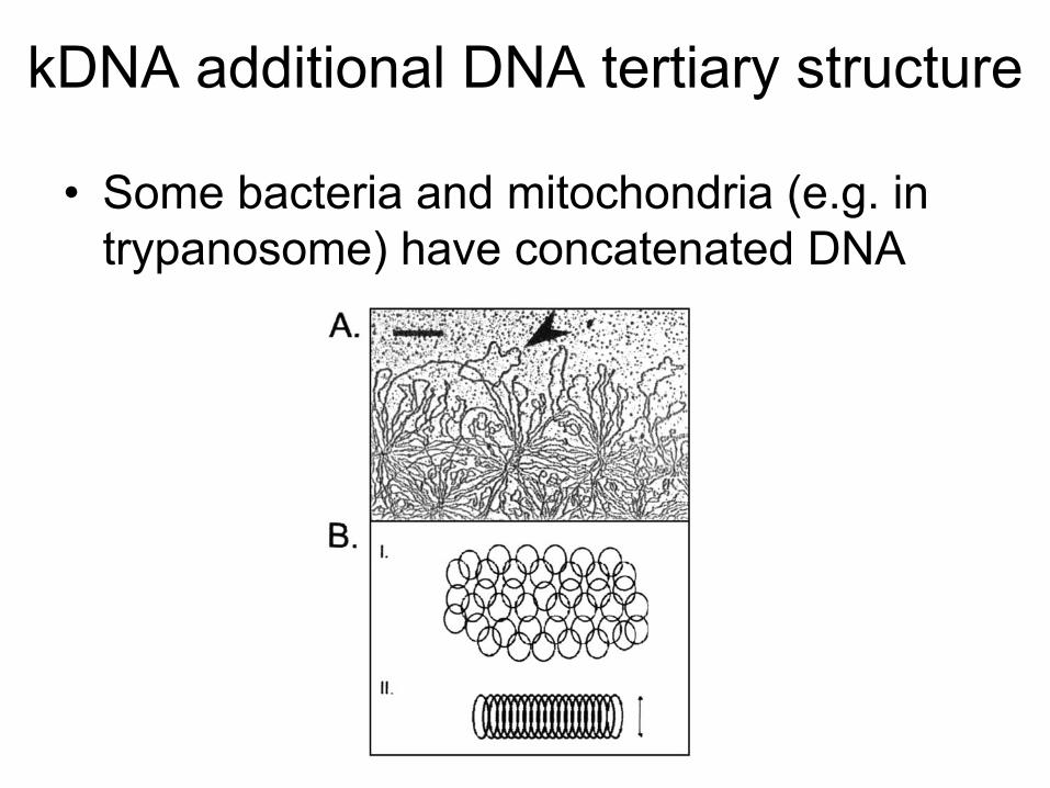
kDNA
additional DNA tertiary structure
•
Some bacteria and mitochondria (e.g. in trypanosome) have concatenated DNA

Chromosome Structure -
eukaryotes
•
Human DNA’s total length is ~2 meters!
•
This must be packaged into a nucleus that is about 5 micrometers in diameter
•
This represents a compression of more than 100,000!
•
It is made possible by wrapping the DNA around protein spools called nucleosomes
and then
packing these in helical filaments
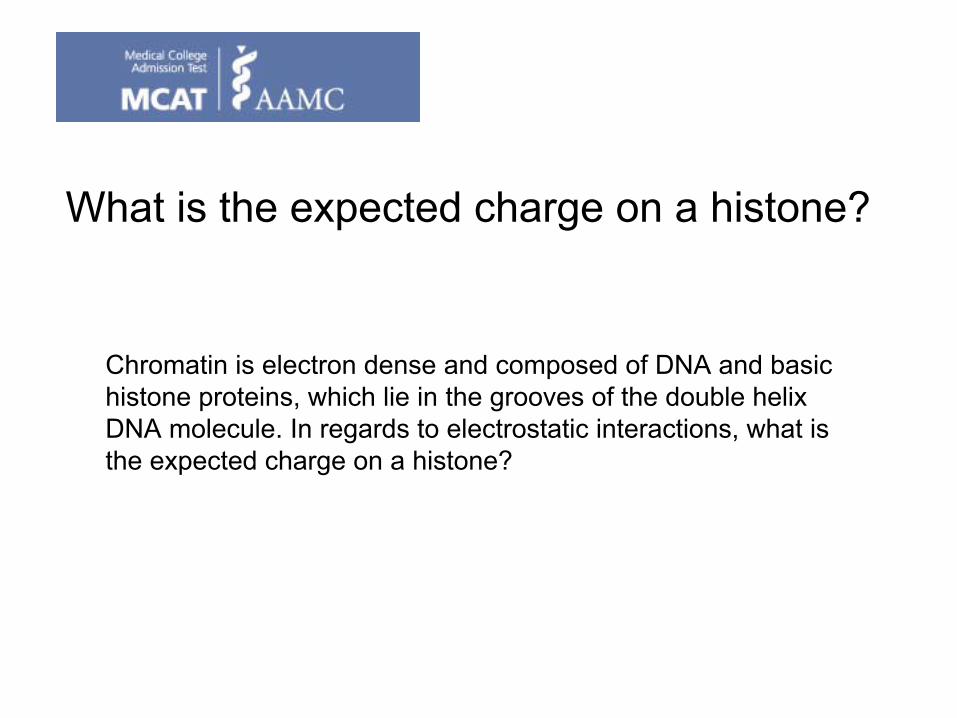
What is the expected charge on a histone?
Chromatin is electron dense and composed of DNA and basic histone
proteins, which lie in the grooves of the double helix DNA molecule. In regards to electrostatic interactions, what is the expected charge on a histone?

Post translational modification of DNA -methylation
Mammalians cells only have 5-
methylcytosine (m5C) -
≈
2 –
7%.It is proposed that methylation
of specific cytosine residues regulates gene expression
methylated
at adenines, N6-methyladenine (m6A), in “GATC”
sites
Higher-order eukaryotes
N4-methylcytosine (m4C). 5-methylcytosine (m5C)
Prokaryotes
Methylation
regulates DNA replication as well as protects DNA from DNA cleaving enzymes

Bacterial m6A methylation
–
additional control of replication
Methylation of the E. coli replication origin creates a refractory period for DNA initiation. DNA methylation
occurs at GATC sequences, 11 of which are found in the origin of replication.About 10 minutes after replication is initiated, the hemimethylated
origins become fully methylated
by a DNA methylase
enzyme.
The lag in methylation
after the replication of GATC sequences is also used by the E. coli mismatch proofreading system to distinguish the newly synthesized DNA strand from the parental DNA strand; in that case, the relevant GATC sequences are scattered
throughout the chromosome.

Correlation with aberrant DNA methylation
and cancer
•
Many tumor-suppressor and other cancer-related genes have been found to be hypermethylated
in human
cancer cells and primary tumors
•
The hypermethylation silences the genes
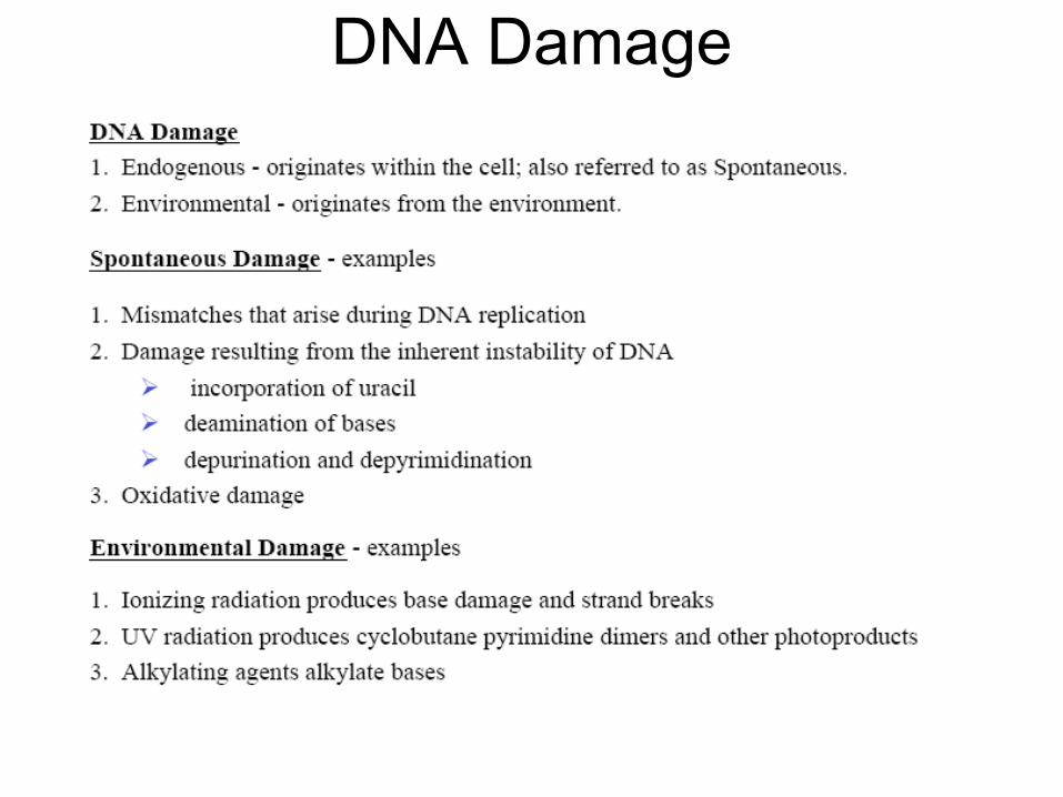
DNA Damage
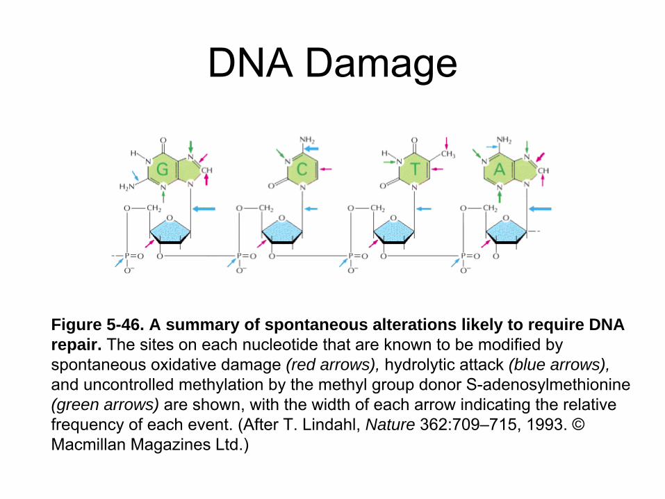
DNA Damage
Figure 5-46. A summary of spontaneous alterations likely to require DNA repair. The sites on each nucleotide that are known to be modified by spontaneous oxidative damage (red arrows), hydrolytic attack (blue arrows), and uncontrolled methylation
by the methyl group donor S-adenosylmethionine
(green arrows) are shown, with the width of each arrow indicating the relative
frequency of each event. (After T. Lindahl, Nature 362:709–715, 1993. ©
Macmillan Magazines Ltd.)
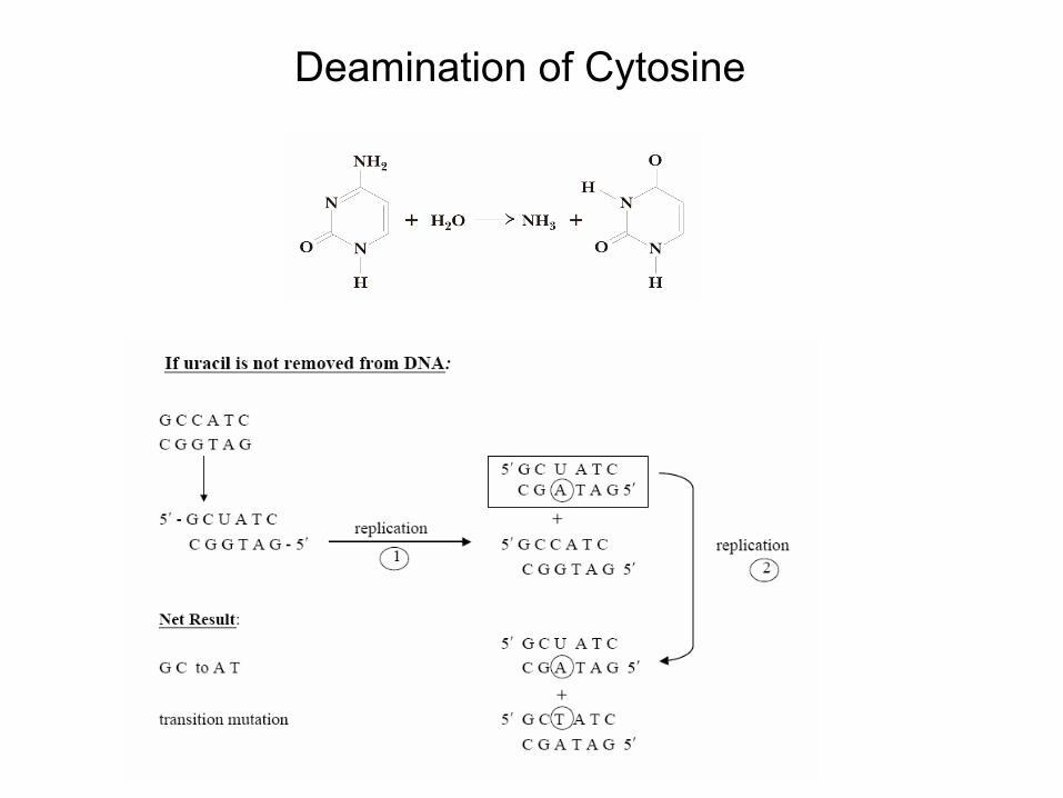
Deamination
of Cytosine

Thyamine
dimers

Nucleases•
Cleave nucleotide sequences
•
DNases
and RNases
and non specific nucleases
•
ss
and ds
specificity•
Exonucleases
(remove nucleotide from the
end)•
Endonucleases
(recognize palindromic
ds
DNA sequences)

Restriction endonucleases
•
Three types (I, II, and III) –
I and III require ATP
•
Type II are used as common molecular biology tools

Type II restriction enzymes
•
Recognize and cleave particular sequencesFor example, BamHIGGATCC
5’-N-N-N-N-G-G-A-T-C-C-N-N-N-N-3’3’-N-N-N-N-C-C-T-A-G-G-N-N-N-N-5’
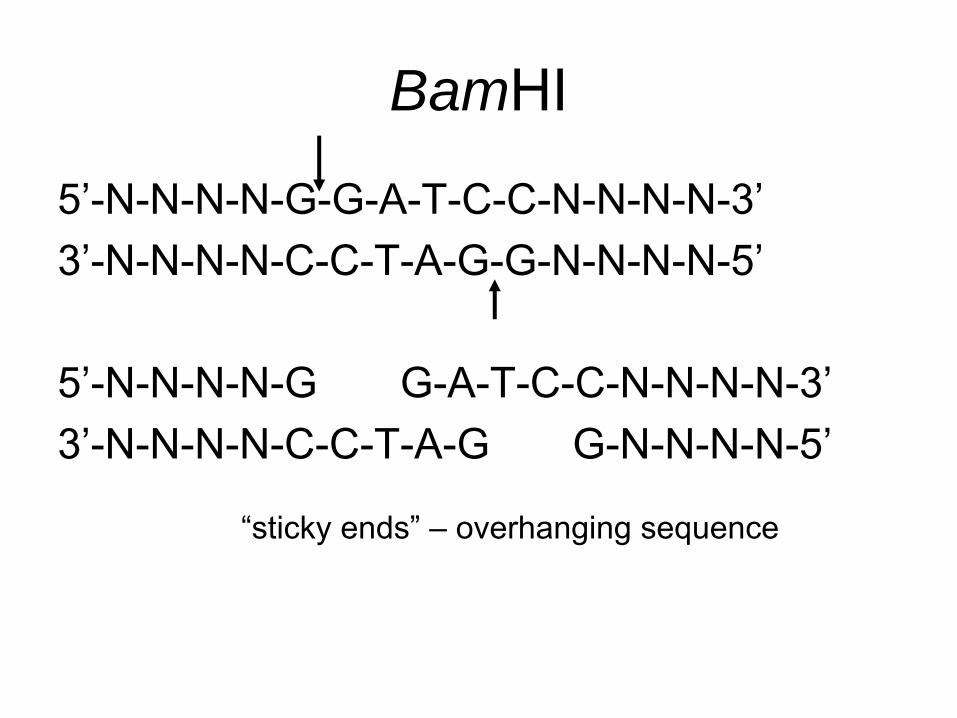
BamHI
5’-N-N-N-N-G-G-A-T-C-C-N-N-N-N-3’3’-N-N-N-N-C-C-T-A-G-G-N-N-N-N-5’
5’-N-N-N-N-G G-A-T-C-C-N-N-N-N-3’3’-N-N-N-N-C-C-T-A-G G-N-N-N-N-5’
“sticky ends”
–
overhanging sequence

Why do bacteria have endonucleases?
How do they avoid digesting their own DNA?
Overview•
DNA structure –
A, B, and Z DNA
•
DNA intercelators
and groove binders•
Thermal melting of DNA
•
DNA tertiary structure•
DNA methylation
•
DNA damage•
nucleases