lecture 1 introduction to general microbiology dr amin
TRANSCRIPT

Lecture 1
Introduction to General
Microbiology
Dr Amin Aqel
Associate Professor

What is a Microbe?
• Smaller than 0.1mm
• Includes viruses, protozoan, bacteria, small
suckers, others
Nomenclature
• Carolus Linnaeus (1735)
• Genus species
• By custom once mentioned can be
abbreviated with initial of genus followed
by specific epithet. E. coli

Why study Microbiology
• Microbes are related to all life.
– In all environments
– Many beneficial aspects
– Related to life processes (nutrient cycling)
– Only a minority are pathogenic.
– Most of our problems are caused by microbes

Classification
of
microorganisms

• Prokaryotes
• Peptidoglycan cell
walls
• Binary fission
• For energy, use
organic chemicals,
inorganic chemicals,
or photosynthesis
Bacteria
Figure 1.1a

• Prokaryotic
• Lack peptidoglycan
• Live in extreme
environments
• Include:
– Methanogens
– Extreme halophiles
– Extreme thermophiles
Archaea:
Halobacteria not
from book

• Eukaryotes
• Chitin cell walls
• Use organic chemicals for
energy
• Molds and mushrooms are
multicellular consisting of
masses of mycelia, which
are composed of filaments
called hyphae
• Yeasts are unicellular
Fungi
Figure 1.1b

• Eukaryotes
• Absorb or ingest
organic chemicals
• May be motile via
pseudopods, cilia, or
flagella
Protozoa
Figure 1.1c

• Eukaryotes
• Cellulose cell walls
• Use photosynthesis for
energy (primary
producers)
• Produce molecular
oxygen and organic
compounds
Algae
Figure 1.1d

• Acellular
• Consist of DNA or RNA
core
• Core is surrounded by a
protein coat
• Coat may be enclosed in a
lipid envelope
• Viruses are replicated
only when they are in a
living host cell
Viruses
Figure 1.1e

• Eukaryote
• Multicellular
animals
• Parasitic flatworms
and round worms
are called helminths.
• Microscopic stages
in life cycles.
Multicellular Animal Parasites
Figure fluke

Knowledge of microorganisms:
• Allows humans to
– Prevent food spoilage
– Prevent disease occurrence
– Others?
• Led to aseptic techniques to prevent
contamination in medicine and in
microbiology laboratories.

• The hypothesis that living organisms arise from
nonliving matter is called spontaneous
generation. According to spontaneous
generation, a “vital force’ forms life.
• The Alternative hypothesis, that the living
organisms arise from preexisting life, is called
biogenesis.
The Debate Over Spontaneous
Generation

Historical background of
Microbiology
• Some highlights
– 1665 Robert Hooke observed fruiting structures of molds and was the first to describe microorganisms
– 1673 van Leeuwenhoek’s microscopes
– 1735 Linnaeus Nomenclature
– 1798 Jenner vaccine
– 1857 Pasteur Fermentation
– 1876 Koch germ theory of disease

Lens
Adjustment

Van Leeuwenhoek’s Microscope

Van Leeuwenhoek’s
drawing on various
organsisms

The Golden Age of
Microbiology
• 1857-1914
• Beginning with Pasteur’s work,
discoveries included the relationship
between microbes and disease,
immunity, and antimicrobial drugs

• 1860s: Joseph Lister used a chemical disinfectant
to prevent surgical wound infections after
looking at Pasteur’s work showing microbes are
in the air, can spoil food, and cause animal
diseases.
• 1876: Robert Koch provided proof that a
bacterium causes anthrax and provided the
experimental steps, Koch’s postulates, used to
prove that a specific microbe causes a specific
disease.
The Germ Theory of Disease
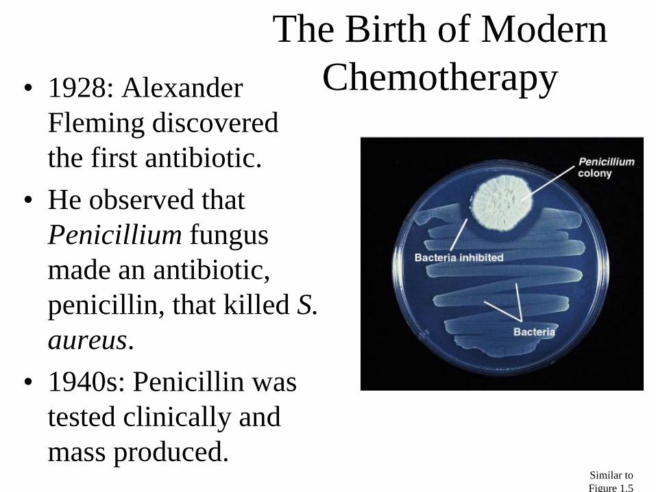
• 1928: Alexander
Fleming discovered
the first antibiotic.
• He observed that
Penicillium fungus
made an antibiotic,
penicillin, that killed S.
aureus.
• 1940s: Penicillin was
tested clinically and
mass produced.
The Birth of Modern
Chemotherapy
Similar to
Figure 1.5

• Bacteriology is the study of bacteria.
• Mycology is the study of fungi.
• Parasitology is the study of protozoa and
parasitic worms.
• Virology is the study of virus
• Recent advances in genomics, the study of an
organism’s genes, have provided new tools for
classifying microorganisms.
• Proteomics is looking at the gene products
Modern Developments in
Microbiology

• Taxonomy: the Science of Classification
– The science of Provides a classifying organisms
– Provides universal names for organisms
– reference for identifying organisms
– Groupings of organisms
– WHY Classify?
– Establish criteria for ID
– Arrange related organisms into groups
– Provide information about evolution of organisms
Taxonomy

Levels of Classification • Kingdom
• Division/Phyta/Phylum
• SubPhylum
• Class
• Order
• Family
• Genus
• Species/Specific Epithet
• Subspecies/Strain

1. Microscopic morphology
2. Macroscopic morphology – colony appearance
3. Physiological / biochemical characteristics
4. Chemical analysis
5. Serological analysis
6. Genetic and molecular analysis • G + C base composition
• DNA analysis using genetic probes
• Nucleic acid sequencing and rRNA analysis
24
Classification Systems in the Procaryotae

Identification Methods
• Morphological
characteristics: Useful for
identifying eukaryotes
• Differential staining:
Gram staining, acid-fast
staining
• Biochemical tests:
Determines presence of
bacterial enzymes
Figure 10.8

Lecture 2
Morphology / Bacterial Structures
Dr Amin Aqel

Morphology / Bacterial Structures

28

29

• Appendages
– two major groups of appendages:
• Motility – flagella and axial filaments (periplasmic flagella)
• Attachment or channels – fimbriae and pili
• Glycocalyx – surface coating
30
External Structures

• 3 parts:
– filament – long, thin, helical structure composed of protein flagellin
– hook- curved sheath
– basal body – stack of rings firmly anchored in cell wall
Flagellar Function -Functions in motility of cell through environment
-Guide bacteria in a direction in response to external stimulus:
31
Flagella

1. Monotrichous – single flagellum at one end
2. Lophotrichous – small bunches arising from one end of cell
3. Amphitrichous – flagella at both ends of cell
4. Peritrichous – flagella dispersed over surface of cell; slowest
Flagellar Arrangements

• Fine, proteinaceous, hairlike bristles from the cell surface
• Function in adhesion to other cells and surfaces
33
Fimbriae

• Rigid tubular structure made of pilin protein
• Found only in Gram negative cells
• Function to join bacterial cells for partial DNA transfer called conjugation
34
Pili

- Coating of molecules external to the cell wall, made of sugars and/or proteins
-Two types:
1-slime layer : loosely organized and attached
2-capsule : highly organized, tightly attached
- Functions: -protect cells from dehydration and
nutrient loss
-inhibit killing by white blood cells by phagocytosis contributing to pathogenicity
◦ attachment - formation of biofilms
35
Glycocalyx

• External covering outside the cytoplasm • Composed of two basic layers:
– cell wall and cell membrane
• Maintains cell integrity • Two generally different groups of bacteria
demonstrated by Gram stain: – Gram-positive bacteria: thick cell wall composed
primarily of peptidoglycan and cell membrane
– Gram-negative bacteria: outer cell membrane, thin peptidoglycan layer, and cell membrane
36
The Cell Envelope

37
Insert figure 4.12 Comparative cell envelopes

• Cell cytoplasm: – dense gelatinous solution of sugars, amino acids, and salts
– 70-80% water • serves as solvent for materials used in all cell functions
• Chromosome – single, circular, double-stranded DNA molecule that
contains all the genetic information required by a cell
– DNA is tightly coiled around a protein, aggregated in a dense area called the nucleoid
38
Bacterial Internal Structures

• Plasmids – small circular, double-stranded DNA – free or integrated into the chromosome – duplicated and passed on to offspring – not essential to bacterial growth and metabolism – may encode antibiotic resistance, tolerance to toxic
metals, enzymes and toxins – used in genetic engineering- readily manipulated
and transferred from cell to cell
39
Bacterial Internal Structures

• Ribosomes
– made of 60% ribosomal RNA and 40% protein
– consist of two subunits: large and small
– procaryotic differ from eucaryotic ribosomes in size and number of proteins
– site of protein synthesis
– present in all cells
40
Bacterial Internal Structures

• Inclusions and granules
– intracellular storage bodies
– vary in size, number and content
– Bacterial cell can use them when environmental sources are depleted.
– examples: glycogen, poly-b-hydroxybutyrate, gas vesicles for floating, sulfur and phosphate granules (metachromatic granules)
41
Bacterial Internal Structures

• Endospores – Inert ,resting, cells produced by some G+ genera:
Clostridium, Bacillus and Sporosarcina • have a 2-phase life cycle:
– vegetative cell – metabolically active and growing
– endospore – when exposed to adverse environmental conditions; capable of high resistance and very long-term survival
– sporulation : formation of endospores • hardiest of all life forms
• withstands extremes in heat, drying, freezing, radiation and chemicals
• not a means of reproduction
– germination- return to vegetative growth
42
Bacterial Internal Structures

43

Chapter 4
Shapes of Bacteria • Coccus
– Chain = Streptoccus
– Cluster = Staphylococcus
• Bacillus – Chain = Streptobacillus
• Coccobacillus
• Vibrio = curved
• Spirillum
• Spirochete
• Square
• Star

Chapter 4

Chapter 4

Lecture 3 Bacterial Physiology
Dr Amin Aqel

Bacterial
physiology

Cell Structure
• Two structural types of cells are recognized:
the prokaryote and the eukaryote.
Prokaryotic cells have a simpler internal
structure than eukaryotic cells, lacking
membrane-enclosed organelles.

Prokaryote cell Eukaryote cell

Cytoplasmic Membrane
The cytoplasmic membrane is a highly selective
permeability barrier constructed of lipids and proteins that
forms a bi-layer with hydrophilic exteriors and a
hydrophobic interior.

Movement of Molecules through
Cytoplasmic Membrane
• The major function of the cytoplasmic
membrane is to act as a permeability barrier,
preventing leakage of cytoplasmic metabolites
into the environment.
• Several ways for molecules to move through
membrane
1. Simple Diffusion
2. Osmosis
3. Facilitated Diffusion
4. Active Transport

Simple Diffusion
• Does not require expenditure of energy
• Process by which some molecules move
freely into and out of the cell
• Small molecules such as carbon dioxide and
oxygen

Microbiology: An Introduction, 9e
by Tortora, Funke, Case
Copyright © 2007 Pearson Education, Inc.,
publishing as Benjamin Cummings.
Figure 4.17: Facilitated diffusion.
Transported
substance Transporter
protein
Outside
Inside
Glucose
Plasma
membrane
Transport proteins (or transporters) responsible for:
Facilitated Diffusion, Active Transport

Cell Wall
• Gram-negative Bacteria have only a few
layers of peptidoglycan , but Gram-positive
Bacteria have several layers.
• In addition to peptidoglycan, gram-negative
Bacteria contain an outer membrane
consisting of lipopolysaccharide (LPS),
protein, and lipoprotein.


Bacterial growth

Bacterial growth curve
• Lag phase
• Exponential phase
• Stationary phase
• Death phase

The Growth Cycle
• Microorganisms show a characteristic growth pattern
when inoculated into a fresh culture medium.

Factors affecting growth
1- Temperature
2- pH
3- Salinity
4- Oxygen
5- Nutrition
6- Osmotic Pressure

Temperature
• Temperature is a major environmental factor
controlling microbial growth. The cardinal
temperatures are the minimum, optimum,
and maximum temperatures at which each
organism grows.


Microorganisms can be grouped by the
temperature ranges they require.

Low or High pH
• The acidity or alkalinity of an environment
can greatly affect microbial growth.
• Organisms that grow best at low pH are
called acidophiles; those that grow best at
high pH are called alkaliphiles.

Salinity
• Some microorganisms (halophiles) have evolved
to grow best at reduced water potential, and some
(extreme halophiles) even require high levels of
salts for growth.

Oxygen
• Aerobes require oxygen to live, whereas
anaerobes do not and may even be killed by
oxygen.
• Facultative: organisms can live with or
without oxygen.
•Aerotolerant anaerobes: can tolerate oxygen
and grow in its presence even though they
cannot use it.
•*Microaerophiles: are aerobes that can use
oxygen only when it is present at levels reduced
from that in air.

Bacterial metabolism
Catabolism: substrate breakdown and conversation into
usable energy
*Anabolism: synthesis of cellular constituents (cell wall,
proteins,fatty acids, nucleic acids
• Bacterial growth requires; a source of energy & raw
materials
* To build the proteins, structures and membranes
* That make up the structure and biochemical machines of
the cell
• Bacteria should obtain or synthesize:
- aminoacids, carbohydrates, lipids as building blocks of
the cell

The minimum requirement for
growth
• Carbon
• Nitrogen
• Energy source
• Water
• Various ions