lead optimization of 2-cyclohexyl-n-[(z)-(3...
TRANSCRIPT

Molecules 2015, 20, 18246-18263; doi:10.3390/molecules201018246
molecules ISSN 1420-3049
www.mdpi.com/journal/molecules
Article
Lead Optimization of 2-Cyclohexyl-N-[(Z)-(3-methoxyphenyl/3-hydroxyphenyl) methylidene]hydrazinecarbothioamides for Targeting the HER-2 Overexpressed Breast Cancer Cell Line SKBr-3
Mashooq A. Bhat 1,*, Abdullah Al-Dhfyan 2,3, Ahmed M. Naglah 4,5, Azmat Ali Khan 1 and
Mohamed A. Al-Omar 4
1 Department of Pharmaceutical Chemistry, College of Pharmacy, King Saud University, P.O. Box
2457, Riyadh 11451, Saudi Arabia; E-Mail: [email protected] (A.A.K.) 2 Stem Cell & Tissue Re-Engineering Program, Research Center, King Faisal Specialized Hospital &
Research Center, MBC-03, P. O. Box 3354, Riyadh 11211, Saudi Arabia;
E-Mail: [email protected] 3 Department of Pharmacology and Toxicology, College of Pharmacy, King Saud University,
Riyadh 11451, Saudi Arabia 4 Department of Pharmaceutical Chemistry, Drug Exploration & Development Chair (DEDC),
College of Pharmacy, King Saud University, Riyadh 11451, Saudi Arabia;
E-Mails: [email protected] (A.M.N.); [email protected] (M.A.A.-O.) 5 Peptide Chemistry Department, Chemical Industries Research Division, National Research Centre,
Dokki, Cairo 12622, Egypt
* Author to whom correspondence should be addressed; E-Mail: [email protected];
Tel.: +966-1-4677-343; Fax: +966-1-4676-220.
Academic Editor: Jean Jacques Vanden Eynde
Received: 19 June 2015 / Accepted: 2 October 2015 / Published: 7 October 2015
Abstract: Lead derivatives of 2-cyclohexyl-N-[(Z)-(3-methoxyphenyl/3-hydroxyphenyl)
methylidene]hydrazinecarbothioamides 1–18 were synthesized, characterized and evaluated in vitro against HER-2 overexpressed breast cancer cell line SKBr-3. All the compounds showed
activity against HER-2 overexpressed SKBr-3 cells with IC50 = 17.44 ± 0.01 µM to 53.29 ±
0.33 µM. (2Z)-2-(3-Hydroxybenzylidene)-N-(3-methoxyphenyl)hydrazinecarbothioamide
(12, IC50 = 17.44 ± 0.01 µM) was found to be most potent compound of this series targeting
HER-2 overexpressed breast cancer cells compared to the standard drug 5-fluorouracil
OPEN ACCESS

Molecules 2015, 20 18247
(5-FU) (IC50 = 38.58 ± 0.04 µM). Compound 12 inhibited the cellular proliferation via
DNA degradation.
Keywords: thiosemicarbazones; HER-2; SKBr-3 cells; BT-474 cells; cancer stem cells
1. Introduction
Thiosemicarbazones constitute an important class of pharmacophore that has been explored by
medicinal chemists owing to its wide range of biological activities, which include antibacterial,
antimalarial, antiviral and antitumor activities [1–3]. Methisazone which was used for the treatment of
smallpox, is an example of a thiosemicarbazone drug [4]. Thiosemicarbazone derivatives having potent
anticancer activities have been reported in the literature [5–8]. Triapine, a potent anti-proliferative is
effective against several cancer types. It obstructs tumor growth by inhibiting ribonucleotide reductase.
Triapine (3-AP, 3-aminopyridine-2-carboxaldehyde thiosemicarbazone) has shown significant
usefulness in the treatment of cancer and is currently in phase II clinical trials [9]. The therapeutic
potential of 3-AP is however limited because of its poor water solubility and toxicity profile.
Worldwide the most common cancer and the second most leading cause of mortality in women is
breast cancer, despite improvements in early detection. Recently the existence of breast cancer stem cells
has been reported. Because of their resistance to conventional treatment, small populations of cells that
are relatively resistant to therapy and able to repopulate in vivo, called cancer stem cells (CSCs), are
believed to be responsible for treatment failures. CSCs have fundamental implications for the early
detection, prevention and treatment of cancer. Drug discovery programs for cancer usually select
compounds which have the property of inducing cytotoxic effects in cancer cell lines [10].
Unfortunately, the cytotoxic effects in vitro and inhibition of tumor growth in vivo is not the end story
for curing cancer in preclinical models because of the existence of CSCs, as tumors are maintained by a
self-renewing CSC population [11]. There are now various research findings which have confirmed
cancer stem cells in leukemia [12], breast [13], brain [14], lung [15], colon [16]. To cause relapse, CSCs
must have survived primary treatment [17]. Studies suggested that aldehyde dehydrogenase 1 (ALDH-1)
is a more potent marker of breast CSCs [18–20]. ALDH-1-positive cells are resistant to conventional
chemotherapy with paclitaxel and epirubicin [21]. ALDH-1-positive breast cancer CSCs can induce
tumor formation with a few as 500 cells. Breast cancer cells that expressed ALDH-1 were more likely
to be estrogen receptor (ER) negative, progesterone receptor (PR) negative and human-epidermal growth
factor receptor-2 (HER-2) positive. Several reports demonstrated that HER-2 regulate CSCs. Cells
displaying stem cell properties such as sphere formation or increased aldehyde dehydrogenase
expression also have increased HER-2 expression compared with bulk cell population [22]. Traditionally
breast cancer can be classified into three main subtypes: luminal, basal like and human epidermal growth
factor receptor-2 positive (HER-2)+. Clinical and laboratory evidences have indicated that
overexpression of HER-2 may render tumor cells resistant to several anticancer drugs [23]. Thus, there
remains an urgent need for new pharmaceutical compounds and compositions to effectively eradicate
and target cancer stem cells. We need to target both proliferating cells as well as cancer stem cells in
order to cure cancer [24].
Molecules 2015, 20 18248
Therefore there is high potential in structural modification of thiosemicarbazone (TSC) derivatives to
improve the existing drug candidates. In our previous research on TSC derivatives bearing a cyclohexyl
moiety, the synthesized compounds showed activity against HER-2 expressed SKBr-3 cells with
IC50 = 25.6 ± 0.07 µM − 61.6 ± 0.4 µM. The two compounds (2-cyclohexyl-N-[(Z)-(3-methoxyphenyl)
methylidene]hydrazinecarbothioamide (C2) and (2-cyclohexyl-N-[(Z)-(3-hydroxyphenyl)-
methylidene]hydrazinecarbothioamide (C10) (Figure 1) showed inhibitory effects on ALDH+
population more effectively than the reference drug, salinomycin. Compound C2 was the most active,
showing a 50% inhibitory effect [25]. These two compounds C2 and C10 were selected as lead
compounds for further derivatization. Modulation of the cyclohexyl moiety was performed using
different groups like phenyl, substituted phenyl, benzyl, alkyl, alkenyl, etc. In order to discover novel
TSC derivatives with significant activity against CSCs. TCS derivatives bearing 3-hydroxy/3-methoxy
phenyl on one side and different groups at the terminal nitrogen were synthesized and their antitumor
activities on SKBr-3 and BT-474 cell line were determined.
Figure 1. Structures of lead compound C2, C10 and newly synthesized compounds 1–18.
2. Results and Discussion
2.1. Chemistry
The synthesis of thiosemicarbazone derivatives 1–18 was carried out in single step as shown in
Scheme 1. Thiosemicarbazides were reacted with 3-methoxybenzaldehyde/3-hydroxybenzaldehyde in
the presence of acetic acid to yield 2-cyclohexyl-N-[(Z)-(3-methoxyphenyl/3-hydroxyphenyl)
methylidene]hydrazinecarbothioamide derivatives 1–18. The purity of the compounds was checked by
TLC and elemental analysis. The compounds were characterized and confirmed by spectral data. In the 1H-NMR spectra, the signals of the respective protons of the derivatives were verified on the basis of
chemical shifts, multiplicity and coupling constants. The spectra of all synthesized compounds showed
a D2O exchangeable singlet at 6.3–8.3 ppm and 11.1–11.7 ppm corresponding to NH protons and
NH NH
S
NR
R1
NH NH
S
NR
modulation of cyclohexyl moiety
C2: R= OCH3; C10: R= OH
R1= -C6H5, 4-Cl-C6H4, -C6H5CH2, -CH2-CH=CH2,
-CH2CH3, 3-OCH3-C6H4, 4-OCH3-C6H4, 4-OC2H5-C6H4.
C2, C10
(1-18)

Molecules 2015, 20 18249
NHC=S protons. The analytical and spectral data of the compounds was in good agreement with the
composition of the synthesized compounds. The physiochemical property data of the of all compounds
is given in Table 1.
R1
NH NH
S
N R
R = 3-O C H 3-C 6H 4; 3-O H -C 6H 4
R1
NH NH
S
NH2
+ RCHOCH3COOH
C2H5OH/H2O
R 1= -C 6H 5, 4-C l-C 6H 4, -C 6H 5C H 2, -C H 2-C H =C H 2, -C H 2C H 3, 3-O C H 3-C 6H 4,
4-O C H 3-C 6H 4, 4-O C 2H 5-C 6H 4.
(1-18)
Scheme 1. Synthetic protocol of compounds 1–18.
Table 1. Physical data of the synthesized compounds 1–18.
Compounds R R1 Molecular Formula Yield % Mp (°C) CLog P
1 3-Methoxyphenyl Phenyl C15H15N3OS 70 161–162 4.42 2 3-Methoxyphenyl 4-Chlorophenyl C15H14ClN3OS 75 197–198 5.41 3 3-Methoxyphenyl Prop-2-en C12H15N3OS 72 115–116 2.70 4 3-Methoxyphenyl Benzyl C16H17N3OS 68 127–129 3.65 5 3-Methoxyphenyl Ethyl C11H15N3OS 72 122–123 2.40 6 3-Methoxyphenyl 3-Methoxyphenyl C16H17N3O2S 65 148–150 4.68 7 3-Methoxyphenyl 4-Methoxyphenyl C16H17N3O2S 70 156–157 4.37 8 3-Methoxyphenyl 4-Ethoxyphenyl C17H19N3O2S 68 160–161 4.91 9 3-Methoxyphenyl 3-Chlorophenyl C15H14ClN3OS 70 138–140 5.45 10 3-Hydroxyphenyl Phenyl C14H13N3OS 78 212–213 3.95 11 3-Hydroxyphenyl 4-Chlorophenyl C14H12ClN3OS 65 222–224 4.94 12 3-Hydroxyphenyl 3-Methoxyphenyl C15H15N3O2S 70 185–186 4.21 13 3-Hydroxyphenyl Prop-2-en C11H13N3OS 60 94–97 2.22 14 3-Hydroxyphenyl Benzyl C15H15N3OS 65 130–132 3.17 15 3-Hydroxyphenyl 4-Methoxyphenyl C15H15N3O2S 72 182–183 3.90 16 3-Hydroxyphenyl 4-Ethoxyphenyl C16H17N3O2S 70 200–202 4.43 17 3-Hydroxyphenyl 3-Chlorophenyl C14H12ClN3OS 75 198–200 4.98 18 3-Hydroxyphenyl Ethyl C10H13N3OS 60 152–154 1.93
2.2. Anti-Proliferative in Vitro Activity
In vitro anti-proliferative activity was measured by the cell growth inhibition assay. For the
determination of IC50 for each compound, WST-1 reagent was used according to the protocol (Table 2).
From our previous experience, TCS derivatives showed selectivity against HER-2 overexpressed cancer
cells over luminal and basal subtypes. All the compounds showed activity against HER-2 overexpressed
SKBr-3 cell with IC50 values ranging between 17.44 ± 0.01 µM to 53.29 ± 0.33 µM. Compound 12
(IC50 = 17.44 ± 0.01 µM) was found to be most potent compound of this series targeting HER-2
overexpressed breast cancer cells compared to the standard drug 5-fluorouracil (5-FU) (IC50 = 38.58 ±

Molecules 2015, 20 18250
0.04 µM). To gain insight into the anti-proliferation mechanism, the effect on cell cycle distribution was
investigated by fluorescence-activated cell sorting (FACS) analysis. SKBr-3 cells were exposed to
10 µM of compound 12 for 48 h and the result was the accumulation of the cells on DNA degradation
phase, which is a strong indication that the treatment induced apoptosis by breakdown of the cells’ DNA.
This was also accompanied by a compensatory decrease in G1, S and M phase cells. Histograms show
the number of cells per channel (vertical axis) vs. DNA content (horizontal axis). The values indicate the
percentage of cells in the relevant phases of the cell cycle. The analysis shows increase in apoptosis of
cells (DNA degradation) by 8 folds compared with untreated cells (Figure 2).
Table 2. In vitro cytotoxic activity of compounds against breast cancer cell line SKBr-3.
Compounds a IC50 (µM)
SKBr-3 1 45.67 ± 0.18 2 23.69 ± 0.06 3 17.89 ± 0.03 4 34.52 ± 0.18 5 23.00 ± 0.16 6 24.55 ± 0.02 7 25.00 ± 0.03 8 23.76 ± 0.01 9 27.00 ± 0.03 10 22.32 ± 0.02 11 26.49 ± 0.02 12 17.44 ± 0.01 13 29.26 ± 0.05 14 27.74 ± 0.03 15 53.29 ± 0.33 16 24.75 ± 0.08 17 18.28 ± 0.05 18 22.33 ± 0.03
5-FU 38.58 ± 0.04 a IC50: Concentration of the compound (μM) producing 50% cell growth inhibition after 48 h
of compound exposure.
These results suggested that compound 12 inhibited the cellular proliferation via DNA degradation.
The apoptotic effect of compound 12 was evaluated using annexin-V staining. Cells were harvested after
treatment by compound 12 (10 µM) for 48 h and incubated with annexin V-FITC and PI as described in
the Experimental Section. Ten thousand cells were analyzed per determination. Dot plots show annexin
V-FITC binding on the X axis and PI staining on the Y axis. Dots represent cells as follows: lower left
quadrant, normal cells (FITC−/PI−); lower right quadrant, apoptotic cells (FITC+/PI−); upper left
quadrant, necrotic cells (FITC−/PI+), upper right quadrant late apoptotic (FITC+/PI+) (Figure 3).

Molecules 2015, 20 18251
Figure 2. Cell cycle specific blocking activity of compound 12 on SKBr-3 cells.
Figure 3. The apoptotic effect of compound 12 using Annexin-V staining of SKBr-3 cells.
The apoptotic population increased from 16.3% in the control group to 49.14% and late
apoptosis population increased from 10% to 26% increased after 48 h exposure time using 10 µM of
compound 12 for incubation. The cytotoxic effect of compound was not correlated to the increase of
necrotic cell population.
The most active compound 12 presented apoptotic effects on overexpressed HER-2 oncogene and
little sensitivity was measured on HER-2 negative MDA-MB-231 cells (Figures 4 and 5). Cell adhesion
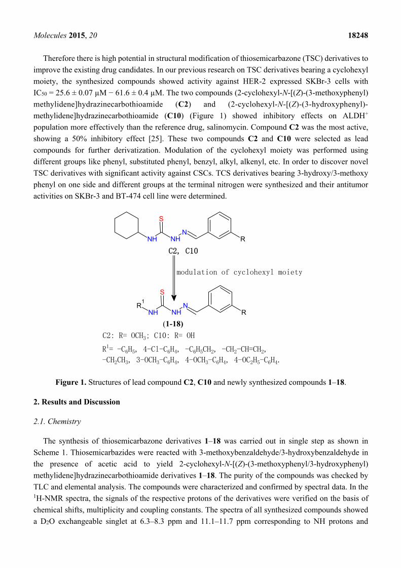
inhibition studies were also carried out using the HER-2 positive SKBr-3 and BT-474 cancer cell lines.
Compound 12 at tested concentrations significantly inhibited cell adhesion of SKBr-3 and BT-474
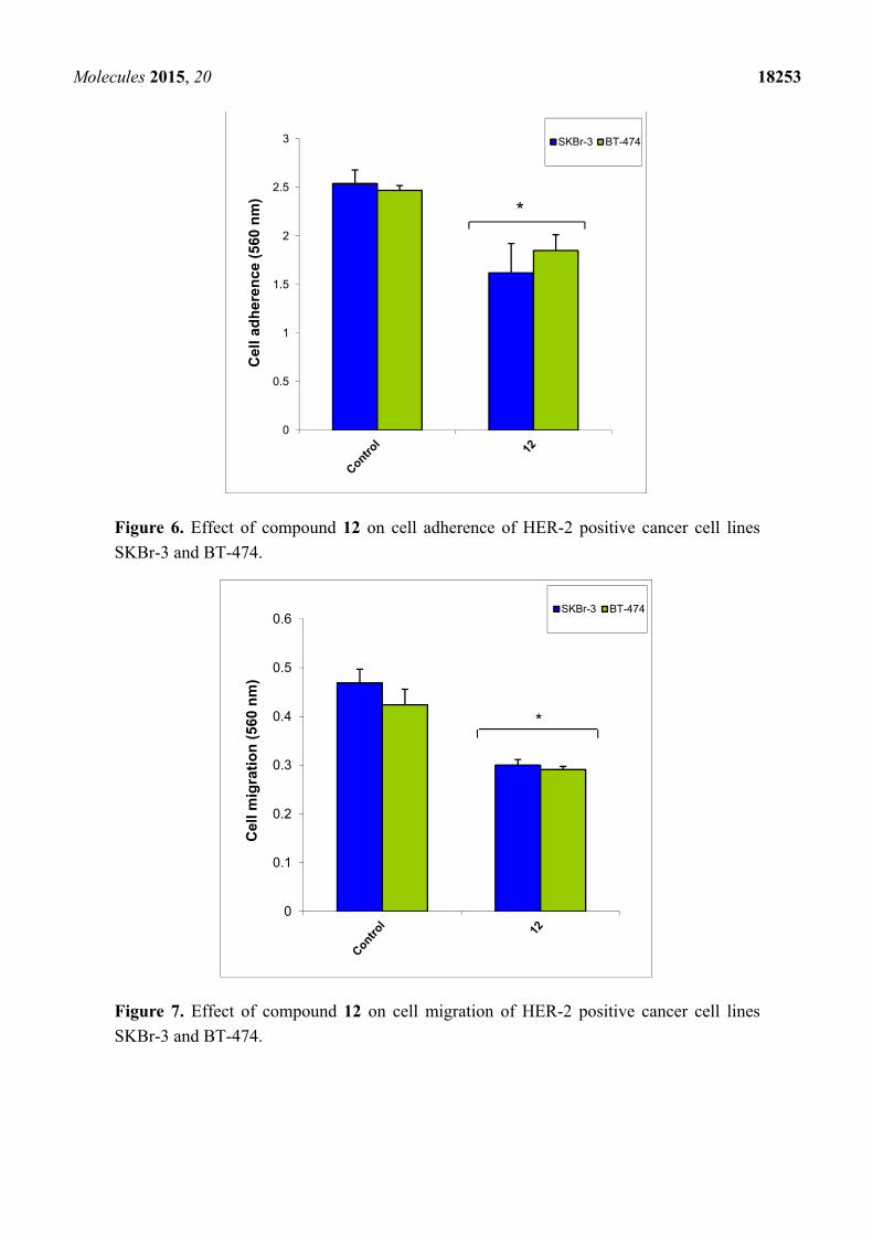
(p < 0.05), (Figure 6). The results shown in Figure 7 demonstrate that compound 12 had a maximum
effect on cell migration of SKBr-3 and BT-474 cancer cells. It significantly inhibited cell migration of
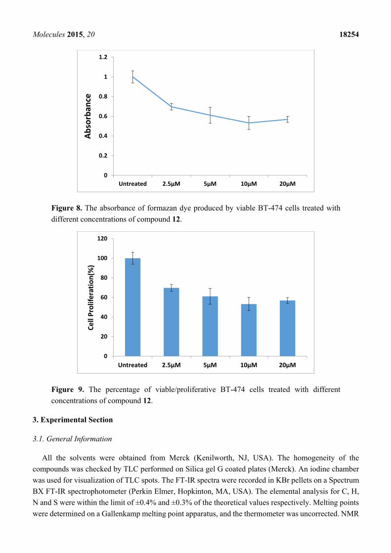
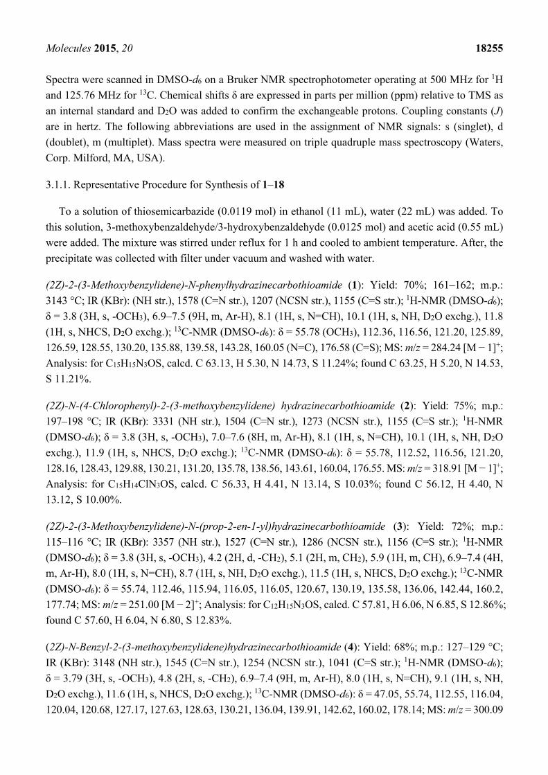
SKBr-3 and BT-474 (p < 0.05). Percentages of viable/proliferative BT-474 cells treated with different
concentration of compound 12 were determined (Figures 8 and 9). Cell proliferation inhibition was found
to be significant at 10 µM concentration of compound 12.

Molecules 2015, 20 18252
Figure 4. The apoptotic effect of compound 12 on HER-2 positive BT-474 and Her-2
negative MDA-MB-231 cells
Figure 5. Histogram showing the % apoptosis of compound 12 on HER-2 negative
MDA-MB-231 cells and HER-2 positive BT-474.
0
5
10
15
20
25
30
35
40
45
50
Untreated C12 Untreated C12
Apop
tosi
s (%
)
MDA-MB-231 BT-474
P=0.0007
Molecules 2015, 20 18253
Figure 6. Effect of compound 12 on cell adherence of HER-2 positive cancer cell lines
SKBr-3 and BT-474.
Figure 7. Effect of compound 12 on cell migration of HER-2 positive cancer cell lines
SKBr-3 and BT-474.
0
0.5
1
1.5
2
2.5
3
Cel
l ad
her
ence
(56
0 n
m)
SKBr-3 BT-474
*
0
0.1
0.2
0.3
0.4
0.5
0.6
Cel
l mig
rati
on
(56
0 n
m)
SKBr-3 BT-474
*
Molecules 2015, 20 18254
Figure 8. The absorbance of formazan dye produced by viable BT-474 cells treated with
different concentrations of compound 12.
Figure 9. The percentage of viable/proliferative BT-474 cells treated with different
concentrations of compound 12.
3. Experimental Section
3.1. General Information
All the solvents were obtained from Merck (Kenilworth, NJ, USA). The homogeneity of the
compounds was checked by TLC performed on Silica gel G coated plates (Merck). An iodine chamber
was used for visualization of TLC spots. The FT-IR spectra were recorded in KBr pellets on a Spectrum
BX FT-IR spectrophotometer (Perkin Elmer, Hopkinton, MA, USA). The elemental analysis for C, H,
N and S were within the limit of ±0.4% and ±0.3% of the theoretical values respectively. Melting points
were determined on a Gallenkamp melting point apparatus, and the thermometer was uncorrected. NMR
0
0.2
0.4
0.6
0.8
1
1.2
Untreated 2.5µM 5µM 10µM 20µM
Abso
rban
ce
0
20
40
60
80
100
120
Untreated 2.5µM 5µM 10µM 20µM
Cell
Prol
ifera
tion(
%)
Molecules 2015, 20 18255
Spectra were scanned in DMSO-d6 on a Bruker NMR spectrophotometer operating at 500 MHz for 1H
and 125.76 MHz for 13C. Chemical shifts δ are expressed in parts per million (ppm) relative to TMS as
an internal standard and D2O was added to confirm the exchangeable protons. Coupling constants (J)
are in hertz. The following abbreviations are used in the assignment of NMR signals: s (singlet), d
(doublet), m (multiplet). Mass spectra were measured on triple quadruple mass spectroscopy (Waters,
Corp. Milford, MA, USA).
3.1.1. Representative Procedure for Synthesis of 1–18
To a solution of thiosemicarbazide (0.0119 mol) in ethanol (11 mL), water (22 mL) was added. To
this solution, 3-methoxybenzaldehyde/3-hydroxybenzaldehyde (0.0125 mol) and acetic acid (0.55 mL)
were added. The mixture was stirred under reflux for 1 h and cooled to ambient temperature. After, the
precipitate was collected with filter under vacuum and washed with water.
(2Z)-2-(3-Methoxybenzylidene)-N-phenylhydrazinecarbothioamide (1): Yield: 70%; 161–162; m.p.:
3143 °C; IR (KBr): (NH str.), 1578 (C=N str.), 1207 (NCSN str.), 1155 (C=S str.); 1H-NMR (DMSO-d6);
δ = 3.8 (3H, s, -OCH3), 6.9–7.5 (9H, m, Ar-H), 8.1 (1H, s, N=CH), 10.1 (1H, s, NH, D2O exchg.), 11.8
(1H, s, NHCS, D2O exchg.); 13C-NMR (DMSO-d6): δ = 55.78 (OCH3), 112.36, 116.56, 121.20, 125.89,
126.59, 128.55, 130.20, 135.88, 139.58, 143.28, 160.05 (N=C), 176.58 (C=S); MS: m/z = 284.24 [M − 1]+;
Analysis: for C15H15N3OS, calcd. C 63.13, H 5.30, N 14.73, S 11.24%; found C 63.25, H 5.20, N 14.53,
S 11.21%.
(2Z)-N-(4-Chlorophenyl)-2-(3-methoxybenzylidene) hydrazinecarbothioamide (2): Yield: 75%; m.p.:
197–198 °C; IR (KBr): 3331 (NH str.), 1504 (C=N str.), 1273 (NCSN str.), 1155 (C=S str.); 1H-NMR
(DMSO-d6); δ = 3.8 (3H, s, -OCH3), 7.0–7.6 (8H, m, Ar-H), 8.1 (1H, s, N=CH), 10.1 (1H, s, NH, D2O
exchg.), 11.9 (1H, s, NHCS, D2O exchg.); 13C-NMR (DMSO-d6): δ = 55.78, 112.52, 116.56, 121.20,
128.16, 128.43, 129.88, 130.21, 131.20, 135.78, 138.56, 143.61, 160.04, 176.55. MS: m/z = 318.91 [M − 1]+;
Analysis: for C15H14ClN3OS, calcd. C 56.33, H 4.41, N 13.14, S 10.03%; found C 56.12, H 4.40, N
13.12, S 10.00%.
(2Z)-2-(3-Methoxybenzylidene)-N-(prop-2-en-1-yl)hydrazinecarbothioamide (3): Yield: 72%; m.p.:
115–116 °C; IR (KBr): 3357 (NH str.), 1527 (C=N str.), 1286 (NCSN str.), 1156 (C=S str.); 1H-NMR
(DMSO-d6); δ = 3.8 (3H, s, -OCH3), 4.2 (2H, d, -CH2), 5.1 (2H, m, CH2), 5.9 (1H, m, CH), 6.9–7.4 (4H,
m, Ar-H), 8.0 (1H, s, N=CH), 8.7 (1H, s, NH, D2O exchg.), 11.5 (1H, s, NHCS, D2O exchg.); 13C-NMR
(DMSO-d6): δ = 55.74, 112.46, 115.94, 116.05, 116.05, 120.67, 130.19, 135.58, 136.06, 142.44, 160.2,
177.74; MS: m/z = 251.00 [M − 2]+; Analysis: for C12H15N3OS, calcd. C 57.81, H 6.06, N 6.85, S 12.86%;
found C 57.60, H 6.04, N 6.80, S 12.83%.
(2Z)-N-Benzyl-2-(3-methoxybenzylidene)hydrazinecarbothioamide (4): Yield: 68%; m.p.: 127–129 °C;
IR (KBr): 3148 (NH str.), 1545 (C=N str.), 1254 (NCSN str.), 1041 (C=S str.); 1H-NMR (DMSO-d6);
δ = 3.79 (3H, s, -OCH3), 4.8 (2H, s, -CH2), 6.9–7.4 (9H, m, Ar-H), 8.0 (1H, s, N=CH), 9.1 (1H, s, NH,
D2O exchg.), 11.6 (1H, s, NHCS, D2O exchg.); 13C-NMR (DMSO-d6): δ = 47.05, 55.74, 112.55, 116.04,
120.04, 120.68, 127.17, 127.63, 128.63, 130.21, 136.04, 139.91, 142.62, 160.02, 178.14; MS: m/z = 300.09

Molecules 2015, 20 18256
[M + 1]+; Analysis: for C16H17N3OS, calcd. C 62.92, H 7.59, N 3.76, S 10.50%; found C 62.87, H 7.57,
N 3.75, S 10.53%.
(2Z)-N-Ethyl-2-(3-methoxybenzylidene)hydrazinecarbothioamide (5): Yield: 72%; m.p.: 122–123 °C;
IR (KBr): 3351 (NH str.), 1544 (C=N str.), 1272 (NCSN str.), 1153 (C=S str.); 1H-NMR (DMSO-d6);
δ = 1.15 (3H, t, -CH3), 3.6 (2H, q, CH2), 3.8 (3H, s, -OCH3), 6.9–7.3 (4H, m, Ar-H), 8.0 (1H, s, N=CH),
8.5 (1H, s, NH, D2O exchg.), 11.4 (1H, s, NHCS, D2O exchg.); 13C-NMR (DMSO-d6): δ = 15.07, 55.72,
112.35, 116.05, 120.62, 130.21, 136.07, 142.22, 160.01, 177.16; MS: m/z = 237.07 [M]+; Analysis: for
C11H15N3OS, calcd. C 55.67, H 6.37, N 17.71, S 13.51%; found C 55.87, H 6.35, N 17.73, S 13.48%.
(2Z)-2-(3-Methoxybenzylidene)-N-(3-methoxyphenyl)hydrazinecarbothioamide (6): Yield: 65%; m.p.:
148–150 °C; IR (KBr): 3308 (NH str.), 1599 (C=N str.), 1286 (NCSN str.), 1156 (C=S str.); 1H-NMR
(DMSO-d6); δ = 3.8 (6H, s, -OCH3), 6.9–7.5 (8H, m, Ar-H), 8.1 (1H, s, N=CH), 10.0 (1H, s, NH, D2O
exchg.), 11.8 (1H, s, NHCS, D2O exchg.); 13C-NMR (DMSO-d6): δ = 55.77, 104.15, 107.40, 110.66,
111.98, 112.44, 115.42, 116.55, 118.48, 121.17, 129.88, 130.69, 134.06, 135.83, 140.66, 141.79, 143.35,
148.55, 159.49, 160.11, 165.69, 176.31. MS: m/z = 315.44 [M]+; Analysis: for C16H17N3O2S, calcd. C
60.93, H 5.43, N 3.32, S 10.17%; found C 60.70, H 5.40, N 3.30, S 10.15%.
(2Z)-2-(3-Methoxybenzylidene)-N-(4-methoxyphenyl)hydrazinecarbothioamide (7): Yield: 70%; m.p.:
156–157 °C; IR (KBr): 3304 (NH str.), 1542 (C=N str.), 1236 (NCSN str.), 1152 (C=S str.); 1H-NMR
(DMSO-d6); δ = 3.8 (6H, s, -OCH3), 6.9–7.5 (8H, m, Ar-H), 8.1 (1H, s, N=CH), 10.0 (1H, s, NH, D2O
exchg.), 11.7 (1H, s, NHCS, D2O exchg.); 13C-NMR (DMSO-d6): δ = 55.69, 55.71, 55.78, 112.27,
113.76, 114.11, 116.50, 121.17, 126.58, 128.27, 130.18, 132.48, 132.71, 135.96, 143.03, 156.99, 157.51,
160.06, 176.98, 180.69; MS: m/z = 315.00 [M]+; Analysis: for C16H17N3O2S, calcd. C 60.93, H 5.43, N
13.32, S 10.17%; found C 60.71, H 5.42, N 13.34, S 10.15%.
(2Z)-N-(4-Ethoxyphenyl)-2-(3-methoxybenzylidene)hydrazinecarbothioamide (8): Yield: 68%; m.p.:
160–161 °C; IR (KBr): 3155 (NH str.), 1540 (C=N str.), 1274 (NCSN str.), 1115 (C=S str.); 1H-NMR
(DMSO-d6); δ = 1.3 (3H, t, -CH3), 3.8 (3H, s, -OCH3), 4.0 (2H, q, -CH2), 6.9–7.5 (8H, m, Ar-H), 8.1
(1H, s, N=CH), 10.0 (1H, s, NH, D2O exchg.), 11.7 (1H, s, NHCS, D2O exchg.); 13C-NMR (DMSO-d6):
δ = 15.16, 55.78, 63.63, 112.23, 114.23, 114.59, 116.51, 121.17, 128.24, 130.17, 132.35, 135.96, 142.99,
156.77, 160.05, 176.93; MS: m/z = 329.23 [M]+; Analysis: for C17H19N3O2S, calcd. C 61.98, H 5.81, N
12.76, S 9.73%; found C 61.75, H 5.84, N 12.74, S 9.75%.
(2Z)-N-(3-Chlorophenyl)-2-(3-methoxybenzylidene)hydrazinecarbothioamide (9): Yield: 70%; m.p.:
138–140 °C; IR (KBr): 3421 (NH str.), 1545 (C=N str.), 1269 (NCSN str.), 1152 (C=S str.); 1H-NMR
(DMSO-d6); δ = 3.8 (3H, s, -OCH3), 7.0–7.7 (8H, m, Ar-H), 8.1 (1H, s, N=CH), 10.1 (1H, s, NH, D2O
exchg.), 11.9 (1H, s, NHCS, D2O exchg.); 13C-NMR (DMSO-d6): δ = 55.78, 112.63, 116.57, 121.20,
124.88, 125.52, 125.84, 130.04, 130.21, 132.65, 135.74, 141.06, 143.80, 160.05, 176.40; MS: m/z =
319.00 [M]+; Analysis: for C15H14ClN3OS, calcd. C 56.33, H 4.41, N 13.14, S 10.03%; found C 56.50,
H 4.40, N 13.15, S 10.00%.

Molecules 2015, 20 18257
(2Z)-2-(3-Hydroxybenzylidene)-N-phenylhydrazinecarbothioamide (10): Yield: 78%; m.p.: 212–213 °C;
IR (KBr): 3311 (NH str.), 1542 (C=N str.), 1275 (NCSN str.), 1163 (C=S str.); 1H-NMR (DMSO-d6);
δ = 6.8–7.6 (9H, m, Ar-H), 8.1 (1H, s, N=CH), 9.5 (1H, s, -OH, D2O exchg.), 10.1 (1H, s, NH, D2O
exchg.), 11.7 (1H, s, NHCS, D2O exchg.); 13C-NMR (DMSO-d6): δ = 114.31, 117.78, 119.39, 125.74,
126.19, 128.31, 128.53, 128.75, 130.13, 135.76, 139.56, 143.69, 158.08, 176.43; MS: m/z = 265.30
[M − 6]+; Analysis: for C14H13N3OS, calcd. C 61.97, H 4.83, N 15.49, S 11.82%; found C 61.97, H 4.83,
N 15.49, S 11.82%.
(2Z)-N-(4-Chlorophenyl)-2-(3-hydroxybenzylidene)hydrazinecarbothioamide (11): Yield: 65%; m.p.:
222–224 °C; IR (KBr): 3277 (NH str.), 1591 (C=N str.), 1276 (NCSN str.), 1196 (C=S str.); 1H-NMR
(DMSO-d6); δ = 6.8–7.6 (9H, m, Ar-H), 8.0 (1H, s, N=CH), 9.5 (1H, s, -OH, D2O exchg.), 10.1 (1H, s,
NH, D2O exchg.), 11.8 (1H, s, NHCS, D2O exchg.); 13C-NMR (DMSO-d6): δ = 114.35, 117.81, 119.41,
127.86, 128.39, 129.70, 130.11, 135.66, 138.57, 144.00, 158.07, 176.41; MS: m/z = 304.13 [M − 1]+;
Analysis: for C14H12ClN3OS, calcd. C 54.99, H 3.96, N 13.74, S 10.49%; found C 54.78, H 3.95, N
13.73, S 10.47%.
(2Z)-2-(3-Hydroxybenzylidene)-N-(3-methoxyphenyl)hydrazinecarbothioamide (12): Yield: 70%; m.p.:
185–186 °C; IR (KBr): 3312 (NH str.), 1546 (C=N str.), 1277 (NCSN str.), 1155 (C=S str.); 1H-NMR
(DMSO-d6); δ = 3.8 (6H, s, 2×-OCH3), 6.5–7.5 (8H, m, Ar-H), 8.1 (1H, s, N=CH), 9.6 (1H, s, -OH, D2O
exchg.), 10.0 (1H, s, NH, D2O exchg.), 11.8 (1H, s, NHCS, D2O exchg.); 13C-NMR (DMSO-d6):
δ = 55.62, 111.16, 11.58, 114.29, 115.11, 117.81, 118.08, 119.41, 121.59, 129.22, 130.14, 130.77,
135.71, 138.13, 140.65, 143.76, 158.08, 159.48, 176.14; MS: m/z = 302.30 [M + 1]+; Analysis: for
C15H15N3O2S, calcd. C 59.78, H 5.02, N 13.94, S 10.64%; found C 59.57, H 5.00, N 13.96, S 10.62%.
(2Z)-2-(3-Hydroxybenzylidene)-N-(prop-2-en-1-yl)hydrazinecarbothioamide (13): Yield: 60%; m.p.:
94–97 °C; IR (KBr): 3357 (NH str.), 1585 (C=N str.), 1222 (NCSN str.), 1144 (C=S str.); 1H-NMR
(DMSO-d6); δ = 4.2 (2H, d, -CH2), 5.1 (2H, m, =CH2), 5.9 (1H, m, =CH), 6.9–7.2 (4H, m, Ar-H), 7.9
(1H, s, N=CH), 8.6 (1H, s, -OH, D2O exchg.), 9.6 (1H, s, NH, D2O exchg.), 11.4 (1H, s, NHCS, D2O
exchg.); 13C-NMR (DMSO-d6): δ = 46.19, 113.80, 116.03, 117.60, 119.14, 130.17, 135.42, 135.85,
142.98, 157.97, 177.59; MS: m/z = 235.12 [M]+; Analysis: for C11H13N3OS, calcd. C 56.15, H 5.57, N
17.86, S 13.63%; found C 56.26, H 5.55, N 17.87, S 13.60%.
(2Z)-N-Benzyl-2-(3-hydroxybenzylidene)hydrazinecarbothioamide (14): Yield: 65%; m.p.: 130–132 °C;
IR (KBr): 3360 (NH str.), 1551 (C=N str.), 1301 (NCSN str.), 1147 (C=S str.); 1H-NMR (DMSO-d6);
δ = 4.8 (2H, s, -CH2), 6.8–7.3 (9H, m, Ar-H), 8.0 (1H, s, N=CH), 9.0 (1H, s, -OH, D2O exchg.), 9.6 (1H,
s, NH, D2O exchg.), 11.5 (1H, s, NHCS, D2O exchg.); 13C-NMR (DMSO-d6): δ = 18.99, 47.05, 56.55,
114.00, 117.58, 119.10, 127.20, 127.67, 128.64, 130.13, 135.90, 139.90, 143.09, 158.03, 177.99; MS: m/z = 285 [M]+; Analysis: for C15H15N3OS, calcd. C 63.13, H 5.30, N 4.73, S 11.24%; found C 63.36,
H 5.32, N 4.75, S 11.22%.
(2Z)-2-(3-Hydroxybenzylidene)-N-(4-methoxyphenyl)hydrazinecarbothioamide (15): Yield: 72%; m.p.:
182–183 °C; IR (KBr): 3169 (NH str.), 1581 (C=N str.), 1283 (NCSN str.), 1022 (C=S str.); 1H-NMR
(DMSO-d6); δ = 3.7 (6H, s, 2× -OCH3), 6.8–7.4 (8H, m, Ar-H), 8.0 (1H, s, N=CH), 9.5 (1H, s, -OH,

Molecules 2015, 20 18258
D2O exchg.), 9.9 (1H, s, NH, D2O exchg.), 11.7 (1H, s, NHCS, D2O exchg.); 13C-NMR (DMSO-d6):
δ = 55.69, 113.72, 114.11, 114.28, 114.66, 117.66, 118.86, 119.31, 126.58, 127.96, 130.09, 132.09,
132.45, 132.70, 135.84, 143.39, 156.99, 157.39, 158.06, 176.79, 180.67; MS: m/z = 302.49 [M + 1]+;
Analysis: for C15H15N3O2S, calcd. C 59.78, H 5.02, N 13.94, S 10.64%; found C 59.55, H 5.00, N 13.92,
S 10.66%.
(2Z)-N-(3-Methoxyphenyl)-2-(3-hydroxybenzylidene)hydrazinecarbothioamide (16): Yield: 70%; m.p.:
200–202 °C; IR (KBr): 3138 (NH str.), 1510 (C=N str.), 1280 (NCSN str.), 1039 (C=S str.); 1H-NMR
(DMSO-d6); δ = 3.8 (3H, s, -OCH3), 4.0 (2H, q, -CH2), 6.9–7.5 (8H, m, Ar-H), 8.1 (1H, s, N=CH), 9.5
(1H, s, -OH, D2O exchg.), 10.0 (1H, s, NH, D2O exchg.), 11.7 (1H, s, NHCS, D2O exchg.); 13C-NMR
(DMSO-d6): δ = 15.16, 55.78, 112.23, 114.23, 114.59, 116.51, 121.17, 128.24, 130.17, 132.35, 135.96,
142.99, 156.77, 160.05, 176.93; MS: m/z = 315.10 [M]+; Analysis: for C16H17N3O2S, calcd. C 60.93, H
5.43, N 13.32, S 10.17%; found C 60.70, H 5.41, N 13.30, S 10.14%.
(2Z)-N-(3-Chlorophenyl)-2-(3-hydroxybenzylidene)hydrazinecarbothioamide (17): Yield: 75%; m.p.:
198–200 °C; IR (KBr): 3281 (NH str.), 1589 (C=N str.), 1276 (NCSN str.), 1172 (C=S str.); 1H-NMR
(DMSO-d6); δ = 6.8–7.7 (8H, m, Ar-H), 8.1 (1H, s, N=CH), 9.5 (1H, s, -OH, D2O exchg.), 10.1 (1H, s,
NH, D2O exchg.), 11.9 (1H, s, NHCS, D2O exchg.); 13C-NMR (DMSO-d6): δ = 114.38, 117.88, 119.46,
124.56, 125.37, 125.54, 130.02, 130.12, 132.60, 135.61, 141.06, 144.18, 158.07, 176.24; MS: m/z = 304.58
[M − 1]+; Analysis: for C14H12ClN3OS, calcd. C 54.99, H 3.96, N 13.74, S 10.49%; found C 54.76, H
3.94, N 13.76, S 10.46%.
(2Z)-N-Ethyl-2-(3-hydroxybenzylidene)hydrazinecarbothioamide (18): Yield: 60%; m.p.: 152–154 °C;
IR (KBr): 3358 (NH str.), 1557 (C=N str.), 1239 (NCSN str.), 1168 (C=S str.); 1H-NMR (DMSO-d6); δ
= 1.15 (3H, t, CH3), 3.6 (2H, q, CH2), 6.8–7.2 (4H, m, Ar-H), 7.4 (1H, s, N=CH), 8.4 (1H, s, NH, D2O
exchg.), 9.5 (1H, s, -OH, D2O exchg.), 11.3 (1H, s, NHCS, D2O exchg.); 13C-NMR (DMSO-d6):
δ = 15.07, 113.96, 117.46, 118.95, 130.09, 135.97, 142.60, 158.04, 177.10; MS: m/z = 223.18 [M]+;
Analysis: for C10H13N3OS, calcd. C 53.79, H 5.87, N 8.82, S 14.36%; found C 53.58, H 5.85, N 8.84,
S 14.34%.
3.2. Cell Lines
SKBr-3, BT-474 and MDA-MB-231 breast cancer cell lines were purchased from the American Type
Culture Collection (0801 University Boulevard, Manassas, VA, USA). SKBR-3 cells were cultured in
McCoy’s 5A (GIBCO, 8717, Grovement Cir, Gaitherberg, MD, USA), and BT-474, MDA-MB-231 cells
were cultured in DMEM (Sigma, 82024 Taufkirchen, Germany). The media supplemented with 10%
FBS (Cambrex Bio Science, Baltimore, MD, USA), 100 IU/mL penicillin and 100 mg/mL streptomycin.
Cell viability was assessed by trypan blue exclusion analysis. Cell numbers were determined by using a
hemacytometer.
3.2.1. WST-1 Cell Proliferation Assay
Cells were seeded into 96-well plates at 0.4 × 10⁴/well and incubated overnight. The medium was
replaced with fresh one containing the desired concentrations of the compounds. After 48 h, 10 μL of

Molecules 2015, 20 18259
the WST-1 reagent was added to each well and the plates were re-incubated for 4 h at 37 °C. The amount
of formazan was quantified using an ELISA reader at 450 nm.
3.2.2. Measurement of IC50
Cells were seeded into 96-well plates at 0.4 × 10⁴/well and incubated overnight. The medium was
replaced with fresh one containing the desired concentrations of the compounds. After 48 h, 10 μL of
the WST-1 reagent was added to each well and the plates were re-incubated for 4 h at 37 °C. The amount
of formazan was quantified using an ELISA reader at 450 nm. For the compounds and the reference
chemotherapeutic agent 5-FU, cells were cultured one day before treatment. Fresh media with fixed dose
of 20 µM were replaced. IC50 was mathematically calculated as IC50 = fixed dose (20) × 50/(formazan
quantity of treated cells/formazan quantity of untreated cells) × 100.
3.2.3. Flow Cytometric Analysis of Cellular DNA Content
2 × 10⁶ cells were fixed in 1 mL ethanol (70%) for 60 min at room temperature. Harvested cells were
re-suspended in 1 mL Na citrate (50 mM) containing 250 μg RNase A and incubated at 50 °C for 60 min.
Next, cells were re-suspended in the same buffer containing 4 μg propidium iodine (PI) and incubated
for 30 min before being analyzed by flow cytometry (Becton Dickinson, San Jose, CA, USA). The
percentage of cells in various cell cycle phases was determined by using Cell Quest Pro software
(Becton Dickinson).
3.2.4. Measurement of Annexin-V Binding by Flow Cytometry
It has been shown that loss of phospholipid asymmetry of the plasma membrane is an early event of
apoptosis. The annexin-V binds to negatively charged phospholipids, like phosphatidylserine. During
apoptosis, the cells react to annexin-V once chromatin condenses but before the plasma membrane loses
its ability to exclude propidium iodide (PI). Hence, by staining cells with a combination of fluorescein
isothiocyanate (FITC) annexin-V and PI, it is possible to detect non-apoptotic live cells, early apoptotic
cells and late apoptotic or necrotic cells. Annexin-V staining was performed by using Vybrant Apoptosis
Assay Kit # 2 (Molecular Probe, Eugene, Oregon, 97402-0469) following the manufacturer’s
recommendations. Annexin-V stained cells were analyzed by flow cytometry, measuring the fluorescence
emission at 530 and less than 575 nm.
3.2.5. Cancer Cell Migration Assay
Cell migration assay was performed according to the standard protocol. Three concentration 25 µM
of compound was taken for testing. The lower well of the migration plate was supplemented with 500 μL
of DMEM containing 10% fetal bovine serum with or without (vehicle control ethanol only) test
compound. To the inside of each insert 100 μL of 0.5–1.0 × 10⁶ cells/mL of SKBr-3/BT-474 cell
suspension was added separately. The plates were incubated for 8 h at 37 °C in a humidified CO2
incubator. After incubation, the media from inside of the inserts was carefully aspirated and the
non-migratory cells were removed using cotton-tipped swabs. The inserts were transferred to a clean
well containing 400 μL of cell stain solution and incubated for 10 min at room temperature. The stained

Molecules 2015, 20 18260
inserts were gently washed several times and then transferred again to an empty well. Finally, 200 μL of
extraction solution per well was added and incubated for 10 min on an orbital shaker. From each sample,
100 μL was taken in a 96-well microtiter plate and the absorbance at 560 nm was read in a plate reader.
3.2.6. Cancer Cell Adhesion Assay
Under sterile conditions, the adhesion plate was allowed to warm up at room temperature for 10 min.
150 μL of 0.1–1.0 × 10⁶ cells/mL of SKBr-3/BT-474 cell suspension in serum free media with vehicle
control ethanol only or test compound (25 µM) was added to the inside of each well. The plates were
incubated for 30–90 min in a CO2 incubator. The wells were washed three times with PBS and the
adhered cells were stained with 200 μL of cell stain solution for 10 min at room temperature. The excess
stain was removed by washing 4–5 times with distilled water. After air drying the wells, 200 μL of
extraction solution per well was added and then incubated for 10 min on an orbital shaker. The 150 μL
from each extracted sample was transferred to a 96-well microtiter plate and the absorbance at 560 nm
was read in a plate reader. Absorbance of dye in the control (vehicle-treated) cells was regarded as 100%
adherence and the percentage adherence of treated cells was calculated in comparison with that of the
control cells. Cell Migration and Cell Adhesion Assay kits were obtained from Cell Biolabs, Inc. (San
Diego, CA, USA).
3.2.7. The Effect of Different Concentration of Compound 12 on BT-474 Cells Proliferation
The cytotoxic effects of the compound 12 on BT-474 cell line was assayed by the MTT assay. The
cells were seeded at a density of 5 × 10⁴ cells/well. The compound was serially diluted to final
concentration of 20 µM, 10 µM, 5 µM and 2.5 µM. 200 μL liquid of each concentration was applied to
the wells of a 96-well plate containing confluent cell monolayers (six wells per concentration). The
dilution medium without the sample served as a control. After 48 h of incubation, MTT solution
(5 mg/mL) was then added to each well, and the formazan precipitate was dissolved in 200 μL dimethyl
sulfoxide after 4 h incubation. The content of the wells was homogenized on a microplate shaker for
5 min. The optical densities (OD) were measured on a microplate ELISA reader at 492 nm. All tests and
analyses were run in triplicate and mean values were recorded. The cell survival curves were calculated
after comparing with the control. The percentage viability was calculated as follows:
%viability = mean absorbance of treated wells × 100 mean absorbance of untreated wells
4. Conclusions
In conclusion, we focused on the design and synthesis of 2-cyclohexyl-N-[(Z)-(3-methoxyphenyl/3-
hydroxyphenyl)methylidene]hydrazinecarbothioamides 1–18, which were fully characterized by
spectral analysis. The synthesized compounds were screened in vitro against HER-2 overexpressed
breast cancer cell lines SKBr-3. Compound 12 presented the most significant activity against HER-2
over expressed breast cancer cell lines SKBr-3 and BT-474. Compound 12 significantly inhibited the
cell migration and cell adhesion of breast cancer cell lines. Compound 12 was found to most active
compound of this series and represents a good lead for development of drugs, targeting HER-2 over-
expressed breast cancer cell lines.

Molecules 2015, 20 18261
Acknowledgments
The authors would like to extend their sincere appreciation to the Deanship of Scientific Research at
King Saud University for funding this Research group No. (RG 1435-006).
Author Contributions
Design and synthesis of the compounds was performed by MA Bhat; A Al-Dhfyan performed the
anti-proliferative activity, AM Naglah contributed in the spectral analysis of the compounds, AA Khan
performed the cell adhesion and cell migration assays, MA Al-Omar helped in the preparation of
the manuscript.
Conflicts of Interest
The authors declare no conflict of interest.
References
1. Pelosi, G. Thiosemicarbazone metal complexes: from structure to activity. Open. Crystallogr. J. 2010, 3, 16–28.
2. Dilović, I.; Rubcić, M.; Vrdoljak, V.; Pavelić, S.K.; Kralj, M.; Piantanida, I.; Cindrić, M. Novel
thiosemicarbazone derivatives as potential antitumor agents: Synthesis, physicochemical and
structural properties, DNA interactions and antiproliferative activity. Bioorg. Med. Chem. 2008, 16,
5189–5198.
3. Kovacevic, Z.; Chikhani, S.; Lui, G.Y.; Sivagurunathan, S.; Richardson, D.R. The iron-regulated
metastasis suppressor NDRG1 targets NEDD4L, PTEN, and SMAD4 and inhibits the PI3K and Ras
signaling pathways. Antioxid. Redox Signal. 2013, 10, 874–887.
4. Heiner, G.G.; Fatima, N.; Russell, P.K.; Haase, A.T.; Ahmad, N.; Mohammed, N.; Thomas, D.B.;
Mack, T.M.; Khan, M.M.; Knatterud, G.L.; et al. Field trials of methisazone as a prophylactic agent
against smallpox. Am. J. Epidemiol. 1971, 94, 435–449.
5. Jutten, P.; Schumann, W.; Hartl, A.; Dahse, H.M.; Grafe, U. Thiosemicarbazones of formyl benzoic
acids as novel potent inhibitors of estrone sulfatase. J. Med. Chem. 2007, 50, 3661–3666.
6. Yogeeswari, P.; Sriram, D.; Thirumurugan, R.; Raghavendran, J.V.; Sudhan, K.; Pavana, R.K.;
Stables, J. Discovery of N-(2,6-dimethylphenyl)-substituted semicarbazones as anticonvulsants:
Hybrid pharmacophore-based design. J. Med. Chem. 2005, 48, 6202–6211.
7. Greenbaum, D.C.; Mackey, Z.; Hansell, E.; Doyle, P.; Gut, J.; Caffrey, C.R.; Lehrman, J.; Rosenthal,
P.J.; McKerrow, J.H.; Chibale, K. Synthesis and structure-activity relationships of parasiticidal
thiosemicarbazone cysteine protease inhibitors against Plasmodium falciparum, Trypanosoma
brucei, and Trypanosoma cruzi. J. Med. Chem. 2004, 47, 3212–3219.
8. Neve, R.M.; Chin, K.; Fridlyand, J.; Yeh, J.; Baehner, F.L.; Fevr, T.; Clark, L.; Bayani, N.;
Coppe, J.P.; Tong, F.; et al. A collection of breast cancer cell lines for the study of functionally
distinct cancer subtypes. Cancer Cell 2006, 10, 515–527.

Molecules 2015, 20 18262
9. Finch, R.A.; Liu, M.; Grill, S.P.; Rose, W.C.; Loomis, R.; Vasquez, K.M.; Cheng, Y.; Sartorelli, A.C.
Triapine (3-aminopyridine-2-carboxaldehyde- thiosemicarbazone): A potent inhibitor of ribonucleotide
reductase activity with broad spectrum antitumor activity. Biochem. Pharmacol. 2000, 59, 983–991.
10. Winquist, R.J.; Furey, B.F.; Boucher, D.M. Cancer stem cells as the relevant biomass for drug
discovery. Curr. Opin. Pharmacol. 2010, 10, 385–390.
11. McDermott, S.P.; Wicha, M.S. Targeting breast cancer stem cells. Mol. Oncol. 2010, 4, 404–419.
12. Bonnet, D.; Dick, J.E. Human acute myeloid leukemia is organized as a hierarchy that originates
from a primitive hematopoietic cell. Nat. Med. 1997, 3, 730–737.
13. Al-Hajj, M.; Wicha, M.S.; Benito-Hernandez, A.; Morrison, S.J.; Clarke, M.F. Prospective
identification of tumorigenic breast cancer cells. Proc. Natl. Acad. Sci. USA 2003, 100, 3983–3988.
14. Singh, S.K.; Clarke, I.D.; Terasaki, M.; Bonn, V.E.; Hawkins, C.; Squire, J.; Dirks, P.B.
Identification of a cancer stem cell in human brain tumors. Cancer Res. 2003, 63, 5821–5828.
15. Ho, M.M.; Ng, A.V.; Lam, S.; Hung, J.Y. Side population in human lung cancer cell lines and
tumors is enriched with stem-like cancer cells. Cancer Res. 2007, 67, 4827–4833.
16. Ricci-Vitiani, L.; Lombardi, D.G.; Pilozzi, E.; Biffoni, M.; Todaro, M.; Peschle, C.; de Maria, R.
Identification and expansion of human colon-cancer-initiating cells. Nature 2007, 445, 111–115.
17. Bomken, S.; Fiser, K.; Heidenreich, O.; Vormoor, J. Understanding the cancer stem cell. Br. J. Cancer
2010, 103, 439–445.
18. Ginestier, C.; Hur, M.H.; Charafe-Jauffret, E.; Monville, F.; Dutcher, J.; Brown, M.; Jacquemier, J.;
Viens, P.; Kleer, C.G.; Liu, S.; et al. ALDH1 is a marker of normal and malignant human mammary
stem cells and a predictor of poor clinical outcome. Cell Stem Cell 2007, 1, 555–567.
19. Morimoto, K.; Kim, S.J.; Tanei, T.; Shimazu, K.; Tanji, Y.; Taguchi, T.; Tamaki, Y.; Terada, N.;
Noguchi, S. Stem cell marker aldehyde dehydrogenase 1-positive breast cancers are characterized
by negative estrogen receptor, positive human epidermal growth factor receptor type 2, and high
Ki67 expression. Cancer Sci. 2009, 100, 1062–1068.
20. Charafe-Jauffret, E.; Ginestier, C.; Iovino, F.; Tarpin, C.; Diebel, M.; Esterni, B.; Houvenaeghel, G.;
Extra, J.M.; Bertucci, F.; Jacquemier, J.; et al. Aldehyde dehydrogenase 1-positive cancer stem cells
mediate metastasis and poor clinical outcome in inflammatory breast cancer. Clin. Cancer Res. 2010,
16, 45–55.
21. Tanei, T.; Morimoto, K.; Shimazu, K.; Kim, S.J.; Tanji, Y.; Taguchi, T.; Tamaki, Y.; Noguchi, S.
Association of breast cancer stem cells identified by aldehyde dehydrogenase 1 expression with
resistance to sequential Paclitaxel and epirubicin-based chemotherapy for breast cancers.
Clin. Cancer Res. 2009, 15, 4234–4241.
22. Magnifico, A.; Albano, L.; Campaner, S.; Delia, D.; Castiglioni, F.; Gasparini, P.; Sozzi, G.;
Fontanella, E.; Menard, S.; Tagliabue, E. Tumor-initiating cells of HER2-positive carcinoma cell
lines express the highest oncoprotein levels and are sensitive to trastuzumab. Clin. Cancer Res. 2009, 15, 2010–2021.
23. Knuefermann, C.; Lu, Y.; Liu, B.; Jin, W.; Liang, K.; Wu, L.; Schmidt, M.; Mills, G.B.;
Mendelsohn, J.; Fan, Z. HER2/PI-3K/Akt activation leads to a multidrug resistance in human breast
adenocarcinoma cells. Oncogene 2003, 22, 3205–3212.
24. Compton, C.C. Colorectal carcinoma: diagnostic, prognostic, and molecular features. Mod. Pathol. 2003, 16, 376–388.

Molecules 2015, 20 18263
25. Bhat, M.A.; Al-Dhfyan, A.; Khan, A.A.; Al-Harbi, N.; Manogaran, P.S.; Alanazi, A.M.; Fun, H.K.;
Al-Omar, M.A. Targeting HER-2 over expressed breast cancer cells with 2-cyclohexyl-N-[(Z)-
(substituted phenyl/furan-2-yl/thiophene-2-yl)methylidene]hydrazinecarbothioamide. Bioorg. Med. Chem. Lett. 2015, 25, 83–87.
Sample Availability: Samples of the compounds (1–18) are available from the authors. The compounds
are solid, crystalline in nature with ≥ 99% purity.
© 2015 by the authors; licensee MDPI, Basel, Switzerland. This article is an open access article
distributed under the terms and conditions of the Creative Commons Attribution license
(http://creativecommons.org/licenses/by/4.0/).