late miocene rhinocerotids (mammai ia) from yulafli ... · late miocene rhinocerotids (mammai ia)...
TRANSCRIPT

LATE MIOCENE RHINOCEROTIDS (MAMMAI IA) FROM YULAFLI (( ORLU THRACE/TURKEY)
TANJU KAYA, KURT HEISSIG
Kaya, T., Heissig, K., 2001; Late Miocene Rhinocerotids (Mammalia) from Yulafli (~orlu-Thrace/Turkey). [Rhino- cerotidae (Mammalia) du Miocene Sup~rieur de Yulafli (Thrace/Turquie)]. Geobios 34 (4), 457-467. Villeurbanne, te 30.09.2001.
Manuscrit d~pos~ le 25.04.2000; accept~ d~finitivement le 29.11.2000.
ABSTRACT - New Rhinocerotidae remains from the Upper Miocene in Yulafli (~orlu-Thrace/Turkey) include Aceratherium incisivum KAUP, 1832, Acerorhinus zernowi (BomssIAK, 1914) and Dihoplus schleiermacheri (KAuP, 1832). A. incisivum and D. schleiermacheri are first documented in Turkey. A. zernowi is already known in Turkey but is first described from Yulafli. This faunal assemblage indicates a Turolian (MN ll-MN 12) age. A. incisivum and D. schleiermacheri are consistent with a European paleobiogeography. The presence of A. zernowi in Thrace indicates a paleobiogeographical crossroad from Anatolia to Europe. © 2001 Editions scientifiques et m~dicales Elsevier SAS
KEYWORDS: RHINOCEROTIDAE, LATE MIOCENE, THRACE, TURKEY.
Rt~SUM]~ - Les restes de Rhinocerotidae nouvellement trouv~s dans la faune de Yulafli (~orlu-Thrace/Turquie) sont Aceratherium incisivum KAUP, 1832, Acerorhinus zernowi (BoRmSIAK, 1914) et Dihoplus schleiermacheri (KAuP, 1832). Les esp~ces A. incisivum et Dihoplus schleiermacheri sont trouv~es pour la premiere fois en Turquie. Alors que A. zernowi est une esp~ce d~j~ connue en Turquie, elle est nouvelle pour Yulafli. Toutes ces esp~ces prouvent que la faune de Yulafli date du Turolien (MN 11- MN 12). A. incisivum et D. schleiermacheri sont des esp~ces d'ori- gine europ~enne. La presence d'A. zernowi d~montr6e en Thrace indique l'existence d'un passage entre l'Anato|ie et l'Europe. © 2001 Editions scientifiques et m~dicales Elsevier SAS
MOTS-CL]~S: RHINOCEROTIDAE, MIOCI~NE SUPI~RIEUR, THRACE, TURQUIE.
INTRODUCTION
The Anatolian rhinoceroses are studied by Sen (1970), Heissig (1974, 1975, 1976), Fortelius (1990), Sara~ (1987, 1994), Geraads (1994) and Kaya (1993, 1994). The Yulafli fauna (~orlu - Thrace/ Turkey) (Fig. 1) was first described by Sara~ (1987) to be characterised by Hipparion sp., Chilotherium sp. (isolated P3, P4, and M1) and Palaeotragus sp. (astragalus). New mammalian fossils in large amounts and with good preservation quality were collected from the Yulafli sand quarry in 1998. The fauna includes Ursavus sp. (lower mandible), Hipparion sp. (isola- ted teeth and limb bones), Aceratherium incisivum KAUP, 1832, Acerorhinus zernowi (BomsSIAK, 1914), Dihoplus schleiermacheri (KAuP, 1832), Deinothe- rium gigantissimum STEFANESCU, 1892 (lower man- dible, isolated upper teeth P3-4, M1-2, tusks and limb bones), Tetralophodon longirostris (KAuP, 1832) (isolated teeth and lower mandible), Micros- tonyx erymanthius (RoTH & WAGNER, 1854) (symph- sis region) and Palaeotragus sp. (4 Mc-III, radius, ulna, catcaneum, tibia and Mt-III). The rhinocerotid remains are the objective of this study. The Yulafli fauna is comprised by the Middle to Late Miocene (Sara~ 1987) Ergene formation (Fig. 2). This formation consists primarily of yellowish gray, sandy to muddy fluvial deposits, and rests
[ . . . . f ' ~ ' \ - \ i . , , ; , ~ " ~.-~" --" BLACK
i t _
FIGURE 1 - Location map. Carte de localisation.
unconformably o n the Sarmatian (Rfickert-Ulkfi- men 1990) Danisment formation. The mammalian fossils occur in a channel-filling sand horizon of the Ergene formation.
Terminology and taxonomy follow Heissig (1972, 1989). Dental measurements are according to Heis-
458
o o . i J Z ~ i " " . * ~ " " Ursavussp.
uJ
i Acerorhin~ls zemo~ (Borissiak 1914) ! ~ C) " ~ , " ". Dihoplus schlele~macheri (Kaup 183~:) LL ~ ~ ~ DineChet~um gigan#ssimur~ {stefanesceJ 1892)
TetraJophodo~ toJ~giros~s (K~up 1832) C t--Ltl :~.:'Z~:'~'~'~ " .~=~'xt~9 Micro~tony~ e~mat~thiu$ (Roth & Wag.er 1854
.... i ~ ~ - ~ , . .
m
I-- w!O ~ < ~ _ Z
0 -J
a z
FIGURE 2 - Generalized strat igraphic section after Sara~ (1987). Colonne stratigraphique gdndralisde d'apres Saraf (1987).
sig (1975) and Fortelius (1990). Measurements are given in mm. The material is deposited in the Natu- ral History Museum (Ege University-Izmir/Turkey). Abbreviations: NHM, Natural History Museum-Izmir/ Turkey; ~Y ~orlu-Yulafli; BL buccal length; LL lingua] length; MB mesial breadth; DB distal breadth; BH buc- cal height; W dia diagonal width; P/p, M/m, D/d, I/i for upper and lower teeth.
shaped, and narrows backward. The parietal crests are separated by the sagittal plane. The nasals are moderately long and flat. They ex- tend backwards above the front of the first premo- lars. The nasals become narrower from the back to the front, and taper at the end. The tip of the nasals is in a somewhat high position. The lateral sides of the nasals are transversally convex. On the dorsal surface the suture between the nasals is distinct. The lower surface of the nasals is convex along the median valley. In lateral view, the nasal notch is deep and quadrate-shaped. Its posterior border is situated above the boundary between P4 and M1. There are two infraorbital foramina. The larger one is situated behind the nasal notch, and above P4. The smaller infraorbital foramen is located below the larger one. The anteorbital area is smooth, and no facial crest can be distinguished. The zygomatic arches are strongly structured and deep. The frontal is flat and slightly concave, and nar- rows backwards. It is broadest at the supraorbital processes. The suture between the nasals and the frontals consists of two composite anteriorly conca- ve lines. The orbits are in a moderately high posi- tion, just below the frontal. The anterior border of the orbit is above the anterior part of M2. The supraorbital processes are well developed. The occipital region is broken away. The postglenoid processes are moderately long, and curve anteriorly and inwards. The joint surfaces with the mandible are flat and oval-shaped. The tip of the posttympa- nic process is broken. However, it is clearly in contact with the postglenoid process. The condyles are oval-shaped and oblique in orientation. U p p e r T e e t h - All the teeth are covered with cement on the labial surface. The length of premolars is equal in size to that of molars (Tab. 1).
SYSTEMATIC PALEONTOLOGY
Order PERISSODACTYLA Owen, 1848 Family RHINOCEROTIDAE Gill, 1872
Subfamily ACERATHERINAE Dollo, 1885 Tribe ACERATHERINI Dollo, 1885
Genus A c e r a t h e r i u m KAUP, 1832
A c e r a t h e r i u m i n c i s i v u m KAUP, 1832 Figs 3.1-3; 4.1-3
M a t e r i a l - Skull wi th r ight and left P1-M3 (1988-~Y/1); left juvenile maxil la wi th DP2-DP4, e rup t ing M1 (1988-~Y/2); r ight juvenile mandible wi th dp2-dp4 (1988-~Y/4); proximal pa r t of left rad ius (1988-~Y/10).
Description T h e s k u l l - The skull (1988-~Y/1) is slightly distor- ted along the midline. The top of the skull is shifted towards the right, and the right toothrow towards the left. The right toothrow has a lower level than the left toothrow, to be related to distortion. An adult individual is apparent. There is no indication of a frontal or nasal horn on the skull. A part of the right maxilla and both premaxillae are broken away. In lateral view, the profile of the skull is flat. It becomes gradually higher from the front to the rear. In dorsal view, the roof of the skull is lozenge-
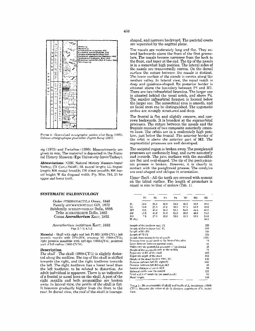
P1 P2 P3 P4 M1 M2 M3
BL 19.0 28.5 32.0 34.0 40.5 43.5 49.0 LL 18.0 27.0 31.5 30.0 37.0 38.5 49.0 MB 13.5 41.0 50.0 53.5 54.8 54.2 47.0 DB 17.5 41.6 51.0 51.0 49.6 49.9 34.0 BH 7.0 17.0 16.0 19.0 19.0 23.0 24.0 W (dia) 49.0
Length of the toothrow excl. P1 Length of the toothrow incl. P1 Length of M1-M3 Length of P2-P4 Length from occiput to tip of nasals Distance from nasal notch to the front of the orbit Least distance between parietal crests Width over the postorbital processes of the frontal Height of the zygomatic arch at the middle Zygomatic width of the skull Zygomatic depth of the skull Height of the skull (Gu~rin 1980, 25) Distance between left P2- right P2 Distance between left M3-right M3 Interior distance of the condyll External width over the condyli Basal width of nasals (at the nasal notch) Nasal length
220 235 124 101
(485) 73 41 18 71
290 252 140 (64)
86 38
111 92
148
TABLE 1 - Measurements of skull and teeth ofA. incisivum (1988- ~Y/1). Mesures du cr~me et de la denture supdrieure d'A. incisi- v u m ,

459
FIGURE 3 - 1-3. Aceratherium incisivum KAuP, 1832.1. Skull, with right and left P1- M3 (1988-~Y/1), lateral view. 2. Same spe- cimen, dorsal view. 3. Same specimen, occlusal view. 1-3. Aceratherium incisivum KAVP, 1832, crdme muni de P1-M3 droite et gauche (1988-~Y/1). 1. Vue latgrale. 2. Vue supdrieure. 3. Vue occlusale.
Premolars - P1 is small and triangular-shaped. The occlusal surface shows no more pattern due to strong wear. P2-P4 are quadrate-shaped, large and structurally similar to each other. The posterior premolars are semimolariform. The labial wall of the teeth is slightly convex, and is devoid of promi-
nent styles. Anterior and posterior protocone val- leys are developed only on P4. The parastyle valley is shallow and short. The medisinus is sigmoidally curved. Protoloph and metaloph are united with the lingual bridge, and their breadths are equal in size. The lingual bridge is higher than the lingual

460
• 2 3cm
5 c m Z.
FIGurE 4 - 1-3. Aceratherium incisi- rum K~ue, 1832. 1. Left juvenile maxilla with DP2-DP4, M1 erupting (1988-QY/2), occlusal view. 2. Right juvenile mandible, with dp2-dp4 (1988-¢Y/4), occlusal view. S. Proximal part of left radius (1988- CY/10) dorsal view. 4-6. Acerorhinus zernowi (BoR~SS~AK, 1914). 4. Left mandible with symphysis and p3-m2 (1988-QY/3) lacking anterior part (male), lateral view, 5, Same speci- men, occlusal view. 6, Left juvenile mandible with dp2-p4, ml (1988- ¢¥/6) (female), lateral view. 1-3. Aceratherium incisivum KAue, 1832. 1. Maxillaire juuenile munie de DP2- DP4, M1 gauche en eruption (1988- QY / 2), cue occlusale. 2. S4rie dental- re avee dp2-dp4 droite (1988-~Y / 4), rue occlusaIe. 3. Extrgmitd proximale de radius droite (1988-~'Y/10), rue dorsale. 4-6. Acerorhinus zernowi (BoR~ss~, 1914). 4~ Sgrie dentaire avec p3-m2 gauche (1988-QY/3), rue latdrale. 5. Idem, rue occlusale. 6. MandibuIe juvenile avec dp2~dp4, m l gauche (1988-~Y/6) (femelle), rue lat~rale.
cingulum and narrower on P4 than on P3. The lin- gual surfaces of the lophs are rounded. The lingual cingulum forms a continuous shelf-like wall, high above the base of the teeth. The labial cingulum is weak between the paracone and meta- cone.
M o l a r s - The labial wall of the teeth is convex at the front and concave at the rear. The lingual side of the protocone is flattened. Its lateral sides are rounded. The protocone is quite broad. Its anterior and posterior valleys are well developed. The para- cone is weak. The metacone forms a bulge at the

461
base. The crochet is present. There is no crista. The antecrochet is strongly developed in the molars. The parastyle valley is shallow and short. The anterior valley of the hypocone is present. The entrance of the medisinus is open. The M3 is trian- gular-shaped. The labial wall of the ectometaloph is convex, and extends to the lingual side. The ecto- metaloph angle is obtuse. The antero-lingual cingulum forms a shelf-like wall, and extends to the entrance of the medisinus. The labial cingulum is slightly developed, and bow- shaped at the base of the teeth. The talon of the M3 forms a low and vertical prominence. The postero- labial cingulum is well developed. Upper M i l k Teeth - The teeth are low-crowned (Tab. 2). The DP2 is triangular-shaped in outline and long anteriorly. The labial wall of DP2 is slightly convex. It has two depressions, and poor indica- tions of the paracone and metacone. The parastyle valley is shallow, and extends to the base of the tooth. The base of the metacone is flat and slightly concave on DP4. The anterior hypocone valley is located within the medisinus. A cingular cusp is situated in the entrance of the widely open medisi- nus. The crochet is long and united to the ectoloph on DP2, but not on DP3-4. The crista is not united to the crochet on DP2. The medifossette is closed on DP2. The postfossette is triangular-shaped, and enlarges to the top. The anterior cingulum extends along the protoloph. It forms a high and wavy structure at the base of the tooth. There is a sharp break in the cingulum at the entrance of the medisinus. The posterior cingu- lum surrounds the postfossette. The labial cingu- lum is faintly developed, and surrounds conti- nuously the base of the tooth. J u v e n i l e m a n d i b l e - The symphysis region is bro- ken. The symphysis reaches posteriorly beside the
DP2 DP3 DP4
BL 32.7 33.4 37.8 LL 30.4 28.5 36.0 MB 28.5 36.1 38.5 DB 31.4 33.8 37.6 BH 20.5 21.7 32.2
TABLE 2 - Measurements of upper milk teeth of A. incisivum (1988-~Y/2). Mesures des dents de lair supdrieures d'A. incisivum.
dp2 dp3 dp4
BL 31.5 35.4 36.2 LL 30.8 35.0 36.0 MB 12.9 16.7 20.0 DB 14.9 19.5 21.6 BH 19.0 16.0 21.8
Height of the corpus mandibulae (between dp2-dp3) 52.5 Diameter of the corpus mandibulae (between dp4-ml) 34.0
TABLE 3 - Measurements of the juvenile mandibulae and teeth of A. incisivum (1988-~Y/4). Mesures de la mandibule juvdnile et de la denture d'A. incisivum.
talonid valley of dp2. The big foramen mentale is located below the protoconid of dp2. The small fora- men mentale is situated below a short diastema. The corpus mandibulae is low. The lower border of the mandible is convex, and curves slightly upwards in the anterior part. The lateral face of the alveolus of I2 is angled. L o w e r m i l k teeth - There is no dpl. The teeth are low-crowned (Tab. 3). The dp2 is triangular-shaped in outline and long. The protoconid forms an occlu- sally high point. The paralophid is long, and extends anteriorly. The hypoconid is recurved. The trigonid basin is quite shallow. The talonid basin is narrow and v-shaped. The anterior labial valley is narrow and shallow. The labial valley (ectoflexid) is narrow, deep and vertically orientated on dp2, and large and oblique on dp3-4. There is a strong proto- conid pli along the anterior part of the labial valley. The talonid is larger than the trigonid, particularly on dp3-4. There is a small lingual cingulum at the anterior part of the trigonid valley. The labial cingulum is poorly developed. The labial and lingual enamel surfaces show fine growth lines. R a d i u s - The proximal facet for the humerus consists of two concave parts. The medial part is larger than the lateral one. The tuberositas radii is smooth. The lateral tuberosity is slightly develo- ped. The ulna facets are separated but near to each other. The lateral facet is large. The medial one is band-shaped. Below these facets a long and rou- ghened attachment surface serves for the proximal ligamentum interosseum. The cross section of dia- physis is triangular. Comparison The skull from Yulafli resembles A c e r a t h e r i u m i n c i s i v u m I~vP from Eppelsheim (Kaup 1832: P1. 10, Figs 2, 2b, P1. 14, fig. 5) with respect to the general morphology and teeth. These include the moderately long nasals with slightly upward trend and a strong supraorbital process. The teeth are characterized by large premolars, an equal breadth for the lophs on P2 and P3, the flat lingual side of the protocone, and the broad protocone on the molars. Differences exist between the Yulafli skull and A. i n c i s i v u m from Eppelsheim. In the latter case, lingual cingulum is weak on the premolars, the lingual bridge is narrow, and the foramen infraorbitale is located above P3. The general cha- racteristics of the Yulafli skull is also coincident with A. i n c i s i v u m KAUP from Tchobroutchi (Pavlow 1914: P1. 5, Figs 1,1a, lb, 3) and from HSwenegg (Htinermann 1989: Figs. 4, 9). 'A. i n c i s i v u m ' from Tchobroutchi is, however, removed from the genus A c e r o r h i n u s , and described to be closer to C h i l o t h e r i u m (Cerdefio 1996). The Tchobroutchi material differs from that of Yulafli by having a quite flat, long skull and narrow nasals. In the case of HSwenegg, the parastyle valley is deep on the molars, the cingulum is absent on the molars, and the foramen infra orbitale is small and located above M1. A. i n c i s i v u m from Montredon (Gu~rin 1980: P1. 8, Fig. A) is coincident with the skull from Yulafli with respect to the general shape of the teeth, but differs in having a longer P1, a narrower

462
protocone in the molars, and an anteriorly placed posterior edge of the nasal notch. The skull from Yulafli is similar to Aceratherium depereti BORISSIAK from the Turgai region (Boris- siak 1927: P1. 1) with respect to the large premo- lars, and strongly developed lingual cingulum in the premolars, and the large lingual bridge. However, there are major differences between these two species. In the case of A. depereti, the nasals are remarkably long and narrow, the dorsal profile of the skull is concave, and the sagittal crest and a small horn are present. The Yulafli material is comparable with Acerorhi- nus palaeosinensis (RINGSTR6M) from Shansi (China). Both species have a retracted nasal notch, a high occiput and a strong lingual cingulum in the premolars. However, A. paleosinense greatly differs by having a four-fold foramen mentale (RingstrSm 1924: Fig. 70; Bohlin1937: Figs 88, 163), and a nar- rower and shorter skull, a thinner zygomatic arch and a higher orbit. The skull presents similarities with Acerorhinus tsaidamensis (BoHLIN) from Qaidam (China) (Boh- lin 1937: P1. 8, Figs 1, 2, 3). They share the retrac- ted nasal notch, moderately long nasals, high occi- put, and flattened lingual side of protoloph. On the other hand, there are several differences between these species. As for A. tsaidamensis, the distance between the parietal crests is lesser (10 mm), the zygomatic arch is slender, and the upper margin of the zygomatic arch is horizontal below the orbit. The skull from Yulafli is similar to Acerorhinus zer- nowi (BoRmSIAK) from Sebastopol at t h e Black Sea (Borissiak 1915: P1. 2), Konya-Kayadibi (Heissig 1975: P1. 1, fig. 2), and Ankara-Sinap (Sara~ 1994). They share the strong lingual cingulum in the pre- molars, a strong supraorbital process, and a strong postglenoid process. A. zernowi differs from the Yulafli specimen in having a rounded protocone, longer nasals, a strong and vertical facial crest, and a longer and narrower P2. The suture of the Yulafli skull between the frontal and nasal also resembles that of A. zernowi (Borissiak 1915: Pt. 2, Fig. C). The Yulafli material greatly differs from A. zernowi from Tung-gur, which is synonymized with Hoploa- ceratherium, (Cerdefio 1996). The Tung-gur mate- rial is characterised by quite long nasals, a concave dorsal profile of the skull, and the presence of a sagittal crest and a shallow nasal notch. The morphology of the Yulafli skull is similar to Aceratherium kiliasi GERAADS • KOUFOS from Pen- talophos-1 (Geraads & Koufos 1990: P1.2, Figs 1, 2, P1.3, fig. 4). A. kiliasi (= Chilotherium kiliasi: Heis- sig 1999) is characterized by the absence of frontal and nasal horns, the retracted nasal notch, the strongly developed supra orbital process, and the flattened lingual side of the protoloph. It differs from the Yulafli specimen by having a v-shaped nasal notch, a quite flat skull, a weak cingulum on the premolars, a larger P1, and considerably shor- ter premolars. The skull differs from Chilotherium RINGSR(SM, 1924. In Chilotherium, the facial crest forms a right angle, the orbit is high, the skull is low, long and
Proximal width 90.8 Proximal diameter 61.7 Width of the proximal facet 88.0 Diameter of the proximal facet 54.0 Width of the diaphsis 44.5 Diameter of the diaphysis 34.2 Height of the ligament attachment surface 107.0
TABLE 4 - Measurements of the radius of A. incisivum (1988- ~Y/10). Mesures du radius de A. incisivum.
broad, the distance between the nasal notch and orbit is large, and the premolars are short. The upper teeth from Yulafli are smaller than those ofA. incisivum from Dorn-Diirkheim 1 (Germany) (Cerdefio 1997). The upper teeth from Yulafli are similar to Acerorhinus hipparionum (KoKEN) from Gansu (China) (RingstrSm 1924) with respect to the presence of the strong lingual cingulum. A. hip- parionum differs from the Yulafli material by having more massive and hypsodont teeth and rounded hypocone on the premolars. The upper milk teeth from Yulafli are similar to A. zernowi from Kayadibi (Heissig 1975: P1. 5, figs 1, 2), with respect to the general shape of the teeth and the presence of a depression of metacone at the base. However, A. zernowi differs from the Yulafli material by having a split up prefossette, a strong lingual cingulum, and a hypocone valley outside the medisinus. The upper milk t ee th differ from Chilotherium species. In C. samium from ~ankiri- ~orakyerler (Heissig 1975: P1. 5, fig. 4) and Samos (Weber 1905: Tab. 9, Fig. 4) the teeth are large, the labial wall of DP2 is quite convex, and the prefos- sette is narrow. In C. schlosseri from Afyon: I~mk (Heissig 1975: P1. 5, fig. 3) the DP2 is small and tri- angular-shaped, and the paracone is located rather distally. The upper milk teeth from Yulafli differ from A. paleosinense (RingstrSm 1924: P1. 11, figs 1, 2, 3) which has strong anterior and posterior protocone valleys and a strongly developed ante- crochet. The radius from Yulafli resembles A. incisivum from HSwenegg (Hiinermann 1989: Fig. 11) and from Montredon (Gu~rin 1980: Fig. 33B). They match in the weakly developed tuberositas radii and the short ulna facet. However, the size of the Montredon specimen is smaller (Tab. 4) and the lateral tuberosity is stronger. A. incisivum differs from A. zernowi from Kayadibi (Heissig 1975: Figs 33a, b) which has a shorter ulna articulation surfa- ce, a deep tuberositas radii, and separate ulna facets. A. incisivum also differs from C. samium from Kayadibi (Heissig 1975: Figs 35a, b) having the deeply hollowed tuberositas radii, and the sepa- rated ulna facets.
Acerorh inus zernowi (BoRISSIAK, 1914) Figs 4.4-6; 5.1-4
M a t e r i a l - Left mandible with symphysis and p3-m2 (1988- ~Y/3 ), lacking anterior part (male); left mandible with sym- physis and p2-p3 (1988-~Y/5) (male); left mandible with m2- m3 (1988-~Y/7); left juvenile mandible with dp2-p4, M1 (1988- ~Y/6).

463
2
FIGUPJ~ 5 - 1-4. Acerorhinus zernowi (BoaIss~K, 1914). 1. Left mandible with symphysis and p3-m2 (1988- ~Y/3 ) lacking anterior part (male), anterior view. 2. Left mandible with symphysis and p2-p3 (1988-~Y/5) (male), anterior view. 3. Same speci- men, occlusal view (1988-~Y/5). 4, Left mandible with m2-m3 (1988- ~Y/7), occlusal view. 5. Dihoplus sehleiermacheri (KAuP, 1832), left mandible with m2-m3 (1988-~Y/8), occlusal view. 1-4. Acerorhinus zer- nowi (Bo~ZSrAK, 1914). 1. Sgrie den- taD~ avec p3-m2 gauche (1988- ~ Y / 3), rue ant~rieure. 2. Sdrie den- taire avec p2-p3 gauche (1988-~Y / 5), rue antdrieure. 3. ldem, rue occlusa- le. 4. Sgrie dentaire avec m2-m3 gauche (1988-~Y/ 7), rue occlusale. 5. Dihoplus schleiermacheri (KAuP, 1832), sdrie dentaire avec m2-m3 gauche (1988-~Y / 8), rue occlusale.
D e s c r i p t i o n M a n d i b l e - The symphysis is robust-and narrow, and begins curving upwards below p2. It extends posteriorly beside p3. The symphysis includes two divergent sheaths of the tusks. The rounded alveo- li of the tusks are filled by the roots. These tusks are very close to each other (about 16 mm on ~Y/3, 14 mm on ~Y/5, Tab. 5), and curve strongly upwards. Their long axes are in an oblique position in ~Y/3, and in a more vertical direction on ~Y/5.
The foramen mentale is located below p3. There is a small foramen below the diastema. The diastema is also rather short. The corpus mandibulae is deep, and displays a uniform height. Its lower border is convex on ~Y/3 and flattened on ~Y/5 and ~Y/7. The margo interalveolaris shows a distinct crest. P r e m o l a r s - The p l is lost, but its alveolus is pre- served. The teeth are high-crowned and oriented obliquely to the jaw. The p2 is triangular-shaped

464
Nr. to ta l L
he igh t of the d iamete r of corpus below corpus
p2/3 p/m m l / 2 p/m m l / 2
root of I2 1 2 Symphysis r amus coronoid dis tance W mand ibu lae process
L W H H
1988 ¢Y/3 1988 ¢Y/5
1988 QY/6 384
83 86 87 42.5 44.5
67 (p3/4) 42
73 69 76 37 38
38 x 31
41 x 30
16
14
94
(104) 20 174 (161)
TABLE 5 - Measu remen t s of mand ibu l ae and incisors ofA. zernowi. Mesures de la mandibule d'A. zernowi.
Tooth/Nr 1988-~Y/3 1988-~Y/5 1988-~Y/7 1988-~Y/6, juv. dpl-4
p2 dpl BL 29.5 33.0 LL 28.9 32.7 MB 18.9 14.9 DB 19.7 16.5 BH 31.3 18.6
p3 BL 33.5 33.8 LL 33.1 33.1 MB 22.8 22.2 DB 23.4 24.3 BH 29.5 28.0
p4 BL 37.2 LL 37.8 MB 24.3 DB 26.8 BH 28.5
ml BL 41.9 LL 39.9 MB 25.1 DB 25.8 BH 29.5
m2 BL 42.0 42.8 LL 42.8 42.5 MB 27.1 28.2 DB 25.8 27.8 BH 28.9 29.2 LH 19.0
m3 BL 42.0 LL 41.4 MB 25.6 DB 24.3 BH 28.1 LH 18.0
dp2
17.2
20.5
dp3 39.5 40.0 20.1 20.9 24.6
dp4 41.0 41.4 24.8 26.9 35.8
TABLE 6 - Measurements of lower teeth ofA. zernowi. Mesures de la denti- tion infdrieure d'A. zernowi.
and long. The paralophid is long, and contains a high anterior point. The anterior labial valley is shallow in p2. The premolars are large. The labial wall of the teeth is rounded in ~Y/3 and flattened in ~Y/5. The labial valley is shallow, and has a slightly oblique inclination. The protoconid angle is recognisable. The trigonid valley is shallow and v- shaped. The talonid valley is rather large, u-shaped
in ~Y/3 and narrower in ~Y/5. These valleys are closed, and do not reach the base of the lingual side. The hypolophid is recurved, and united with the metalophid. The antero-lingual cingulum is well developed, and extends falling obliquely to the anterior border of the trigonid valley. Faint horizontal growth lines occur on the labial and lingual enamel surfaces. M o l a r s - The molars are longer than the premolars (Tab. 6). The trigonid valley is v-shaped and shal- low. The talonid valley is large and u-shaped. The labial wall of the teeth is rounded. The labial valley is remarkably oblique to the occlusal surface. The paralophid is short, and does not extend to the lin- gual side. The metalophid is long in mesio-distal direction. The hypolophid is recurved. The protoco- nid angle is weaker than that of the premolars. The anterio-lingual cingulum is strongly developed as in the premolars. J u v e n i l e m a n d i b l e - The corpus mandibulae is thin and low and of constant height. Its lower border is convex. The foramen mentale is located below the dp2. There is no pl, but its alveolus is preserved. The symphysis is narrow and short, and curves upwards below dp2. The length of the symphysis extends posteriorly besides p2. The ramus mandi- bulae turns steeply upwards. The condyle is round. The coronoid process is missing. The mandibular notch is quite narrow. The lateral surface of the ramus mandibulae (fossa masseterica) is flattened. L o w e r m i l k t e e t h - The dpl is absent, but its alveo- lus is preserved. The teeth are low-crowned. The dp2 is triangular-shaped and long. The labial wall of the tooth is roughened, and bears two valleys. The flat anterior labial valley runs vertically up to the enamel base. The labial valley is deep and has an oblique direction. The paralophid is short. The trigonid valley is shallow. The talonid valley is nar- row and u-shaped. The protoconid pli is strong, and extends parallel to the labial valley in dp2. The antero-lingual cingulum extends along the basis of the anterior part of tooth. There is a verti- cal cingulum between the trigonid and talonid val- leys on dp2. The antero-labial cingulum is short. There is no lingual cingulum. C o m p a r i s o n The general shape and morphology of the mandible from Yulafli bear similarities to those ofA. z e r n o w i

(BoRISSIAK) from Kayadibi and Konya-Kayadibi- Sarisikinleri (Heissig 1975). The common features are the following: the symphysis is narrow (94 mm) and curves steeply upwards; the tusk roots are rounded; the premolars are large and long; the paralophid is short; the orientation of the too- throws is oblique. However, some differences in morphology and size are remarkable. In A. zernowi from Kayadibi the labial cingulum is present on the anterior lobe, the symphysis is narrower (83 mm), the teeth are broader, and the distance between the tusks is larger. The material also presents similari- ties with A. zernowi from Tung-gur (Cerdefio 1996: Fig. 3) having a small pl, large premolars, sym- physis of about 92 mm width, and a great angle bet- ween the symphysis and corpus mandibulae. The Yulafli specimens resemble the A. kiliasi man- dible (= Acerorhinus zernowi: Heissig 1999) from Pentalophos-1 (Geraads & Koufos 1990: P1. 3, figs 2, 3, 5) with respect to the narrow symphysis, the large premolars, and the ovoid-shaped and oblique alveoli of the tusks. They differ from A. kiliasi which has a greater distance between the tusks and a strong cingulum in p2. Several aspects of A. zernowi compare with A. palaeosinensis from Shansi (Ringstr6m 1924: P1. 10, fig. 4; Bohlin 1937: P1.7, fig. 10). These include a narrow symphysis (respectively, 94 mm and 95 mm), a larger and triangular p2, and an oblique tusk position. In the case of A. palaeosinensis, the tusks are flat in the crown cross-section (Bohlin 1937: p. 70, 47 x 24 mm), there is a large distance between the tusks (Bohlin 1937: p. 70, 45 mm), and the horizontal tusks are slightly upturned. A. tsaidamensis from Qaidam (Bohlin 1937: Figs 92, 93, 164, 165) is similar to the material from Yulafli with respect to narrow symphysis (75-95 ram), large premolars, highly upturned symphysis, and a narrow distance (Bohlin 1937: p. 70, 25 mm) between tusks. It differs in having a ventrally smooth symphysis. The mandible from Yulafli greatly differs from Chilotherium. In Chilolherium, the symphysis is broader anteriorly and hollowed ventrally, the tusks are horizontally curved and with upturned medial flanges (Heissig 1989, 1999), and the axes of the tusks are oblique and far from each other. The premolar series are shorter than the molar series, particularly in Ch. kowalevskii (PAVLOW). The Yulafli specimens resemble A. incisivum (Kaup 1832: P1. 14, fig. 9) with respect to several features of the teeth and mandible. These include the convex lower border of the corpus mandibulae, the long p2, and a shallow and u-shaped trigonid val- ley. They differ greatly from A. incisivum having horizontal tusks and a ventrally smooth symphysis.
Subfamily RHINOCEROTINAE Dollo, 1885 Tribe RHINOCEROTINI Dollo,1885
Subtribe RHINOCEROT1NA Dollo, 1885 Genus Dihoplus BRANDT, 1878
Dihoplus schleiermacheri (KAUP, 1832) Fig. 5.5
465
m2 m3
BL 47.6 47.5 LL 48.5 47.2 MB 32.6 29.2 DH 31.0 26.5 BH 28.3 28.7 LH 23.0 27.5
Height of the corpus mandibulae (m2-m3) 78.5 Diameter of the corpus mandibulae 51.4
T̂ BLE 7 - Measurements of lower teeth and mandible of D. schleiermacheri (1988-(~Y/8). Mesures de la mandibule et de la denture d'D. schleiermacheri.
M a t e r i a l - Left mandible with m2-m3 (1988-~Y/8). D e s c r i p t i o n Mandible - The mandible is larger than that of the other rhinocerotid remains. In the labial side, the height of the corpus increases posteriorly, and in the lingual side, the height of the corpus is constant. The lower edge of the corpus is convex. The ramus is quite flattened laterally, and rises abruptly above the toothrow. Teeth - The teeth are rather high-crowned (Tab. 7). The paralophid is long and narrow. The labial walls of the teeth are characterized by the deep labial valley with oblique orientation, the angled metalo- phid, and the slightly angled hypolophid. The pro- toconid fold is distinct. The trigonid valley is shal- low and u-shaped. The talonid valley is large, shal- low and u-shaped. In m3 there is a small tubercle in the talonid valley. The antero-labial cingulum continues lingually along the anterior surface, and ends at the entran- ce of" the trigonid valley. The lingual cingulum is slightly developed at the base of the lingual side of the teeth. C o m p a r i s o n Dihoplus schleiermacheri was first erected by Grooves (1983) for Dicerorhinus schleiermacheri (KAuP, 1832) from Eppelsheim. The Eppelsheim material was described as Dicerorhinus schleierma- cheri (Gu~rin 1980), and as Dihoplus schleierma- cheri (Geraads & Koufos 1990). Cerdefio (1995a) synonymized 'Dicerorhinus' schleiermacheri with Lartetotherium schleiermacheri.
The mandible and teeth resemble in morphology and size those of Dihoplus schleiermacheri from Eppelsheim (Kaup 1832: P1. 11, fig. 8) and from Soblay (Gu~rin 1980: P1.9, fig. E, tab. 49), with res- pect to the presence of the long paralophid, the angled metalophid and the convex lower edge of the corpus mandibulae. Some morphological diffe- rences recognized. In the Eppelsheim material include the v-shaped trigonid valley and the longer paralophid. The lower teeth are larger than L. schleiermacheri from Dorm-Dtirkheim 1 (Germany) described by Cerdefio (1997). D. schleiermacheri resembles Lar- tetotherium sansaniense (LARTET) from Sandelzhau- sen with respect to the long paralophid, weakly

466
developed anterolabial cingulum, and the deep and oblique labial valley. It differs from L. sansaniense from Sandelzhausen which is smaller in size and has a v-shaped trigonid valley.
PALEOECOLOGY
Deinotherium, which is the most common element of the Yulafli fauna indicates a forested habitat. The scarcity of Hipparion in the Yulafli fauna implies a restricted amount of steppe or savannah areas. A. incisivum, as a medium-sized form, has a habitat of swamp and open forest with probably patches of grassland, in a warm temperature (Gu~rin 1980). D. schleiermacheri, which is a large form, indicates a habitat of open forest intervened by some grassland and humid environment (Gu~rin 1980). Ceratotherium neumayri and the Chilotherium spe- cies, which are grazer-like forms with semihypso- dont teeth, are the common elements of the Late Miocene faunas of Anatolia. Their habitat is savan- nah environment with patches of bushes (Heissig 1975; Kaya 1994). They are not present in the Yulafli fauna. The paleocology of the Yulafli fauna (Thrace region) is different from that of the Anatolian localities. The faunal elements of the Yulafli fauna correlate bet ter with those of European localities (such as, Eppelsheim, Concud), Dorn-Dtirhkeim 1) (Gu~rin 1980; Cerdefio 1997) than with Anatolian ones.
CONCLUSIONS
The Rhinocerotidae remains from Yulafli (~orlu) include Aceratherium incisivum and Acerorhinus zernowi, and Dihoplus scheiermacheri. Aceratherium incisivum was first recorded in Tur- key by Ozansoy (1965) in a faunal list regarding the ~obanpinar area, Ankara. It is known from several Late Miocene localities in Central and Western Europe, such as Eppelsheim (MN 9) (Kaup 1832), Ht}wenegg (MN 9) (Htinermann 1989), Montredon (MN 10) (Gu~rin 1980), Masia del Barbo (MN 10), La Roma 2 (MN 10), Cerro de los Batallones (MN 10), Dorn-Dfirkheim 1 (MN 11), Concud (MN 12) (Cerdefio 1992, 1997; Cerdefio & Sanchez 1998), Rudabanya (MN 9) (Heissig 1996, 1999) and Tcho- broutchi (Pavlow 1914). The overall stratigraphic range is MN 9 to MN 13 (Gu~rin 1982; Cerdefio 1998; Heissig 1999). Acerorhinus zernowi was first defined in Sebastopol (MN 9) (Borissiak 1914). It -is known from Konya- Kayadibi and Kayadibi-Sarisikinleri (both MN 11) (Heissig 1975), Ankara- Sinap (MN 9) (Sara~ 1994) and Tung-gur (MN 8) (Cerdefio 1996). The strati- graphic range is MN 9 to MN 11 to Heissig (1999), and. early Vallesian for Europe and late Astaracian for Asia (Cerdefio 1998). Dihoplus scheiermacheri is the first occurrence in Turkey. It is known from several Late Miocene loca- lities in Europe such as Eppelsheim (MN 9),
Gauweinheim (MN 9), Esselborn (MN 9), Soblay (MN 9), Luberon (MN 9) (Gu~rin 1980), Masia del Barbo (MN 10), La Roma 2 (MN 10), Pierra (MN 11), Dorn-Dfirkheim 1 (MN 11), and Concud (MN 12) (Cerdefio 1992, 1997). Its stratigraphic range is MN 9 to MN 13 (Gu~rin 1982; Heissig 1999). The coexistence of A. incisivum and A. zernowi is recognized for the first time. This is not recorded in Central or Western European localities, where A. zernowi is unknown. A. incisivum and D. schleier- macheri occur together in Eppelsheim, Montredon, Concud, La Roma 2, Dorn-Dfirkheim 1 and Masia del Barbo (Gu~rin 1980; Cerdefio 1992; Cerdefio 1995b; Cerdefio 1997). The common presence of the above species points to a Turolian age (MN l l -MN 12). The other faunal elements, such as large-sized Deinotherium gigantissimum and Microstonyx ery- manthius are indicative of the Turolian age. A. incisivum and D. schleiermacheri are European taxa, while A. zernowi is an Eurasian one. The for- mer species are widespread in Europe during the Late Miocene. A. zernowi is significant for a paleo- biogeographical crossroad sense of Thrace to Europe.
Acknowledgements - This study has been supported by Deut- scher Akademischer Austauschdienst (DAAD), and by an Ege University grant (TTM/001/1998). We thank to Prof. Dr. Dr. h. c. mult. T. Berchem (President of DAAD) and Prof. Dr. R. Leinfelder (Director of the Institut und Staatssammlung ftir Paltiontologie und Geologie, MOnchen), Dr. W. Werner, Dr. H. Mayr, Dr. G. Schairer, and Dr. N. Rtickert-UlkOmen (Munich). Thanks to Dr. C. Gu~rin (Lyon) and Dr. E. Cerdefio (Argentine) for critical reviews, and I. Benbanaste (~orlu), S. Ersen (~orlu) and E. Koralay (Izmir) for collection of fossils. Thanks are extended to K. Dossow and G. Bergmeier (Munich) and S. Mayda (Izmir) for technical assistance, and to N. McAndrew-Yllmaz and E. Akyol (Izmir) for linguistic support.
REFERENCES
Borissiak, A., 1914. Mammif'eres fossiles de Sebastopol I. Trudy geolegicheskago komiteta, Novaja seria 87, 1-154.
Borissiak, A., 1915. Mammif'eres fossiles de Sebastopol II. Trudy geolegicheskago komiteta, Novaja seria 137, 1-45.
Borissiak, A., 1927. Aceratherium depereti nov. sp. from the Jilancik beds. Bulletin de l'Acad~mie des Sciences de I'URSS 21 (6), 769-786.
Bohlin, B., 1937. Eine tertitire St~ugetier-Faunas Tsaidam. Palaeontologia Sinica 14 (1), 1-109.
Cerdefio, E., 1992. Spanish Neogene Rhinoceroses. Palaeontology, 35 (2), 297-308.
Cerdefio, E., 1995a. Cladistic analysis of the family Rhinoce- rotidae (Perissodactyla). American Museum Novitates 3143, 1-25.
Cerdefio, E., 1995b. Changes in western European Rhinocero- tidae related to climatic variations. Palaegeography, Palaeo- climatology, Palaeoecology 114, 325-338.
Cerdefio, E., 1996. Rhinocerotidae from the middle Miocene of the Tung-gur formation, inner Mongolia (China). American Museum Novitates 3184, 1-43.
Cerdefio, E., 1997. Rhincoretidae from the Turolian site of Dorn- Dtirkheim 1 (Germany). Courier Forschungs-Institut Sen- ckenberg 197, 187-203,
Cerdefio, E., 1998. Diversity and evolutionary trends of the fami- ly Rhinocerotidae (Perisoodactyla). Palaegeography, Palaeo- climatology, Palaeoecology 141, 13-34.
Cerdefio, E., Sanchez, B., 1998. Aceratherium incisivum (Rhi- nocerotidae) en el Mioceno Superior de Cerro de los Batal-

4 6 7
lones (Madrid). Revista Espanola de Paleontologia 13 (1), 51- 60.
Fortelius, M., 1990. Rhinocerotidae from Pasalar, middle Mio- cene of Anatolia (Turkey). Journal of Human Evolution 19, 489-508.
Geraads, D., 1994. Les gisements de mammif'eres du Mioc6ne sup6rieur de Kemiklitepe, Turquie: Rhinocerotidae. Bulletin du Mus6um national d'Histoire naturelle 4 (16), 81-95.
Geraads, D., Koufos, G., 1990. Upper Miocene Rhinocerotidae from Pentalophos-1, Macedonia, Greece. Palaeontographica A, 210, 151-168.
Groves, C.P., 1983. Phylogeny of the living species of rhinoceros. Zeitschrift ftir zoologische Systematic und Evolutions- forschung 21 (4), 293-313.
Gu6rin, C., 1980. Les rhinoc6ros (Mammalia, Perissodactyla) du Miocene terminal au Pleistoc6ne sup6rieur en Europe Occi- dentale. Comparaison avec les esp6ces actuelles. Documents du Laboratoire de G6ologie de Lyon 79 (1, 2, 3), 1-1184.
Gu6rin, C., 1982. Les Rhinocerotidae (Mammalia, Perissodac- tyla) du Mioc6ne terminal au Pleistoc6ne sup6rieur d'Europe occidentale compar6s aux esp~ces actuelles: tendances evolu- rives et relations phylog6n6tiques. Geobios 15 (4), 599-605.
Heissig, K., 1972. Paltiontologische und geologische Untersu- chungen im Tertitir von Pakistan 5. Rhinocerotidae (Mamm.) aus den unteren und mittleren Siwalik-Schichten. Abhand- lungen der Bayerischen Akademie der Wissenschaft Math- Naturw. K1. NF 152, 1-112
Heissig, K., 1974. Neue Elasmotherini (Rhinocoretidae, Mam- malia) aus dem Obermiozfin Anatoliens. Mitteilungen der Bayerischen Staatssammlung ftir Palaontologie und histo- rische Geologie 14, 21-35.
Heissig, K., 1975. Rhinocerotidae aus dem Jungtertitir Anato- liens. Geologisches Jahrbuch B (15), 145-151.
Heissig, K., 1976. Rhinocerotidae (Mammalia) aus der Anchi- therium-Fauna Anatoliens. Geologisches Jahrbuch B (19), 3- 121.
Heissig, K., 1989. The Rhinocerotidae. In: Prothero, D.R., Schoch, R.M. (Eds), The evolution of Perissodactyls. New York, Oxford University Press, 399-417.
Heissig, K., 1996. The stratigraphical range of fossil rhinoceroses in the late Neogene of Europe and the Eastern Mediter- ranean. In : Bernor, R.L., Fahlbusch, V., Mittmann, H-W. (Eds), The evolution of Western Eurasian Neogene Mammal Faunas. New York, Columbia University Press, 339-347
Heissig, K., 1999. Family Rhinocerotidae. In : R6ssner, G.E., Heissig, K. (Eds), The Miocene Land Mammals of Europe. Verlag Dr. Friedrich Pfeil, Mtinchen, 175-188.
Htinermann, K.A., 1989. Die Nashornskelette (Aceratherium incisivum KAUP, 1832) aus dem Jungtertitir vom HSwenegg im Hegau (Stidwestdeutschland). Andrias 6, 5-116.
Kaup, J.J., 1832. Uber Rhinoceros incisivus Cur. und eine neue Art Rhinoceros schleiermacheri. Isis 8, 898-904.
Kaya, T., 1993. The Late Miocene Perissodactyla in Sazak (Kale- Denizli). Bulletin Mineral Research and Exploration Insti- tute, Turkey 115, 23-31.
Kaya, T., 1994 - Ceratotherium neumayri (Rhinocerotidae, Mam- malia) in the Upper Miocene of Western Anatolia. Turkish Journal of Earth Sciences 3 (1), 13-22.
Ozansoy, F., 1965. ]~tude des gisements continentaux et des Mammif'eres du C6nozo~que de Turquie. M6moires de la Soci6t6 G6ologique de France N.S, 44, 1-92.
Pavlow, M., 1914. Mammif~res tertiaires de la Nouvelle Russie. 26 Partie Aceratherium incisivum, Hipparion, Proboscidea, Carnivora. Nouveaux M6moires de la Soci6t6 Imp6riale des Naturalistes de Moscou 174, 1-78.
RingstrSm, T.J., 1924. NashSrner der Hipparion-Fauna Nord Chinas. Palaeontologica Sinica C1 (1), 1-159.
Riickert-Olkfimen, N., 1990. Neue Ergebnisse zum Alter der mioz~nen Fisch-Schichten in Nord-Thrakien (Ttirkei). Strati- graphie I. Mitteilungen der Bayerischen Staatssammlung ftir Palgontologie und historische Geologie 30, 27-37.
Saraq, G., 1987. Kuzey Trakya bSlgesinde Edirne-Klrklareli, Saray-~orlu, UzunkSprti-Dereikebir ySresinin memeli paleo- faunasi. Ankara University Science Faculty, Master's thesis (unpublished), 110 pp.
Sara~, G., 1994. The biostratigraphy and palaeontology of the Rhinocerotidae (Mammalia, Perissodactyla) of the continen- tal Neogene sediments in the Ankara region. Ankara University, Ph D thesis (unpublished), 217 pp.
Sen, S., 1970. Tfirkiye Miyosen ve Pliyosen Rhinoceros'larmln odontolojik ozellikleri. Ankara University, Master's Thesis (unpublished).
Weber, K.M., 1905. Ueber tertitire Rhinocerotiden vonder Insel Samos II. Bulletin de la Soci4t6 des Naturalistes de Moscou, Nouvelle S6rie 18, 344-363.
T. KAYA Ege University Tabiat Tarihi Mtizesi
35100 Bornova-Izmir, Turkey E-mail: [email protected]
K. I-IEISSIG Bayerische Staatssammlung ffir
Paltiontologie und Geologie Richard-Wagner-Strabe 10
D-Miinchen E-mail: [email protected]