late cretaceous and early tertiary turtles from the big
TRANSCRIPT

LATE CRETACEOUS AND EARLY TERTIARY TURTLES FROM
THE BIG BEND REGION, BREWSTER COUNTY, TEXAS
by
SUSAN LEIGH TOMLINSON, B.F.A., M.S.
A DISSERTATION
IN
GEOSCIENCE
Submitted to the Graduate Faculty of Texas Tech University in
Partial Fulfillment of the Requirements for
the Degree of
DOCTOR OF PHILOSOPHY
Approved
Accepted ^pi
May, 1997

Copyright 1997, Susan L. Tomlinson

ACKNOWLEDGEMENTS
I would hke to thank the members of my committee. Dr. Sankar Chatterjee,
Dr. George Asquhh, Dr. James Barrick, Dr. Richard Strauss, and the committee
chairman. Dr. Thomas Lehman for theh contributions to this dissertation. In particular,
I'd like to thank Dr. James Barrick and Dr. Richard Strauss for their valuable insight m
the many discussions we had about certain parts of this study.
Tom Lehman was instmmental in starting me on the path of the turtle. Without
his support and invaluable knowledge about Big Bend stratigraphy and vertebrate
paleontolgy, this project would not have been started, nor completed. I look forward
to many more years of Big Bend collaboration with hhn.
There were a number of people who helped m both big and small ways. I thank
the Texas Memorial Museum, the Louisiana State University Museum of Natural
Science, and the Museum at Texas Tech University for making their collections
available to me. JuUa Sankey at LSU also helped by tracking down map locations For
their help in the field I thank Bill Straight, James Browning, Dave Evans, Heather
Beatty, and Dr. Elisabeth Wheeler. I'd hke to thank Dr. Howard Hutchison for sharing
the University of California Museum of Paleontology turtle collection with me for my
edification (and also for his many explanations). I also thank the following for then-
help: Anne Weil, Dr. Kathleen Nichols, and Jill Haukos.
Most of all, I thank my husband, Walt Schaller. I wouldn't have finished
without his support.
u

TABLE OF CONTENTS
ACKNOWLEDGMENTS u
ABSTRACT iv
LIST OF TABLES v
LIST OF FIGURES vi
CHAPTER
L INTRODUCTION 1
IL STRATIGRAPHY 5
m. SYSTEMATIC PALEONTOLGY 21
IV. STRATIGRAPHIC DISTRIBUTION OF TURTLES 117
V ECOLOGICAL DFVERSITY OF THE BIG BEND
TURTLE FAUNA 125
VI. MORPHOMETRIC ANALYSIS OF PIT VARIANCE IN
TRIONYCHID SHELLS 148
REFERENCES 177
APPENDIX 184
lU

ABSTR.ACT
Fossil turtles are abundant in the Upper Cretaceous and Lower Tertiary
sediments of the Aguja, Javelina, and Black Peaks Formations m Big Bend National
Park, Brewster County, Texas. Nine genera of freshwater and terrestrial turtles have
been identified from these deposits, including Bothremys, "Baena ", Neurmikylus.
Compsemys, Adocus, BasUemys. "Aspideretes", '"^Eelopanoplid^ and Hoplochelys
A marine turtle from the Aguja Formation represents a new genus.
Turtles are most abundant in the marginal marine, brackish, and freshwater
floodplain deposhs of the Aguja Formation, and within the Aguja, they are most
abundant in the upper shale member. 'Aspideretes' is the dominant genus in the
Upper Cretaceous sediments, followed by "Baena " Turtle fossils are rare in the
fluvial floodplain deposhs of the Javehna Formation, where the dominant genus is also
Aspideretes." There is a shght increase m abundance and diversity of turtles in the
fluvial floodplain deposits of the overlying Black Peaks Formation, where Eoplochelys
is the dominant genus. The decrease in numbers and diversity of turtles in the Javelina
and Black Peaks formations, compared to the Aguja, was probably the resuh of a
change to dry inland environments less hosphable to turtles.
The diversity level of the upper shale member of the Aguja Formation is
comparable to that in the correlative Fmhland and Khlland formations of the San Juan
Basin in New Mexico, and to a modem assemblage of turtles m the Brazos River
IV

drainage basm in Texas. A comparison of the shell morphologies exhibhed in the
Aguja fauna and in the Brazos River fauna also suggests that the diversity level is
comparable. A morphometric technique was used to determine whether variabihty in
the ornamentation patterns in trionychid shells is usefial for taxonomic discrimination
Preliminary results suggest that discrete shell ornamentation patterns are
discriminatory and non-random. This technique may be useful for identifying levels of
fossil trionychid diversity
V

LIST OF TABLES
4.1 Turtles of the Aguja, Javehna and Black Peaks formations 120
5.1 Genera of turtles represented in the modem Brazos River drainage of Texas, the Aguja fauan of Big Bend, and the Fmitiand and Kirtland fauna of New Mexico 131
5.2 Comparison of Brazos River drainage fauna and Aguja fauna 143
5.3 Comparison of Brazos River drainage fauna and Aguja fauna 144
5.4 Taxa and number of individuals from the upper shale member of the
Aguja Formation used for Simpson's Index calculations 146
6.1 Variables used m morphometric study 155
6.2 TOTTURTdata 158
6.3 RANDTURTNC data 165
6.4 ALLDATA 168
VI

LIST OF FIGURES
2.1 Upper Cretaceous and lower Tertiary strata in Big Bend (after Maxwell etal., 1967 and Lehman, 1985) 7
2.2 Stratigraphy of the Upper Cretaceous and lower tertiary sediments (after Maxwell et al., 1967) 8
2.3 The Aguja Formation as subdivided by Lehman (1985) 11
3 1 Carapace and plastron bones and scutes (diagram after Zangeri, 1969) 22
3.2 Bothremys sp., TMM 43381-1, left first costal, dorsal and visceral views 26
3.3 Bothremys sp., TMM 43469-2, right hyo- (above) and hypoplastron (below), visceral view 28
3.4 Bothremys sp., TMM 43469-2, right hyo (above) and hypolpastron (below), ventral view 29
3.5 Bothremys sp 32
3.6 A comparison of the plastron of Bothremys, Taphrosphys, and the Big Bend specimen 34
3.7 ''Baena'' cf "5 " omata, LSUMG V-1136, carapace in dorsal view 37
3.8 ''Baena'' cf "^" omata, LSUMG V-1136, plastron m ventral view 39
3.9 "Baena" cf "5" nodosa, TMM 42536-1, carapace m dorsal view 43
3.10 "Baend' cf "B " nodosa, TMM 42536-1, plastron m ventral view 44
Vll

3.11 "Baena" cf "B " nodosa, TMM 43251 -1, carapace fragments 45
3.12 "Baend' cl "B" nodosa, TMM 432351-1, plastron fragments 46
3.13 "Baena" sp., carapace fragments of TTU 5-104-47 in dorsal \^ew 51
3.14 "Baena" sp., TTU 5-104-47, plastron in ventral view 52
3.15 "Baena" sp., LSUMG V-1168, plastron in ventral view 56
3.16 Neurankylus eximius, TMM 43385-1, carapace in dorsal view 63
3.17 Neurankylus eximius, TMM 43385-1, plastron in ventral view 65
3.18 Cheloniidae, n. gen., n. sp., TMM 43072-1,
lower mandible in oral and side views 68
3.19 Chelomidae, n. gen., n. sp., TMM 43072-1, carapace fragments 70
3.20 Cheloniidae, n. gen., n. sp., TMM 43072-1, plastron in ventral view 71
3.21 Cheloniidae, n. gen., n. sp., TMM 43072-1, left scapula, ventral view 72
3.22 Cheloniidae, n. gen., n. sp., TMM 43072-1, left scapula, dorsal view 73
3.23 Cheloniidae, n. gen., n. sp., TMM 43072-1, left and right humeri, dorsal view 75
3.24 Cheloniidae, n. gen., n. sp., TMM 43072-1, left and right humeri, ventral v ew 76
3.25 Cheloniidae, n. gen., n. sp., TMM 43072-1, right ulna 77
viu

3.26 Cheloniidae, n. gen., n. sp., TMM 43072-1, right radius 78
3.27 Cheloniidae, n. gen, n. sp , TMM 43072-1, right femur 80
3.28 Comparison of plastrons of Chelonioidea 82
3.29 Comparison of humeri of Chelonioidea 83
3.30 ^£/ocw5 sp, TMM 4257-3, first costal 87
3.31 Trionychidae, TMM43380-5, partial carapace 92
3.32 Trionychidae, TMM 42335-8, left nuchal 93
3.33 Trionychidae, TMM 43534-2, right fifth costal 95
3.34 Trionychidae, TMM 43534-8, right hypoplastron 96
3.35 Trionychidae, TMM 422880-6, right hyoplastron 98
3.36 Trionychidae, TMM 42539-5, left hypoplastron, left xiphiplastron
and the distal end of a humems 100
3.37 Trionychidae, TMM 43057-324, left hypoplastron 102
3.38 Trionychidae, TMM 42874-1, right xiphiplastron 103
3.39 Trionychidae, TMM 41400-5, left humems 105
3.40 Representative specimens of Trionychidae 107
3.41 Representative specimen of Trionychidae 108
3.42 Representative specimens of Trionychidae 109
3.43 Representative neurals from Hoplochelys sp. specimens 113
3.44 Representative costals from Hoplochelys sp. specimens 114
3.45 Representative peripherals from Hoplochelys sp. specimens 115
IX

4.1 Stratigraphic distribution of turtles in the Aguja, Javelina,
and Black Peaks formations 118
4.2 Stratigraphic distribution of turtles in the Aguja Formation 119
5.1 Drainage area estimated for this study 134
5.2 Drainage area estimated for this study 136
5.3 Method of measurement for morp :ology study 141
6 1 Plot ofPRIN2*PRIN1, TOTTURTdata 159
6.2 Plot of PRIN3*PRIN2, TOTTURTdata 161
6.3 Plot of PRIN2*PRrNl, RANDTURTNC data 165
6.4 Plot of PRrN3*PRIN2, RANDTURTNC data 166
65 Plot of PRJN2*PRIN1, ALLDATA 169
6.4 Plot of PRIN3*PRIN2, ALLDATA 171
X

CHAPTER I
ESITRODUCTION
General Background of Study
Upper Cretaceous and lower Tertiary sedimentary rocks exposed in Big Bend
National Park (Texas) accumulated at the southernmost extreme of the Western
Interior Province, and the westernmost extreme of the Gulf Coast Province. These
sediments record the final transgressive/regressive sequence of the Western Interior
Cretaceous Seaway and the subsequent transhion from a marine to a terrestrial
environment. The total area of exposure of Upper Cretaceous and lower Tertiary
sediments in Big Bend is small compared to correlative exposures of these strata
elsewhere in North America. In spite of this, numerous invertebrate and vertebrate
fossils have been collected from the park since the first geologic work was done there
around the turn of the century. Collection of vertebrate fossils began with Bamum
Brown working under the aegis of the American Museum of Natural History , and has
continued through the years through the efforts of many workers (Langston,
Standhardt, and Stevens, 1989). The Big Bend area represents one of the
southernmost exposures of Upper Cretaceous and lower Tertiary sediments in North
America, and the rarity of exposures underscores the importance of all the vertebrate
material that has been collected and studied from Big Bend.
Fossil turtles are relatively abundant in these strata, but have often been
overlooked in preference to other fossil vertebrates. The turtles, while occasionally

collected and identified as parts of various other studies, have never been
systematically collected and studied as a group. Turtles were an important part of the
Late Cretaceous and early Tertiary terrestrial and aquatic ecosystems. Work by
Hutchison and Archibald (1986) suggests that ttartles could provide important clues to
what happened to flora and fauna during the puzzling K/T transition. And, because
they survived the transition, and are in fact a "living fossil" organism, modem turtles
offer us an opportunity to understand their ecology in a way that other fossils do not
Therefore, h is useful to study turtles as a group on their own merit in order to gain
more knowledge about the ecology of that time. The Big Bend turtle fauna, then, is an
important component in that body of work. This study focuses on the identification,
stratigraphic distribution, and diversity of the Big Bend turtle fauna m order to add to
that knowledge.
Purpose and Scope of Study
The purpose of this report is three-fold. First, h documents the occurrence of
fossil turtles in Big Bend National Park. Secondly, this study attempts to assess the
level of diversity of the Late Cretaceous and early Tertiary turtle fauna by comparing
the Big Bend fauna to turtle faunas both from stratigraphically equivalent intervals and
from a modem assemblage. Finally, an exploratory morphometric study is used to
determine the level of variability in ornamentation patterns on trionychid turtle shells.
This technique may allow discrimination between soft-shelled turtle taxa, and thereby
give us a more accurate assessment of the level of diversity.
2

Methods and Techniques
Existmg collections of fossil turtle material from the Texas Memorial Museum
(TMM), the Museum of Nattiral Science at Louisiana State University (LSUMG), and
the Museum at Texas Tech (TTU) were borrowed for study, and in some cases,
preparation. Complete information on the geographic localities and stratigraphic
poshions for each of the TMM, TTU, and LSUMG ttirtle fossil specimens are
available at the respective institutions.
In addition to studying collections already in existence, field excursions were
made to the park for the purpose of collecting additional turtle fossils. In collecting the
new turtle specimens, a deliberate attempt was made not to "high grade" the
collection. Rather, any and all material, even fragmentary, was collected, in order to
better estimate the relative abundance of turtle genera. Standard field techniques,
including the use of stabiUzing agents such as "Butvar" (a polymer dissolved in
acetone) and plaster jackets, were used in the collection of large turtle fossils before
removal and transport from the field. Fossil localities were plotted on USGS
topographic quadrangles at 1:24,000 scale. All new localhies collected are registered
at the Texas Memorial Museum and the specimens were reposited with the Texas
Memorial Museum at the conclusion of this study.
The new specimens were prepared at Texas Tech University. An effort was
made to reconstmct fragmented fossils where possible, and to stabilize those that were
too poorly preserved to reconstmct. Broken fragments were bonded with epoxy and
plaster, and surfaces coated with Butvar. In some cases, hard concretionary sandstone

coatings or friable gypsum and calcite coatings obscured surface detail These
coatings were removed, in part, with the use of an airscribe, the sparing use of acetic
acid, and careful application of various dental tools.

CHAPTER n
STRATIGRAPHY
Regional Geology
Late Cretaceous and early Tertiary sedimentary rocks are exposed in the fill of
a basin that formed in Big Bend National Park during Laramide time. This large
intermontane basin, covering much of the Trans-Pecos region, is known as the
Tomillo Basin. The Tomillo Basin is bounded on the west by the Chihuahua Tectonic
Beh, and on the east by the Marathon Uplift (Lehman, 1985, 1991). The Tomillo
Basin is a strongly asymmetric sedimentary basin with the thickest accumulation of
sediment along the eastern margin (Lehman, 1991). This basin was dismpted by
extensional fauhing in late Tertiary time, resuhing in the "Big Bend Sunken Block"
(Udden, 1907), a large graben in which Big Bend National Park now sits.
In Big Bend, the fauh-bounded Sunken Block comprises a valley
approximately 25 km wide, bounded on the east by the Sierra del Carmen, a west-
dipping fauhed monocline, and the Santiago Mountains, a overturned fauhed anticline
(Maxwell, Lonsdale, Hazzard, and Wilson, 1967). The Sierra del Carmen and the
Santiago Mountains border the southwestern margin of the Marathon Uplift The
Sunken Block graben is bounded on the west by Mesa de Anguila, a northwest-
trending tihed fault block, and the Terlingua Uplift, a broad arch with a steeply dipping
southwestern margin (Maxwell et al., 1967). Most of the major folds and thmst fauhs
bounding the uplifts and within the basm trend northwest and were formed during the

Laramide Orogeny (Maxwell et al., 1967). The Teriingua-Sohtario Uplift, a large
dome resuhing in part from the intmsion of a laccohth in middle Tertiary time, exhibhs
evidence for left-lateral transpressive deformation during Laramide time (Maxwell et
al., 1967, Erdlac, 1990; Lehman, 1991).
Upper Cretaceous and Lower Tertiary Strata
The Upper Cretaceous and lower Tertiary strata in Big Bend National Park
have been largely covered by middle Tertiary volcanic rock and Quaternary sediment,
have been intmded by numerous middle Tertiary plutons, and dismpted by late
Tertiary fauhing (Lehman, 1991). However, exposures of these strata occur in
numerous areas within the park (Figure 2.1). These sediments record the transhion
from the last major marine regression of the Western Interior Cretaceous Seaway to
the fluvial-dominated terrestrial environments of latest Cretaceous to earliest Tertiary
time. Udden (1907) was among the first to map the strata in the Tomillo Basin. At
that time, strata that are now known to be Paleocene and Eocene in age (Black Peaks
and Hannold HiU Formations) were included with those of the Upper Cretaceous
(Udden, 1907). These strata have more recently been subdivided into five formations
that are, in ascending order, the Aguja, Javelina, Black Peaks, Hannold Hill, and
Canoe Formations (Figure 2.2). These formations (excluding the Aguja) together
comprise the "Tomillo Clays" of Udden (1907), and were subsequently renamed the
Tomillo Formation by Adkms (1933). Maxwell et al. (1967) redefined the Tomillo
Formation as the Tomillo Group, and subdivided the Tomillo Group into the Javelina,

Figure 2.1. Upper Cretaceous and lower Tertiary strata in Big Bend, after Maxwell et al., 1967 and Lehman, 1985. Kag = Aguja Formation; Kjv = Javelina Formation; Tbp = Black Peaks Formation.
M
RT
IAR
Y
LU
AC
EO
US
1 -UJ
O
Eo
ce
ne
loc
en
e P
ale
B r i d g e r-ian
W a s a t c h ian
Tiff anian
Torreionian
Puercan
C A N O E F O R M A T I O N
H A N N O L D H I L L F O R M A T I O N
BLACK PEAKS
FORMATION
JAVELINA
FORMATION
AGUJA
FORMATION
Figure 2.2. Stratigraphy of the Upper Cretaceous and lower Tertiary sedhnents (after MaxweU, et al., 1967).
8

Black Peaks, and Hannold Hill Formations. The status of these units (the formations
of Maxwell et al., 1967) as formations has recently been disputed (Lawson, 1972;
Schiebout, Rigsby, Rapp, Hartnell, and Standhardt, 1987; Lehman, 1988). This
dispute has been primarily concerned v th the recognition of boundaries and
mappability of the units. Schiebout et al. (1987) have proposed that the formational
subdivisions of Maxwell et al. be reduced to member status, and that the Tomillo
Group be reduced in rank to the Tomillo Formation However, this issue is largely
beyond the scope of this study, and the formations of the Tomillo Group as originally
designated by Maxwell et al.(1967), are used for the purpose of determining
stratigraphic poshion of the Late Cretaceous and early Tertiary turtle specimens.
Turtle specimens described in this report were recovered from the Aguja and
Javelina Formations, and the lower part of the Black Peaks Formation. The history of
the nomenclature and stratigraphic divisions of each formation are described below.
Detailed lithologic descriptions are not given because the Hthologic descriptions and
measured sections of these formations have already been well-documented by
numerous workers (Maxwell et al, 1967; Lawson, 1972; Schiebout, 1970; Lehman,
1985; and Straight, 1996). The descriptions of lithologies and contacts given by these
authors will be adopted for this sttidy. In all instances, the lithology and contacts
between the Aguja, Javelina, and Black Peaks Formations will be as defined and
described by Maxwell et al. (1967) and Lehman (1985). Turtle specimens have been
found in only certain intervals of each formation, and in differing abundances, and so

the lithology of the sections in which the turtle fauna occurs is discussed more
thoroughly below.
The Aguia Formation
The Aguja Formation was originally named the "Rattlesnake Beds" by Udden
(1907), and was subsequently renamed by Adkins (1933) because the name was
already in use. Adkins (1933) used the name Aguja for these beds, in reference to
Sierra Aguja (Needle Peak), which is near Udden's original type section at Rattlesnake
Mountain. However, Adkins did not measure a type section there, rendering study of
the original type section difficult. Lehman (1985) proposed that several sections
measured by Udden in the Chisos Pen area can be combined to form a composite type
section for the Aguja.
The Aguja Formation was first subdivided by Maxwell et al.(1967) into four
informal units, consisting of (1) a basal sandstone unconformably overlying the Pen
Formation; (2) a fossiliferous marine clay; (3) a unit of alternating marine sandstone
and clay that grades upward into; (4) a unit of non-marine alternating sandstone and
clay. Further refinement of subdivisions of the Aguja have been proposed by three
workers, Kovschak (1973), Knebusch (1981), and Lehman (1985), all loosely based
on Maxwell et al.'s (1967) original lithologic subdivisions. For the purpose of this
study, the subdivisions of Maxwell et al. (1967), and Lehman (1985) will be followed.
Lehman (1985) redefined the subdivisions of the Aguja into six informal
members (Figure 2.3). In ascending order they are: the basal sandstone member,
10

UPPER SHALE MEMBER
TERLINGUA CREEK SANDSTONE MEMBER
MIDDLE SHALE MEMBER
RATTLESNAKE MOUNTAIN SANDSTONE MEMBER
LOWER SHALE MEMBER
BASAL SANDSTONE MEMBER
Figure 2.3. The Aguja Formation as subdivided by Lehman (1985).
11

lower shale member, Rattlesnake Mountain sandstone member, middle shale member,
Teriingua Creek sandstone member, and upper shale member. In addition, Lehman
recognized that the lower part of the Aguja and the upper part of the Pen were both
gradational and intertonguing, and that the fossiliferous marine clay of Maxwell et al.
(1967) was an tongue of the underlying Pen Formation. This fossiliferous marine clay
was subsequently renamed the McKinney Springs tongue of the Pen Formation by
Lehman (1985). Lehman (1985) considered the McKinney Springs tongue of the Pen
and the middle shale member of the Aguja to be stratigraphically equivalent. The fauna
of the Aguja indicates a middle to late Campanian age (Lehman, 1985). The Aguja
records the parahc deposits of the final two transgressive-regressive cycles of the
Western Interior Cretaceous Seaway and the subsequent transhion to non-marine
sedimentation (Lehman, 1985). The contact between the Aguja Formation and the
underlying Pen Formation was originaUy identified as unconformable by Maxwell et al.
(1967). Lehman (1985), however, has determined that this contact is gradational and
intertonguing rather than unconformable. The contact is placed at the lowest
"substantial" sandstone bed in a group of intertonguing sandstones that are present in
most places in the uppermost part of the Pen (Lehman, 1985). The contact of the
Aguja with the overlying Javelina Formation is placed at the top of the highest
sandstone bed above which the mudstones are predominantly variegated (Maxwell et
al., 1967; Lehman, 1985). This contact is gradational, and h is not always easy to
recognize the transition from the Aguja to the Javehna.
12

The entire Aguja Formation was measured by Lehman (1985) for a maximum
thickness of 242-285 meters in the west part of Big Bend National Park in the
Rattlesnake Mountain-Santa Elena Canyon area. It thins to 118-135 meters in the
Grapevine Hills-McKinney Springs area in the eastern part of the park. The upper part
of the Aguja, the Teriingua Creek sandstone member and the upper shale member (see
below), retain a more or less constant thickness of 118-172 meters These
measurements exclude the McKinney Springs tongue of the Pen, which intertongues
with the Aguja and is probably equivalent to the middle shale member in the western
part of the park. Turtle specimens have been found in the lower shale member, the
Rattlesnake Mountain sandstone member, the Teriingua Creek sandstone member, and
the upper shale member.
Lower Shale Member
The lower shale member overlies the basal sandstone member. Accordmg to
Lehman (1985), it is present only on the western side of the park and farther west,
outside the study area. The upper contact of this member is with the overlying
Rattlesnake Mountain Sandstone Member at the base of a white, fossiliferous, "ledge-
forming" sandstone.
The lower shale member consists of carbonaceous claystone, hgnite, and coal
interbedded with lenticular sandstone. Where exposed, the shale weathers to a brown,
tan, and gray badlands topography. Limonite after pyrite is common throughout the
clay shale. Red, black, and yellow Fe and Mg oxide-rich "ironstone" concretions
13

appear in many horizons throughout this member This unit has been interpreted by
Lehman (1985) as a coastal marsh and swamp deposh with associated esttiarine
channels, shoreface sands, and inner shelf sand sheets. Vertebrate and invertebrate
fauna found in this unit include a wide variety of marine and brackish water molluscs,
as well as turtles, crocodilians and dinosaurs. These sediments accumulated in two
depositional environments: in the interdistributary zones of prograding deltas, and in
the delta plains and adjacent coasts after deha abandonment.
Rattlesnake Mountain Sandstone Member
The lower shale member is in sharp contact with the overlymg Rattlesnake
Mountain sandstone member, a marine sandstone exposed only m the western part of
the park. The upper contact of the Rattlesnake Mountain sandstone member is
gradational with the overlying middle shale member in the west, and with the
McKinney Springs Tongue of the Pen in the east. The Rattlesnake Mountam
sandstone member is composed ahnost enthely of laterally extensive, thin, sheet-like
sandstones. Local zones of calche-cemented fine sandstone and shell hash
conglomerate are common. The abundance of fossil oysters, notably Flemingostrea
pratti, Flemingostrea subspatulata, Crassostrea cusseta, and Crassostrea trigonalis,
is highly characteristic, but not necessarily diagnostic, of this member. This faunal
assemblage suggests a brackish-water environment. Lehman (1985) and Macon
(1994) have interpreted the deposhional environment to be an inner shelf sand sheet
14

and shoal facies, a facies that represents the accumulation of sediment from
transgressive erosion of the shoreline and delta plain environments.
Middle Shale Member/McKinnev Springs Tongue of the Pen
The middle shale member overiies the Rattlesnake Mountain sandstone
member along a sharp contact in the western part of the park. It is laterally
gradational with and equivalent to the McKinney Springs tongue of the Pen Formation
in the east. The McKinney Springs tongue represents a member of the Pen Formation
that extends laterally as a southwesterly-thinning wedge in the lower part of the Aguja
This was formerly included as part of the Aguja Formation (Maxwell et al., 1967).
This member contains dark gray-, brown-, and black-weathering carbonaceous
clay stones, lignite, and coal interbedded with light gray or yellow shale. The shale and
claystone of this member resemble the claystone of the lower shale member. No
ironstone concretions are present, however.
Lehman (1985) has mterpreted the depositional setting to be coastal swamp
and marsh wdth associated tidal channel deposhs in the lower part of the member.
These facies grade laterally and upward into an overlying prodehaic-deha front and
muddy shelf facies.
Teriingua Creek Sandstone Member
The Teriingua Creek sandstone member is in gradational contact with the
underlying middle shale member and McKinney Springs tongue of the Pen, but the
15
contact is locally sharp. It is in gradational contact with the overiying upper shale
member.
This member is an extensive marine sandstone of varying thicknesses, ranging
from 2 meters to over 30 meters. In most areas it is a single sandstone body, but it can
locally contain as many as three separate sandstones units. The sandstone is fine
grained, and consists of a lower, fiiable, white-weathering unit generally overiain by a
calche-cemented dark-brown unit. Reworked oyster hash deposits, "ironstone"
concretions, and clasts of gray claystone and lignite are found along erosional surfaces
v^thin the sandstone. Lehman (1985) has interpreted the sand bodies of this member
to be prodehaic-delta front, distributary channel and mouth bar, progradational
shoreface, and inner shelf sand sheet deposhs.
Upper Shale Member
The upper shale member is in gradational contact with the underlying Teriingua
Creek sandstone. The contact with the underlying Terhngua Creek sandstone member
is placed at the top of a laterally continuous sandstone above which the sediments are
predommantly mudstones that contain only lenticular sandstone bodies. The
mudstones m the lower part of this member are drab gray to olive m color and contain
"ironstone" (sideritic) concretions. The upper part of this unit is banded light or dark
gray, purple, and maroon. The mudstones are interbedded with lenticular tan or
reddish brown sand bodies that hold up a series of distinctive, easily recognizable
hogbacks. The upper contact with the overlying Javelina Formation is gradational and
16

sometimes difficult to locate precisely. The mudstones of the upper shale member,
however, are characteristically different form the overlying mudstones of the Javelina
in containing more abundant lenticular sandstones. These sandstones differ in color
from the tan, pale green and dark brown sandstones of the Javelina. The color
banding is also more drab in the upper shale member than in the highly variegated
purple and gray mudstones of the Javelina. In addhion, the Javelina mudstones
contain abundant calcareous concretions, rather than sideritic concretions. These
concretions are easily recognized in the field, and serve as a good marker of location in
the section. The contact with the Javelina is arbhrarily placed at the top of the highest
sandstone above which the mudstones change from a drab to predominantly variegated
banding.
The upper shale member contains a diverse vertebrate fauna and brackish-
water molluscan fauna. The upper shale member has been interpreted by Lehman
(1985) as the deposits of fluvial environments that are in close association with coastal
environments. The lower part of the unit represents dehaic coastal plam deposhion and
the sediments in the upper part of the unit represent inland fluvial floodplam
deposition.
The Javelina Formation
Overlying the Aguja Formation is the Javelina Formation, named for Javelina
Creek in the northeastern part of Tomillo Flat (Maxwell et al., 1967). It is the lowest
of the three formations included in the original Tomillo Clays of Udden (1907), and
17

later, included in the Tomillo Formation of Adkins (1932), and the Tomillo Group of
Maxwell etal. (1967; Figure 2.3). Udden (1907) believed that all of the Tomillo
Clays were Cretaceous in age. However, the discovery of Tertiary mammals in the
upper part of the Tomillo Clays indicated that the Cretaceous/Tertiary boundary falls
whhin this umt (see Schiebout, 1974; Schiebout et al., 1987). Consequently, Maxwell
et al. (1967) described the Javelina Formation as the Cretaceous part of the Tomillo
Group, and the Black Peaks and Hannold Hill Formations as the Paleocene and
Eocene parts of the Tomillo Group respectively.
The Javelina Formation conformably overiies the upper shale member of the
Aguja Formation. It lies at the top of the highest sandstone above which the
mudstones are predominantly variegated. Lehman (1985) divided the Javehna into two
broad stratigraphic intervals, with the lower of the two intervals defined as containing
significantly more interbedded sandstone, and with mudstones containing abundant
distinctive calcareous nodules, while the upper interval is mudstone dominated and
lacks nodules. This is significant, because dinosaur bones have been found in the
lower interval and Paleocene mammals occur in the upper interval, effectively placing
the K/T boundary at this level (see Schiebout, 1974; and Schiebout et al., 1987)
Lehman (1985) suggested that the upper boundary with the overlying Paleocene Black
Peaks Formation could be readily placed at the top of the sandstone-dominated unit
above which the mudstone is more abundant and darker in color, the mudstones have
bright maroon-colored bands and darker black and gray bands, and the mudstones lack
calcareous nodules. The formational boundary thus coincides roughly with the
18

Cretaceous/Tertiary boundary. Lehman (1985) has interpreted the Javehna Formation
as representing an inland fluvial floodplain deposit.
The Black Peaks Formation
The Black Peaks Formation overiies the Javelina Formation. The Black Peaks
Formation was named for three small black peaks on Tomillo Flat, northwest of
McKinney Hills (Maxwell et al., 1967). This formation is approximately the middle of
Udden's Tomillo Clay, and later, the middle part of Maxwell's Tomillo Group
(Maxwell et al., 1967). There is some dispute regardmg the nature of the contact
between the Black Peaks and the Javelina Formations. Maxwell et al. (1967) show
the contact to be unconformable, however, Lehman considers h to be conformable
(1985, 1988). The Black Peaks Formation is, like the Javelina, primarily a muhi-
colored shale and mudstone unit. Sandstone beds are less abundant m the Black Peaks
than in the underlying Javehna Formation or the overlying Hannold Hill Formation,
and the Black Peaks mostly lacks in calcareous nodules (Maxwell et al., 1967;
Lehman, 1985; Schiebout, 1974; Schiebout et al., 1987). Fauna from the Black Peaks
Formation indicates a middle to late Paleocene age (latest Torrejonian to Clarkforkian;
Schiebout, 1974; Schiebout et al., 1987; Straight, 1996). The absence of an earliest
Paleocene fauna is, in part, the reason some workers believe an unconformity exists at
the boundary between the Javelina and Black Peaks Formations (Schiebout, 1974;
Schiebout et al., 1987).
19

The lower boundary of the Black Peaks Formation is placed at the top of the
ledge-forming sandstone-dominated part of the Javelina, above which there are
predominantly more mudstones with distinct black bands and which lack the distinctive
calcareous nodules found in the Javelina. This contact includes strata formerly
mapped as part of the upper mudstone-dominated part of the Javelina now identified
with the Black Peaks.
The Black Peaks Formation contains alternating sandstones and mudstones,
which, overall, contains fewer sandstones than the underlying Javehna (Maxwell, et al.,
1967). The sandstones are gray and gray-white, and contain distinctive "cannonball"
concretions, which spHt into platy layers (Maxwell et al., 1967).
Numerous vertebrate fossils have been collected from the Black Peaks
Formation, most notably Paleocene mammals including Periptychus, Mimetodon,
Psittacotherium, Phenocodus, and others (Maxwell et al., 1967; Schiebout, 1974).
Schiebout (1970) has interpreted the Black Peaks sedhnents as representing channel
and flood-plain deposits of a seasonally wet and dry inland fluvial environment.
20

CHAPTER III
SYSTEMATIC PALEONTOLOGY
Introduction
Most of the turtle specimens described in this sttady are fragmentary, and
consist of a few carapace or plastron fragments of several individuals. A few limb
bones, disarticulated vertebral elements, and skull fragments have also been collected.
Current taxonomic and phylogenetic studies rely heavily of skull characters. Therefore,
m most instances specimens are identified only to the generic level in this study on the
basis of their carapace or plastron features. A diagram showing the termmology of the
carapace and plastron is provided to illustrate the placement of these fragments. The
terminology for the dermal bones and epidermal scutes follows that of Zangeri (1969,
Figure 3.1). Measurements of the specimens are given in centimeters. Measurements
are projected straight hne distances where the surfaces are curved.
A few of the Late Cretaceous and early Tertiary turtles from Big Bend
National Park have been described by previous workers m the course of faunal surveys
for other theses and dissertations, but the turtles have not been collectively described.
This chapter of the dissertation provides a systematic description of the turtle fauna
from the Aguja, Javehna, and lower Black Peaks Formations. It is not the purpose of
this study torevise current taxonomy, therefore m most cases I have chosen to use the
the most recent classification schemes for different groups of turtles, for example,
Gaffhey (1972) for the Baenidae, and Hiryama (1994) for the Chelonioidea.
21

cervical
AipramarsiDfti
intargulir
perqihenl
wpiapygal
Figure 3.1. Carapace and plastron bones and scutes. (Diagram after Zangeri, 1969).
22

Not all specimens from Big Bend are described herein, but all taxa found in
each of the stratigraphic units are represented. In choosing which specimens to
describe, I have concentrated on: (a) the most complete specimens, (b) specimens
from taxa that are rarely represented, or (c) specimens that exhibit unusual features.
Many of the specimens from Big Bend are simply fragments, but it is nonetheless
worth noting their presence for the purpose of gaining a clearer picture of the total
diversity. The illustrations given here show the more complete specimens, and the
characteristic shell ornamentation is shown in some areas.
The format of the systematic descriptions follows a nested classification of the
specimens, with synonymies. All specimens are listed by skeletal poshion, in numerical
order where possible. Localhies are listed next, with stratigraphic poshion noted. The
descriptions are followed by a discussion section, and the specimens are discussed m
the order in which they appear in the description section. In a few places, I deviate
slightly from this order so that a discussion of current taxonomic classification may be
inserted.
The following abbreviations are used for museum collections that were studied
during the course of this project: TMM, Texas Memorial Museum; LSUMG,
Louisiana State University Museum of Paleontology; TTU, Texas Tech University.
23

Class REPTILIA
Subclass TESTUDINES
Gigaorder CASICHELYDIA Gaffhey 1975
Megaorder PLEURODIRA GafFeny 1975
Family PELOMEDUSIDAE Cope 1865
Subfamily PELOMEDUSINAE Gaffiiey 1975
Bothremys sp. Leidy 1865
Referred Specimens: TMM 42534-7, TMM 42537-2, TMM 43370-1, TMM
43375-4, TMM 43382-1, TMM 43466-1, TMM 43469-1, TMM 43473-1.
Localhies: TMM 42534-7 is from the southwest side of Rattlesnake Mountain;
TMM 42537-2 is from the northwest side of Grapevine Hills; TMM 43375-4 is from
north of McKinney Springs; TMM 43370-1 is from the east end of the River Road;
TMM 43382-1 is from Dagger Flats; TMM 43466-1, 8, is from the east end of the
River Road.
Stratigraphic Distribution: Aguja Formation, Teriingua Creek Sandstone
Member and the lower part of the upper shale member.
Description
Most of the specimens referred here to Bothremys sp. consist of fragments of
carapace and plastron. Many of these fragments exhibh a distmctive subtle
ornamentation of small shallow, vermiform grooves in a broken, polygonal pattern.
24

Most of the fragments are on the order of 1 cm m thickness, but some probable
plastron fragments are as thick as 2 cm. Several fairly complete elements are
represented m the collection.
TMM 43466-1 is a large left first costal that is longer than h is wide (maximum
length 20 cm, maximum width approximately 11.5 - 12 cm, maximum thickness is 1
cm). If h were complete, h would have a distinctive "wing" shape (Figure 3.2 ). ft is
badly weathered and abraded at the sutural edges, and appears to be missmg
approximately one-fifth of hs total area. The visceral surface exhibhs a strongly
developed costiform process that is partly broken on the left lateral portion of the
process. The left antero-lateral suture (for the first costal/second through fourth
peripherals) and posterior suture ( for the second costal) are present, but the right
antero-lateral (for the nuchal and first peripheral) and medial (for first costal/first
costal) sutures are broken. The left antero-lateral suture tapers to a thm (4 mm) edge.
The dorsal surface of the first costal exhibits the typical shallowly grooved, reticulated
vermiform sculpturing found on other spechnens, but on a sUghtly larger scale.
TMM 43469-1 is a partial right first costal, measuring 14 cm m maximum
length, 10.5 cm in maxhnum width, and approximately 0.9 cm in maxhnum thickness.
It appears as if nearly one-third to one-half of the total area is missing from the medial
portion of the specunen. If this specimen were complete, h would be the near mhror
image of TMM 43466-1 in shape and size. A costiform process is present on the
visceral side, but is mostly covered by a sandstone matrix. The portion of the costal
25

N,
\
\
y
\ \
Figure 3.2. Bothremys sp., ST 94-1, left first costal, dorsal view and visceral view. Bar scale is 10 cm
26

that is present is nearly complete, with the sutures mostly unbroken and unabraded.
The sutural surfaces taper to a thin (-0.5 cm) edge.
TMM 42534-7 is a small peripheral fragment, measuring 4.5 x 2.5 x 1 cm. The
fragment is broken on two sides, tapers to an acute edge on one side, and exhibhs the
terminal edge of a suture on the fourth side.
TMM 43469-2 consists of two large, disarticulated portions of the anterior
and posterior plastral lobes, which are apparently the right hyo- and hypoplastron
(Figures 3.3, 3.4). These pieces are badly weathered and broken. Portions of the
matrix remain on the hyo- hypolastral suture, the medial suture of the hyoplastron, and
the hypo-xiphiplastral suture. The hyoplastron is large (17.5 cm maximum length, 14
cm maximum width, and approximately 1.2 cm maximum thickness, excludmg the
axillary buttress). The broken process of the axillary buttress is present on the visceral
surface, and makes an angle of approximately 80 degrees from the edge of the hyo-
/hypoplastral suture. The medial and hyo-hypoplastral suture is intact, but the
marginal edge, where the hyoplastron would connect with the bridge is broken, and
the suture between the hyoplastron and the entoplastron is badly abraded, or broken
entirely. The position of the pectoral/abdominal sulcus is clearly marked on the ventral
side of the hyoplastron. This sulcus is approximately 5.5 cm from the hyo-
hypoplastral suture, or about one-fourth the total length of the hyoplastron from the
suture. The distmctive, subtle, reticulated sculpturing is present and easily seen on the
ventral side.
27

Figure 3.3. Bothremys sp., ST 94-8a, right hyo- (above) and hypoplastron (below), visceral view. Bar scale is 10 cm.
28

Figure 3.4. Bothremys sp., ST 94-8a, right hyo- (above) and hypoplastron (below), ventral view. Bar scale is 10 cm.
29

The hypoplastron is large, 14 cm in length along the approximated midline,
18.5 cm in maximum width, and approximately 2 cm thick at maximum width, which is
at the medial suture at the level of the inguinal buttress. The medial suture is intact,
the hyo-hypoplastral suture and the hypo-xiphiplastral sutures are badly abraded and
covered with a sandstone matrix. The hypoplastral margin with the inguinal notch is
intact and partly coated with sandstone. The distance from the inguinal notch to the
broken hyo- hypoplastral suture (measured from the mid-point of the notch and m line
with the lateral margin of the plastron), is approximately 8.0 cm, or nearly one-half the
total length of the hypoplastron from the xiphiplastral suture. The abdominal/femoral
sulcus cannot be located, probably owing to the badly weathered surface on the
ventral side of the hypoplastron. A large portion of the inguinal buttress is present,
and h exhibhs an angle of nearly 90 degrees with the lateral margin of the plastron.
The buttress appears thin and somewhat poorly developed in comparison to the overall
size of the hypoplastron. On the ventral surface, the subtle, reticulated sculpturing can
be seen.
A fragment from TMM 43370-1 is probably a portion of the right
hypoplastron. The hypoplastron is broken at a Ime approxhnately one third of the
distance from the hypoplastral/xiphiplastral suture to the hyoplastral/hypoplastral
suture. Part of the mguinal buttress is present, as is the hypoplastral/xiphiplastral and
medial hypoplastral sutures. The distance between the inguinal notch and the medial
suture is 8.5 cm. Maximum thickness at the medial suture is 1.5 cm. A part of the
abdominal/femoral sulcus is present on the antero-medial edge.
30

TMM 42537-2 is also a large plastron fragment (Figure 3.5). ft is flat, 8.5 cm
in width, 7 cm in length, and 1 cm thickness, ft apparently comes from the bridge of
the plastron; one broken side appears to be part of a buttress. The ventral surface
shows inframarginal (?) sulci intersectmg with the abdominal/pectoral (?) sulcus. The
distinctive sculpturing of shallow, vermiform and reticulated grooves mark the ventral
surface.
Discussion
That these specimens all pertain to the same taxon is suggested by the
distinctive vermiform, reticulate sculpturing on the outer surface of the shell
fragments. It is on the basis of this distinctive sculpturing, and the stratigraphic
distribution of the specimens, that these fragments are assigned to Bothremys sp. This
same pattern is illustrated by Hay (1908) and Gaffiiey (1965), and is shovm to occur in
the genera Taphrosphys and Podocnemis, both members of the family Pelomedusidae.
This sculpture pattern is not found in any other Cretaceous or Tertiary famihes. Also,
all of these spechnens were found in marginal marine deposhs, mdicating that they may
pertain to marine turtles, like the pelomedusids. In addition, the "wing-shaped" first
costal seen in these specimens, is found in Bothremys, Taphrosphys, and the modem
Podocnemis, all members of the Pelomedusidae (Hay, 1908; Gaffiiey, 1965). The
present specimens can be confidently assigned to that family, in sphe of the absence of
a preserved xiphiplastron to demonstrate an ischiac scar, or of more diagnostic
specimens.
31

Figure 3.5. Bothremys sp. Top: TMM 42537-2, plastron fragment. Bottom, ST 94-9, plastron fragment. Bar scales are 5 cm.
32

Without a preserved skull or xiphiplastron, assigning these specimens to a
genus is somewhat problematic. The distinctive, reticulate sculpturing, as stated
above, has been illustrated in the Iherature for Taphrosphys and in the modem
Podocnemys, but this same sculpturing has not been illustrated m the literature as
occurring m Bothremys. Taphrosphys is a relatively rare genus, appearing in North
America only in the Cretaceous sedhnents of New Jersey (Gaffhey, 1965, 1975). Two
other genera, Bothremys and Podocnemys, are reportedly very abundant in the
Atiantic Coastal Plain Cretaceous nearshore and marine sediments (Gaffhey, 1965;
Gaffeny and Zangeri, 1968). GafiSiey and Zangeri (1968) have smce determmed,
however, that all of these specimens belong in fact to Bothremys, and none are
referable to Podocnemys, a Uving form that is present today in South America
(Gaffiiey, 1965; Gaffhey and Zangeri, 1968). It is most reasonable, therefore, that the
Big Bend specimens are referable to Bothremys rather than Taphrosphys, shnply on
the basis of the much more abundant occurrence of Bothremys in correlative deposits.
An examination of the anterior and posterior plastral lobes in these specimens also
suggests that this is the case. In Taphrosphys, the pectoral/abdominal sulcus is very
near the hyo-/hypoplastral suture, whereas in Bothremys it is proportionally not as
near to that suture (Figure 3.6). In the anterior lobe contaming this sulcus (TMM
43469-1), the sulcus is at one-fourth the total length. This is proportionally more
distant from the hyo-/hypoplastral suture than would be expected for Taphrosphys, but
h is approximately what is expected for Bothremys (Figure 3.6). In addition to this, in
33

Figure 3.6. A comparison of the plastrons of Bothremys, Taphrosphys. and the Big Bend specunen. Figures (1) and (2) are from Zangeri (1948). (1) Podocnemys (=Bothremys; see Gaffiiey and Zangeri, 1968); (2) Taphrosphys; (3) Big Bend specimen. Arrow pomts to abdommal/pectoral sulcus. Plastrons are not to scale.
34

Taphrosphys the inguinal notch is proportionally closer to the hyo-/hypoplastral suture
than h is in Bothremys. In TMM 43469-1-a the poshion of the inguinal notch position
is more similar to Bothremys than Taphrosphys (Figure 3.6). The better preserved
plastral lobes and first costal specimens are more readily identified as Bothremys.
Many of the remaming fragments, however, are badly weathered and abraded,
especially those that were found in coastal deposhs. Most of these are unidentifiable,
but some clearly exhibh the same reticulate sculpturing seen on TMM 43466-1 and
TMM 43469-1. On the basis of the thickness and the sculpturing of these fragmentary
specimens, they are also assigned to the genus Bothremys.
Suborder CRYPTODIRA Cope 1871
Superfamily BAENOIDEA (Cope 1882) Wilhams 1950
Family BAENIDAE Cope 1882
Subfamily BAENINAE (Cope 1882)
"Baena"
There is considerable variabihty in the shells of Cretaceous specimens assigned
to the genus Baena, and few associated skulls have been described. Gaffiiey (1972)
noted that two Eocene species, Baena arenosa and Chistemon undatum, have very
different skulls, but their shell morphology is remarkably similar. Also, there is
considerable morphological variation among mdividuals of Baena arenosa. Therefore
it is difficuh to determme which of the taxa assigned to Baena are actually valid
species. However, as Gaffhey (1972) points out, there is also a wide geographic and
35

geologic range, and h is unlikely that only one species exists for this genus in the
Cretaceous. Therefore, Gaffiiey (1972) suggests considering these taxa indeterminate,
and assigning them to the form-genus "Baena".
"Baena" cf B. omata Gilmore, 1935
Type specimen: USNM 13229 (Gilmore, 1935, p. 165, figs. 7, 8, pi. 14),
carapace and plastron.
Referred specimen: LSUMG V-1136.
Localhies: LSUMG V-1136 is from southwest of Sombrero Peak.
Stratigraphic Distribution: lower part of the Paleocene Black Peaks formation.
Description
LSUMG V-1136 has a sub-rounded, nearly oval carapace that is nearly as wide
as it is long, and has its broadest transverse diameter at midlength, or just below
midlength (Figure 3.7). The posterior end is deeply emarginated below the sulci for
the fifth vertebral scute. The posterior edge is also scalloped with sharply pomted
notches beginnmg at about the fourth peripheral, and extendmg to the rear
emargmation. A slight dorsal keel extends discontmuously along the midhne the enthe
length of the specimen, with the most pronounced portion of the keel being near the
posterior margin. The axillary and inguinal buttresses are present, and very thin-
walled. The length of the bridge from axillary notch to mguinal notch is a little less
than one-half the total length of the carapace. The first and second dorsal vertebrae
36

Figure 3.7. "Baena" cf Baena omata, LSUMG V-1136, carapace in dorsal view. Bar scale is approximately 10 cm.
37

are present. Strongly developed costiform processes are on the first costal bones that
articulate with the first dorsal vertebra. Strongly developed costiform processes are
on the eighth costal bones, for the attachment of the pedicle which articulates with the
ilium.
The ornamentation consists of longitudinal ridges and distinct bumps, or nodes
that cover most of the carapace surface. The ridges are concentrated near the midhne,
and grade into nodes toward the lateral margins. Superimposed over the large-scale
ornamentation of the ridges and nodes is a finer scale surface of pinhead-sized
irregular bumps, such as those seen in other baenids. Sutures are, whh one exception,
completely coalesced on the carapace, but are discemable in many cases through the
presence of fine transverse lines. Sulci are visible, however. The specimen possesses
sulci delineating a total of five vertebral scutes that are broader than long, with a
rectangular first vertebral bordered by what are either paired accessory scutes or
supramarginal scutes, or oddly triangular-shaped, large marginal scutes. There is no
supracaudal scute; instead, the fifth vertebral simply ends at the posterior margin.
The plastron is moderately robust, with a bridge that is approximately one third
the total width of the plastron (Figure 3.8). The plastron is thick, as in other baenids,
but not disproportionately so. The buttresses are thin relative to the overall
thickness of the plastron. The edges are missing from the anterior and posterior
plastral lobes, making their relative proportions difficuh to assess. However, a rough
estimate suggests that they are approximately equal in length. The anterior lobe
38

Figure 3.8. "Baena" cf Baena omata, LSUMG V-1136, plastron in ventral view Bar scale is approximately 10 cm.
39

appears to be slightly narrower than the posterior, and tapers to a rounded point The
posterior lobe is slightly broader, and somewhat tapered. Exact configurations of the
terminal margms cannot be determined. There is no apparent concavity to the ventral
surface of the plastron. Sulci and sutures are both easily traced, but owmg to the
missing termmal margms, the gular sulci, epiplastral sutures, and part of the
entoplastral sutures cannot be delineated. Heterotopic sulci are present along the
midhne in the form of short lateral lines in both the anterior and posterior lobes.
Ornamentation on the plastron consists of smaU, pmhead-sized irregular bumps.
Discussion
The mgose shell ornamentation consisting of nodular bumps and ridges seen
on this specimen is characteristic of only two species m the form-genus "Baena ",
"B." nodosa and "B". omata (Gaffiiey, 1912). Gihnore (1916, 1935) suggests that
these two species can be separated on the basis of shell shape, vertebral scute shape,
and the presence or absence of triangular accessory scutes lateral to the first vertebral.
LSUMG V-1136 varies shghtly from the diagnosis of both "B. " nodosa and
"B. " omata. In most respects LSUMG V-1136 is more like B. " omata than "B. "
nodosa, especially in the shape of the shell. Although the sheU is more rounded and
quadrangular, rather than triangular, the greatest transverse width is shghtly below
midlength, giving h an uneven "egg" shape. Gihnore (1935) mentions one mdividual
of "B. " nodosa which is more rounded in the anterior portion of the carapace, but no
illustration is provided. Gilmore (1935) also ches Wiman's (1933) observation that
40

many individuals referred to "B. " nodosa are variable in scute pattern and shell shape.
The vertebral scutes on LSUMG V-1136 are quadrangular, including the first
vertebral, and have a width greater than their length. This corresponds with Gihnore's
diagnosis of "B. " omata (1935). However, this specimen possesses oddly shaped
marginal scutes, which may or may not be triangular accessory scutes (Figure 3.7). I
cannot discern any addhional sulci that would separate these scutes from tme
margmals, therefore h appears that this is some sort of scute abnormaUty. This type of
abnormahty ehher represents an addhion of supracostal scutes in "B. " omata, or the
loss of one of the borders of the marginal scutes m "B. " nodosa. The addhion or loss
of scutes in a particular species is not considered uncommon (Zangeri and Johnson,
1957). Therefore, littie taxonomic weight can be placed on the presence of this
character on this specimen. On the basis of the overaU shape of the shell, the
quadrangular shape of the first vertebral, and the overall shape of the vertebrals, this
specunen is tentatively referred to "Baena " cf "B. " omata.
41

"Baena" cf "B." nodosa Gilmore 1916
Type specimen: USNM 8345 (Gilmore, 1916, figs. 34, pi. 76), nearly complete
shell.
Referred specimens: TMM 42536-1; TMM 43251-1; TMM 42533-3.
Localhies: Rattlesnake Mountain; Windy City SE locality, east of Rattlesnake
Mountain.
Stratigraphic Distribution: lower and upper shale members, Aguja Formation.
Description
TMM 42536-1 is a poorly preserved, partial carapace and plastron. The shell
is round, and missing the anterior and posterior one-fourth of the carapace (Figure
3.9). The surface is very friable and weathered, and the sulci and sutures cannot be
located. A rough ornamentation of nodular bumps and ridges is visible.
The plastron is largely covered by matrix, but the outhne of the posterior lobe
is discemable, and h extends well beyond the broken edge of the carapace (Figure
3.10). It is partially broken at the posterior edge, but h is appears to be narrow and
tapers to a squared terminal margin. The anterior lobe is completely broken off below
the inguinal notch. No sulci or sutures are visible on the plastron. Where the surface
is exposed, small, irregular bumps are visible on the plastron.
TMM 43251-1 consists of fragments of badly weathered carapace and plastron
(Figures. 3.11,3.12). Breaks on the fragments appear fahly fresh, but the surfaces of
42

Figure 3.9. "Baena " cf B. nodosa, TMM 42536-1, carapace in dorsal view. Bar scale is 10 cm.
43

Figure 3.10. "Baena" cf B. nodosa, TMM 42536-1, plastron m ventral view. Bar scale is 10 cm
44

X
\
Figure3.11. "Baena" cf B. nodosa, TMM 43251-1, carapace fragments. Bar scale 10 cm. is
45

/
/ y
I
\
\
^ ^ - ^
Figure 3.12. "Baena" cf B nodosa, TMM 43251-1, plastron fragments. Bar scale is 10 cm.
46

both carapace and plastron fragments show considerable abrasion. The carapace
fragments consist of a piece from the left lateral anterior margin, parts of the medial
portion of the carapace, including the neural bones, parts of the costal bones with
broken tubercles, and part of the posterior margin, including the pygal bone. The
dorsal carapace surface exhibhs large-scale nodular bumps and ridges. The surface is
too weathered to definitively determine finer scale ornamentation. However, on the
medial pieces there are some small, irregular bumps that might be remnants of the fine
ornamentation. No sutures are visible. Sulci are visible on some of the fragments.
Marginal and vertebral sulci are longer than they are wide.
Plastron fragments include parts of the right and left hypoplastron. Sulci are
partially visible on these fragments. The right hypoplastron has part of the
abdominal/femoral sulcus, and the left hypoplastron has parts of the abdominal/femoral
and femoral/anal sulci (Figure 3.12). No apparent ornamentation is visible, but this
might be the resuh of post-deposhional abrasion.
Discussion
The mgose shell ornamentation consisting of nodular bumps and ridges seen
on these three specimens is characteristic of only two species in the form-genus
"Baena", "B." nodosa and "B". omata. Gilmore (1916, 1935) suggests that these
two species can be separated on the basis of sheh shape, vertebral scute shape, and the
presence or absence of triangular accessory scutes lateral to the first vertebral. Gilmore
(1935) describes the carapace shape of "B. " nodosa as triangular, with the widest
47

transverse measurement posterior to the inguinal notches, and the shape of the
carapace in "B. " omata as more quadrangular, with the greatest transverse width
being at midlength. In spite of having broken anterior and posterior edges, it is clear
that TMM 42536-1 has a shell that is more quadrangular than triangular. However, h
is unhkely that this species would extend from the early Campanian to early Paleocene.
Because the sulci and suttare patterns are completely obscured by the weathering of the
surface and the hgnite matrix, this specimen is referred to the "Baena" cf "B. "
nodosa on the basis of the shell ornamentation and stratigraphic poshion alone.
Unfortunately, TMM 43251-1 does not contain enough material to determine
the enth-e shape of the shell. However, when comparing "B. " nodosa to "B. "
omata, Gilmore (1935) states that the former can be distinguished from the latter by
the shape of the vertebrals, and m particular, the first vertebral scute, among other
things. "B. " nodosa is said to have vertebral scutes that are longer than wide, as
opposed to "B. " omata, which has vertebrals that are wider than long. The first
vertebral scute of "B. " nodosa is more triangular in shape than that of "B. " omata. It
is also apparent from Gilmore's (1935) text and figures that "B. " nodosa is more
Ukely to have intergular sulci than "B. " omata. On the basis of vertebrals that are
longer than wide, on the triangular shape of the first vertebral scute, and the apparent
mtergular sulcus, TMM 43251-1 is referred to "Baena" cf B. nodosa.
48

"Baena " sp.
Referred specimens: TTU 5-104-47, a partial carapace and a nearly complete
plastron, TMM 42533-3, TMM 42534-3, TMM 42534-4, TMM 43380-4, TMM
43380-6, TMM 43380-7, LSUMG V-1081, LSUMG V-863, LSUMG V-1168.
Locahties: TTU 5-104-47 is from Glenn Draw, 4.3 miles southeast of Glenn
Springs, TMM 42533-3, TMM 42534-3 and TMM 42534-4 are from Rattlesnake
Mountam; TMM 43380-4, TMM 43380-6, and TMM 43380-7 is from north
Grapevine Hills; LSUMG V-1081 and V-863 are from Dawson Creek, Dogie
Mountam area; LSUMG V-1168 is from Sombrero Peak.
Stratigraphic Distribution: Cretaceous Aguja Formation and early Paleocene
Black Peaks Formation.
Description
The overall shell shape of TTU 5-104-47 is difficuh to determine, owing to the
fragmentary nature of the carapace. Some pieces have been fitted together, however,
and the sulci are, in part, traceable on these larger pieces. The pieces that fit together
include much of the anterior medial, as well as the right and left lateral portions of the
carapace, mcludmg the axillary notches. The anteromedial portions of the carapace
hiclude the broken costal processes of the third (?) through sbcth (?) costals, and parts
of the thh-d (?) through seventh (?) costals, and third (?) through seventh (?) neurals.
49

Part of the anterior edge of the carapace is also present, and it exhibits a
strongly developed right (?) costiform process on the first costal. This piece is very
distorted, and h is impossible to orient it correctly, because the matching costiform
process is missing. The distortion is pronounced, and is probably due in part to post-
depositional processes. However, the deformation is so great on this piece, that some
of h must be pathological. The other portions that fit together also exhibit some
deformation. The right lateral margin of the carapace clearly exhibits sulci for
marginal scutes as well as for parts of the first two supramarginal scutes. Sulci for
marginal scutes are also visible on the other edges of the carapace (Figure 3.13). The
portion of the carapace with the anterior edge is unusually pointed, but some of this
may be post-mortem deformation. It also exhibits what appears to be a supracostal
sulcus on the first vertebral. Other visible sulci on the carapace fragments are not
definitive. Sutures are completely coalesced, and owing to the poor preservation of
the specimen, are not determinable by transverse lines. Ornamentation appears to
consist of rough, poorly defined, longitudmal ridges, and very small, pinhead-sized
irregular bumps.
The plastron is nearly complete and the sulci are clearly visible, but the sutures
are not (Figure 3.14). The plastron is convex in shape on hs ventral surface and very
thick and robust. The anterior lobe of the plastron is shorter and narrower than the
posterior, and tapers to a rounded point. The posterior lobe is wide, broadens toward
the posterior margin, and is squared at the end, with no evidence of a xiphiplastral
notch. The bridge is robust, and the length of the bridge is a little less than one half of
50

/
/ /
N
/
^/'
Figure 3.13. "Baena" sp., carapace fragments of TTU 5-104-47 m dorsal view. Bar scale is 10 cm.
51

I
Figure 3.14. "Baena" sp., TTU 5-104-47, plastron in ventral view. Bar scale is 10 cm.
52

the total length of the plastron. The intergular and gular sulci are paired, bilaterally
symmetrical, and meet at the midline. The intergulars are very small, and are only
about one-eighth the size of the gulars. The anal/femoral sulcus is not visible in its
entirety, but appears to be somewhat squared at the midline. Sutures are not
discernible. Ornamentation consists of small pin-head-sized irregular bumps and lines.
TMM 42533-3 consists of one or more costals, probably from a smgle
specimen. The dorsal surface exhibhs large-scale nodular bumps and ridges.
TMM 42534-3 is a single specimen consisting of several broken and
disarticulated carapace and plastron fragments. Many of the fragments were coated
with concretion, which preserved the surface of the specimen. Other fragments are
badly weathered, and surface features are gone. Carapace fragments consist mostly of
parts of costals. Two pieces are from the margm. The other is a large fragment from
the right edge, just below the midline, and contains both partially broken peripheral
bones and costal bones. Sulci are visible on only two fragments, marginal and pleural
sulci on the large fragment from the edge, and indeterminate sulci on a smaller
fragment. Sutures are fully coalesced, and not discemable on the dorsal side of the
carapace fragments, but are visible in some places on the ventral side. The
ornamentation on the carapace fragments consists of rough, nodular bumps and ridges.
There are four small fragments from the plastron, two are probably from either the
hyoplastron or hypoplastron, judging from theh sizes. No terminal edges, sulci,
sutures, or ornamentation are present.
53

TMM 42534-4 is a small fragment from the medial portion of the carapace,
and includes at least two, possibly three neural bones, and parts of three (?) costals.
Broken costal tubercles are present on one half of the ventral side of the fragment.
One tubercle is clearly visible, two more are broken off, but their poshions can be
extrapolated. The fragment appears to be broken down the midline of the site of
attachment of the neurals to the neural arch. Sulci and sutures are not visible.
Ornamentation consists of rough, nodular bumps and ridges.
TMM 43380-4is a small fragment composed of part of a costal from the medial
part of the carapace. A broken costal tubercle is present on the ventral side of the
fragment. A pleural/pleural sulcus and a pleural/vertebral sulcus are visible on the
dorsal side. Sutures are not present. Ornamentation consists of rough, nodular bumps
and ridges.
LSUMG V-1081 is a single specimen composed of eight small fragments of
carapace, and two small fragments of plastron. Two of the carapace and one of the
plastron fragments contain part of a buttress. This is probably the right inguinal
buttress, because parts of the abdominal/femoral and the femoral/anal sulci are visible
on this fragment. It is not possible to teU which buttress is present on either of the
carapace fragments. One small piece of a costal exhibhs a broken costal tubercle.
Ornamentation consists of large nodular bumps and ridges on the carapace fragments.
LSUMG V-863 is a single specimen, badly weathered and fragmented. This
specimen consists of the first through fourth neural bones, parts of the first through
fourth costal bones, and parts of two more neurals. There is also part of a vertebra
54

with two pairs of transverse processes, part of a neural arch (?), and other small
fragments of carapace. The first costal bones exhibit well developed costiform
processes for articulation with the first dorsal vertebra, as well as the broken proximal
ends of the axillary buttresses. The surface is very badly weathered and all of the
surface detail is gone. It is possible, however, to see the remnants of large nodular
bumps on some fragments.
LSUMG V-1168 is a nearly complete plastron and fragments from the
carapace. The carapace fragments are badly weathered and broken. Sulci and sutures
are not visible. The carapace fragments exhibh a surface with large-scale nodular
bumps and longitudinal ridges. The plastron has a complete anterior and posterior
lobe, and a nearly complete right bridge (Figure 3.15). The plastron exhibhs no
ventral concavity. The plastron shows paired, bilaterally asymmetrical gulars meeting
medially. No intergulars are present. Sutures are closed and no transverse lines are
visible. The surface is mostly worn smooth, but in small areas, reticulated ridges and
bumps can be seen.
TMM 43380-6 is a broken ?right hypoplastron. The abdominal/femoral sulcus
is visible, and ornamentation consists of small, irregular bumps. TMM 43380-7 is part
of a small plastron, probably the right hyoplastron. Two sulci are visible, and are
probably the pectoral/humeral and humeral gular sulci. No ornamentation is visible.
55
y
Figure 3.15. "Baena" sp., LSUMG \'-l 168, plastron m ventt-al view. Bar scale is 5 cm.
56

Discussion
The distorted nature of TTU 5-104-47 makes h very difficuh to determine hs
identity. The most puzzling area is the anterior end of the carapace. Ehher the
costiform processes are abnormally oriented, or the usual longitudmal ornamentation
of the carapace instead mns diagonally. Nehher condition would be normal. The
distortions seen in the carapace cannot be fully explamed by post-deposhional
processes, and h is apparent that this specimen has some irregularities that must be
pathogenic. Therefore, other hregularities, such as supramarginal scutes and
supracostal scutes, must be considered as possibly pathogenic as well, and not
necessarily taxonomically diagnostic.
Lawson (1972) originally assigned this specunen to the Baenidae on the basis
of the length of the bridge in proportion to the length of the anterior lobe, and the
broad, non-tapered shape of the posterior lobe. Lawson (1972) identified the
specimen as Thescelus sp. on the basis of the shape of the plastron and the very small
size of the intergulars. These plastral characters superficially resemble those of
Thescelus as described by Hay (1908). The configuration of the mtergular and gulars
m Thescelus, as well as their size, are notable. When the mtergulars are present they
are pah-ed and meet each other medially, and do not meet the gulars. This condition is
found m Thescelus, Boremys, "Baena " marshi, and "Baena " escavada , but
accordmg to the hterature apparently not m "Baena" hatcheri, "Baena" callosa,
"Baena" nodosa OT "Baena" omata (Hay, 1908; Gilmore, 1916, 1935; Gafftiey,
1972). Most workers beheve that the presence or absence of intergulars and the
57

intergular/gular relationship is a highly variable condition even within a single species
(Hay, 1908; Gaffhey, 1972). As such, this character is not sufficient to identify this
specimen as Thescelus. The one indisputable character that would identify this
specimen as Thescelus is the projection of the anterior lobe of the plastron beyond the
anterior edge of the carapace. Because of the fragmentary nature of the specimen, it is
impossible to determine whether this is the case.
There are two other characters that might offer clues to the identity of this
specimen, the sulci for the supracostal(?) scute and the supramarginal scutes on the
carapace. The odd distortion of the fragment containing sulci for this scute is
discussed above. The orientation of this portion of the carapace is probably shown
correctly (Figure 3.13), but owing to the reasons listed above, some doubt exists. If
the orientation is correct, then this is probably a triangular supramargmal scute.
Supracostal scutes may be present or absent in "Baena " callosa, "Baena " nodosa,
"Baena " escavada, Boremys, and Thescelus. They are absent in all others listed
previously. Again, this character seems to be variable, in particular for the form-genus
'Baend
The supramarginal scutes may be more diagnostic. The Iherature makes h
clear that only four genera possess supramarginal scutes, the Triassic Proganochelys,
Jurassic Platychelys, Cretaceous Boremys, and present day Macroclemmys (Lambe,
1906; Gaffney, 1972). This would seem to indicate that the TTU specimen is Boremys
by defauh. However, given the unusual condition of this specimen, the possibility of
shell abnormality must be considered. Zangeri and Johnson (1957) studied scute
58

abnormalhies and found that "abnormalities involving three or more scales in
continuous aberration were the rarest of the types." In addhion, multiple abnormalities
occur more frequently in larger individuals. But the addition of one or more scales is
more common. Because of the manner in which the present specimen is fractured, it is
difficuh to tell if more than two supemumerary scutes are involved. Abnormalhies
occur less frequently in aquatic species than in semi-aquatic or terrestrial. Considering
the observations of Zangeri and Johnson (1957), and considering that this individual
has other demonstrable deformities, h is difficuh to dismiss the possibility that these
supramarginal scutes are abnormal.
Some parts of the carapace exhibh a subtle ornamentation of bumpy ridges. It
is more subtle than the spechnens identified in this report as "Baena " cf nodosa, or
"B. " cf omata, but nevertheless resembles that type of ornamentation. Therefore,
the possibility is strong that this individual may be an abnormal representative of either
"B. " nodosa or "B. " omata.
The other specimens are all referred to the form-genus "Baena", on the basis
of the nodular bumps and ridges on the carapace fragments. This type of
ornamentation is characteristic of both "B. " nodosa and "B. " omata. TMM 42534-
3, TMM 42534-4, and LSUMG V-1081 had previously been referred to the species
"B. " nodosa, and LSUMG V-863 had previously (on the specimen labels) been
referred to the species "B. " omata. However, none of the diagnostic characters
needed for the specific identification of these spechnens is present. Therefore they are
referred here to "Baena " sp.
59

Subfamily indeterminate
Compsemys victa Leidy, 1856
Compsemysparva Hay, 19\0, p. 308.
Compsemys vafer Hay, 1910, p. 311.
Compsemyspuercensis GilmoTe, 1919, p. 19.
Compsemys torrejonensis GilmoTe, 1919, p. 21.
Referred specimens: LSUMG V-1354, LSUMG V-1476, LSUMG V-5004.
Localities: LSUMG V-1354 is from "Running Lizard" locaUty, LSUMG V-
1476 is from the "Tom's Top" locality on Dawson Creek, and LSUMG V-5004 is
from south of Dogie Mountam.
Stratigraphic Distribution: Cretaceous Aguja Formation and early Paleocene
Black Peaks Formation.
Description
LSUMG V-1354, LSUMG V-1476, and LSUMG V-5004 are small costal
fragments, no larger than 2 cm in width, and approximately 0.5 cm in thickness. Two
partial costal processes are visible on the visceral surface of V-1354, and the dorsal
surface over one of the ribs ends in an uncoalesced margin. Nearly one-half of the
dorsal surface on this specimen is worn, with most of the ornamentation missing.
However, all of the specimens bear a distinct ornamentation consists of small, low,
densely spaced tubercles arranged in a random order.
60

Discussion
Gaffhey (1972) places Compsemys in an indeterminate subfamily, and regards
all named species as junior synonyms of Compsemys victa. This is because most of the
known specimens of Compsemys lack any diagnostic characters that would allow
diagnosis of separate species (1972). LSUMG V-1354 was identified as Compsemys
victa by Standhardt (1986) on the basis of the ornamentation that is visible on the
outer surface of the costal bone. This distinctive ornamentation is unique to the genus
Compsemys, and sufficient to identify these fragments (Gaffhey, 1972). LSUMG V-
5004 is a single fragment in a much larger collection of small, badly weathered and
abraded, generally indistmguishable fragments (probably pertaining to Eoplochelys) in
the LSUMG collection marked V-5004, "Turtle".
Neurankylus Lambe, 1902
Neurankylus eximius Lambe, 1902
Charitemys captans Hay, 1908, p.98.
Neurankylus baueri Gilmore, 1916c, p.290.
Baenafluviatilis Parks, 1933, p. 19.
Referred specimens: TMM 43385-1, a mostly complete carapace and plastron;
TMM 43467-1, a mostly complete carapace and plastron.
Localhies: TMM 43385-1 is from near the east end of the River Road; TMM
43467-1 is from the summit of Rattlesnake Mountain.
61

Stratigraphic Distribution: the upper shale member of the Aguja Formation.
Description
TMM 43385-1 is a mostly complete carapace and plastron (Figure 3.16). The
carapace is oval with parallel sides. The shell has been partially flattened and
distorted, but not severely. The carapace has a posterior margin exhibhing a sUght
scalloping, and an anterior margm that is rolled at the edges, forming a shallow gutter.
The shell is very thin for hs size, with the thickest part of the carapace at the anterior
end, thinning to an acute edge at the posterior margin. A narrow keel extends from
approximately the sixth neural to the second suprapygal along the midhne on the
posterior half of the carapace. A well-developed costiform process is present on the
left first costal. Moderately developed costiform processes occur on the eighth
costals. Fragments of dorsal vertebrae three through six are present.
A portion of the anterior edge of the carapace is missmg, eliminatmg useful
information about the first vertebrals of the carapace. Most of the rest of the shell is
fractured, but mtact. The sulci are easily traceable. The shell is completely co-
ossified, as is the case with all aduh baenids (Archibald, 1977). However, some of the
sutures can be determmed by tracing of fine transverse hues.
The nuchal scute is missmg, as well as part of the first vertebral. The carapace
is very fractured at the anterior end, further obscuring detail that would help to identify
the second through fourth neurals, the first through third costals, and the fh-st and
second vertebral scutes. The details of nearly aU the peripherals and the margmal
62

Figure 3.16. Neurankylus eximius, TMM 43385-1, carapace m dorsal vaew. Bar scale is 10 cm.
63

scutes have been obscured, with the exceptions of the seventh through ninth
peripherals and the tenth through twelfth marginal scutes.
The anterior lobe of the plastron is partially missing, but h is evident that it was
probably squared, and the posterior lobe is somewhat narrow and tapering to a
rounded point (Figure 3.17). There is no xiphiplastral notch. The plastron is thin for a
baenid of this size, with the thickest part being the anterior lobe, and thinning to an
acute edge at the posterior margin. The anterior portion containing the gular and
intergular scutes, and the entoplastron and epiplastral bones are missing, ahhough a
small portion of the right epiplastron is preserved. However, sulci for the anal,
femoral, abdominal, and parts of the pectoral scutes are present and easily identifiable.
Sulci for the axillary, inguinal, and marginal scutes are obscured by fractures. The
sutures are not very clearly delineated, but h is possible to make out the xiphiplastral,
hypoplastral, and part of the mesoplastral sutures by use of the transverse hues. The
surfaces of both the carapace and plastron are relatively smooth. Ornamentation,
where h exists, consists of very small, irregular bumps.
Discussion
Gaffhey (1972) hsts Neurankylus as subfamily indeterminate, and refers aU
described species as junior synonyms of Neurankylus eximius. The general shape and
configuration of the shell bones and scute sulci that are identifiable agrees with the
diagnosis for Neurankylus as given by Gilmore (1916), and Gaffiiey (1972): (1) the
64

Figure 3.17. Neurankylus eximius, TMM 43385-1, plastron in ventral view. Bar scale is 10 cm.
65

vertebral scutes are wide compared to their length, with bracket-shaped sides, (2)
there are no supramarginal scutes, (3) the last vertebral scute is closed posterioriy, (4)
the last pair of marginal scutes meet medially behind the vertebral, (5) the last
margmals are rectangular, and (6) the posterior edge of the carapace is not
emargmated.
The similarities between Neurankylus, Glyptops, and Trinitychelys have been
thoroughly examined by both Gaffhey (1972) and Archibald (1977). Gaffhey states
that Trinitychelys and Neurankylus in particular have very similar shells, and that,
although the presence or absence of a keel is variable among individuals of
Neurankylus, this character is present in most mdividuals of Neurankylus, and may be
used to separate h from Trinitychelys. Neurankylus also lacks the pronounced
GlyptopS'type omamentation seen on the shell of Trinitychelys. Archibald (1977)
further states that Neurankylus has a co-ossified shell as an adult, while Glyptops and
Trinitychelys do not, and that the anterior lobe of the plastron is squared in
Neurankylus and rounded in Glyptops and Trinitychelys. Also, the scalloping typically
seen in Neurankylus is lacking in Glyptops, and it is not known whether h exists in
Trinitychelys. On the basis of these characters, TMM 43385-1 can confidently be
assigned to Neurankylus eximius, and TMM 43467-1 is very probably referable this
species as well.
66

Superfamily CHELONIOIDEA Baur 1893
Family (?)CHELONIIDAE Gray 1825
New Genus, New Species
Referred specimen: TMM 43072-1
Locality: TMM 43072-1 is from near Santa Elena Canyon.
Stratigraphic Distribution: Rattlesnake Mountain Sandstone Member of the
Aguja Formation.
Description
TMM 43072-1 is a large, incomplete specimen consisting of the lower
mandible and hyoid bone, a portion of the margin of the carapace, a large part of the
right side of the plastron and a smaller portion of the left side of the plastron, left
scapula and acromial process, right and left humeri, (?) right (?) uha, (?) right (?)
radius, proximal portion of the right femur, distal portion of the (?) right femur, and
assorted fragments and disarticulated elements of mdeterminate nature.
The lower mandible is slender. The right ramus consists of the complete
dentary, angular, splenial, surangular, and prearticular bones. The Mekehan foramen,
and the adductor and glenoid fossae are well preserved. A large part of the left ramus,
includmg all of the symphyseal region and triturating surface is present. The symphysis
is completely fused, and hs antero-posterior length is short (Figure 3.18). A low,
serrated labial ridge is present on the triturating surface, and a subtle lingual ridge is
67

•<i,-:ii
> \ -
Figure 3.18. Chelomidae, n. gen., n. sp., TMM 43072-1, lower mandible in oral and right lateral views. Bar scale is 10 cm.
68

present on the proximal halves of the rami. The floor of the trimrating surface adjacent
to the ridges is smooth. The two mandibular rami form an angle of approximately 50°.
There are two large sections of the lateral margin of the carapace (Figure
3.19). One measures 38 cm. m length and the other is 19 cm. in length. Both are
relatively straight, although the larger one exhibits a subtle curve The longer one is
probably from the left margm of the carapace. The position of the smaller piece is
uncertain. They are badly weathered, so surface detail is obscured.
The plastron is broken, mcomplete, and weathered. It is massive, elongate, and
exhibhs no evidence for reduction or development of fontanelles (Figure 3.20). It
appears to be shghtly flattened to concave anteromedially, and somewhat less so
posteromedially. The concave shape may be a postmortem feature. The right side is
more complete than the left, and measures approxhnately 53 cm from the axillary to
the mgumal notch. The axillary buttress is robust, while the mgumal buttress is slightly
less robust. The plastron is thickened approxhnately 1.5 to 2 cm at the margms, but
thms medially to a thickness of 0.5 cm in places. The left side consists primarily of the
anterior thh-d of the plastron, mcludmg the axillary notch. The plastron is completely
coossified; no sutures are visible.
The left scapula and acromial process are large, and form an angle of
approximately 110° to each other (Figure 3.21, 3.22). The scapula measures 28 cm
from the distal end of the blade tip to a pomt at the base of the acromial process. The
acromial process is 24.5 cm m length from the point in the center of hs union with the
69

Figure 3.19. Cheloniidae, n. gen., n. sp., TMM 43072-1, carapace fragments m dorsal view. Fragments are from the lateral edge of the carapace, but exact position is unknown. Bar scale is 10 cm.
70

1
Figure 3.20. Chelomidae, n. gen., n. sp., TMM 43072-1, plastron m ventral view. Bar scale is 10 cm.
71

Figure 3.21. Chelomidae, n. ge., n. sp., TMM 43072-1, left scapula, ventral view. Bar scale is 10 cm.
72

Figure 3.22. Chelomidae, n. gen., n. sp., TMM 43072-1, left scapula, dorsal view. Bar scale is 10 cm.
73

scapula, to the distal tip. The scapula is distally flattened and blade-like. The acromial
process is cylindrical in shape, and rounded on the distal end. There is a large, slightly
flattened tubercle at the base of the acromion process. The dorsal surface of the
tubercle is rough. The glenoid fossa is narrow, about 2 cm wide and approximately 8
cm long. The surfaces of the scapular and acromial shafts are striated.
The left and right humeri are large, 32 and 30 cm in length, respectively
(Figure 3.23, 3.24). The shaft is slender and dehcate, measuring approximately 5 cm
m width at hs narrowest point. The proximal end is shghtly curved anterodorsally. The
head is robust, and is nearly twice as large as the deltopectoral crest (= radial
tuberosity). The lateral tubercle, is large and pointed, extending beyond the head a
length approximately equal to the length of the head. The shaft narrows sharply below
the union with the process posteroventrally. The surface of the shaft has a striated
texture. The humems is flattened on the distal end, with a robust ectepicondyle. The
ectepicondylar foramen is small. The (?) right (?) ulna is robust, sub-hexagonal m cross
sectional shape, and approximately 14 cm m length (Figure 3.25). It is badly weathered
and abraded, and the olecranon process is missing. The distal end is flattened and
flares laterally.
The (?) right (?) radius is well preserved, ft is large and about 14.5 cm long
(Figure 3.26). It is narrow in the middle of the shaft and flared at the distal and
proximal ends. The surface is striated.
The (?) right femur is badly weathered and abraded, and consists of the
proximal end, which is broken near the union of the head with the shaft at the fourth
74

Figure 3.23. Cheloniidae, n. gen., n. sp., TMM 43072-1, left and right humeri, dorsal view. Bar scale is 10 cm.
75

a
Figure 3.24. Cheloniidae, n. gen., n. sp., TMM 43072-1, left and right humeri, ventral view. Bar scale is 10 cm.
76

Figure 3.25. Cheloniidae, n. sp., TMM 43072-1, ?right uhia. Bar scale is 10 cm.
77

/Sir-.: • • . •. N >, •-.•."
• . > ... .».A
Figure 3.26. Chelomidae, n. gen., n. sp., TMM 43072-1, right radius in lateral view. Bar scale is 10 cm.
78

trochanter, and another segment of part of the shaft and the distal end of the same
femur (Figure 3.27). The articular head is large, pointed, and flattened, and the
intertrochantic fossa is shallow but well defined. Both branches of the ventral ridge
system are broken, but h appears that the internal trochanter is slightly narrower than
the proximal posterior branch. The adductor ridge appears to be worn away. The
articular surfaces for the tibia and fibula are badly worn, and little can be determmed
about theh tme shape. There are a number of large, flat, and massive pieces of bone
that are probably from the carapace and plastron. There are also pieces of the ends of
epipodial bones, as well as smaller parts of phalanges. At this time, however, the
skeletal position of each is indeterminate, m large part because of their pooriy
preserved condition.
Discussion
Hhyama (1994) divided the marine turtles into three famihes, the Chelomidae,
Protostegidae, and Dermochelyidae. The family Toxochelyidae of Zangeri (1953) has
been placed within the Chelomidae primarily on the basis of cranial features. Gaffiiey
and Meylan (1988) have also redefined the Chelonioidea primarily, but not exclusively,
on the basis of cranial morphology. Although the only cranial feature present in this
specimen is the lower mandible, other skeletal characters are useful in determining the
classification of this specimen, at least to the family level.
TMM 43072-1 is a very large turtle that has a number of unusual characters.
The slender mandibular rami and shortened symphyseal region are similar in form to
79

Figure 3.27. Cheloniidae, n. gen., n. sp., TMM 43072-1, right femur in medial view. Bar scale is 10 cm.
80

some members of the Cheloniidae, for example, Ctenochelys, and Toxochelys The
angle of the rami, however, is narrower than that of Toxochelys, and the overall size of
the Big Bend mandible is greater than either Toxochelys or Ctenochelys. The
shortened symphysis is unlike the longer symphysis of protostegids.
The plastron is probably the most unusual feature of TMM 43072-1 because of
hs massive, elongate morphology. In size, h is probably closest to some of the
Protostegidae, such as Protostega, but it lacks the radiating stellate pattern of the
plates that comprise the hyo- and hypoplastron, and exhibhs no evidence for
development of fontanelles. Its morphology appears similar to the Australian
flatbacked turtle, Natator (Zangeri, Hendrickson, and Hendrickson, 1988), and also to
the Miocene Syllomus (Weems, 1974) (Figure 3.28). Natator and Syllomus are sister-
taxa in the family Chelonhdae (Hhyama, 1994).
The scapula is large in TMM 43072-1, and exhibits an unusual tubercle on the
acromial process. This tubercle is apparently a character that is not found in any of the
late Cretaceous marine turtles. It is found, however, in the modem leatherback turtle,
Dermochelys (Walker, 1973). Only one representative of the Dermochelyidae is
known from the Cretaceous, Protosphargis, from Upper Cretaceous of Italy, but this
turtle has a highly reduced plastron and carapace (Mlynarski, 1976).
The slender humeri have a morphology similar to Toxochelys, Lophochelys,
Osteopygis, and Rhinochelys (Figure 3.29) (Hiryama, 1994; Zangeri, 1953b). They
are slightly curved, rather than straight, which is a synapomorphy found in those
81

(a) (b)
(c)
Figure 3.28. Comparison of plastrons of Chelonioidea. (a) = Syllormis aegypticus (b) -Natator depressus; ( c) = TMM 43072-1. Syllomus and Natator after Hiryama (1994). Plastrons are not to scale
82

(a) (b)
(c)
Figure 3.29. Comparison of humeri of Chelonioidea. (a) = Toxochelys latiremus; (b) = Osteopygis emarginatus; ( c) = Rhinochelys sp.; (d) = TMM 43072-1, Toxochelys and Osteopygis after Zangeri (1953b); Rhinochelys after Hiryama (1994). Bar scale is 5 cm; Rhinochelys not to scale.
83

genera, and differ from those of other chelonioids. The deltopectoral crest is most like
Rhinochelys in hs placement; h lies more distal to the head than in Toxochelys,
Lophochelys, Osteopygus, but more proximal to the head than in other chelonioids
(Zangeri, 1953a, 1953b, 1960). Zangeri (1953b) beheved that the forelimb of
Toxochelys was unusual, and is intermediate between the chelomids and chelydrids, a
view with which Gaffiiey and Meylan (1988) concur, ft appears that TMM 43072-1
shares these characters with Toxochelys, Lophochelys, Osteopygis, sister-taxa in the
family Cheloniidae, and Rhinochelys, which is in the family Protostegidae (Hiryama,
1994).
The humeral characters suggest a relationship with Toxochelys and other
turtles mcluded in the Toxochelyidae, and now in the Cheloniidae, and with
Rhinochelys, which is in the family Protostegidae (Hiryama, 1994). The plastron
appears similar to Syllomus and Natator, which have been placed m the family
Chelomidae, and unlike the characteristic protostegid plastron (Hiryama, 1994). These
features suggest that the Big Bend marine turtle should be placed in the family
Cheloniidae. The combination of characters found in this specunen is not seen in any
other known Cretaceous marine turtle. Therefore, TMM 43072-1 likely represents a
new genus and species.
84

Superfamily TRIONYCHOIDEA Gray 1873
Family ADOCIDAE Cope 1870
Discussion
Specimens from two related genera, Adoais and Basilemys, are present in the
Big Bend collection. The relationship between these two genera has been controversial
(Wieland, 1904; Hay, 1908; GUmore, 1919; Hutchison and Bramble, 1981; Gaffiiey
and Meylan, 1988; Meylan and Gaffiiey, 1989). All workers consider the two genera
related, because among other things, they both share a unique neural formula
(6>4<6>6>6>6>6). But while theh relationship to each other is accepted, their
relationship to other turtles at the family level has been debated. Most workers place
them in the family Dermatemydidae (Hay, 1908; Gihnore, 1919; Hutchison and
Bramble, 1989). Gaffiiey and Meylan (1989) revised the taxonomy of Adocus and
Basilemys in a phylogenetic analysis of the superfamily Trionychoidea. In the
revision, they removed Adocus and Basilemys from the family Dermatemydidae and
revived the family Adocidae (first proposed by Cope, 1870). The family Adocidae is
placed within the superfamily Trionychoidea, primarily on the basis of cranial and shell
morphology. An evaluation of this revision is beyond the scope of this study.
However, in order to be consistent with the taxonomic framework estabhshed for the
Baenidae, I will use the phylogenetic hypothesis developed by Gaffhey and Meylan
(1988).
85

Genus Adocus Cope 1868
Adocus sp.
Referred specimens: TMM 42534-5, TMM 42537-3, TMM 41838-14, TMM
43476-1.
Localities: TMM 42534-5 is from the southwest side of Rattlesnake Mountain;
TMM 42537-3 is from northwest of Grapevine HiUs; TMM 41838-14 is from Sierra
Aguja; TMM 43476-1 is from Paint Gap Hills.
Stratigraphic Distribution: lower part of the upper shale member. Cretaceous
Aguja Formation.
Description
All of these specimens are single, small, isolated fragments. Two fragments are
concave in shape, and one, TMM 4257-3 exhibhs a sutural margin (Figure 3.30).
Both of these are fragments of costals, and TMM 4257-3 is probably a second costal.
The other fragment, TMM 43476-1 is too small to determine its poshion on the shell.
No costal tubercles or costiform processes are present. No sulci or sutures are
present. The external surface of all specimens exhibit minute tubercles that are densely
patterned. The intervening pits have a subrectangular, sub-oval shape, and are aligned
in linear rows. These fragments all have an omamentation pattern characteristic for
Adocus (Hay, 1908; Gilmore, 1919; Meylan and Gaffhey, 1989). No other characters
are present that would allow a species-level identification.
86

Position of costal
Figure 3.30. Adocus sp., TMM 4257-3, right second costal. Bar scale is 5 cm.
87
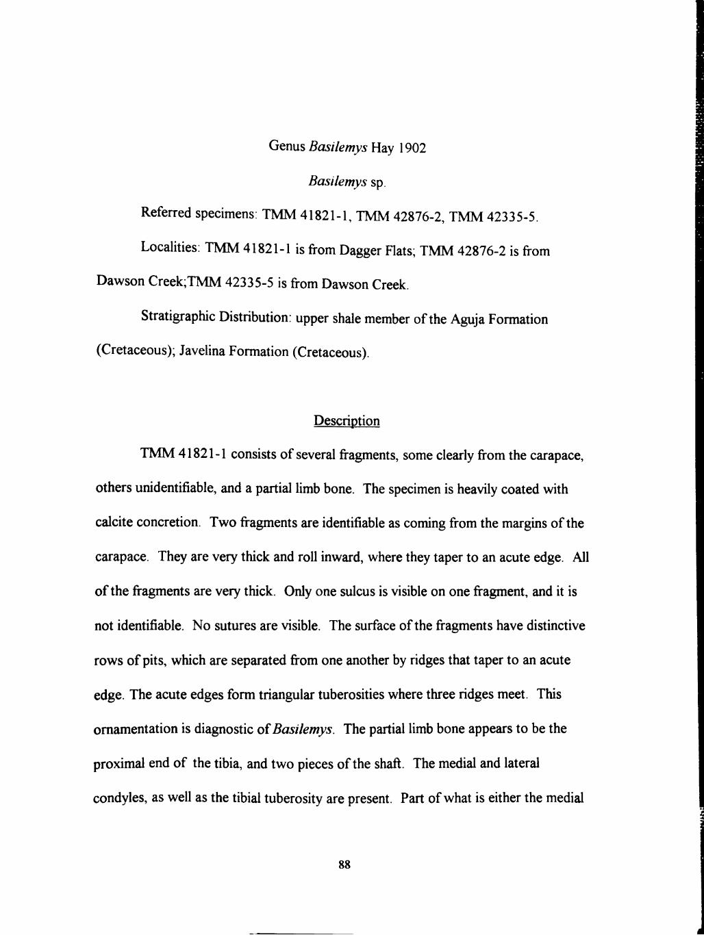
Genus Basilemys Hay 1902
Basilemys sp.
Referred specimens: TMM 41821-1, TMM 42876-2, TMM 42335-5.
Localities: TMM 41821-1 is from Dagger Hats; TMM 42876-2 is from
Dawson Creek;TMM 42335-5 is from Dawson Creek.
Stratigraphic Distribution: upper shale member of the Aguja Formation
(Cretaceous); Javelina Formation (Cretaceous).
Description
TMM 41821-1 consists of several fragments, some clearly from the carapace,
others unidentifiable, and a partial limb bone. The specimen is heavily coated with
calche concretion. Two fragments are identifiable as coming from the margms of the
carapace. They are very thick and roll inward, where they taper to an acute edge. All
of the fragments are very thick. Only one sulcus is visible on one fragment, and h is
not identifiable. No sutures are visible. The surface of the fragments have distinctive
rows of pits, which are separated from one another by ridges that taper to an acute
edge. The acute edges form triangular tuberosities where three ridges meet. This
omamentation is diagnostic of Basilemys. The partial limb bone appears to be the
proximal end of the tibia, and two pieces of the shaft. The medial and lateral
condyles, as well as the tibial tuberosity are present. Part of what is ehher the medial
88

or the lateral condyle is fractured. There is a foramen in the proximal end of the tibial
tuberosity.
TMM 42876-2 consists of three fragments that are probably from the same
specimen, and one addhional fragment. One of the fragments is from the margm of the
carapace. It is thick, shghtly rolled, and tapers to an acute edge. The other fragments
are much thinner. No sulci or sutures are visible. The surfaces of three of the
fragments have the diagnostic sculpturing of Basilemys. The fourth fragment, has a
somewhat similar sculpturing, but on a much smaller, and more subtle scale.
TMM 42335-5 consists of four fragments of the same specimen. The
fragments are probably from the carapace. One of the fragments is from the margin
and tapers to an acute edge, and appears to come from either the anterior or posterior
margm. All of the fragments are thick. No sulci or sutures are visible.
Discussion
These specimens aU possess the distinctive sculpturing and thick shell generally
attributed to the genus Basilemys (Hay, 1908; Langston, 1956; Brinkman and
NichoUs, 1993). Four species have been recognized for this genus, Basilemys
variolosa, B. sinuosa, B. nobilis, and B. praec/ara (Langston, 1956). None of the
specimens have the diagnostic characters necessary to assign them to any species.
There are no identifiable sulci or sutures, and the most complete specimen is largely
covered with calche concretion. All of this makes h difficult to identify these
specimens beyond the genus level; therefore they are identified herein as Basilemys sp.
89

Family TRIONYCHIDAE Fitzinger 1826
Subfamily TRIONYCFIINAE Fitzinger 1826
?Aspideretes spp.
?Eelopanoplia sp.
Referred specimens: TMM 41400-5, TMM 42335-8, TMM 42534-2, TMM
42534-8, TMM 422880-6, ST 7-16b, TMM 42539-5, TMM 42874-1, TMM 43057-
324, TMM 43380-5, TMM 43386-1; LSUMG V-1232.
Localities: TMM 41400-5 and TMM 42335-8 are from Dawson Creek; TMM
42534-2 and TMM 42534-8 are from Rattlesnake Mountam; TMM 42539-5 and
TMM 42874-1 are from Sierra Aguja; TMM 43057-324 is from Terhngua Microshe;
TMM 422880-6 is from the west fork of Alamo Creek; TMM 43379-1 is from
Grapevme hills; TMM 43380-5 is from Grapevine Hills; TMM 43386-1 is from
Pterosaur Ridge; and LSUMG V-1232 is from the "Gringo" locahty..
Stratigraphic Distribution: Upper shale member, Aguja Formation; lowermost
Javehna Formation; uppermost Javehna Formation; Black Peaks Formation.
Description
TMM 43380-5 is a partial carapace (Figure 3.31). It is fairly large, measuring
approximately 34 cm from the margm of the nuchal to the broken edge of the fifth
costal. It is sub-oval in shape, with a wide anterior end tapering to the posterior. The
90

nght lateral half of the carapace is nearly intact, whh a complete right half of the
nuchal and partial left half of the nuchal, through a nearly complete right fifth costal.
A fragmentary sixth costal, a preneural, and neurals one and two are also present, as
well as parts of ribs one through five. Neural/costal sutures for costals three through
five are intact, but worn. Moderately well developed first costiform processes are
present on the visceral side of the carapace. /Ul sutures are unfiised. A small,
suprascapular fontanelle is present. Omamentation on the dorsal surface is very worn
in places, but appears to consist of small, sub-rounded pits separated by narrow,
pomted ridges. The tapered edge of the ridges gives the illusion that the phs are
unusually deep m places. Omamentation does not extend to the margins, stoppmg
histead about 2.0 to 2.5 cm from the lateral periphery, and 3.0 to 3.5 cm from the
anterior margin of the nuchal..
TMM 42335-8 is the right half of a nuchal bone (Figure 3.32). ft is large, 17.5
in maximum length, 7.5 cm in maximum width, and approximately 2 cm thick. If h
were complete, the whole nuchal length would be at least four times the width when
measured at its wadest point. It is completely coated and partially replaced by gypsum.
Both the dorsal and visceral surfaces are very weathered, and h is difficult to
determine much about the surface omamentation. It is apparent, however, that the
dorsal surface had phs that were fairly large, and that they had coalesced in some
places. The anterior edge of the nuchal bone is shghtly curved in the center of the
bone, but is primarily straight, and does not exhibh a medial emargination.
91

-CJ
Figure 3.31. Trionychidae, TMM 43380-5, partial carapace. Bar scale is 10 cm.
92

Poshion of nuchal fragment
3
Figure 3.32. Trionychidae, TMM 42335-8, right half of a nuchal. Bar scale is 10 cm.
93
TMM 42534-2 is a well preserved right 5th costal (Figure 3.33). It is very
large, measuring 23.5 cm in length from a point on the lateral margin midway from the
anterior and posterior costal sutures to a point on the medial suture that is midway
between the anterior and posterior costal sutures. Its width at the lateral margin is 14
cm, and at the midline the width is 4.5 cm from the anterior costal suture to the
posterior costal suture. It is approximately 1 cm in average thickness. The costal has
a pronounced curve at the 6th costal suture, and a slight curve at the 4th costal suture.
Omamentation consists of very large round to sub-rounded, irregular phs,
some of which are fully coalesced into linear furrows and ridges that extend the width
of the costal. The phs extend from the medial suture to approximately 3.5 cm from
the peripheral edge. The omamentation appears worn, but does not exhibit extensive
signs of weathering or post-mortem abrasion. Sutural edges are intact. The visceral
surface exhibits the proximal end of a costiform process.
LSUMG V-1232 is a fragment from a costal. Omamentation consists of tiny,
reticulated, disconnected pin-sized ridges and tubercles. There are no phs.
TMM 43379-1 is an islolated fragment from a costal. Omamentation consists
of medium sized sub-rounded pits, narrowly spaced.
TMM 42534-8 is a broken, right hypoplastron (Figure 3.34). It is small, and
measures 0.7 cm in width. The anterior and posterior processes appear to be equal in
size. Omamentation consists of tiny, reticulated, disconnected pin-sized ridges and
tubercles. There are no pits.
94

i
Figure 3.33. Trionychidae, TMM 43534-2, right fifth costal. Bar scale is 10 cm.
95

Figure 3.34. Trionychidae, TMM 43534-8, right hypoplastron. Bar scale is 3 cm.
96

TMM 422880-6 consists of one nearly complete, weh preserved right
hyoplastron, one small fragment from the lateral posterior portion of a hypoplastron,
one peripheral edge of the fourth or fifth costal, and a small, unidentifiable piece of a
plastral element (Figure 3.35), as well as numerous other, largely unidentifiable
fragments from different individuals. The hyoplastron measures 25 cm from the medial
edge to a pomt on the lateral edge just posterior to two lateral anterior processes, ft
measures 8.6 cm in maximum width, and 6 cm in minimum width. It is approximately
1 cm in thickness, excluding the thickened portion of the axillary bridge. The hyo-
hypoplastral suture is mtact, unfused, and exhibits no evidence of post-mortem
abrasion. The plastral callosity does not extend all the way to the lateral margm of the
hyoplastron. The epiplastral anterior process which would project mto the median
fontanelle is broken, but h was fairly substantial, straight, and would be approximately
4.5 to 5.0 cm in width. The lateral anterior processes, while wide, are thin, measuring
only 0.5 to 0.7 cm in thickness. The axillary notch is moderately emarginate at a point
approximately one-fourth of the total distance from the lateral margin to the medial
margin. Sculpturing on the callosity consists of very reticulated pits, some of which
are coalesced, and separated by narrow ridges. Sculpturing does not extend all the
way to the edge of the callosity, but stops about 0.3 cm inside the edge. The right
lateral, posterior fragment of hypoplastron is well preserved, with little evidence of
post-mortem abrasion. Both lateral posterior processes are mostly intact. The
sculpturing on the callosity consists of very reticulated sub-rounded to vermiform phs
separated by thin ridges. Sculpturing does not extend to the margins of the callosity,
97

Figure 3.35. Trionychidae, TMM 422880-6, right hyoplastron. Bar scale is 10 cm.
98

but grades into a more or less smooth surface approximately 1.5 to 2 cm from the
edge.
The well preserved costal fragment consists of the distal end of the fifth costal.
The costal would have been large; the peripheral edge measures 8.5 cm in width. Both
costal sutures are intact. They and the broken edge show no signs of post-mortem
abrasion. Omamentation consists of large reticulated, sub-rounded pits that do not
extend aU the way to the margm, instead stopping approxhnately 2 cm from the edge.
TMM 42539-5 consists of the distal portion of the left hypoplastron, a partial
left xiphiplastral element, the distal end of the (?)right humems, and an element that
appears to be a portion of the pelvis that contamed the left (?) acetabulum (Figure
3.36). The distal end of the hypoplastron is preserved, and exhibhs one mtact lateral
process and one broken one. The digital processes are very robust, with the anterior
one shghtly thinner than the posterior process. The posterior and left lateral margins
are intact, but the hypoplastron is broken off about midway through the mgumal notch.
The broken edges do not appear to correspond to any sutures. The margin of the
hypoplastron tapers to a thin edge near the mguinal notch. The anterior end of the
xiphiplastron is intact and robust, measuring 10 cm in maximum length (from the left
margin to the midline suture), and 1.5 cm in average thickness. The callosity of the
hypoplastron and xiphiplastron is approximately 0.5 cm thick on average, and appears
to mn from edge to edge on all sides of both elements. The caUosity would contact
the right xiphiplastral element at the midlme. Sculpturing of the plastral calloshies
consists of phs of unequal sizes separated by ridges that taper to a sharp edge.
99

Figure 3.36. Trionychidae, TMM 42539-5, left hypoplastron, left xiphiplastron, and the distal end of a humems. Bar scale is 5 cm.
100

The humems is large, with the distal end measuring nearly 4.5 cm in width, and
the shaft measuring about 1.5 cm in diameter. The humems is broken at a point about
8 cm above the tip of the lateral condyle. The humems is slightly curved toward the
dorsal surface.
The broken portion of the pelvis containing the acetabulum is also large,
measurmg about 5 cm at maxhnum length and 4 cm at maxhnum width. This
specimen is badly weathered and the surface is covered with a calcareous concretion.
The broken surfaces of the ischium, ilium, and pubis are badly weathered and abraded,
but the acetabulum is largely mtact.
TMM 43057-324 is a broken but otherwise well preserved left lateral portion
of a hypoplastron (Figure 3.37). It is broken toward the medial edge of the ingumal
notch and near the hyo- hypoplastral suture. The mguinal notch is robust, measuring
2 cm in width on average. The lateral posterior and anterior processes are unequal m
size, with the anterior lateral process more slender than the posterior process.
Omamentation of the plastral caUosity consists of large coalesced phs.
TMM 42874-1 is a weU preserved, partially broken right xiphiplastron (Figure
3.38). It is roughly triangular in shape, and large, measuring 11 cm from the tip of the
anterior projection to the tip of the posterior medial projection. Projections are
xiphiplastral callosity was probably a moderately robust portion of the plastron. The
xiphiplastron is thin relative to hs overall size, measuring approximately 1 cm in
average thickness, mcludmg the plastral callosity. The callosity is approximately 0.4
101

Figure 3.37. TMM 43057-324, left hypoplastron. Bar scale is 10 cm.
102

Figure 3.38. Trionychidae, TMM 42874-1, right xiphiplastron. Bar scale is 3 cm.
103

slender and short relative to the overall size of the callosity, indicating that the cm
thick at the lateral anterior edge, and tapers to 0.2 cm at the medial edge. The
callosity extends to the medial edge, but does not give any indication that it would
meet the left xiphiplastral callosity across the midline Sculpturing of the callosity
consists of sub-rounded phs separated by thick ridges, which give the phs a shallow
appearance. The phs are largely of similar size, but become slightly smaller toward the
edges, or disappear altogether near the edge of the anterior medial projection.
TMM 41400-5 is a mostly intact, medium-sized left humems that measures 10
cm in length (Figure 3.39). The head, proximal articulation, and mtemal trochanter are
broken, but they appear slender and not robust. The mtemal fossa is shallow. The
fourth trochanter is abraded, but visible. The distal end is broken just above the lateral
and medial condyle, as are the articulation surfaces for the ulna and radius.
Discussion
The phylogenetic relationships of the family Trionychidae are complex,
primarily because useful taxonomic criteria are still being determmed (Webb, 1990).
Meylan (1987) has instituted a major revision of the family using cladistic analysis in
recent years, and Gardner and Russell (1994) and Gardner, Russell and Brinkman
(1995) have done a great deal of work on the identification and classification of the
Cretaceous trionychids, but there is still much that is unresolved, especially regarding
fossil species. It is not the purpose of this study to conduct a taxonomic revision of
this family; the specimens from Big Bend are not sufficient for this task. The work of
104

Figure 3.39. Trionychidae, TMM 41400-5, left humems. Bar scale is 5 cm.
105

Gardner and Russell (1994) and Gardner et al. (1995) may ultimately prove useftil in
identifying the Big Bend material to a species level. Certainly there are fragments
which are suggestive of Aspideretoides foveatus, A. splendidus, and A. allani
(Gardner et al., 1995). Insufficient material exists at this time to make a diagnostic
assignment concerning the specimens that resemble these species. In many instances
there are noticeable differences in shell morphology in trionychids from the Aguja,
Javelina, and Black Peaks Formations. While h is tme that some omamentation is
related to hs poshion on the shell, in many cases omamentation on two separate
fragments is so different as to be unlikely that the fragments came from the same
species. This seems confirmed by the recent work of Gardner and Russell (1994) and
Gardner et al. (1995). For the purpose of assessing the diversity of the fauna, h is
useful to make a conservative estimate of the number of different species in each
formation. For this reason, I will separate the trionychid fauna into forms below on the
basis of ornamentation, shell thickness, and overall size. These are illustrated in
Figures 3.40, 3.41, 3.42. Generic classifications are based on Hay (1908).
?Aspideretes sp. "A": Small with thin costal plates; omamentation consists of
very small, uniformly sized, uniformly rounded, uniformly and widely spaced pits. This
omamentation is is similar to that described for Aspideretes foveatus by Gardner et al.
(1995). Referred specimens: TMM 43473-2, Aguja Formation.
?Aspideretes sp. "B": Small with thin costal plates; omamentation consists of
medium sized, variably sized, rounded to sub-rounded, narrowly spaced phs. This
106

f . *
• * '^;^ti %. » « * .
(1)
(2)
(3)
Figure 3.40. Representative specimens of (1) ?Aspideretes sp. "A ", (2) "^Aspideretes "B", and (3) ?Eelopanoplia. Bar scales are 3 cm.
107

Figure 3.41. Representative of ?Aspideretes sp. "C". Bar scale is 10 cm.
108

(1)
"»?.•«*•.
«9 # * # « » # «
• ^ ^ *
(2)
area shown in Figure 2
Figure 3 42. Representatives of (1) ?Aspideretes sp. "D". and (2) ?A. sp. "E" (detail of carapace of TMM 43380-5, Figure 3.31). Bar scales are 3 cm.
109

omamentation is similar to that described for Aspideretes allani (Gardner et al, 1995)
Referred specimens: T\LM 42880-3, TMM 42534-6, T\LM 43379-1.
?Aspideretes sp. "C". Large with thick costal plates and plastrons;
omamentation consists of large, variably sized, sub-rounded, variably spaced pits that
are coalesced near the lateral margm. Referred specimen: TMM 42534-2. Aguja
Formation.
?Eelopanoplia sp. ; Small, thin hypoplastron; omamentation consists of small,
hregular. pin-head or smaller sized tubercles that sometimes coalesce to form a narrow
ridge. The omamentation is shnilar to that described by Hay (1908) Referred
spechnens: TMM 42534-8, LSL^G V-1232, Aguja Formation.
?Aspideretes sp. "D". Small with thin costal plates; omamentation consists of
smaU, uniformly sized, sub-rounded to rounded phs, spaced uniformly, at a distance
that is approximately one-fourth the diameter of a ph. Referred specimen: TMM
43386-1, Javehna Formation.
'^Aspideretes sp. "E"\ Large, thm carapace; omamentation consists of large,
variably sized, sub-rounded, variably spaced phs. Referred specunen: TMM 43380-5,
Black Peaks Formation. This specunen strongly resembles the Paleocene species
Aspideretes puercensis (Hay, 1908) with the sub-oval shape of the carapace (as seen
m Figure 3 31). Aspideretes puercensis also has suprascapular fontanelles, but they
are much more pronounced than the Big Bend specunen In fact, the nuchal does not
contact the first costal at all m 4. puercensis. Other authors have noted, however, that
m other spechnens where a suprascapular fontaneUe is present that there is significant
110

variation in this character among individuals (Gardner et al., 1995). Given the
resemblance of the overah shape, the suprascapular fontanelles, and the equivalent
stratigraphic poshion of . peurcensis and the Big Bend specimen (TMM 43380-5), it
is tempting to assign the Big Bend specunen to A. puercensis sensu (Hay, 1908).
Family DERMATEMYDIDAE Gray 1870
Genus Eoplochelys Hay 1905
Eoplochelys sp.
Referred spechnens: TMM 41400-19, TMM 41400-24, TMM 42957-2,
LSUMG V-979, LSUMG V-1004, LSUMG V-1476, LSUMG V-1504, LSUMG
5004, LSUMG V-5005, NGH 4, NGH 1-3, TMM 43476-2, ST no number.
Locahties: TMM 41400 19, 24 are from Dawson Creek; TMM 42957-2 is
from the Dogie area; LSUMG V-979 is from the "Tom's Top" locality; LSUMG V-
1004 is from the "Joe's Bonebed" locahty; LSUMG V-1476 is from the "Tom's Top"
locahty; LSUMG V-1504 is from the "Snail's Place" locahty; LSUMG V-5004 is from
the Dogie area; LSUMG V-5005 is from the "Tom's Top"; NGH 4 and NGH 1-3 are
from north Grapevine Hills; TMM 43476-2 is from the type section for the Black
Peaks Formation, and ST no number is from north Grapevine Hills.
Stratigraphic Distribution: lower part of the Paleocene Black Peaks Formation
111

Description
These specimens are all collections of fragments. There are several neurals,
peripherals, and costals. All exhibh a characteristic fine punctate sculpturing that is
visible with a handlens. The neurals are typically marked by deep sagittal sulci or deep
transverse sulci which bisect dorsal carinae (Figure 3.43). Specimen collections with
neurals mclude TMM 41400-19, LSUMG V-979, and LSUMG V-5005. There is one
suprapygal bone in TMM 41400-24.
Costals also typically exhibh longitudinal carinae bisected by deep transverse
sulci (Figure 3.44). Costals are found in the specimen collections of TMM 41400-19,
41400-24, LSUMG V-979, V-1004, LSUMG V-1476, and LSUMG V-5005.
Peripheral bones are thick, have ehher an upturned or non upturned edge, with
edges that range from acute to rounded (Figure 3.45). Deep marginal sulci are often
present. Peripheral fragments from TMM 41400-19 and TMM 41400-24 have a
subtle vmnklmg on the surface. Peripherals are found in the collections of TMM
41400-19, TMM 41400-24, TMM 42957-2, LSUMG V-979, and LSUMG V-1004.
Plastron fragments come from the hyo- and hypoplastron. They are thin
compared to the carapace bones, and where present, the buttress is remarkably non-
robust, given the thickness of some of the carapace fragments Sutures and sulci are
plainly visible. TMM41400-19 and LSUMG V-1476 contain fragments from the
plastron.
112

Position of neurals
?N3 ?N6
?N7 ?N8
Figure 3.43. Representative neurals from Eoplochelys sp. specunen TMM 41400-19 Bar scale is 3 cm.
113
Istm .s*m
Position of costal
?C3
Figure 3 44. Representative costal from
Bar scale is 3 cm.
//.p/oc/.e/,.sp. specimen TMM 41400-19.
114
Position of peripherals
?P4 (1) ?P3 (2)
Figure 3.45. Representative peripherals from Eoplochelys sp. specimens (1) TMM 41400-19 and (2) LSUMG V-979. Peripherals are shown m side view
Bar scale is 3 cm.
115
Discussion
All of these fragmentary turtle specimens are referred to the genus
Eoplochelys, primarily on the basis of the strongly developed dorsal carinae, the deep
sulci, and the thick, roUed edges of some of the peripherals. These diagnostic
characteristics match the descriptions of Eoplochelys found in Hay (1908) and
Gilmore (1919). The distinctive punctate sculpturing is also described by Gilmore
(1919), and is found on all the fragments described. Therefore, those fragments
without the dorsal carina, deep sulci, and rolled edges are also assigned to this genus.
116

CHAPTER IV
STRATIGRAPHIC DISTRIBUTION OF TURTLES
Introduction
Fossil turtles are, in general, far more abundant in the marginal marine,
brackish, and freshwater flood-plain deposhs of the Aguja Formation than in other
facies of the Aguja, or the fluvial flood-plain deposhs of the overlying Javehna and
Black Peaks Formations. A discussion of the turtles present in each formation is given
below, and a hstmg of the turtle genera and where they occur in the stratigraphic
sequence is provided (Figure 4.1). A more detailed breakdown of where each genus
occurs within the Aguja Formation (Figure 4.2), and a table showing the stratigraphic
distribution of each genus is also provided (Table 4.1). In some cases, as in
Bothremys, it is easy to esthnate the minimum number of mdividuals (MNI) because
the spechnens came from separate localhies. But because most of the material studied
is fragmentary, it is difficult to assess the MNI when many specimens are found at the
same localities. Thus, the numbers shown in the figures represent, in some cases, only
the occurrence of each genus at different locahties, and only for the three collections
studied. It is therefore probably a gross under-estimation of total number of individuals
present. Additionally, the numerous fragments of trionychid sheUs were separated
qualitatively mto groups suspected of being single individuals. These fragments were
used in the morphometries study (Chapter 6).
117

Kl
*%. ^
TIA
R
GC m h-
co 3
o LU o <
H LU cr o
<D C 0 O o LU
ce
ne
o 0) CO Q.
B r i d g e r-ian
W a s a t c h ian
TIffanian
Torrejonian * » ^
^ V ^ ^s ,
Puercan
C A N O E F O R M A T I O N
H A N N O L D HILL F O R M A T I O N
BLACK PEAKS
FORMATION
JAVELINA
FORMATION
AGUJA
FORMATION
M « p ' d « r « f « s 5p.
Hoploehmlyt ap.
8m»nm et. B. ornatm
7AapldmT9tma ap.
Baallamya ap.
tothramfa ap.
Bamnm ap.
Baana ct. B. nodoaa
Naurankylua ap.
Baallamya ap.
Compaamya ap.
Adoeua ap.
TAapldarataa ap.
THmlopanoplla sp.
TChmlnlldaa n. gan^ n. ap.
Figure 4.1. Stratigraphic distribution of turtles m the Aguja, Javehna, and Black Peaks Formations.
118
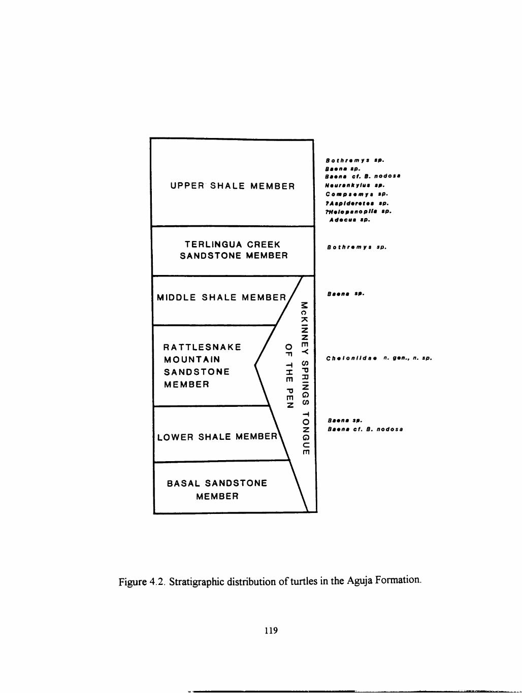
UPPER SHALE MEMBER
TERLINGUA CREEK SANDSTONE MEMBER
MIDDLE SHALE MEMBER
RATTLESNAKE MOUNTAIN SANDSTONE MEMBER
LOWER SHALE MEMBER
BASAL SANDSTONE MEMBER
B othram y a ap. Baanm ap. Baana et. B. nodoaa Naurankylua ap. Compaamy a ap. TAapldarataa ap. THmlopanoplla ap.
Adoeua ap.
B othram y a ap.
Baana ap.
Cha Ionlldaa n. gan., n. sp.
Baana ap. Baana cf. B. nodosa
Figure 4.2. Stratigraphic distribution of turtles in the Aguja Formation.
119
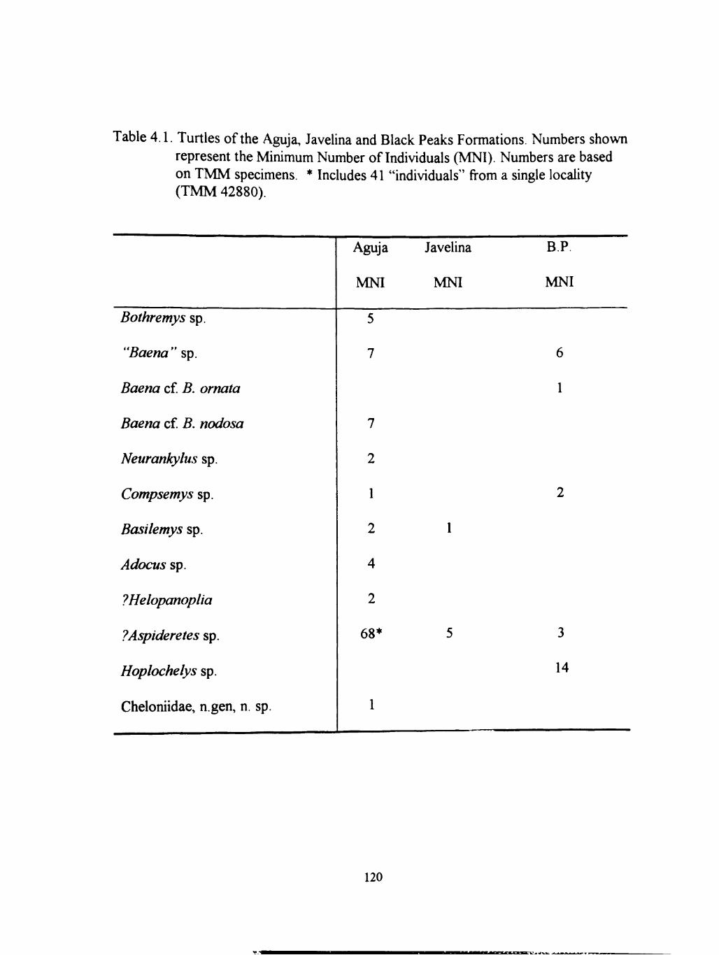
Table 4.1. Turtles of the Aguja, Javelina and Black Peaks Formations. Numbers shown represent the Minimum Number of Individuals (MNI). Numbers are based on TMM specimens. * Includes 41 "individuals" from a single locality (TMM 42880).
Bothremys sp.
"Baena" sp.
Baena cf B. omata
Baena cf B. nodosa
Neurankylus sp.
Compsemys sp.
Basilemys sp.
Adocus sp.
?Eelopanoplia
7Aspideretes sp.
Eoplochelys sp.
Cheloniidae, n.gen, n. sp.
Aguja
MNI
5
7
7
2
1
2
4
2
68*
1
Javelina
MNI
1
5
BR
MNI
6
1
2
3
14
120

Distribution and Abundance of Turtle Fauna
Turtles are relatively common in the Aguja Formation, and within the Aguja
they occur most commonly in the upper shale member. Of the two units in the upper
shale member - the lower, dehaic coastal plain deposits and the upper, fluvial channel
and flood-plain deposhs ~ most of the turtle fossils are concentrated in the lower unit.
Nearly all the turtles are thought to represent freshwater taxa. However, one marine
turtle was collected from the Rattlesnake Mountain Sandstone Member (TMM
42880). In addhion to this marine turtle there are several specimens of the
pelomedusid Bothremys from brackish water and coastal deposits of the upper shale
member. Pelomedusids have been found elsewhere in North America and the world m
both brackish and marine envhonments. The Aguja specimens (Bothremys) seem to be
equally distributed between the lower unit of the upper shale member, and the upper
part of the underlying Teriingua Creek sandstone member.
Of the freshwater turtles, those belonging to the family Trionychidae are the
most abundant, not only in the Aguja, but in all of the formations. These specimens are
represented mostly by broken, and sometimes reworked carapace and plastron
fragments. Most of the trionychids are probably referable to the genus 1 Aspideretes,
or the poorly documented ?Eelopanoplia, but the fragmentary nature of the material
makes h difficuh to determine whether other genera of this family are present. Current
taxonomic classification relies on skull and other shell characters besides sculpturing
for diagnosis at the generic or specific level. Nevertheless, much of the shell
sculpturing on the fragments collected from different localhies in the Aguja are
121

distinctive enough to be easily grouped into pattems. It seems reasonable to surmise
that at least Aspideretes is present, and that more than one species of this genus is
present, or possibly more than one genus of Trionychidae are present in these strata.
Differences in shell omamentation pattems could be explained by other factors, of
course, mcluding the age of the individual, the original poshion of the fragment on the
shell, or the amount of transport and weathering. The problems associated whh trying
to use shell sculpturing to determine the level of taxonomic diversity are discussed in
the taxonomic and morphometric sections.
The second most abundant group of turtles from the Aguja Formation belongs
to the family Baenidae. These are represented by "Baena" cf B. nodosa, "Baena" sp.,
Compsemys victa, and Neurankylus eximius (Table 4.1). The baenids are also more
commonly found as intact shells than any of the others. This is probably because the
shells of baenids are fully coossified by the time they are adults, making the shells less
Ukely to disarticulate after death, and more likely to be preserved intact. A nearly
complete carapace and plastron of 5. nodosa was collected from the lower shale
member. A complete carapace and plastron of the baenid Neurankylus cf A . eximius
was found intact in the upper unit (fluvial flood-plain deposhs) of the upper shale
member of the Aguja. Another nearly complete turtle, also referable to Neurankylus,
was found in the upper shale member. Compsemys victa is also present in the Aguja
Formation but is very rare.
The family Adocidae is represented in the Aguja by the presence of a few
isolated fragments of Adocus and marginal and costal fragments from Basilemys.
122

There are four fragments of Adocus, all from the upper shale member of the Aguja
Formation. There are three specimens of Basilemys, two of which are from the upper
shale member of the Aguja Formation.
There is a dearth of fossil turtle material from the Javelina Formation. Only
Aspideretes and Basilemys appear to be present, and these are very rare. The few
specimens that have been found are from fluvial flood-plam deposhs.
The abundance of fossil turtle material increases in the overlying Black Peaks
Formation. The most abundant form is Eoplochelys sp., followed by Aspideretes sp.,
and Baena cf B. omata.
Conclusion
The Aguja Formation contains the most diverse turtle fauna and the greatest
number of specimens, probably as a resuh of having been deposhed in a coastal plain
environment. This served as an excellent preservational environment, as well as being
an environment that probably hosted a large number of mdividuals (similar to the high
level of diversity which is found in coastal environments today).
There is a notable drop in both diversity and number of individuals found m the
Javelma Formation. This does not seem to be a function of taphonomic bias, because
numerous other vertebrate taxa, such as Quetzalcoatlus, Alamosaurus, Torosaurus,
and a variety of other dinosaurs, crocodilians and fish are present in moderate
abundance (Lehman, 1988). Indeed, the paleoenvironment, that of inland fluvial flood-
plain, would seem to be conducive to the preservation of vertebrate bones. The
123

paucity of turtle specimens could simply indicate that the environment was not one
where turtles were living in the latest Cretaceous. The turtle fauna increases sUghtly in
number and diversity in the Black Peaks Formation. This could be in part because the
environment returned to one that was slightly more favored by turtles during
Paleocene time.
124
" . - • - - ^ — - ^ - ^ - ^ - ' ^ - • • • - ' - •

CHAPTER \
ECOLOGICAL DIVERSITY OF THE BIG BEND TLHTLE F.AL^A
Introduction
The level of diversity of a fauna, however it is measured, is more easily
ascertained in modem faunal assemblages than in vertebrate fossil assemblages.
Vertebrate fossils, seldom present in the rock record in abundance, are often
mcompletely represented in the fossil record (and thus m museum collections) owmg
to numerous factors that include bias m collecting methods, bias in the preservational
modes, and varied amount of exposure of the strata m question. Such factors typically
result in an underesthnate of the diversity of a fossil fauna relative to its Uving
counterpart. All of these factors resuh in biasmg the fossil turtle fauna collected from
the Big Bend region compared to Uving faunas, thus making it difficult to determine
the tme level of diversity of the Big Bend turtle fauna.
A simple assessment of the relative abundance of the different taxa suggests
which turtles were present in greater numbers. Knowmg only the relative abundances
of the taxa does not give us much useful information about the level of diversity. To
determine that we must compare the Big Bend fauna both to typical modem faunas
and to other turtle assemblages from equivalent strata.
125

Methods
To limit the impact of biasing factors on the interpretation of the diversity of
the Big Bend turtle fauna, I have selected for study only the stratigraphic interval
where both preservation and coUection has been most thorough, the upper shale
member of the Aguja Formation (see Chapter IV). Although the exposure area of the
upper shale member is only a small fraction of that found m equivalent strata elsewhere
m North America, h is the richest level for turtles in Big Bend. This richness is
possibly a resuh of a good preservational environment, as weU as being an
environment that was originally one favored by turtles.
The upper shale member is also perhaps the most thoroughly coUected
stratigraphic horizon of all the mtervals covered in this report. I am aware of most, if
not aU of the specimens coUected thus far from this horizon. For the purposes of this
study only specimens coUected by the author and spechnens in the Texas Memorial
Museum collection were used. Surface collection was used by the author; aU
fragments of turtles found were coUected, and none were cuUed. The TMM specimens
were also surface coUected, and the entire coUection from this horizon was made
avaUable. Specimens from other collections involved other coUection methods, such as
screen washing, and were available only on a more Umited basis. For these reasons
they were not included as part of the diversity study.
126

Methods of Diversitv Estimation
There are primarily three components to species diversity species evenness, or
how evenly the taxa are distributed in the ecosystem under study; species richness, or
the number of species in a given area; and heterogeneity, which measures a
combination of both richness and evenness (Magurran, 1988). This study utilized
species richness to determine the level of diversity of the Big Bend fauna.
Most indices used in diversity measurement utilize the species as the
operational unit. Because many of the fossil taxa are represented only by fragments in
this study, and are not diagnostic at the species level, it is difficuh to identify the
number of species actuaUy represented m a genus. Because of this, I compare only the
genera m the different assemblages. Therefore, instead of usmg the term species when
discussmg the methods used for assessmg diversity, I will hereafter use the term genus
or genera.
Generic Richness
Measured generic richness is probably the simplest and most commonly used
method of determining the level of diversity in an ecosystem. It is a good way to
compare assemblages from different geographic locahties, and as I use h in this study,
potentially useful for comparing fossil assemblages to their modem counterparts.
Richness indices are based simply on the number of different taxa over a
geographic area sampled (Magurran, 1988; Rosenzweig, 1995) However, the larger
the area sampled, the greater the expected number of taxa represented. This has been
127

demonstrated to be tme through empirical means, and is typically represented by a
logarithmic species-area curve (Ricklefs, 1990; Rosenzweig, 1995) Therefore,
samples from a large geographic area are more likely to contain a greater number of
taxa. It is also tme that as areal coverage increases, the expected number of taxa found
m a sample approaches the number of taxa that exist m that environment. That is, new
taxa wiU not be added when the actual limit on the number of taxa m that envh-onment
is reached. The ideal study would strike a balance between the two extremes of not
sampUng enough to account for all the possible taxa in an area, and a needless waste of
time and resources continumg sampUng when aU the taxa are present and accounted
for. The difficulty, obviously, Ues in determming when aU the taxa have probably been
recorded. The sample size for the Aguja fauna is smaU (approximate minimum number
of individuals <50 for the upper shale member), covers only one sample area (Big
Bend, about 800 km^ of outcrop), and contains few taxa (seven genera). The exposure
area of the Big Bend strata is miniscule compared to equivalent strata elsewhere in
North America (Lehman, 1996). While the approximate minimum number of
mdividuals is known for the Big Bend fauna, that value cannot now be determined
from the literature for other assemblages, modem or otherwise, in order to make
meaningful comparisons in terms of minimum number of mdividuals.
The principle correction that must be made when comparing different modem
or fossil assemblages using generic richness indices is that the sampling m each
assemblage needs to be comparable in order to be meaningful (Magurran, 1988).
When comparing different geographic areas, h is safest, of course, to use areas of
128

comparable size. All other things being equal (such as amount of time spent sampling,
taphonomic biases, etc.), having comparable geographic areas somewhat ensures the
probability of comparable sample size, and therefore, one sample area will not be
"oversampled" or "undersampled" compared to the others. In the following sections, I
have compared the Big Bend fossU turtle fauna to other faunas from both
stratigraphically equivalent strata, and to a turtle fauna from an environmentaUy
comparable modem river drainage.
Comparison of the Big Bend Fauna to a Stratigraphically Equivalent Fauna
Turtles from equivalent strata, such as the Fmhland Formation and Kirtland
Shale of the San Juan Basin m northwestern New Mexico, come from much larger
exposures of Upper Cretaceous strata (about 4000 km of outcrop to about 800 km
for the Aguja, Lehman, 1996). Therefore, these strata are Ukely to preserve a richer
fauna than that of the upper shale member of the Aguja Formation. In some cases, if
the number of mdividuals coUected from the equivalent strata is known, a technique
caUed rarefaction can be used, which effectively normalizes sample sizes (Magurran,
1988). The rarefaction technique is not appUcable to this study, however, because h
depends on knowing the number of mdividuals in a sample, and this mformation is
unavailable from the literature concerning equivalent strata of the Fmhland and
Khtland formations.
A direct comparison of the generic richness of the Big Bend fauna and the
fauna from the Fmhland and Kirtland Shale formations is problematic. Nevertheless, 129

a genus for genus comparison with the Fmhland and Kirtland Shale fauna has been
made (Table 5.1).
Comparison of the Aguia Fauna to a Modem Assemblage
Another useful measure of diversity is to compare the Aguja turtle fauna to a
modem assemblage. When comparing the Big Bend fauna to modem assemblages h is
again necessary to have comparable geographic areas, for the same reasons given
above. In this mstance, however, a rough geographic equivalence is obtamed
esthnatmg the total geographic area represented by the upper shale member in both
space and time, and using this to extrapolate the comparable area m a modem
geographic area, i.e., to "fit" the modem assemblage to the fossU assemblage. This is
achieved in the foUowing manner.
The upper shale member does not represent only one habitat through the entire
duration of its deposition, but instead represents a coastal/ marginal marine
environment that eventuaUy graded over time mto an mland fluvdal floodplain
environment (Lehman, 1985). A comparable modem envhonment must also include
the same diversity of habhats. The climate and geography of the Big Bend area was
probably very much like the Central American peninsula (Mexico, Guatemala,
Honduras, Nicaragua, Costa Rica, and Panama) during the thne of Aguja deposhion
(Lehman, 1996), an area which includes the same coastal to inland fluvial habhats
found in the Aguja. However, the mformation about specific drainage basms
130

Table 5.1, Genera of turtles represented in the modem Brazos River drainage of Texas, the Aguja fauna of Big Bend, and the Fmhland and Kirtland fauna of New Mexico.
l ^ ^ ^ ^ ^ ^ ^ ^ ^ ^ i ^ W W ^ M W V
Brazos River Drainage Upper Shale Member Fmhland and Kirtland Shale
Caretta Lepidochelys Dermochelys Trionyx Gophems Geochelone Trachemys Terrepene Pseudemys Malaclemmys Graptemys Dehochelys Stemothems Kinostemon Chelydra
?Aspideretes ?Helopanoplia Baena Adocus Basilemys Neurankylus Compsemys Bothremys
Aspideretes Baena Adocus Basilemys Neurankylus Boremys Plastomenus Thescelus
,j^finnnriMtifxnjv^nnnfirinsmium^ • • » • • • • • » • . • •H>»^<%»^A<W*AMMA^^^»W»A<^^ .W^I^^<VW%VWWWWWV^^ An^AAAMAAAMMAf
131

in Central America, and the turtles found in those basins is unavailable. The generic
level of diversity for turtles in the entire Central American peninsula is easy to
ascertain, however, from Ernst and Barbour (1989). There are 12 freshwater or
terrestrial genera and 33 species found in Central America today. In Mexico alone
there are 12 freshwater or terrestrial genera and 29 species (Ernst and Barbour, 1989).
This suggests that the level of diversity for genera is relatively constant on the
continental landmass. If we compare the level of turtle diversity in the state of Texas
to that of Mexico, we find that there are 12 freshwater or terrestrial genera and 28
species found in Texas (Dixon, 1987), suggesting that the level of generic diversity for
the two landmasses is equivalent in sphe of the differences in size and climate.
Dixon (1987) provides a detaUed Usting of the turtles found in each county in
Texas, and in this manner h is easy to determine what turtles are found along a
particular drainage. Because the level of turtle diversity in Texas is comparable to that
of Central America, which in turn has a comparable geography and cUmate to the
Aguja, the Brazos River drainage in Texas, from hs headwaters to the coast was
chosen to represent a modem turtle habitat for comparison. But this presents a
problem: the total area of the Brazos River drainage basin far exceeds that of the
upper shale member, and to compare it directly to the upper shale member would
incorrectly inflate the generic richness of the turtles in the Aguja fauna. That is,
suppose the number of genera is the same for the Brazos River dramage and the upper
shale member. Let us use an example of five taxa. Then there would be five genera per
25,280 (632 (length) x 40 (width))square miles for the Brazos River dramage, or
132
0.0002 genera per square mile, and five taxa per 1600 (40 x 40) square miles for the
upper shale member, or 0.003 genera per square mile. This would erroneously lead us
to believe that the upper shale member has a richer number of taxa per square mile.
An estimation of the poshion of the coastline at the beginning of deposition of
the upper shale member and again at the end should provide a tmer estimate of the
geographic area that is being measured. That is, the length as measured from where
the upper shale member is exposed today, to the approximate position of the coastline
at the end of deposition of the upper shale member. This extends a length of 276 miles
(Figure 5.1), should give an estimate of how far inland the outcrop area of the upper
shale member was at the end of hs deposhion. The width is represented by the width
of the exposure today, or approximately 40 mUes. If the same length and width is
applied to the Brazos River, measuring from the coastline to a point inland that equals
the length applied to the upper shale member drainage, this yields a total area of
11,060 square miles for the area compared. It should be noted that the majority of the
genera along the Brazos River drainage occur within the first two or three counties
adjacent to the coast. However, to record all twelve, h is necessary to go 90 or more
miles inland, a distance that is still less than hah" of the distance measured for the length
of the Aguja drainage. This suggests that the distance measured for the Brazos River
drainage is still over-inflated, and in order to compare it more accurately to the Aguja,
h would be useful to know how far inland along the Aguja drainage h is necessary to
go in order to have sampled all of the genera. This information is not available,
therefore we will consider an area of 11,060 square miles for both drainages.
133

Figure 5.1. Dramage area estimated for this study. Upper shale member dramage is represented by the stippled pattern and the Brazos River drainage area is represented by the hachured Unes. Paleogeographic position of the shoreUne (dashed hne) during the middle of the Maastrichtian is from Lehman, 1985.
134

The upper shale member also represents approximately two million years of
geologic time, from the Late Campanian to Early Maastrichtian stage (Lehman, 1996).
So a comparison to the modem fauna must also include any turtles from the last two
million years (as an approximation, including the Pleistocene) of Texas. This fauna
would thus include Trachemys (the slider ttirtle), Chelydra (the snapping ttirtle), and
Geochelone (the extinct giant tortoise) (Hohnan, 1964,1969). Geochelone is the only
genus extinct today, and because relative abundance is unimportant in a taxon richness
study, h is necessary only to note the occurrence of this genus.
This same technique is appUed to the Fmhland/Kirtland Formations of the San
Juan Basm by measuring the linear distance derived from the position of the coastUne
at the beginning of the Campanian, and agam at the beginnmg of the Maastrichtian.
This distance is then applied to the Brazos River (Figure 5.2). A shnple comparison of
generic richness between the Aguja turtle fauna and modem fauna is provided below.
The total area of the Fmitiand and Kirtland Formations is derived by measuring the
distance between the poshion of the coastline of the Cretaceous Seaway during the
"middle" Campanian and the position of the coastline during the Late Maastrichtian,
and muhiplying by the width of the Fmhland and Kirtland Formation outcrops. The
total length is approximately 700 miles and the width is approximately 80 miles,
making the total area 56,000 square miles. The total length of the Brazos River
dramage is only roughly 600 miles, so the total area measured is 600 x 80 = 48,000
square mUes. Although smaller, the Brazos River dramage area is roughly comparable.
135

Figure 5.2. Drainage area esthnated for this study. The Fmitland/Kirtland dramage is represented by the stippled pattern and the Brazos River area is represented by the hachured lines. Position of the paleoshoreUne (dashed line) during the middle of the Maastrichtian is from Lehman, 1985.
136
Resuhs
The generic richness for the Aguja turtle fauna is eight genera (including one
marine taxon) per 11,000 square mUes, while the generic richness measurement for the
modem Brazos River drainage basin is a total of twelve genera (without the marine
taxa) per 11,000 square miles, and fifteen genera per 11,000 square miles with marine
taxa included (Dixon, 1987; Ernst, Lovich, and Barbour, 1994).
The taxon richness for the Fmhland and Kirtland Formations is eight genera
for 48,000 square miles (Gilmore, 1916, 1919, 1935). The taxon richness for the
Brazos River for 48,000 square miles is the same as h is for 11,000 square mUes,
twelve non-marine genera. The taxa known for the San Juan Basm, Big Bend, and the
counties of the Brazos River drainage basin are Usted in Table 5.1.
In the taxon richness comparison, the modem fauna appears to be 50 percent
more diverse than the Aguja fauna, with 12 freshwater and terrestrial turtles per
11,000 square miles, and three marine turtles to the Aguja fauna's 8 freshwater turtles
and one marine for the same area. A comparison of the Fmhland and Kirtland
Formation's assemblage yields approximately the same levels of diversity (Table 5.1).
It is mterestmg to note, however, that although the exposure of the Fmitiand and
Kirtland Formations is nearly five times that of the upper shale member (Lehman,
1996), the number of taxa represented is comparable. This impUes ehher that the
Aguja fauna is more adequately represented, in sphe of hs Umited exposure, or that the
Aguja fauna is potentiaUy even richer than that of the Fmitiand and Kirtland
Formations.
137
Morphology as a Diversitv Measure
At first glance, h is tempting to conclude that the comparable modem fauna is
substantially more diverse than the Aguja fossil fauna. However, an alternative way to
compare the diversity is to compare whether the diversity of the ecological niches
filled is the same. Assummg that a turtle's mode of life or ecological setting is reflected
in hs sheU morphology, a diversity assessment could be based on similar representation
of turtle "morphotypes" in comparable modem and fossil faunas. In some cases, for
example, in the trionychids, h is easy to see that the morphotypes are the same in both
modem and fossU forms. For the other freshwater aquatic taxa, however, it is more
difficuh to assess whether or not they fiU a comparable number of ecologic niches.
Of course, ecologic niches are complex, and include aspects that would be
impossible to determine in a fossU fauna, such as whether the turtle was nocturnal or
diumal, whether it preferred burrowing in mud to swimming in clear waters, or
whether h fed at the top, middle, or bottom of the water column. However, ecologists
have long understood that morphology is selected by environmental pressures, and
that the morphology of an organism reflects its poshion m the habhat (Ricklefs, 1990).
An advantage in using the morphology of an organism is that morphological
"measurements are independent of habhat context" (Ricklefs, 1990, p. 734). In other
words, it is not necessary to know whether a fossU turtle fed from the top, middle or
bottom of the water column; the organism has hs own niche in morphological "space."
An alternative measure of diversity, then, is whether the Aguja turtle fauna has a level
138

of diversity assessed in terms of morphological niche space comparable to a modern
assemblage.
The part of the turtle that is most easily preserved, the sheU, provides a very
broadly defined niche space in the form of the morphology of the sheU. In modem
faunas, the largest group of freshwater turtles, the Emydidae, can be grouped into two
categories according to a preference for fast or slow moving water (rivers or ponds).
In general, those turtles whh high-domed carapaces prefer slow moving water, such as
in ponds, marshes, dhches, or in the quieter bottom water of streams. Those having a
depressed carapace are more likely to be found in the fast moving water of rivers than
the high-domed turtles, and they will also occur in ponds, marshes and dhches (Ernst
et al., 1994). In terms of fluid mechanics, a high-domed sheU has greater surface area
than a depressed sheU of the same circumference. Greater surface area in tum offers
more resistance to stream flow owing to greater drag. To Uve in a fast-moving body of
water then, a high-domed turtle would probably have to expend more energy. It is less
clear how the overaU size of the shell would determine preference for bodies of water,
but as discussed below, there are some clear preferences here as well.
Because the size and shape of the sheU might hold clues to the number of niche
spaces available, the modem turtle fauna were divided into nine categories. The first
six include the Emydidae, the largest and most diverse group. The emydids are
arbitrarily divided into small (<17 cm), medium (17-40 cm), and large sizes (>40 cm).
These size groups are in tum divided into two groups each on the basis of the ratio of
total shell width (measured by holding one end of a string to the lateral edge of the
139

carapace at a point about midway between the anterior and posterior ends, and
mnning the string across the dorsal portion of the carapace to the equivalent
peripheral) to a straight-line measurement of width at the same point (Figure 5.3).
Those turtles with a carapace height to width ratio greater than 1.5:1 are placed into
the high-domed category, while those with a ratio of less than 1.5:1 are considered to
have a depressed carapace. This classification of recent turtles into six categories is
shown in Table 4 I was only able to directly measure shells on six genera of the
modem turtles. The other turtles are placed in their categories according to
measurements found in the Iherature, and using photographs. I was able to measure
sheUs on all of the fossil turtle genera except Compsemys. The recent and fossU
specimens that I measured were ehher specimens used in this study, or specimens from
museum coUections. It is important to note that fossU turtles are often found in an
artificially depressed state owing to cmshing from burial compaction. This must be
taken into account when assessing the validity of these categorizations, and this was
done qualhatively by estimation. When the emydids are classified in their appropriate
morphological categories h becomes apparent that the modem turtles that prefer
ponds and still waters (Stemotherus, Kinostemon, Terrepene) are turtles with small,
high-domed shells. The medium and large turtles with depressed shells (Pseudemys,
Graptemys, Trachemys, Deirochelys, Malaclemmys, and Chelydra) can be found in
almost any body of water, fast or slow moving. It is interestmg to note that there are
no large, high-domed freshwater turtles. The other three groups are the marine turtles
and the Trionychidae, which are more directly comparable to fossil forms than the
140

Figure 5.3. Method of measurement for morphology study. Dashed Une represents measurement. Turtles are drawn m the manner of Sonrel (1857).
141

Emydidae are to the Aguja fauna, and the tortoises, Geochelone and Gopherus The
nine different categories are listed in Table 5.2. It is possible to compare the
morphological estimates of niche space for the modem and Aguja faunas. The six
categories of emydids are compared with the freshwater turtles of the Aguja fauna on
the basis of the shell size and shape, and the other three categories of marine,
trionychid and tortoise are also compared. Of the six emydid (or, in the case of the
fossil turtles, freshwater) categories, all but the small depressed and large, high-
domed forms have representatives in both the modem and Aguja fauna. The other
categories of morphological niches can be directly compared. For example, the marine
turtle Bothremys, and the trionychids (lAspideretes and lEelopanoplia) each have
modem counterparts or living representatives, so the presence or absence of a
morphotype is easily ascertained. There is no Late Cretaceous representative for the
tortoise morphotype; tortoises themselves did not appear in the fossil record untU the
Eocene (Hay, 1908), but the tortoise morphotype appeared in the Paleocene in the
form of Eoplochelys (Hay, 1908).
When totaled, the modem fauna has seven morphological categories
represented, and the Aguja fauna has six (Table 5.3). Where the freshwater turtles are
concerned, including the Trionychidae, the modem fauna and the Aguja fauna have
equal numbers of four morphological niches represented. So although there are nearly
twice as many genera in the modem fauna, there are a comparable number of
morphological niches (on the basis of shell size and shape) in both groups, suggesting
that the Aguja fauna is probably adequately represented with respect to diversity
142

Table 5.2. Comparison of Brazos River drainage fauna and Aguja fauna. Asterisks denote turtles that have not been physically measured.
Size Shape
SmaU <17cm >1.5/1 Domed
Small <17cm <1.5/1
Flat
Medium 17-40 cm
>1.5/1 Domed
Medium 17-40 cm
<1.5/1 Flat
Large >40cm >1.5/l Domed
Large >40 cm <1.5/1
Flat
EXTANT
FOSSIL
Stemotherus
Kinostemon
Terrepene
Compsemys*
Deirochelys
Baena
Pseudemys*
Graptemys*
Trachemys*
Adocus
Malaclemmys*
Chelydra
Neurankylus
Basilemys
143

Table 5.3. Comparison of Brazos River drainage fauna and Aguja fauna.
General morphology Modem Fossil
Small, high domed
Small, depressed
Medium, high domed
Medium, depressed
Large, high domed
Large, depressed
Marine
Trionychid
Tortoise
X
X
X
X
X
X
X
X
X
X
X
X
X
144

measured in morphological "space." This is consistent with current thinking in
morphological study, which holds that the number of niches in morphological space
(i.e. "species packing") is relatively constant (Ricklefs, 1990, p. 734).
Simpson's Index of Diversity
Simpson's Index of Diversity (SI) is a commonly used diversity index in which
D = I ((w , -ni)/(N^ - N)), where «, is the number of individuals in the ith taxon, and
A' equals the total number of individuals. As D increases, diversity decreases
(Magurran, 1988; Pielou, 1975). The upper limh ofD is set at 1.0 (Rosenzweig,
1995). Unfortunately, there are no studies of which I am aware that use Shnpson's
Index on either fossil or recent turtles, so there is no D value to which I could compare
the D value of the Aguja fauna. However, for any future studies that may use this
index, I have calculated the SI of the fauna from the upper shale member of the Aguja
Formation. For this fauna, D = 0.3487. A Ust of minimum number of individuals for
this fauna is shown in Table 5.4.
Conclusions
Although a generic richness comparison of the Aguja fossil turtle fauna with a
modem assemblage of turtles suggests that the Aguja fauna is only half as diverse, a
comparison whh equivalent strata like the Fmitiand and Khiland Formations (which
have much larger area of exposure) shows a very similar level of diversity. A
comparison of the Aguja fauna diversity expressed as shell morphotypes with a
145

Table 5.4. Taxa and number of individuals from the upper shale member of the Aguja Formation used for Simpson's Index calculations.
Genus
?Aspideretes
Baena
Adocus
Bothremys
BasUemys
Neurankylus
?Helopanoplia
Compsemys
Total
Minimum Number of Individuals
23
5
4
2
2
2
1
1
40
146

modem assemblage suggests that, although not as many taxa are represented m the
upper shale member, neariy as many shell forms are present in the fossil assemblage as
are present in the modem assemblage. As mentioned previously, the Aguja fauna was
selected for the diversity study, in part, because the collecting and preservational
biases were at a minimum. Because of this, the comparison of the this fauna to both
stratigraphicaUy equivalent fauna and to a modem assemblage points to a moderate
level of diversity for the Aguja fauna.
147

CHAPTER VI
MORPHOMETRIC ANALYSIS OF PIT VARIANCE
IN TRIONYCHID SHELLS
Introduction
Carapace and plastron fragments of turtles representing the soft-shelled family
Trionychidae are abundant throughout the Late Cretaceous and early Tertiary strata of
North America. Current taxonomic work on turtles relies heavily on skull characters,
as well as other skeletal features, for the diagnosis of species (Gaffiiey 1975; Gaffhey
and Meylan, 1988; Meylan 1987; Meylan and Gaffiiey, 1989). WhUe there is good
reason for this, h is not always useful for the stratigraphic paleontologist,
paleoecologist, or biostratigrapher faced with a paucity of well-preserved cranial
material. In many cases, while it is evident from fragments of sheUs that soft-sheUed
turtles were abundant at some localities, v thout the skuUs or some other diagnostic
skeletal character these turtles can only be identified to the familial or generic level.
This obscures a great deal of useful information that could be gleaned from the she
about the level of diversity of the fauna.
For many years, pattems in the distinctive phs on the carapace or plastral
calloshies of soft-shelled turtles were considered to be specifically diagnostic features
(Gilmore, 1919, 1946; Hay, 1908). However, this practice has fallen into disfavor
(Meylan, 1984, 1989). Recent work by Gardner and others (Gardner and RusseU,
148

1994; Gardner, RusseU, and Brinkman, 1995) suggests, however, that the pattems of
the phs on the sheU might be of use in taxonomic diagnosis. To use the phs as a
diagnostic character the assumption must be made that intraspecific variability in the
pattems is low; that is, pattems are not highly variable among individuals, but are
discriminatory at some taxonomically significant level, such as species or genus.
The assumption of non-discriminatory variability in ph pattems has never been
assessed quanthatively. If h could be found that characteristic ph pattems, however
subtle, can be identified for distinct groups (perhaps reflecting different genera or
species), the potential for identifying fragments as belonging to different species of
turtles increases, as does the potential for usmg these fragments in ecological or
stratigraphic studies.
A number of factors must be considered before attempting to quantify ph
variability. Variability m pattems may be related to (1) where the phs are located on
the sheU, or to (2) sexually dimorphic, (3) ontogenetic, or (4) taphonomic processes.
Before any study like this can be interpreted whh confidence, such "noise" factors
must be identified and partitioned or fihered out. As a first step a model should be
developed which would assess whether any pattems of a discriminating nature at all
are distmguishable. If no such pattems are found then the assumption of a
discrimmatory variabUity is shown to be false, and no further testing would be
necessary. If a discriminatory pattern is found, however, then further testmg is desired
on a larger data set in order to determine whether h is possible to fiher out the noise
factors, and if so, whether h is then possible to use this method for taxonomic
149
purposes. Furthermore, if a pattern is shown to exist, h is important to determine
whether the pattern or pattems that are discemed are simply an artifact of the
statistical analysis or something with a biological basis. This can be done by generating
random numbers that are similar in character to the numbers used to describe the phs.
Morphometric studies are muhivariate analyses used to describe biological
forms, as weU as to discriminate among biological forms (Bookstein, Chemoff, Elder,
Humphries, Smith, and Strauss, 1985; Pimentel, 1979). One of the methods long used
by biologists to compare forms is to place them into morphological space, or
"morphospace" (Bookstein et al., 1985). This idea, used by Raup in a landmark study
on the coiUng of molluscs (1966), takes natural forms and compares them to imagined
or unobserved forms by placing them into a multi-dimensional space that geometrically
describes the forms. The pits on trionychid turtle sheUs are forms that wUl fit
somewhere in morphospace. Ideally, the differences in the shapes, sizes, and spacmg
of the pits on the sheUs, will fit in different places m this muhi-dimensional
morphospace. Then, if the variability is discriminatory (i.e., if there is a discemable
pattern for a given taxonomic level, like species or genus), the differences in pattems
will show up as different places in morphospace. If this should be the case, we might
be able to verify, at the very least, the presence of more than one taxonomic group in a
fauna, even if their actual identities are unknown.
The following is a preUminary study utilizing some fiindamental morphometric
techniques to determine whether any discriminatory patterns in the phs of trionychid
shells can be identified. If the resuhs indicate that pattems are discemable in the phs,
150

then fiirther studies will be conducted at a later date in order to determine what these
pattems represent, and whether they will be of any biostratigraphic or paleoecological
use determining stratigraphic correlations or levels of diversity in a turtle fauna.
Methods
Morphometric measurements were made on fragments of Late Cretaceous and
early Tertiary turtle sheUs coUected from Big Bend, using Image-pro Plus Image
Processmg System software (Media Cybernetics, 1992). A digitized measurement of
the total area and perimeter was made first for each fragment. Then each individual ph
was measured, taking the outside perimeter measurement and deriving the area from
that. Where two or more phs had apparently grown together (coalescing to form one
pit), the original individual phs were measured first separately, and then the coalesced
measurement was taken. The coalesced measurements were kept in a separate data set
in order to determine the effect they might have on principle components.
Of the avaUable fragments, 24 were from the same locaUty (TMM 42880) in
coastal floodplain facies of the upper shale member of the Aguja Formation. These
fragments, although from the same locality, are not all from the same individual. This
assumption is based on a taphonomic assessment of where the fragments were located
at the she, a quaUtative assessment of ph pattems and appearances, and a distinct
difference in the preservational appearance of the fragments. However, a minimum
number of individuals is not determinable. Standards for comparison were chosen from
two other localhies; these included 3 addhional trionychid shell fragments, 2 from a
151

locaUty within the Javelina Formation, and I from a Paleocene locality within the
overlying Black Peaks Formation A fourth standard was chosen from the Javelina
Formation from a specimen representing the family Dermatemydidae, and the genus
Basilemys
Principle components analysis (PC A) was chosen as a method of measuring
variance/covariance of the variables in the data set. PC A is often used as a starting
pomt in any analysis of biometric data, partly because the mterpretation of the data is
often straightforward and uncompUcated (Davis, 1986). In principle components
analysis selected variables (charaaers that are being measured) and their covariances
are plotted on a matrix. For example, the total area and perimeter of a section on the
sheU containing a coUection of pits would be two variables (or components). The
covariance between these two measurements is then calculated. The variance-
covariance matrix can then be mterpreted geometricaUy, and the principle components
analysis measures the axes and their magnitudes on an x/y plot. In a muhivariate data
matrix PC A isolates the principle components, and plots them in muhidimensional
space in a series of x/y plots.
The primary value in PC A Ues in being able to isolate those variables that are
most (and least) significant. In PC A the components are weighted factors, with each
factor comprismg a percentage of the total variance (Kachigan, 1991). For example.
principle component 1 (PCI) might account for 80% of the total variance. This
identifies PCI as the smgle most important variable measured. PC A can thus be used
to identify the most (and least) hnportant variables being measured.
152
The computed scores of the eigenvectors are called "loadings", and are the
coefficients of the linear equation which defines the eigenvector (Davis, 1986). A high
negative or positive loading for a variable indicates that it is highly correlated with the
prmciple component. A component having both negative and poshive loadings is
considered bipolar, while one with a single sign is a general size factor (Bookstein et
al, 1985). A bipolar component is usually a character that is not related to overall size
(e.g., simply measuring the difference between a large and small specimen of the same
organism), but measures the relationship between other variables, such as size and
shape.
One advantage of using PC A in this application is to identhy the most
hnportant contributions to size/shape variance among the phs on trionychid sheUs.
Also, because h is not known how many groups are actuaUy present in the data set,
PCA can also be used to see whether there is a pattem to the distribution that suggests
when more than one group is present.
Data Analysis of Big Bend Trionychid SheU Fragments
Principle components analysis was applied to the data set usmg nine variables.
These variables were chosen to describe the morphology of the pits by measuring size
of the individual phs, complexity of shape, and distance between mdividual phs. The
nme variables included: total fragment area (TOT ARE A), total fragment perimeter
(TOTPERIM), mean areas (MAREA) and perimeters (MPERIM) of individual phs
within the total area measured, standard deviations of the areas (SAREA) and
153

(SPERIM) perimeters of the individual phs within the total measured area, sums of
the areas of the individual phs subtracted from the total measured area (SPACE), and
the covariance between area and perimeter of the individual phs (SQCOV) (Table
6.1). All variables were transformed to natural logarithms. Complexity of form is
assessed by comparison of the standard deviation of ph perimeter to area of the ph
measured. Variability in the size of phs is measured by comparison of the standard
deviation in area of phs measured to the mean size of the phs in the area measured.
Distance between phs is measured by subtracting the sum of the area of the pits on a
specimen from the total area measured on the specimen. The program was written in
the SAS programming language. This program is provided in the Appendix and is
called "Totturt.SAS". The resuhs are discussed in the Resuhs section below.
Randomized Data Analysis
The purpose for generating random data is to determine whether the pattems
seen in the PCA are biological or geometrical in origin. In Raup's (1966) study with
the coilmg of moUusc shells he was able to demonstrate that one could predict where a
biological form would plot in morphospace by plotting geometric forms. Real data
from a real form would then hypothetically plot somewhere within that space defined
by geometrically generated data. If data were genuinely random in nature, and the real
data being tested were not random in origin, but had biological constraints, then the
two data sets should plot differently. The ideal study would first try to generate
random data, within a range similar to the real data, to determine whether the two sets
154

Table 6.1 Variables used in morphometric study.
Total area
Total perimeter
Mean, area of individual phs
Mean, perimeter of individual phs
Standard deviation, area of individual pits
Standard deviation, perimeter of individual phs
Space between individual phs
Covariance between pit area and perimeter
TOTAREA
TOTPERIM
MAREA
M_PERIM
S_AREA
S_PERIM
SPACE
SQCOV
155

of data plot similarly. If they do not, then this would suggest that the real data are
measuring variables that are somehow constrained biologically. That is to say, random
forms represent the null hypothesis of non-discriminating variation.
Two SAS programs were written to compare the observed data to randomly
generated data. In the first of these, a random set of data was generated for
comparison to the real data in order to determine whether any pattems that arose from
the data were statistical or biological in nature. In the second program, random data
and real data were merged m order to isolate any biological constraints that would
determine where the real data would plot in morphologic space. These programs are
provided in the Appendix and are caUed "RandturtNC.SAS" and "AUdata.SAS"
respectively. The resuhs are provided in the resuhs section below
In the program to generate representative random data the foUov^ng
constraints were placed on the data: ph perimeter and area had to be of a comparable
size to the real data; ph number per specimen had to be of comparable size and vary in
number from 4-10; random numbers generated for the perimeter have a normal
distributution; perimeter/area is variable. Fifty "spechnens" were generated for a total
of 259 observations. This program provided in the Appendix and is caUed
"Randturtnc.SAS'.
The second program merged random data with the real data. In this program
constramts were placed on the random data. Because resuhs from real data suggest
that the variance in the shape and size of the ph is inversely proportional to the size of
the ph, this was included as a constraint on both the area and perimeter of each ph. An
156
additional constraint factor was added for the perimeter in order to limit the variance
in the complexity of the shape. These data were then merged with real data to compare
the resuhs in PCA loadings and plots. This program is included in the Appendix and is
called "AUdata.SAS".
Resuhs
Data Analysis of Big Bend Trionychid Shell Fragments
Two initial sets of analyses were mn on the SAS program, one to account for
the coalescence of phs, the other to remove the coalescence factor. No differences in
resuhs were observed. The first principle component (PCI) reflects -50% of the total
variance among the phs (Table 6.2). This axis is a bipolar axis with strong poshive
loadings for the standard deviations of the areas and perimeters of the pits, and strong
negative loadings for the means of the areas and perimeters of the phs, suggesting that
h measures variabUhy in the size of the phs within the area measured.
PC2 reflects -32% of the total variance. This axis exhibhs aU poshive
loadings, and is therefore a general size component. PC3 accounts for -16% of the
total variance, and is a bipolar axis. A very strong poshive loadmg, with a value near
1.0, is registered for the space factor. This component, then, measures the distance of
the space between the phs. Principle components 4 through 7 accounts for less than
3% of the total variance, and are therefore considered to have Uttle influence on the
total variance. The plots of PCI versus PC2, and PC2 versus PC3 are shown in
Figures 6.1 and 6.2, respectively.
157

Table 6 2 TOTTLUT Data.
Mean StD
M.AREA
0.8431 0.2610
S_AREA
0.2567 0.1006
M_PER1M
2.268 0.2673
Mean StD
S.PERIM SPACE SQCOV
0.2881 0.1310
59.18 28.36
0.2678 0.1088
M_AREA S.AREA M_PERIM S.PERIM SPACE SQCOV
Correlation Matrix
M_AREA S_AREA M_PER1M
1.000 -.1869 0.9909 -.0956 0.2129 -.1578
-.1869 1.000 -.1882 0.9220 0.1137 0.9857
0.9909 -.1882 1.000 -.0937 0.1553
.1539
the mean, AREA the standard deviation, AREA the mean, PERIM the standard deviation, PERIM
S PERIM SPACE SQCOV
M_AREA S_AREA M_PERIM S_PERIM SPACE SQCOV
-.0956 0.9220 -.0937 1.0000 -.0502 0.9711
0.2129 0.1137 0.1553 -.0502 1.0000 0.0362
-.1578 0.9857 -.1539 0.9711 0.0362 1.0000
the mean, AREA the standard deviation, AREA the mean, PERIM the standard deviation. PERIM
Eigenvalues of the Correlation Matrix
Eigenvalue Difference Proportion Cumulative
PRINl PRIN2 PRIN3 PRIN4 PRIN5
3.042 1.9450 0.9479 0.05651 0.00756
1.097 0.9970 0.8914 0.04895 0.0066
0.5070 0.3242 0.1580 0.009418 0.0013
0.5070 0.8312 0.9892 0.9986 0.9998
158
•A^^fe^AAAari B^^M
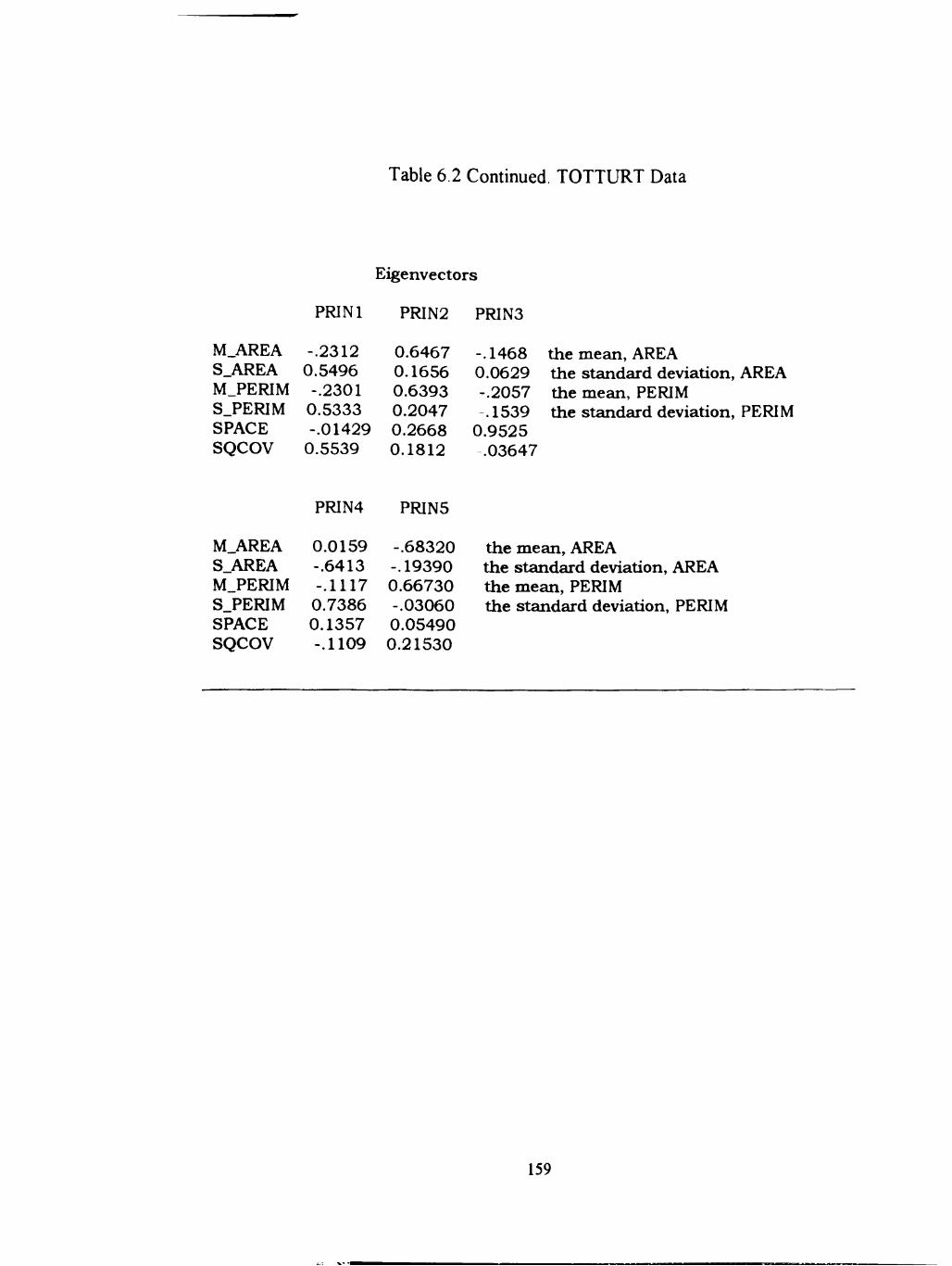
Table 6.2 Continued. TOTTURT Data
M.AREA S_AREA M_PERIM S.PERIM SPACE SQCOV
PRlNl
-.2312 0.5496 -.2301
0.5333 -.01429
0.5539
Eigenvectors
PR1N2
0.6467 0.1656 0.6393 0.2047 0.2668 0.1812
PRIN3
-.1468 0.0629 -.2057 .1539
0.9525 .03647
the mean, AREA the standard deviation, AREA the mean, PERIM the standard deviation, PERIM
PR1N4 PRIN5
M_AREA S_AREA M_PERIM S.PERIM SPACE SQCOV
0.0159 -.6413 -.1117
0.7386 0.1357 -.1109
-.68320 -.19390 0.66730 -.03060 0.05490 0.21530
the mean, AREA the standard deviation, AREA the mean, PERIM the standard deviation, PERIM
159

Figure 6.1. Plot of PRJN2*PRJN1, TOTTURT Data. Bold, underlined characters are standards used, which include Basilemys sp. TMM 42335-5 (z), Aspideretes sp., Javelina Formation TMM42874-1(A), Trionychidae(a) TMM 42876-1 (B), and Trionychidae(b) TMM 42876-1 (C). PRINl represents variability in ph size; PRIN2 represents overall size of ph.
160

PRIN2
3 +
X
2 +
1 +
0 +
- 1 +
- 2 +
- 3 +
-4 + I
V
a r u in
t b
w
B
+-
4
- + •
- 3
-+
-2
- +
• 1
+
2
+-
3
PRINl
161

Figure 6.2. Plot of PRJN3*PRIN2, TOTTURT Data.. Bold, underlined characters are standards used, which include Basilemys sp. TMM 42335-5 (z), Aspideretes sp., Javelma Formation TMM42874-1(A), Trionychidae(a) TMM 42876-1 (B), and Trionychidae(b) TMM 42876-1(C).
162

PRIN3
3 . 0
2 .5
2 . 0
1.5 +
1.0
0.5 +
0 .0 +
- 0 . 5 +
- 1 . 0 +
- 1 . 5 + I -+-
-4 - 3
k C
m
n
g b
i c d
w
a tu r
V
y e
X
- 2
- + +-
- 1
PRIN2
163

Random Data Analysis
Random data generated separately did not plot similarly to the trionychid data
(Figures 6.3, 6.4). Loadings are shown in Table 6.3. When the random data were
merged with the real data (with constraints) PCI accounted for -43% of the total
variance, PC2 for -23%, and PC3 for -17% (Table 6.4). Loadings were very similar
to that of the real data. When the data are plotted according to PCI versus PC2, and
PC2 versus PC3, the random data and the real data appear to plot similariy in
morphospace (Figures 6.5, 6.6).
Discussion and Conclusion
Discussion
In the analysis of the real trionychid data there were no discemable differences
in the numerical or graphical resuhs of the analyses between the data set of coalesced
phs and the data set m which this factor had been removed. This suggests that the
coalescence of pits as a factor is too small to be measured as a component of variance
in this data set. However, coalescence of phs appears to typically occur along the
margins of carapaces, so it is possible that with a larger data set, this component could
take on more significance as an indicator of location of the specimens on the carapace.
Consequently, h would be advisable to continue measuring coalesced phs separately
and using them for comparison with a data set in which this factor is removed. Two
164

Table 6.3. RANDTLHTNC Data.
M_AREA S_AREA M_PERIM S_PER1M SQCOV
Mean 1.4064 StD 0.0448
0.1777 0.0585
2.293 0.0251
0.0966 0.0201
0.0223 0.0737
Correlation Matrix
M_AREA S_AREA M PERIM
M.AREA S_AREA M_PERIM S.PERIM SQCOV
1.0000 0.6114 0.1159 -.0530 0.0539
0.6114 1.000
0.1189 0.1127 0.3837
0.1159 0.1189 1.0000 -.1299 -.1669
the mean, LOGAREA the standard deviation, LOGAREA the mean, LOGPERIM the standard deviation, LOGPERIM
S.PERIM SQCOV
M.AREA S.AREA M.PERIM S.PERIM SQCOV
.0530 0.1127 -.1299 1.0000 0.4290
0.0539 0.3837 -.1669 0.4290
1.0000
the mean, LOGAREA the standard deviation, LOGAREA the mean, LOGPERIM the standard deviation, LOGPERIM
Eigenvalues of the Correlation Matrix
PRINl PRIN2 PRIN3 PRIN4
Eigenvalue
1.821 1.457 0.8662 0.5659
Difference
0.3642 0.5903 0.3002 0.2755
Proportion
0.3641 0.2913 0.1732 0.1132
Cumula
0.3641 0.6555 0.8287 0.9419
Eigenvectors PRINl PRIN2 PRIN3 PRIN4
M AREA S AREA M.PERIM S.PERIM SQCOV
0.4935 0.6436 0.01293 0.3128 0.4942
0.4581 0.2398 0.4938 -.5454 -.4374
-.2976 -.07284 0.8562 0.3992 0.1170
0.3861 .1427
-.1028 0.6631 -.6167
mean, LOGAREA std. dev., LOGAREA mean, LOGPERIM std. dev., LOGPERIM
165

2 +
PRIN2
A A
A A
0 +
A
A
- 2 +
-4 +
A BA
A A
AA
•-+ + + + + + + +-
- 3 - 2 - 1 0 1 2 3 4
Figure 6.3. Plot of PRJN2*PRINL RANDTURTNC Data. Legend: A = 1 obs, B = 2 obs, etc.
166

2 . 0 +
1.5
1 .0
PRIN3
0.5
0 .0 +
- 0 . 5 +
- 1 . 0 +
- 1 . 5 +
- 2 . 0 + • - + -
- 3
A A
A A A
AA
A A
A
A A A A
A
A A
A A
A
A
A A
-+-
•2
-- + -
- 1
- + •
0
- +
2
PRIN2
- + -
3
• + -
4
Figure 6.4. Plot of PIUN3*PRIN2, RANDTURTNC. Legend: A = 1 obs, B = 2 obs, etc.
167
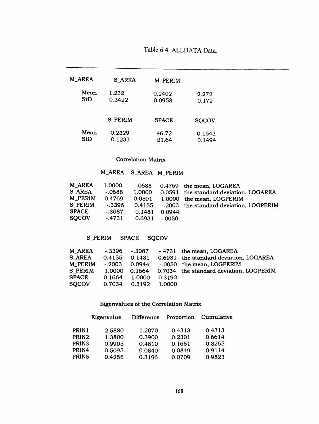
Table 6.4. ALLDATA Data.
M.AREA
Mean StD
Mean StD
S.AREA
1.232 0.3422
S.PERIM
0.2329 0.1233
M.PERIM
0.2402 0.0958
SPACE
46.72 21.64
2.272 0.172
SQCOV
0.1543 0.1494
Correlation Matrix
M.AREA S.AREA M.PERIM
M.AREA S.AREA M.PERIM S.PERIM SPACE SQCOV
1.0000 -.0688 0.4769 -.3396 -.3087 -.4731
-.0688 1.0000 0.0591 0.4155 0.1481 0.6931
0.4769 0.0591 1.0000 -.2003 0.0944 -.0050
the mean, LOGAREA the standard deviation, LOGAREA the mean, LOGPERIM the standard deviation, LOGPERIM
S.PERIM SPACE SQCOV
M.AREA S.AREA M.PERIM S.PERIM SPACE SQCOV
-.3396 0.4155 -.2003 1.0000
0.1664 0.7034
-.3087 0.1481 0.0944 0.1664 1.0000 0.3192
-.4731 0.6931 -.0050
0.7034 0.3192 1.0000
the mean, LOGAREA the standard deviation, LOGAREA the mean, LOGPERIM the standard deviation, LOGPERIM
Eigenvalues of the Correlation Matrix
PRINl PRIN2 PRIN3 PRIN4 PR1N5
Eigenvalue
2.5880 1.3800 0.9905 0.5095 0.4255
Difference
1.2070 0.3900 0.4810 0.0840 0.3196
Proportion
0.4313 0.2301 0.1651 0.0849 0.0709
Cumulat
0.4313 0.6614 0.8265 0.9114 0.9823
168

Table 6 4 Continued ALLDATA Data.
Principal Component Analysis
Eigenvectors
PRINl PRIN2
M.AREA S.AREA M.PERIM S.PERIM SPACE SQCOV
-.3932 0.4168 -.1505 0.4949 0.2735 0.5737
0.51280 0.41270 0.72230 0.00740 0.08856 0.19250
PR1N3
-.24540 .29260
0.25860 -.25790 0.84620
.06868
the mean, LOGAREA the standard deviation, LOGAREA the mean, LOGPERIM the standard deviation, LOGPERIM
PR1N4 PRIN5
M.AREA S.AREA M.PERIM S.PERIM SPACE SQCOV
0.05945 -.64250 0.31800 0.68770 -.08930 0.04036
0.61390 0.10260 -.47730 0.33700 0.43950 -.27940
the mean, LOGAREA the standard deviation, LOGAREIA the mean, LOGPERIM the standard deviation, LOGPERIM
169

Figure 6.5. Plot of PIUN2*PRINl, ALLDATA. Bold, underUned characters are standards used, which include Basilemys sp. TMM 42335-5 (z), Aspideretes sp., Javelina Formation TMM42874-1(A), Trionychidae(a) TMM 42876-1 (B), and Trionychidae(b) TMM 42876-1 (C). Randomly generated data are represented by an asterisk (*). PRINl represents variabUity in ph size for the trionychid data; PRIN2 represents overall pit size in the trionychid data.
170
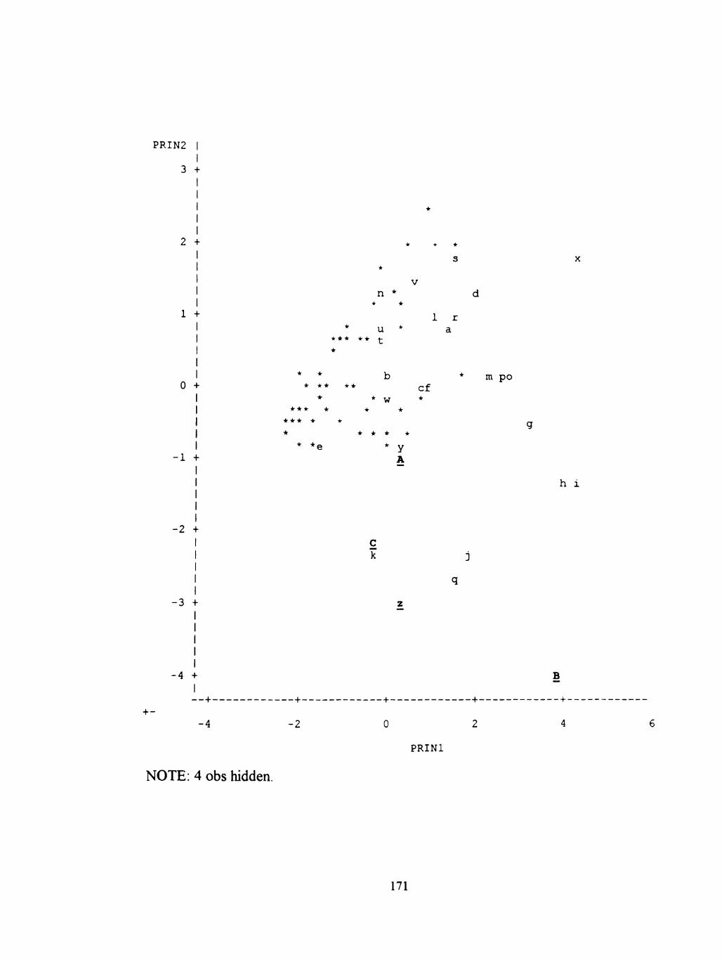
PRIN2
3
0 +
- 1 +
-2 +
- 3 +
-4 + I
s
V
n *
u 1 r
a
* * * • • * *
* * • * * • * •
* * • * *
b c f
w *
* in p o
• • , y A
h i
C k
+-—+-
-4 -2 0 2
PRINl
B
4
NOTE: 4 obs hidden.
171

Figure 6.6. Plot of PRIN3*PRES[2, ALLDATA. NOTE: 13 obs hidden. Bold, underlined characters are standards used, which include Basilemys sp. TMM 42335-5 (z), Aspideretes sp., Javelina Formation TMM42874-1(A), Trionychidae(a) TMM 42876-1 (B), and Trionychidae(b) TMM 42876-1 (C). Randomly generated data are represented by an asterisk (*).
172

PRIN3
4 +
m
+ n
b P
kC w
- 1 +
-2 + B
e * • * • * • * •
•
* • • • g * •
y* h * * • •
* *
-+-•4
• + -
•2 • + -
0 -+-2
• + -
4
PRIN2
173

plots were produced as a resuh of the analysis of the trionychid data (Figures 6.1 and
6.2). In the first, principle component 1 (PCI) plotted variability of size of the phs
against overall size (PC2). One prominent cluster appears in the upper left quadrant of
the plot. Specimens with large phs of mostly uniform size appear in this area. A check
of the specimens against the plot confirms this. A less well defined cluster of outliers
appears in the lower left quadrant of the plot. Two of these were standards used for
comparison to the 24 Aguja specimens. Accordmg to the plot, these specimens have
small phs that are mostly uniform in size, which is apparent in actual specimens. A
check of the other specimens which appear as outUers on the plot confirm that their
positions in morphospace are consistent with the ph pattems.
PC2 and PC3 characterize the overaU size of the pits with the space between
the phs. Specimens with large spaces between small phs plot in the upper left
quadrant. A check of the specimen labeled "q" confirms this. Likewise, a specimen
with large pits and little space between them plots in the lower right, as does specimen
"x".
Having already determined from the real data that the variability of the pit size
was a fiinction of the overall ph size, two sets of random forms were generated. The
first (named "RandataNC") was to determine whether numbers that fell within a
reasonable range of constraints, and would produce phs of comparable size and
number for imagined species, would produce the same results as real data. These
random data did not closely mimic the real data (Figures 6.3, 6.4).
174

The second set, "/Vlldata", merged the random data with the real data, and
placed biological constraints on the random data. The second data set confirmed that
the biological constraints on the placement of the phs in morphospace were, in fact,
the variability in the size of the phs compared to their overall size. The loadings and
proportions of the principle components are very similar to the real data set (Table 8),
and the plots of PCI versus PC2, and PC2 versus PC3 demonstrate that the random
data seem to plot in the same area in morphospace (Figures 6.5, 6.6).
Conclusions and Future Work
This exploratory use of principle components analysis is promising, but far
from complete. The purpose of this exercise was to determme whether a model could
be developed which might uhimately determine whether the observed pit pattems were
significantly non-random and, and if so, where they could be expected to appear in
morphospace. This was successfiilly done, whh the resuhs shown here indicatmg that
definite pattems are apparent m the pits on trionychid sheUs.
It is not possible to determine from these resuhs whether the pattems are
related to ontogenetic variables, placement of the phs on the sheUs, taphonomic
influences, sexual dimorphism, or whether these pattems reflect real taxonomic
differences. Several things might help determine the origin of the pattem. The next
step in the study would be to increase the size of the original data set whh more
specimens from the same locality, and to provide a more comprehensive set of
standards. Whh an increased data set, and more standards to use for comparison, h
175

might be possible to determine influences on vanance The standards should include
not only specimens that are known to represent different genera and species, but also
specimens origmating from different poshions on the shells. These standards should
mclude specimens that are stratigraphically comparable.
Modem trionychid specimens should also be used as standards. The modem
specimens should include different genera, different species of the same genus, and
several individuals representing the same species (this last m order to determine what
variabUity is tmly individual). Once individual variabihty is accounted for, then
ontogeny, sexual dhnorphism, and ph poshion can be studied in both modem and
paleontologic spechnens. Taphonomic bias can then be studied by analyzmg the
influence of abrasion on modem specimens.
By increasing the size of the data set and providing more standards for
comparison, presumably denser clusters can be produced, and, with the other sources
of "noise" such as pit poshion, and taphonomic, sexuaUy dhnorphic, and ontogenetic
bias muted, these clusters in tum might indicate taxonomic separation. Actual use of
morphometries on trionychid sheU phs for systematic studies may not be possible.
However, it would be useful for studies in diversity if h could be determined that there
were more than one species present on a stratigraphic level. Preliminary resuhs from
quantitative analysis of these phs are therefore promising.
176

REFERENCES
Adkins, W S., 1933. Mesozoic system in Texas, In EH Sellards, W.S. Adkins, and F.B. Plummer (eds), The Geology of Texas, vol. 1, Stratigraphy, The Univ of Texas BuU. 3232. pp. 240-518.
Archibald, J.D., 1977. Fossil mammalia and testudines of the HeU Creek Formation, and the Geology of the Tullock and Hell Creek Formations, Garfield County, Montana, 2 vols. Unpubl Ph.D. dissertation, Univ. Cal., Berkeley. 694 pp.
Bookstein, F.L., Chemoff, B., Elder, R.L., Humphries, Jr., J.M., Smith, G.R., Strauss, RE., 1985. Morphometries in evolutionary biology. Acad. Nat. Sci. Philadelphia Spec. Pub. No. 15. 277 pp.
Brinkman, D., and NichoUs, E. L., 1993. New Specunen of BasUemys praeclara Hay and hs bearing on the relationships of the Nanhsiugchelydae (ReptUia: Testtidmes). J. Paleont., vol. 67(6), pp. 1027-1031.
Cope, E.D., 1870. On the Adocidae. Proc. Am. Phil. Soc, vol. 11, pp. 547-553.
, 1871. On the homologies of some of the cranial bones of the reptilia, and on the systematic arrangements of this class. Proc. Amer. Assoc. Adv. Sci., 19'*' meeting, August, 1870, pp. 194-247.
, 1882. Contributions to the history of the vertebrata of the Lower Eocene of Wyoming and New Mexico, made during 1881. Proc. Amer. PhU Soc. Vol. 17, pp. 139-197.
Davis, J.C, 1986. Statistics and data analysis in geology. 2' ed. WUey and Sons, New York.
Dixon, JR., 1987. Amphibians and reptUes of Texas. Texas A&M University Press, CoUege Station. 425 pp.
Emst, C.H., and Barbour, R.W., 1989. Turtles of the world. Smithsonian Institution, Washington D.C. and London. 313 pp.
Emst, C.H., Lovich, J.E., and Barbour, R.W., 1994. Turtles of the United States and Canada. Smithsonian Institution Press, Washington and London. 578 pp.
177

Fasset and Hinds, 1971. Geology and ftiel resources of the Fmhland Formation and Kirtland Shale of the San Juan Basin, New Mexico and Colorado. Geol. Surv Prof Pap. 676. Washington. 76 pp.
Fisher, R.A., Corbet, A.S., and WiUiams, C.B., 1943. The relation between the number of species and the number of individuals in a random sample of an animal population. J. Anim. Ecol., vol. 12, pp. 42-58.
Gaffhey, E.S., 1965. The genus Taphrosphys Cope (Pelomedusidae: Reptiha). Unpublished undergraduate thesis. Rutgers University, New Bmnswick, New Jersey. 71 pp.
^ 1972. The systematics of the North American family Baenidae (Reptilia, Cryptodira). BuU. Am. Mus. Nat. Hist. vol. 147, pp.245-315.
, 1975a. A phylogeny and classification of the higher categories of ttirtles. BuU. Am. Mus. Nat. Hist. vol. 155, pp.387-436.
, 1975b. A revision of the side-necked turtle Taphrosphys sulcatus (Leidy) from the Cretaceous of New Jersey. Amer. Mus. Novitates. No. 2571, pp. 1-24
, 1979. Description of a large trionychid turtle sheU from the Eocene Bridger Formation of Wyoming. Contr. To Geol. Univ. of Wyoming, vol. 17, no.l, p. 53-57.
, and Meylan, P. A, 1988. A phylogeny of turtles. In M.J. Benton (ed.). The phylogeny and classification of tetrapods, vol. 1, Amphibians, reptiles, birds, pp. 157-219. Oxford:Clarendon Press.
, and Zangeri, R., 1968. A revision of the chelonian genus Bothremys (Pleurodira: Pelomedusidae). Fieldiana, Geology, Field Mus. Nat. Hist, vol 16, no.7, pp. 193-239.
Gardner, J.D., and RusseU, A.P., 1994. Carapacial variation among soft-shelled turtles (Testudines: Trionychidae), and its relevance to taxonomic and systematic studies of fossU taxa. N. Jb. Geol. Palaont., Abh., vol. 193, pp. 209-244. Stuttgart.
Gardner, J.D., RusseU, A.P., and Brinkman, D.B.. 1995. Systematics and taxonomy of soft-shelled turtles (Family Tionychidae) from the Judhh River Group (mid-Campanian) of North America. Can. J. Earth Sci. vol. 32, pp. 631-643.
178

Gilmore, C.W., 1916. Contributions to the Geology and paleontology of San Juan County, New Mexico. II. Vertebrate faunas of the Ojo Alamo, Kirtland, and Fmitiand formations. U.S. Geol. Surv. Prof Paper, no 98-Q. pp. 279-308.
, 1919. Reptilian faunas of the Torrejon, Puerco, and underlying Upper Cretaceous formations of San Juan County, New Mexico USGS Prof Paper 119.
_ , 1935. On the reptilia of the Kirtland Formation of New Mexico, with descriptions of new species of fossU ttirtles. Proc. U.S. Nati. Mus , vol. 83., pp. 159-188.
, 1949. ReptUian fauna of the North Horn Formation of central Utah. USGS Prof Paper 210-C.
Hay, O.P., 1908. Fossil ttirtles of North America. Carnegie Inst. Wash. PubUcation vol. 75, pp. 1-568.
, 1910. Descriptions of eight new species of fossU turtles from west of the one hundredth meridian. Proc. U.S. Nat. Mus., vol. 38, pp. 307-326
Hhyama, R., 1994. Phylogenetic systematics of chelonioid sea turtles. The Island Arc, vol. 3, pp. 270-284.
Holman, J.A.H., 1964. Pleistocene amphibians and reptiles from Texas. Herpetologica, vol. 20, no. 2, pp. 73-83.
, 1969. The Pleistocene amphibians and reptUes of Texas. Publ. of the Mus.-Mich. St. Univ. Biol. Ser. vol. 4, no.5, pp. 161-192.
Hutchison, J.H, and Archibald, J.D., 1986. Diversity of turtles across the Cretaceous/Tertiary boundary in northeastern Montana. Paleogeography, Plaeoclimatology, Paleoecology, 55, pp. 1-22.
Hutchison, J.H., and Bramble, D.M., 1981. Homology of the plastral scales of the Kinostemidae and related turtles. Herpetologica, vol. 37(2), pp. 73-85.
Image - pro Plus Image Processing System Software, 1992. Media Cybemetics, SUver Springs, MD.
Kachigan, S.K., 1991. Muhivariate Statistical Analysis, 3"* ed. Radius Press, New York. 303 pp.
179
I I — M M 1 '

Knebusch, W.E., 1981. Evidence for deltaic environment of deposhion for Aguja Formation (Upper Cretaceous), southwest Texas. Amer. Assoc. Pet. Geol. BuU. (abstr), vol. 65, p. 764.
Kovshak, A. A., 1973. Igneous and stmctural geology of the Grapevine HiUs, Big Bend National Park, Brewster County Texas. Univ. of Texas at Arlington, unpubl. M.S. thesis. 107 pp.
Lambe, L.M., 1902. On the Vertebrata of the mid-Cretaceous of the Northwest Territory. Contr. Canadian Paleont., vol. 3, pp. 25-81.
, 1906. Boremys, a new chelonian genus from the Cretaceous of Alberta. Ottawa Nat., vol. 19, no. 10, pp. 232-234.
Langston, Jr., W., 1956. The shell of Basilemys varialosa (Cope). Bull. Nat. Mus Canada, No. 142., pp. 155-165.
, Standhardt, B. Stevens, M., 1989. Fossil vertebrate coUecting in the Big Bend - History and retrospective. In: A. Buseby III and T. Lehman (eds). Vertebrate Paleontology, Biostratigraphy and Depositional Environments, Latest Cretaceous and Tertiary, Big Bend Area, Texas. 90 pp.
Lawson, DA., 1972. Paleoecology of the Tomillo Formation, Big Bend National Park, Brewster County, Texas. Unpubl. M.S. thesis. The Univ. of Texas at Austin. 182 pp.
Lehman, T.M., 1985. Stratigraphy, sedhnentology, and paleontology of Late Cretaceous (Campanian-Maastrichtian) sedimentary rocks in Trans-Pecos Texas. Unpubl. Ph.D. dissertation, Univ. of Texas at Austin. 310 pp.
, 1988. Stratigraphy of the Cretaceous-Tertiary and Paleocene-Eocene transition rocks of Big Bend: discussion. J.Geol., vol. 96, pp. 627-631.
, 1990. Paleosols and the Cretaceous/Tertiary transhion m the Big Bend region of Texas. Geology, vol. 18, pp. 362-364.
, 1991. Sedimentation and tectonism in the Laramide TomiUo Basin of West Texas. Sedimentary Geology, vol. 75, pp. 9-28.
180

, 1996. Late Campanian dinosaur biogeography in the Western Interior of North America. In. D. Wolberg and E. Stump, eds., Dinofest International. Proceedings of a symposium sponsored by Arizona State University. Special Publication, Academy of Natural Sciences, Philadelphia, PA.(in press).
Leidy, J. 1856. Notices of extinct Vertebrata discovered by Dr. F.V. Hayden, during the expedhion to the Sioux country under the command of Lieut. G.K. Warren. Proc. Acad. Nat. Sci. Philadelphia, vol. 8, pp. 311-312.
Macon, C.C, 1994. Facies analysis and sedimentology of Transgressive shoreline deposhs in the Aguja Formation (upper Cretaceous), Trans-Pecos Texas. Unpubl. M.S. thesis, Texas Tech University, Lubbock. 212 pp.
Magurran, A.E., 1988. Ecological diversity and hs measurement. Princeton Univ. Press, Princeton, New Jersey. 179 pp.
Maxwell, R.A., Lonsdale, J.T., Hazzard, R.T., and WUson, J.A., 1967. Geology of Big Bend National Park, Brewster County, Texas. The University of Texas Publ. No. 6711, Bureau of Eonomic Geology, Austin. 320 pp.
Meylan, P.A., 1987. The phylogenetic relationships of soft-sheUed turtles (family Trionychidae). BuU. Am. Mus. Nat. Hist. vol. 186, pp. 1-101.
Meylan, P. A, and Gaffhey, E.S., 1989. The skeletal morphology of the Cretaceous cryptodiran turtle, Adocus, and the relationships of the Trionychidae. Am. Mus. Novhates, no. 2941, 60 pp.
Mlynarski, M. 1976. Testudines. In O.Kuhn (ed), Handbuch der Paleoherpetologie 7, pp. 1-130. Gustav Fischer Verlag, Stuttgart.
Pielou, E.G., 1975. Ecological diversity. Wiley, New York. 165 pp.
Preston, F.W., 1962a. The canonical distribution of commonness and rarity. Ecology, vol. 43, pp. 185-215.
Preston, F.W., 1962b. The canonical distribution of commonness and rarity. Ecology, vol. 43, pp. 410-32.
Raup,D.M. 1966. Geometric analysis of sheU coiling: general problems. J. Paleont. Vol. 40, pp 1178-1190.
Ricklefs, RE., 1990. Ecology. 3'*' ed. W.H. Freeman and Company, New York. 896 PP
181
Romer, AS., 1976. Osteology of the reptiles. 3"* ed. University of Chicago Press, 772 PP
Rosenzweig, ML., 1995. Species diversity in space and time. Cambridge University Press, Cambridge, U.K., 436 pp.
Schiebout, J. A., 1974. Vertebrate paleontology and paleoecology of Paleocene Black Peaks Formation, Big Bend National Park, Texas. Texas Mem. Mus., BuU. 88 PP
, Rigsby, C.A., Rapp, S.D., Hartnell, J.A., and Standhardt, B.R., 1987. Stratigraphy of the Cretaceous-Tertairy and Paleocene- Eocene transition rocks of Big Bend National Park, Texas. J.Geol., vol. 95, pp. 359-375.
Sonrel, A., 1857. Young turtles, natural size (lithographs). In. L Agassiz (author). Contributions to the Natural History of the United States, vol. 2. Little, Brown and Company.
Standhardt, B.R., 1986. Vertebrate paleontology of the Cretaceous/Tertiary transition of Big Bend National Park, Texas. Unpubl. Ph.D. dissertation, Louisiana State Univ., Baton Rouge, La. 298 pp.
Straight, W H., 1996. Stratigraphy and paleontology of the Cretaceous-Tertiary Boundary, Big Bend National Park, Texas. Unpubl. M.S. thesis, Texas Tech University, Lubbock. 102 pp.
Sugihara, G., 1980. Minhnal community stmcture and explanation of species abundance pattems. American Naturalist, vol. 116, pp. 770-87.
Udden, J.A., 1907. A sketch of the geology of the Chisos Country, Brewster County, Texas. The Univ. of Texas at Austin BuU. 93. 101 pp.
Webb, R.G., 1990. Trionyx. Catal. Am. Amph. Rept., vol. 487, pp. 1-7
Wieland, R. 1904. Stmcture of the Upper Cretaceous turtles of New Jersey: Adocus, Osteopygis and Propleura. Am. J. Sci., vol. 17, pp. 112-132.
WiUiams, E.E. 1950. Variation and selection in the cervical central articulations of living turtles. BuU. Amer. Mus. Nat. Hist., vol. 94, pp. 505-562.
Wiman, C. 1933. Uber SchiUcroten aus der oberen Kreide in New Mexico. Nova Acta R. Soc. Sci. Upsaliensis, vol. 9, no. 5, pp. 1-34.
182

Zangeri, R., 1948. The vertebrate fauna of the Selma Formation of Alabama, Part 2, The Pleurodiran turtles. Fieldiana Geology, Chicago Nat. Hist. Mus vol.3, no.2.
_, 1953a. The vertebrate fauna of the Selma Formation of Alabama, Part 3, The turtles of the family Protostegidae. Fieldiana Geology, Chicago Nat. Hist. Mus. vol.3, no.3.
^ 1953b. The vertebrate fauna of the Selma Formation of Alabama, Part 4, The turtles of the family Toxochelyidae. Fieldiana Geology, Chicago Nat. Hist. Mus. vol.3, no.4.
, I960. The vertebrates of the Selma Formation of Alabama, Part 5, An advanced chelonhd sea turtle. Fieldiana Geology, Chicago Nat. Hist. Mus. vol. 3, no.5.
, 1969. The turtle shell. In C. Gans and T.S. Parsons (eds). Biology of the Reptiha, I, pp. 311-339. Acdemic Press, New York.
Hendrickson, L.P., and Hendrickson, JR., 1988. A redescription of the Australian Flatback, sea turtle, Natator depressus. Bishop Mus. Bull. Zoology I. Bishop Mus. Press, Honolulu. 69 pp.
, and Johnson, R.G., 1957. The nature of shield abnormalities in the turtle shell. Fieldiana Geology, Chicago Nat. Hist. Mus. vol. 10, no. 29.
183

APPENDIX
SAS PROGRAM FOR MORPHOMETRIC STUDY
TOTTURT SAS
options linesize=79;
* Read text file containing one line per ph, grouped by specimen;
data turtle; infUe 'turtle2.dat'; input labels spec$ totarea totperim ph$ area perim; area = log(sqrt(area)); perim = log(perim); mn;
*proc means data=turtle; * mn;
proc sort data=turtle; by spec; mn;
* Calculate and save univariate statistics for each specimen, across phs; * Save resuhs in individual datasets;
proc univariate data=turtle noprint; var area; by spec; output out=uniarea mean=m_area std=s_area;
mn;
proc univariate data=turtle noprint; var perim; by spec; output out=uniperim mean=m_perim std=s_perim;
184

mn;
data turtle2; infile 'turtle2.dat'; input labels specS totarea totperim ph$ area perim; mn;
proc sort data=turtle2; by spec; mn;
proc univariate data=turtle2 noprint; var area; by spec; output out=space sum=pitarea; mn;
proc print data=space; mn;
proc univariate data=turtle2 noprint; var totarea; by spec; output out=totarea mean=totarea; mn;
proc print data=totarea; mn;
data space; merge space totarea; space = totarea - pharea; mn;
proc print data=space; mn;
* Calculate and save covar between area,perim for each specimen, across phs; * Save results in dataset and fiher for covariance only;
185

proc corr data=turtle cov noprint outp=corrdata; var area perim; by spec; mn;
data corrdata; set corrdata;
•take square root of covarinace to make units equivalent; sqcov = sqrt(abs(perim)); if(perim<0)then
sqcov = -sqcov; keep spec sqcov; drop area perim _type name_; if _type_='COV and _name_='AREA' then output; mn;
* Save the 1-char label for each specimen from original dataset;
data label; set turtle; if _n_=l then specsave = ' '; if spec NE specsave then output; specsave = spec; * keep spec label totarea totperim; keep spec label totarea; keep spec label; retain specsave; mn;
proc sort data=space; by spec; mn;
* Merge all resuhs into common dataset having one line per specunen;
data turtle; merge label uniarea uniperim space corrdata; mn;
proc print data=turtle; run;
186

* Analyses of specimens;
proc princomp data=turtle out=scores; * VAR Marea S_area M_perim S_perim tarea t_perim SQCOV:
VAR Marea Sarea M_perim S_perim space SQCOV; mn;
proc plot data=scores; plot prin2 * print = label; plot prin3 * prin2 = label; mn;
RANDTURTSAS
options linesize=75;
data turtle; do spec = 1 to 50;
* numbers of pits/specimen vary from approx 6-12, centered on 9; do ph = 1 to round(rannor (0) + 9);
* perimeter is normaUy distributed, with a variance that is inversely * proportional to the number of phs (size), and centered arbhrarily * on 10;
* perim = rannor(O) + 10y*no constraint*/ c = 0.2; perim = rannor(0)/(c*pit) + 10;/*constramed*/
* area of circle having this perimeter; area = perim * perim/(4* 3.1415927);
* restrict area to within 0.3-0.9 of maximum; area = area*(0.3+ranuni(0)*6)/ph + 10; /*unconstramed*/ sqarea = sqrt(area); logarea = log(sqarea); logperim = log(perim), output;
end; end;
187

*proc print data=turtle; * mn:
*proc chart data=turtle; * vbararea; * vbar perim;
proc univariate data=turtle noprint; var logarea; by spec; output out=uniarea mean=m_area std=s_area; mn;
proc univariate data=turtle noprint; var logperim; by spec; output out=uniperim mean=m_perim std=s_perim; mn;
proc corr data=turtle cov noprint outp=corrdata; var logarea logperim; by spec; mn;
*proc print data=corrdata; * mn;
data corrdata; set corrdata; * take sqrt of covariance to make units equivalent; sqcov = sqrt(abs(logperim)); if (logperim < 0) then
sqcov = -sqcov; * drop logarea logperim _type name_; keep spec sqcov; * keep only one Ime per specimen; if _type_='COV' and _name_='LOGAREA' then output; mn;
•proc print data=corrdata; * mn;
188

data space; do spec = 1 to 50;
space = 20 + 40*ranuni(0); logspace = log(space);
output; end;
data turtle; merge uniarea uniperim space corrdata; mn;
*proc print data=turtle; *mn;
proc princomp data=turtle out=scores; var marea sarea mjjerim s_perim space sqcov; mn;
proc plot data=scores; plot prin2 * print; plot prin3 * prin2; mn;
ALLDATA SAS
options linesize=75;
data turtle; do spec = 1 to 50;
* numbers of pits/specimen vary from approx 6-12, centered on 9; do ph = 1 to round(rannor (0) + 9);
* perimeter is normally distributed, with a variance that is inversely * proportional to the number of phs (size), and centered arbhrarily * on 10;
* perim = rannor(O) + 10; /* no constramt */ c = 0.2; /* constraint factor, where limit 0 = unconstramed */ perim = rannor(0)/(c*ph) + 10; /* biological constraint */
189
' W l
* area of circle having this perimeter; area = perim * perim/(4* 3.1415927);
* restrict area to within 0.3-0.9 of maximum; * area constrained to be between 0.3-0.9 of max area for given * area = area * ranuni(O); /* unconstrained •/ area = area * (0.2 + ranuni(0)*8)/ph + 10; /* constrained •/
sqarea = sqrt(area); logarea = log(sqarea); logperim = log(perim); output;
end; end;
proc univariate data=turtle noprint; var logarea; by spec; output out=uniarea mean=m_area std=s_area; mn;
proc univariate data=turtle noprint; var logperim; by spec; output out=uniperim mean=mj3erim std=s_perim; mn;
proc corr data=turtle cov noprint outp=corrdata; var logarea logperim; by spec; mn;
data corrdata; set corrdata; * take sqrt of covariance to make units equivalent; sqcov = sqrt(abs(logperim)); if (logperim < 0) then
sqcov = -sqcov; keep spec sqcov;
190
. tfcd

* keep only one line per specimen; if _type_=*COV' and _name_=*LOGAREA' then output; mn;
data space; do spec = 1 to 50;
space = 20 + 40*ranuni(0); logspace = log(space); output;
end;
data randturt; merge uniarea uniperim space corrdata; label = '*•; drop spec; mn;
Read text file containing one Ime per ph, grouped by specimen;
data turtle; infUe 'turtle2.dat'; input labels specS totarea totperim phS area perim;
space = totarea - sum (of area);
area = log(sqrt(area)); perim = log(perim); mn;
proc means data=turtle; mn;
*proc print data=turtle; * mn;
* Sort by specimen;
proc sort data=turtle; by spec; run;
191

* Calculate and save univariate statistics for each specimen, across pits; * Save resuhs in individual datasets;
proc univariate data=turtle noprint; var area; by spec; output out=uniarea mean=m_area std=s_area; mn;
proc univariate data=turtle noprint; var perim; by spec; output out=uniperim mean=m_perim std=s_perim; mn;
data turtle2; infile 'turtle2.dat'; input labels specS totarea totperim phS area perim; mn;
proc sort data=turtle2; by spec; mn;
proc univariate data=turtle2 noprint; var area; by spec; output out=space sum=pharea; mn;
proc univariate data=turtle2 noprint; var totarea; by spec; output out=totarea mean=totarea; mn;
data space; merge space totarea; space = totarea - pharea; mn;
* Calculate and save covar between area,perim for each specimen, across pits;
192

Save resuhs in dataset and fiher for covariance only;
proc corr data=mrtle cov noprint outp=corrdata; var area perim; by spec; mn;
data corrdata; set corrdata; sqcov = sqrt(abs(perim)); if (perim < 0) then
sqcov = -sqcov; drop area perim _type name_; if _type_='COV and _name_='AREA' then output; mn;
* Save the 1-char label for each specimen from original dataset;
data label; set turtle; if _n_=l then specsave = ' '; if spec NE specsave then output; specsave = spec; keep spec label; retain specsave; mn;
* Merge aU resuhs into common dataset having one Ime per specimen;
data realturt; merge label uniarea uniperim space corrdata; drop spec; mn;
* Analyses of specimens;
data turtle; set randturt realturt; run;
proc princomp data=turtle out=scores;
193
•_m.^ - ^ - ^ * » M.^m ».*- --•' *^^ '* -—

VAR Marea S_area MjDcrim S_perim Space SQCOV; mn;
proc plot data=scores; plot prin2 * prinl = label; plot prin3 * prin2 = label; mn;
194
- ^ " ^ ~ ^ t - . J . -" --**--'* * '"'- ''' ^ **
