lasers and laserlike devices: part twodocshare01.docshare.tips/files/28208/282084946.pdf · lasers...
TRANSCRIPT

REVIEW ARTICLE
Lasers and laser-like devices: Part two
Deshan F Sebaratnam,1,2 Adrian C Lim,3 Patricia M Lowe,1,2 Greg J Goodman,4 Philip Bekhor5
and Shawn Richards6
1Department of Dermatology, Royal Prince Alfred Hospital, Camperdown, New South Wales, 2University ofSydney, Camperdown, New South Wales, 3Department of Dermatology, Royal North Shore Hospital, St
Leonards, New South Wales, 4Skin and Cancer Foundation Inc, Carlton, Victoria, 5Department ofDermatology, Royal Childrens Hospital, Melbourne, Victoria, and 6Skin and Cancer Foundation Australia,
Westmead, New South Wales, Australia
ABSTRACT
Part two of this review series evaluates the use oflasers and laser-like devices in dermatology basedon published evidence and the collective experienceof the senior authors. Dermatologists can laser-treata wide range of dermatoses, including vascular,pigmentary, textural, benign proliferative andpremalignant conditions. Some of these conditionsinclude vascular malformation, haemangioma, facialtelangiectases, café-au-lait macules, naevi of Ota,lentigines, acne scarring, rhytides, rhinophyma andmiscellaneous skin lesions. Photodynamic therapywith lasers and intense pulsed light is addressed, withparticular reference to actinic keratosis and actiniccheilitis. A treatment algorithm for acne scarringbased on scar morphology and severity is comprehen-sively outlined. Following from part one, the variousdevices are matched to the corresponding dermato-logical conditions with representative pictorial casevignettes illustrating likely clinical outcomes as wellas limitations and potential complications of thevarious laser and light therapies.
Key words: acne, CO2 laser, Er:YAG laser, KTPlaser, Nd:YAG laser, pigment, QS laser, rhytides,ruby laser, vascular.
INTRODUCTION
The broad principles of dermatological laser therapy havebeen covered in Part one of this review series.1 Part twoexplores the use of lasers in procedural and cosmetic der-matology with a particular focus on conditions that areclinically relevant to dermatologists. We discuss likely clini-cal outcomes as well as the limitations of laser therapy,including its use in darker Fitzpatrick skin types, based onthe literature review and the senior authors’ collectiveexperience.
Vascular conditions
Vascular malformation The most recent CochraneReview2 of laser treatments for capillary malformations(CM) (port-wine stains) included five randomised con-trolled trials (RCT) totalling 103 patients. A pulsed dye laser(PDL) (585 or 595 nm) was employed in all five trials and,depending on the regimen employed, resulted in a
Correspondence: Dr Deshan F Sebaratnam, Department ofDermatology, Royal Prince Alfred Hospital, Missenden Road,Camperdown, NSW 2050, Australia. Email: [email protected]
Deshan F Sebaratnam, MBBS (Hons). Adrian C Lim, FACD.Patricia M Lowe, FACD. Greg Goodman, FACD. Philip Bekhor,FACD. Shawn Richards, FACD.
Conflict of interest: noneSubmitted 28 June 2013; accepted 11 August 2013.
Abbreviations:
AK actinic keratosisALA aminolevulinic acidCM capillary malformationCMN congenital melanocytic naevusCO2 carbon dioxideDST darker skin typeEr erbiumEVLA endovenous laser ablationHHT hereditary haemorrhagic telangiectasiaHQ hydroquinoneHOI haemangioma of infancyIPL intense pulsed lightKTP potassium tritanyl phosphateNd:YAG neodymium-doped yttrium aluminium garnetPDL pulsed dye laserPIH post-inflammatory hyperpigmentationPDT photodynamic therapyQS quality switchedRCT randomised controlled trialTCA trichloracetic acidTTT triple topical therapy
bs_bs_banner
Australasian Journal of Dermatology (2014) 55, 1–14 doi: 10.1111/ajd.12111
© 2013 The Australasian College of Dermatologists

minimum of 25% reduction in redness. Both the pulsedneodymium-doped yttrium aluminium garnet (Nd:YAG)(1064 nm) laser and intense pulsed light (IPL) were effec-tive, but the PDL results were superior and therefore, con-sidered the laser of choice for CM (Fig. 1).3
Head and neck CM respond better than trunk and distalextremity lesions. Nodular, hypertrophic or recalcitrant CMmay not respond to PDL and may be better suited to treat-ment with a pulsed Nd:YAG laser, a PDL and pulsed Nd:YAGlaser combination, a pulsed alexandrite laser (755 nm) orIPL.4 Children under the age of 1 year seem to have the bestresponse and should be treated as early as possible.4 It isimportant to inform patients or parents that gradual recur-rence is likely but tends to be less cosmetically conspicuous.Recently topical rapamycin has been used as an adjuvant tovascular laser to accelerate the clearance of CM with PDL.5
Haemangioma of infancy Laser treatment of haeman-gioma of infancy (HOI) remains controversial. Early studiesusing obsolete, non-cooled lasers and employing excessivefluence and severe purpuric end-points have complicatedthe pursuit of an evidence-based approach to the manage-ment of HOI.6 It has been our experience that the PDL at 6–7J/cm2, 10 mm spot, 1.5 ms pulse width, achieving a transientto minimally purpuric end-point will effectively treat earlyflat HOI present in the superficial dermis. This treatmentprotocol is often used for focal, facial and superficial hae-mangioma in conjunction with timolol; a topical beta-blocker. In general, lasers do not have a major role in raisedor subcutaneous haemangiomas because PDL can pen-etrate to a depth of only 1.2 mm. PDL is useful in selectedcases of involuting and ulcerated haemangiomas. Somecentres use more deeply penetrating wavelengths (IPL andNd:YAG) to treat thicker and deeper lesions. The role oflasers in patients with haemangiomas is set to diminish
further with the advent of systemic beta-blockers as a dra-matically effective medical treatment option.7,8 However,many lesions treated with beta-blockers will leave a super-ficial telangiectatic component that is amenable to PDL.
Facial telangiectases The most commonly used devicesfor facial telangiectases are PDL, potassium tritanyl phos-phate (KTP) (532 nm) lasers and IPL. The role of lasers inreducing telangiectases is well established, with severalstudies demonstrating their treatment efficacy with copper-bromide (578 nm), krypton (520, 530 or 568 nm), KTP andPDL (Fig. 2).9 The 532 nm and 1064 nm tracing lasers areideally suited for targeting discrete facial telangiectases; thelatter being useful for larger calibre blood vessels andvenules. PDL can be used on a range of vascular lesionswith either purpuric (pulse duration < 3ms) or non-purpuric (pulse duration ≥ 3ms) end-points. It has beensuggested that fine telangiectases can be adequately treatedwith non-purpuric parameters while purpuric parametersare more effective at clearing thicker telangiectases.However, thicker lesions can be effectively treated (withoutpurpura) through pulse stacking and multiple passes usingnon-purpuric parameters. Perialar telangiectases can berecalcitrant to laser treatment, thus requiring more ses-sions, and are also more prone to recurrence.10 IPL has alsobeen shown to improve facial telangiectases. Although laseris considered to be superior,11,12 a study comparing PDL andIPL showed no significant clinical difference between thetwo modalities.13 Facial telangiectases frequently occur inthe setting of rosacea. Anecdotally, it has been found thatsome patients will demonstrate a clinical amelioration ofthe rosacea itself but few studies have actually investigatedthis phenomenon.
Hereditary haemorrhagic telangiectasia Hereditary haem-orrhagic telangiectasia (HHT) or Osler–Weber–Rendu syn-drome is an autosomal dominant disorder characterised bycutaneous and visceral telangiectases that require systemicinvestigation. Smaller HHT vascular lesions respond to PDLand KTP lasers while larger and often bleeding lesions
Figure 1 Caucasian male, 3-years old, Fitzpatrick phototype IIwith capillary malformation right cheek and upper lip treated withpulsed dye laser (Candela Vbeam; Candela, Irvine, CA, USA)(595 nm): 7 mm spot, 9 J/cm2 fluence, 1.5 ms pulse duration (mildpurpuric end-point), 30 ms spray 30 ms delay cryogen cooling,single pass with minimal overlap. Showing: (a) baseline and (b) 18months after third treatment undertaken at 6–12 monthly intervals.
Figure 2 Caucasian woman, 57-years old, Fitzpatrick phototypeI–II with rosacea erythema and telangiectases treated with pulseddye laser (Candela Perfecta; Candela, Irvine, CA, USA) (595 nm): 3× 10 mm spot, 13.5 J/cm2 fluence, 20 ms pulse duration, 30 ms spray20 ms delay cryogen cooling, 1–2 passes followed by intense pulsedlight (Sciton BBL Palo Alto, CA, USA): 560 nm filter, 16 J/cm2
fluence, 15–20 ms pulse duration, 15°C, single pass. Showing: (a)baseline and (b) 6 weeks after second treatment.
2 DF Sebaratnam et al.
© 2013 The Australasian College of Dermatologists

require treatment with a Nd:YAG laser. Use of the Nd:YAGlaser within the oral cavity needs to be undertaken withcare as the laser beam can damage natural dentition anddental crowns. Oral mucosal lesions may bleed profusely ifthe treatment power is inadequate and may require over-sewing for adequate haemostasis.
Venous lakes and other vascular lesions Venous lakes arecharacterised by ectatic thin-walled venules in the superfi-cial papillary dermis and, depending on size, can be effec-tively treated with a variety of vascular lasers: Nd:YAG ordiode laser for larger lesions14 and PDL for smaller lesions.15
Nd:YAG laser is the laser of choice with a 94% clearancerate in one case series of 35 patients.16 Other vascularlesions such as cherry angiomas, spider angiomas andangiokeratomas respond satisfactorily to any of the vascularlasers listed above if the laser is selected according to thelesion size and the laser’s depth of penetration.
Leg veins Sclerotherapy remains the gold standard forsmall diameter vessels such as venules and capillaries.However, in the last decade, endovenous laser ablation(EVLA) has replaced surgical stripping as the preferredtreatment option for varicose veins. EVLA consists of a fibre-optic thread inserted into the vein with wavelengths rangingfrom 810–1500 nm. A meta-analysis of 64 clinical studiesevaluating 12 320 limbs concluded that EVLA of lower limbvaricosities was superior to surgical intervention17 and isoften performed in conjunction with ultrasound-guidedsclerotherapy. External beam lasers play a very limited rolein lower limb vessels unless all the larger refluxing veins(both clinical and subclinical) have been adequatelytreated.18 Nevertheless, external beam lasers may on occa-sion be useful for small vessels that are difficult to cannulate,telangiectatic matting not due to underlying venous refluxand for needle-phobic patients. Lasers with longer wave-lengths offer deeper penetration and epidermal sparing,with the most commonly employed lasers being Nd:YAG,KTP, PDL, alexandrite, diode (810 nm) and IPL. The choicewill again be determined by the size and depth of the vascu-lar target.19 Generalised essential telangiectasia is anothercause of telangiectases on the legs and is responsive to lowfluence vascular lasers (PDL and KTP) and IPL.
Pigment-related conditions
Epidermal lesions Lesions such as solar lentigines, lentigosimplex and ephelides consist of epidermal pigment origi-nating from basal layer melanocytes. Any laser that selec-tively targets the melanin chromophore or ablates theepidermis can potentially ameliorate such lesions, typicallyin 1–2 sessions. The mainstay lasers include quality switched(QS) alexandrite, QS ruby (694 nm) and QS frequencydoubled Nd:YAG (532 nm).20 Long-pulsed counterpartsare also effective, less likely to cause post-inflammatoryhyperpigmentation (PIH) and generally preferred forpatients of dark skin types (DST). IPL (filter 500–600 nm) canalso be very effective for this indication, while concurrently
improving telangiectases. Superficial and fractional resur-facing lasers (density > 50%) also treat epidermal pigmenta-tion and ameliorate mild textural photodamage but have arecovery time of approximately 5–7 days.
Café-au-lait macule In our experience, QS lasers yieldthe best results with QS frequency doubled Nd:YAG, QSalexandrite and QS ruby as the modalities of choice. Recur-rences are common and multiple treatment sessions may berequired, but some patients will obtain excellent outcomeswith a long-term significant reduction in colour of themacule if they sun-protect the area.
Becker’s naevus Patients with Becker’s naevus may ask fortreatment for lesional hyperpigmentation and hyper-trichosis. Recurrence and incomplete response arecommon and are believed to be due in part to the sparing ofsanctuary sites of pigmented keratinocytes and melanocytesin the deeper hair follicle.21 At this time there is no reliabletreatment for Becker’s naevus. Different approaches totreatment have been described in the literature but there isno consensus on their safety or effectiveness.21,22 Until betteroutcomes are achieved these patients are usually bestadvised to avoid treatment.
Congenital melanocytic naevus Many different lasershave been employed in the management of congenitalmelanocytic naevus (CMN),23 with the largest series of 52patients with 314 lesions treated with combined ultrapulsedcarbon dioxide (CO2) laser and frequency doubled QSNd:YAG (532 nm), with mean follow up of 8 years.24 Approxi-mately 95% of these lesions had a reduction in pigment,with five patients failing treatment, five experiencing recur-rence and one developing melanoma. While there is someevidence for the role of lasers in the treatment of CMN,surgery remains the gold standard.
Acquired naevus Definitive surgical excision also remainsthe mainstay of treatment for acquired naevi. Shave exci-sion of skin-coloured dermal nevi is accepted practice dueto their low risk of malignant transformation, along withablative laser re-contouring of these lesions. However, flat,pigmented naevi should be excised rather than treated withlaser. Asian patients with a very low risk of melanoma aresometimes considered for QS laser treatment of benignnaevi on the face.25 Laser treatment of pigmented naevi maycomplicate an accurate diagnosis of subsequently re-pigmenting lesions and is not ideal in the setting of mela-noma surveillance.
Naevus of Ota Pigment-selective lasers have largely super-seded other treatments in the management of naevi of Ota.As the melanocytosis in the naevus of Ota is primarilydermal, longer wavelength QS lasers (694 nm, 755 nm and1064 nm) are generally preferred (Fig. 3). Published caseseries support the use of QS ruby,26 QS alexandrite27,28 and
Clinical use of lasers in dermatology 3
© 2013 The Australasian College of Dermatologists

QS Nd:YAG for this condition.29 A study of 602 Chinesepatients treated by QS alexandrite laser reported a cure rateof 92% after 9 treatments, with success significantly relatedto the number of treatment sessions (P < 0.001).27
Naevus of Hori Given the histopathological and clinicalsimilarities between the naevus of Ota and Hori’s naevus(bilateral mid-face macular pigmentation with ocularsparing), QS lasers have been used to treat the latter since itwas first described in 1984. In a case series of 131 Thaifemale patients treated with QS ruby laser there was com-plete clearance of lesions after an average of 2.3 sessions ata mean follow-up period of 2.5 years.30 In contrast, in a studyof 66 patients treated by QS Nd:YAG only 26% of themdemonstrated a 50% improvement after 1–2 treatments,although the authors noted that this may have improvedwith further sessions.31
Melasma Topical treatments such as superficial chemicalpeels, hydroquinone (HQ), kojic acid, tranexamic acid andtriple topical therapy (TTT) (consisting of a mixture of HQ,retinoid and corticosteroid) remain first-line management,with mixed evidence for the effectiveness of lasers (Fig. 4).A RCT of 20 patients with predominantly epidermalmelasma compared TTT with fractional erbium:glass(Er:glass) non-ablative laser (1,550 nm) and demonstratedsimilar efficacy and safety,32 but at 6 month follow up mostof the patients had recurrence. In contrast, a split-face RCTof 29 patients comparing combined TTT and fractionalEr:glass non-ablative laser with TTT monotherapy demon-strated poorer outcomes with laser due to a high frequencyof PIH.33
Topical agents remain the mainstay of treatment formelasma with the benefits offered by laser seeminglyreduced by the risk of PIH and rebound hyperpigmentation.At best, lasers play an adjuvant and supporting role totopical therapy for melasma. Trials comparing TTTmonotherapy with combination TTT and fractional CO2
laser34 and combination TTT and PDL (to treat finetelangiectases commonly accompanying melasma)35 havedemonstrated a beneficial synergistic effect of combining
topical and laser modalities. This finding was also observedin a RCT comparing HQ monotherapy with low fluence QSNd:YAG plus HQ.36 The fractionated thulium (1927 nm)laser, a relative newcomer, has shown early promise in thissetting.37 Ablative lasers may facilitate the delivery of drugsthrough the skin, such as HQ, with a multimodal approachpotentially improving patient outcomes. Laser-assisteddrug delivery may have a role in melasma management aswell as in the broader dermatological arena.38
Post-inflammatory hyperpigmentation As with melasma,PIH can be disappointing to treat and general measuresalong with realistic patient expectations are paramount.There is a paucity of clinical trials investigating the role oflasers in the management of PIH and accordingly it is dif-ficult to provide an evidence-based evaluation of their effi-cacy in this setting. Selected cases of persistent PIH (> 12months) may respond to QS lasers39 but this is fraught withpotential problems and is not routinely recommended.Variable responses of PIH to laser therapy have beenobserved and generally there is incomplete clearance ofpigmentation, along with a risk of the PIH worsening.
Hair removal Permanent hair reduction refers to stable,long-term decreased hair regrowth following laser treat-ment rather than the total elimination of all hairs in thetreatment area.40 A systematic review concluded that epila-tion with lasers resulted in partial short-term hair reductionbeyond 6 months following treatment with alexandrite anddiode lasers, and probably after ruby and Nd:YAG lasertreatment.41 Efficacy was improved with repeated treat-ment, superior to conventional epilation treatments andIPL, with a low frequency of adverse effects being observedacross all laser types. More recent studies have further vali-dated the utility of the diode42–44 alexandrite45,46 and Nd:YAG47
lasers, with hair reduction achieved beyond 12 months in
Figure 3 Asian woman, 33-years old, Fitzpatrick phototype IV withnaevus of Ota, treated with quality switched neodymium-dopedyttrium aluminium garnet laser (Revlite C3; Cynosure, Westford,MA, USA) (1064 nm): 4 mm spot, 6.5–7.5 J/cm2 fluence at 10 Hz.Showing: (a) baseline and (b) after 12 treatments. Figure 4 African woman, 45-years old, Fitzpatrick phototype V–VI
with prominent periorbital melasma effectively controlled withfirst-line topical therapy with 4% hydroquinone, 4% kojic acid, 1%hydrocortisone and 0.05% tazarotene (prescribed separately). Thepigmentation worsened with prior attempted low-fluence qualityswitched neodymium-doped yttrium aluminium garnet laser.Showing: (a) baseline and (b) after 12-months topical therapy.
4 DF Sebaratnam et al.
© 2013 The Australasian College of Dermatologists

some studies,48–50 and this is preferred over the ruby laser.IPL produced a comparable degree of hair reduction tolasers, especially in fair-skinned individuals, but was lesseffective and more likely to cause burns in tanned or darkskin types.41 There is increasing awareness of the problemof paradoxical hypertrichosis (especially in women withpolycystic ovarian syndrome) that can compromise thetreatment of fine facial hair.
Tattoo removal QS lasers are the treatment of choice fortattoo removal and the most commonly employed are theQS ruby, QS Nd:YAG and QS alexandrite.51 The QS Nd:YAGcan treat most colours but blue or green responds best to QSalexandrite, purple or violet responds best to QS ruby andred responds best to QS 532 nm. All lasers perform equallywell at removing black tattoo pigment52 and amateur blacktattoos are the easiest to remove. A recent report advocatedundertaking four treatment sessions in the same day sepa-rated by a 20-min interval for accelerated tattoo clearance.53
However, we have not been able to replicate these impres-sive results with this method of tattoo removal.
Important adverse effects observed with laser treatmentinclude pigmentary changes and irreversible darkening ofcosmetic (e.g., skin-coloured) tattoos. Accordingly, testspots are an important consideration when using lasers forthis indication. Multiple treatments (often > 5 sessions andsometimes between 10–20 sessions) are required to achievethe greatest clearance. However, if a particular laser failsto adequately remove a tattoo, another device can beemployed as multi-modal and multi-wavelength treatmentoften has a synergistic effect on clearance.
Lasers in Darker Skin Types (DST)
Patients with Fitzpatrick phototypes III–VI have greaterquantities of melanin in the stratum basale and thus havean increased risk of non-specific light absorption, leadingto a higher risk of adverse effects including burns,dyspigmentation, textural changes, atrophy and scarring.Concomitantly, competitive absorption by melanindecreases the total amount of energy reaching targettissues, rendering it more of a challenge to obtain desiredclinical outcomes.54 Given the predilection for absorption ofenergy by the epidermal melanin, it is essential to use con-servative power settings and employ effective coolingdevices to counteract the effects of accumulated thermalenergy within the stratum basale in DST patients.
Despite these challenges, patients with DST can be suc-cessfully treated with a variety of lasers using appropriatemodification of laser parameters. The peak absorption ofmelanin lies within the UV range and decreases as wave-length increases. Accordingly, lasers generating longerwavelengths, which are less avidly absorbed by endogenousmelanin, provide improved safety profiles and clinical effi-cacy for DST patients such as the diode, Nd:YAG or IPL withfilter cut-offs at longer wavelengths.55 Devices with longerinfrared wavelengths become colour blind as they have alow affinity to melanin but a high affinity to water, and are
characterised by resurfacing lasers (1550 nm and beyond).Resurfacing lasers (ablative or non-ablative) in DSTpatients have increased potential for PIH and thus peri-procedural bleaching and sun protection is important.
Textural or benign proliferative conditions
Acne scarring Fractional non-ablative Er:glass (1540 nm,56
1550 nm), fractional ablative CO2 laser57 and ablativeEr:YAG58 have all been shown to ameliorate the appearanceof acne scars. Based on qualitative findings from a RCT of 20patients with DST, combination treatment with fractionalablative CO2 plus non-ablative Nd:YAG laser treatmentyielded superior cosmetic results compared with a frac-tional CO2 laser alone.59 While the outcome measuresemployed in the various studies have been diverse, a sys-tematic review of fractional laser for acne scars concludedthat ablative fractional lasers offered an improvement rangeof 26–83% compared to 26–50% with non-ablative fractionalresurfacing.60 It is reasonable to say that of all the conditionsthat fractional lasers can improve, it is acne scarring thatshows the most consistent and remarkable benefits and theprocedure is not only safer than fully ablative lasers butappear to be at least as effective (Fig. 5).
A treatment algorithm has been developed by a seniorauthor stratifying patients according to the grade of acne
Figure 5 Caucasian man, 39-years old, Fitzpatrick phototype IIIwith Grade 3 acne scarring. The patient received treatment withfractionated CO2 laser (Deka Smartxide; Deka, Via Baldanzese,Italy) and fractionated non-ablative erbium: glass laser (1550 nm)(Fraxel Restore; Fraxel, Hayward, CA, USA), as well as hyaluronicacid injections, punch grafting and scar excision. Showing: (a) base-line and (b) after combination treatment.
Clinical use of lasers in dermatology 5
© 2013 The Australasian College of Dermatologists
scarring and burden of disease, which has been reportedpreviously.61 Briefly, the scarring is first graded according tothe morphology of the lesions (Table 1), with treatmentdetermined accordingly (Tables 2–5). In grade 1 scarring,where pathology is mainly flat but dyschromic the emphasisis to even out these discolourations. As the severity ofcontour abnormality increases, preparatory (pre-laser)work is necessary for contour correction before lasertherapy, whose role is to treat the more superficial contourand texture issues. For hypertrophic scarring, lasers mayhave a minor ancillary role in settling the contour but againonly after a preparatory injection and medical therapy.Quite often the preparatory medical and procedural treat-ment will make laser treatment unnecessary. Our manage-ment algorithm is outlined in Tables 2–5.
Rhytides The role of lasers in facial rejuvenation is wellestablished and a range of lasers has been utilised in thetreatment of rhytides. For the past 20 years fully ablativelaser resurfacing with the continuous wave CO2 laser orEr:YAG has been the mainstay of therapy. The popularity ofthese methods of full ablation has waned, in view of longhealing times and the very high incidence of CO2-inducedhypopigmentation. However, advances such as fractionatedlasers and novel technologies (erbium: yttrium scandiumgallium garnet [2790 nm] and plasma skin resurfacing)have further expanded our therapeutic armamentarium.62
Three RCT comparing CO2 lasers with Er:YAG lasers havedemonstrated comparable clinical results.63–65 It has beenproposed that Er:YAG laser is best employed for fine tomedium rhytides and due to its superior safety profile it isbetter suited to DST patients. In comparison CO2 laser issuperior for deep lines and more intensive tissue tighten-ing. In terms of combination therapy, the administration ofbotulinum toxin enhanced cosmetic outcomes in patientsundergoing laser resurfacing,66,67 the application of topical
retinaldehyde increased dermal thickness in patientstreated with the Er:glass laser68 and adjunctive CO2 laserresurfacing improved the overall cosmetic effect forpatients undergoing surgical blepharoplasty.69 Fractionalablative lasers (CO2 and erbium) have an established rolein the management of rhytides and can complement othermodalities ranging from non-ablative IPL to full resurfac-ing (Fig. 6).70,71
Rhinophyma Ablative resurfacing lasers can effectivelyre-contour the rhinophymatous nose.72 CO2 is preferredover erbium lasers because of the bloodless field thataccompanies the coagulation of blood vessels from residualthermal energy (Fig. 7). There have been several small caseseries comparing laser resurfacing to electrosurgery orscalpel excision, all yielding similar patient outcomes.9
Rhinophyma can also be de-bulked and sculpted with aradiofrequency electrosurgery wire loop. It has been rec-ommended that isotretinoin be discontinued 6 −12 monthsprior to resurfacing of rhinophyma to mitigate the risk ofdelayed wound healing or keloid formation.9 There is alsoan increasing trend to employ high-density fractional abla-tive lasers for mild rhinophyma followed by a secondtouch-up treatment if required.
Epidermal naevus Pigmented epidermal naevi can be tar-geted by long pulsed 532 nm lasers. Verrucal epidermalnaevi and related benign proliferative skin disorders can beeffectively controlled with ablative lasers. Depending uponthe type of epidermal naevus, long-term remission can beachieved with a single treatment session. In general, super-ficial seborrhoeic keratosis and acrochordon-like lesions dobest whereas lesions with deep appendageal involvementwill tend to recur unless deep ablative procedures are per-formed, which carry the risk of incomplete ablation, scar-ring and dyspigmentation. Although there are morepublished data on CO2 ablation of epidermal naevi than onablative erbium, the latter is also effective.73 Debulkingcurettage of the lesion immediately prior to ablative laserhastens the procedure and provides a specimen for ahistopathological review.
Other benign skin pathologies Seborrhoeic keratosis andits related variant – dermatosis papulosa nigra – commonlypresent for cosmetic treatment. Many of these are amena-ble to cryotherapy, shave excision, curettage and cautery;all of which are reasonable first-line therapies. However,for cosmetically sensitive locations such as the eyelids,nose and lips, lasers offer superior control and finesse.These growths can be treated with 2–3 mm spot ablativelasers, as well as long pulsed 532nm lasers (Table 6).Common skin-derived tumours such as dermal naevi,fibrous papules angiofibromas and sebaceous hyperplasiaare amenable to ablative laser removal, as areappendageal tumours such as syringomas and depositssuch as xanthelasma. However, many of these tumours,particularly syringomas and angiofibromas, will recur over
Table 1 Grading algorithm for acne scarring according to lesionmorphology
Grade Description
1 Abnormally coloured macular disease: erythematous,hyperpigmented or hypopigmented flat marks visibleat any distance.
2 Mild but abnormally contoured scarring: mild atrophyor hypertrophy that may not be obvious at socialdistances of 50 cm or greater and may be adequatelycamouflaged with makeup, the normal shadow of ashaved beard in men or normal body hair ifextra-facial.
3 Moderately abnormally contoured disease: moderateatrophic or hypertrophic scarring that is obvious atsocial distances of 50 cm or greater and is notcovered easily but flattens substantially by manualstretching of the skin.
4 Severely abnormally contoured disease: severe atrophicor hypertrophic scarring that is obvious at socialdistances greater than 50 cm, is not covered easily.Manual skin stretching cannot flatten it.
6 DF Sebaratnam et al.
© 2013 The Australasian College of Dermatologists

time due to their deep location in the skin. Laser (Er:YAG)is also better suited for patients of DST where excessivetissue injury from non-laser methods can increase the riskof PIH. It is important to acknowledge that the use of
topical rapamycin is a major breakthrough in the manage-ment of angiofibromas associated with tuberous sclerosis.Rapamycin has the potential to significantly reduce therole of laser in this condition.74
Table 2 Treatment algorithm for acne scarring grade 1: macular coloured marks
Scar type Pre-laser treatment plan Appropriate laser treatment
Erythematous flat marks Surface SurfaceSkin care† Vascular lasers (long-pulsed 532 nm or 595 nm)
Fractional non-ablative lasersHyper-pigmented flat marks
(post-inflammatory marks)Skin care†
Optimised home care (bleaching agents, sunprotection etc.) and light-strengthpeels ± microdermabrasion
Possibly fractional 1927 nm laserPigment lasers or intense pulsed light if required
Hypo-pigmented macular scars Skin care,† sunscreens and occasionallybleaching preparations to limit contrast
Occasionally melanocyte transfer procedures
Fractional non-ablative resurfacing
†Retinoids, topical anti-inflammatories and silicon dressings.
Table 3 Treatment algorithm for acne scarring grade 2: minor atrophic or hypertrophic disease
Scar type Pre-laser treatment plan Appropriate laser treatment
Mild rolling atrophic scars Surface SurfaceMultiple treatments of one or more of the following:Skin needling or rollingMicrodermabrasion
Non-ablative fractional resurfacingMid-infrared, non-ablative non-fractional
resurfacing (not as effective as fractional lasers)Volume (to increase)Dermal fillers and superficial dermal fillers
Small soft papular scars andmild hypertrophic disease
Volume (to decrease) Volume (to decrease)Fine wire diathermyIntralesional fluorouracil, intralesional
corticosteroids
Fractional ablative lasers
Table 4 Treatment algorithm for acne scarring grade 3: moderately abnormally contoured disease – moderate atrophic or hypertrophicscarring
Scar type Pre-laser treatment plan Appropriate laser treatment
Moderate rolling,shallow boxcar
Surface SurfaceMedical skin rolling, dermabrasion, chemical peeling, plasma
skin resurfacingThese may be replacement for lasers rather than preparatory
treatment (medical skin rolling is the only currently popularalternative technique)
Fractional resurfacing (ablative ornon-ablative); ablative lasers (CO2 orerbium). All are excellent for this scar typeafter appropriate preparation
Volume (to increase)Focal dermal fillers if localisedConsider volumetric, deeply placed hyaluronic acid, calcium
hydroxylapatite or other stimulatory agents such aspoly-L-lactic acid if more generalised
Volume (to decrease) Volume (to decrease)Intralesional corticosteroids or intralesional fluorouracil Fractional ablative and non-ablative
occasionally usefulPulsed dye laser for residual erythema
MovementBotulinum toxin to muscles in lower face in affected areas (chin,
marionettes) or in sites (glabella, forehead) of maximal musclemovement
SurgerySubcision
Clinical use of lasers in dermatology 7
© 2013 The Australasian College of Dermatologists
Premalignant conditions
Solar radiation is implicated in the pathophysiology ofphotoaging, actinic dysplasia and cutaneous malignancy.In Australia these conditions commonly overlap and lasersare well placed to treat these conditions synchronously.
Actinic keratosis Historically, ablative lasers such as fullCO2 and erbium resurfacing have been used to treat actinickeratosis (AK) and compare favourably with field fluorour-acil (5-FU) and trichloroacetic acid (TCA) (30%) peels.75,76
Fractional resurfacing lasers are currently being evaluatedfor their efficacy in treating AK, with thulium (1927 nm)showing the most promise.77 Amelioration of photoagingwas noted in a RCT comparing photodynamic therapy(PDT) monotherapy with combination fractional CO2 laserand PDT treatment. Combination treatment also resulted ina lower rate of AK recurrence.78 PDT using aminolevulinicacid (ALA) in combination with either PDL (575–595 nm) orIPL can significantly clear AK.79,80 Pretreating the skin withshort contact (1 h) ALA prior to vascular laser or IPL
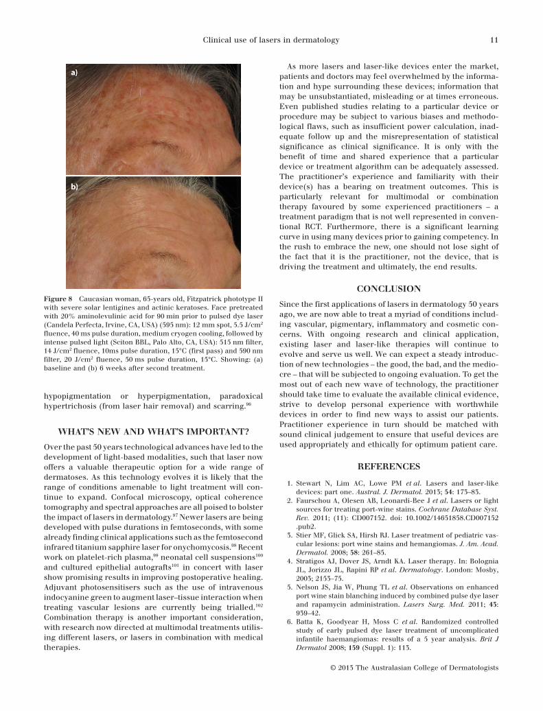
therapy is a useful strategy to reduce any dysplastic lesionsthat may accompany the pigmentary and vascular photo-damage (Fig. 8). For patients seeking treatment of prema-lignant skin lesions as well as photorejuvenation, eitherPDT with non-ablative laser or light devices or fractionalresurfacing lasers (thulium or CO2) may offer a practicaltherapeutic solution.
Actinic cheilitis Ablative laser therapy of actinic cheilitis isan important tool in the dermatologist’s armamentarium,which includes topical therapy, PDT, cryotherapy, curettageand cautery, and surgical vermillionectomy. A prospectivecohort study of 40 patients compared CO2 laser withvermillionectomy, 5-FU or TCA peels in the management ofactinic cheilitis, followed up for 4 years.81 None of thepatients treated with laser or surgical vermillionectomydeveloped clinical recurrence, compared with ≥ 50% recur-rence rates with the other modalities. ALA PDT activated byPDL has also shown promise as an effective intervention forpatients with actinic cheilitis recalcitrant to conventionaltherapies,82 as has fractional thulium laser.83 An important
Table 5 Treatment algorithm for acne scarring grade 4: severely abnormally contoured disease – severe atrophic or hypertrophic scarring
Scar type Pre-laser treatment plan Appropriate laser treatment
Punched outatrophic (deepboxcar), ice pick
Surface SurfaceTrichloroacetic acid (Chemical Reconstruction of
Skin Scars [CROSS] technique if numerous, deepand small)
Fractional resurfacing may be combined with CROSSIf few and broad but still < 4 mm in diameter,
consider punch techniques (float, elevation,excision or grafting) – see surgery, with or withoutsubsequent fractional or ablative resurfacingtechniques
Fractional resurfacing (ablative or non-ablative)All are good for this atrophic scar type but only after
preparatory treatmentAblative lasers (CO2 or erbium) is generally not as
useful as fractional lasers
Marked atrophy Volume (to increase) Volume (to increase)Fat transferVolumetric filling with hyaluronic acid or calcium
hydroxylapatite or stimulatory fillers such asdiluted poly-L-lactic acid
Fractional resurfacing (ablative or non-ablative)All are good for this atrophic scar type but only after
preparatory treatmentAblative lasers (CO2 or erbium) is generally not as
useful as fractional lasers but is better than it iswith punched out scars due to its tightening effecton the skin surface
Significanthypertrophy orkeloid
Intralesional corticosteroids or fluorouracil maybesupplemented with vascular laser
Fractional lasers and vascular lasers may be usefulbut again, preparation must be undertaken foruseful results
Atrophic orhypertrophicdisease
MovementBotulinum toxin often combined with fillers
especially in lower face for atrophic diseaseAs supplement to excision of atrophic or
hypertrophic scars
Fractional lasers are more useful if movement andtension on scars are settled prior to laser therapyeven if only in the few months following treatment
Bridges andtunnels,dystrophic scars
SurgeryExcision Fractional ablative and non-ablative and
non-fractional full ablative lasers are all useful todisguise scars after excision
Punched out scars(deep boxcar)
Punch elevation if scar base suitablePunch excision, punch grafting if scar base poor
Laser use is the same as for bridges and tunnels
Marked saggingand apparentredundancy
Occasionally rhytidectomy Lasers have limited role
CO2, carbon dioxide.
8 DF Sebaratnam et al.
© 2013 The Australasian College of Dermatologists

consideration in treating actinic cheilitis with laser, as withany other ablative modality, is that no tissue specimen isobtained, precluding a histopathological review. In an anec-dotal report on approximately 100 patients with actiniccheilitis treated with CO2 ablation only one case developeda squamous cell carcinoma in the treatment field,84
although rates of up to 5% have been reported. Ongoingsurveillance in this group is essential.
Inflammatory dermatoses
Lasers have been used as a non–first-line therapy forvarious common dermatological conditions such as acneand psoriasis (Table 6). The generic mechanism of action ismost likely related to laser effects on lesion vasculature andthe modulation of underlying cytokines and inflammatorymediators. Due to community concern over the systemictherapy of active acne, laser and light-based treatmentalternatives have gained popularity in recent years. Thesetherapies are thought to ameliorate acne through the inhi-bition of sebum production, the modulation of inflammationand keratinisation, and the conversion of porphyrins natu-rally synthesised by Propionibacterium acnes to bactericidalreactive oxygen species.85 PDL has been shown to reduceacne severity, with the most rapid improvements observedwithin 4 weeks of commencing treatment.86 PDL has beenshown to be as effective as IPL and light-emitting diodephototherapy in the treatment of acne.87 PDL effects wereenhanced in the setting of methyl-aminolevulinate-PDT,88
but conferred no additional benefit when combinedwith clindamycin-benzoyl peroxide topical therapy.89 Theresults for Nd:YAG, KTP and diode (1450 nm) were lessconclusive.90–94 The studies described have employed a widerange of outcome measures, using pooled results for meta-analysis. However, a protocol recently submitted for aCochrane Review on light therapies for acne may yield anevidence-based approach for the use of lasers in thissetting.95 Nevertheless, given that well-established, effectiveand less costly medications are available, consideration forlaser therapy should be reserved for those who fail or havecontraindications to medical therapies.
LASER COMPLICATIONS
The safety of laser therapy is well established although, aswith any intervention, adverse effects are possible. A preop-erative clinical review should include an evaluation ofFitzpatrick phototype, recent or planned sun exposure,recent artificial tan application, immunological or inflam-matory comorbidities, history of herpes simplex, allergy,scarring, previous cosmetic or surgical procedures and amedication history for risk stratification. Pre-procedure andpost-procedure photo-documentation should be mandatory.Common transient side effects include pain, pruritus, ery-thema, purpura, oedema, acne, vesiculation, crusting andpigmentary change. Bacterial, viral and candidal infectionscan complicate resurfacing procedures and prophylacticantimicrobials are often considered. Depending on the laseremployed, potential long-term sequelae include permanent
Figure 6 Caucasian woman, 50-years old, Fitzpatrick phototype IIIwith photodamage and periorificial rhytides. Resurfacing param-eters for: (i) upper eyelids: superficial fractional CO2 (LumenisAcupulse, San Jose, CA, USA) – 100 mJ, 60% density, single pass,(ii) perioral region: deep fractional CO2 – 25 mJ 15%, two passeswith third pass superficial CO2 100 mJ 60% (Lumenis Acupulse),(iii) rest of face: superficial erbium peel 30 microns with 30microns coagulation (Sciton Profile, Palo Alto, CA, USA) withonabotulinumtoxinA (Botox; Allergan, Irvine, CA, USA) injections tofrontalis, corrugator supercilii, procerus and lateral orbicularisoculi (30 units in total). Showing: (a) baseline and (b) 2 months aftertreatment.
Figure 7 Caucasian man, 72-years old, Fitzpatrick phototype IIwith severe rhinophyma. Treated with the Sharplan CO2 (nowLumenis Acupulse; Lumenis, San Jose, CA, USA) laser with com-puterised flash-scanner at 30 w, 3 mm spot, on continuous setting infeather mode. Treatment carried out under nerve block local anaes-thesia. Showing: (a) baseline and (b) after treatment.
Clinical use of lasers in dermatology 9
© 2013 The Australasian College of Dermatologists
Tab
le6
Com
mon
derm
atol
ogic
alco
ndi
tion
sam
enab
leto
lase
rth
erap
y
Der
mat
olog
ical
con
diti
onPa
thol
ogy
Non
-las
erth
erap
yL
aser
ther
apy
Com
men
t
Acn
evu
lgar
isC
omed
ones
,pa
pule
s,pu
stu
les
from
incr
ease
dse
bum
and
Pro
pion
iba
cter
ium
acn
esac
tivi
ty
•T
opic
als
(ben
zoyl
pero
xide
,an
tibi
otic
s,re
tin
oids
)•
Ch
emic
alpe
els
•Sy
stem
ic(a
nti
biot
ics,
reti
noi
ds,
anti
-an
drog
ens)
•L
ED
(blu
e,re
d)•
PDL
/IPL
•±
PDT
wit
hab
ove
Con
ven
tion
alth
erap
yle
ssco
stly
than
lase
r
An
giofi
brom
aPa
pule
sw
ith
incr
ease
dva
scu
latu
rean
dfi
brou
sti
ssu
e•
Ele
ctro
surg
ery
(cau
tery
orh
yfre
cati
on)
shav
eex
cisi
on•
Abl
ativ
ela
ser
(spo
tC
O2
orer
biu
m)
•H
otK
TP
Pote
nti
altu
bero
us
scle
rosi
sli
nk
(con
side
rto
pica
lra
pam
ycin
)PD
Lu
sefu
lon
lyfo
rle
sion
eryt
hem
aD
erm
aln
evu
sSm
ooth
skin
-col
oure
ddo
me-
shap
edn
ests
ofn
aevo
mel
anoc
ytic
cell
s•
Shav
eex
cisi
on;
elec
tros
urg
ery;
cure
tte
•E
xcis
ion
•A
blat
ive
lase
r(s
pot)
Las
eru
sefu
lfo
rfa
cial
lesi
ons
Sebo
rrh
oeic
kera
tosi
sor
derm
atos
ispa
pulo
san
igra
Ben
ign
epid
erm
alh
yper
kera
tosi
san
dac
anth
osis
•C
yrot
her
apy
•Sh
ave
exci
sion
orel
ectr
osu
rger
yor
cure
tte
•A
blat
ive
lase
r(s
pot)
•K
TP
(532
nm
)So
lar
len
tigi
nes
may
bepr
ecu
rsor
lesi
on(t
reat
wit
hn
on-a
blat
ive
devi
ces)
Fla
tle
sion
sm
ayre
spon
dto
pigm
ent
lase
rsor
IPL
Ker
atos
ispi
lari
sru
bra
Fol
licu
lar
kera
tosi
sw
ith
peri
foll
icu
lar
eryt
hem
aon
face
,la
tera
lar
ms
and
thig
hs
•T
opic
alke
rato
lyti
cs•
Gen
tle
mec
han
ical
exfo
liat
ion
•PD
Lor
IPL
•L
aser
hai
rre
mov
alE
ryth
ema
resp
onds
bett
erth
ante
xtu
rero
ugh
nes
s
Pseu
dofo
llic
uli
tis
Infl
amm
ator
yfo
llic
ula
rpa
pule
s,an
dpu
stu
les
from
curl
yh
air
re-e
nte
rin
gth
esk
in
•T
opic
al(b
enzo
ylpe
roxi
de,
anti
biot
ics,
eflor
nit
hin
e)•
Stop
shav
ing
•L
aser
hai
rre
mov
alL
ong
puls
edN
d:YA
Gpr
efer
red
for
pati
ents
wit
hda
rksk
inph
otot
ypes
Psor
iasi
sT
-cel
ldr
iven
hyp
erpr
olif
erat
ive
skin
diso
rder
•T
opic
al•
Phot
oth
erap
y•
Syst
emic
orbi
olog
ics
•30
8n
mex
cim
erla
ser
•PD
LL
aser
opti
onn
otbe
enw
idel
yad
opte
d
Seba
ceou
sh
yper
plas
iaV
isib
leye
llow
enla
rgem
ent
ofse
bace
ous
glan
ds•
Ele
ctro
surg
ery
•PD
L(±
PDT
)•
Dio
de(1
450
nm
)•
Abl
ativ
ela
ser
(spo
t)•
KT
P(5
32n
m)
Rec
urr
ence
com
mon
Syri
ngo
ma
Ben
ign
prol
ifer
atio
nof
swea
tdu
cts
pres
enti
ng
aspe
rioc
ula
rpa
pule
s•
Ele
ctro
surg
ery
•Sn
ipex
cisi
on(f
ew)
•A
blat
ive
lase
r(s
pot)
•F
ract
ion
alab
lati
vela
ser
•O
ccas
ion
ally
KT
Pla
ser
Rec
urr
ence
expe
cted
Vit
ilig
oF
ocal
orge
ner
alis
edlo
ssof
skin
mel
anoc
ytes
and
pigm
ent
•T
opic
alim
mu
nos
upp
ress
ion
•Ph
otot
her
apy
•A
uto
logo
us
graf
ts
•30
8n
mex
cim
erla
ser
•A
blat
ive
wit
hpi
gmen
ttr
ansf
erte
chn
iqu
es
Las
erop
tion
not
been
wid
ely
adop
ted
War
t(v
erru
cae)
Hu
man
papi
llom
avi
rus
indu
ced
epid
erm
alh
yper
kera
tosi
s•
Cry
oth
erap
y•
Ele
ctro
surg
ery
•T
opic
alch
emoc
aute
ry•
Imm
un
oth
erap
y
•PD
L(±
PDT
)•
Lon
g-pu
lse
Nd:
YAG
(106
4n
m)
•A
blat
ive
lase
r(s
pot
CO
2)
Non
-sca
rrin
gm
eth
ods
pref
erre
d
Xan
thel
asm
aC
hol
este
rol
depo
siti
onar
oun
dth
em
edia
lca
nth
us
•T
rich
loro
acet
icac
id(3
0–50
%)
•E
lect
rosu
rger
y•
Exc
isio
n
•A
blat
ive
lase
r(s
pot)
•F
ract
ion
alab
lati
vela
ser
Ch
eck
seru
mli
pids
CO
2,ca
rbon
diox
ide;
IPL
,in
ten
sepu
lsed
ligh
t;K
TP,
pota
ssiu
mtr
itan
ylph
osph
ate;
LE
D,l
igh
t-em
itti
ng
diod
e;N
d:YA
G,n
eody
miu
m-d
oped
yttr
ium
alu
min
ium
garn
et;P
DL
,pu
lsed
dye
lase
r;PD
T,
phot
odyn
amic
ther
apy.
10 DF Sebaratnam et al.
© 2013 The Australasian College of Dermatologists
hypopigmentation or hyperpigmentation, paradoxicalhypertrichosis (from laser hair removal) and scarring.96
WHAT’S NEW AND WHAT’S IMPORTANT?
Over the past 50 years technological advances have led to thedevelopment of light-based modalities, such that laser nowoffers a valuable therapeutic option for a wide range ofdermatoses. As this technology evolves it is likely that therange of conditions amenable to light treatment will con-tinue to expand. Confocal microscopy, optical coherencetomography and spectral approaches are all poised to bolsterthe impact of lasers in dermatology.97 Newer lasers are beingdeveloped with pulse durations in femtoseconds, with somealready finding clinical applications such as the femtosecondinfrared titanium sapphire laser for onychomycosis.98 Recentwork on platelet-rich plasma,99 neonatal cell suspensions100
and cultured epithelial autografts101 in concert with lasershow promising results in improving postoperative healing.Adjuvant photosensitisers such as the use of intravenousindocyanine green to augment laser–tissue interaction whentreating vascular lesions are currently being trialled.102
Combination therapy is another important consideration,with research now directed at multimodal treatments utilis-ing different lasers, or lasers in combination with medicaltherapies.
As more lasers and laser-like devices enter the market,patients and doctors may feel overwhelmed by the informa-tion and hype surrounding these devices; information thatmay be unsubstantiated, misleading or at times erroneous.Even published studies relating to a particular device orprocedure may be subject to various biases and methodo-logical flaws, such as insufficient power calculation, inad-equate follow up and the misrepresentation of statisticalsignificance as clinical significance. It is only with thebenefit of time and shared experience that a particulardevice or treatment algorithm can be adequately assessed.The practitioner’s experience and familiarity with theirdevice(s) has a bearing on treatment outcomes. This isparticularly relevant for multimodal or combinationtherapy favoured by some experienced practitioners – atreatment paradigm that is not well represented in conven-tional RCT. Furthermore, there is a significant learningcurve in using many devices prior to gaining competency. Inthe rush to embrace the new, one should not lose sight ofthe fact that it is the practitioner, not the device, that isdriving the treatment and ultimately, the end results.
CONCLUSION
Since the first applications of lasers in dermatology 50 yearsago, we are now able to treat a myriad of conditions includ-ing vascular, pigmentary, inflammatory and cosmetic con-cerns. With ongoing research and clinical application,existing laser and laser-like therapies will continue toevolve and serve us well. We can expect a steady introduc-tion of new technologies – the good, the bad, and the medio-cre – that will be subjected to ongoing evaluation. To get themost out of each new wave of technology, the practitionershould take time to evaluate the available clinical evidence,strive to develop personal experience with worthwhiledevices in order to find new ways to assist our patients.Practitioner experience in turn should be matched withsound clinical judgement to ensure that useful devices areused appropriately and ethically for optimum patient care.
REFERENCES
1. Stewart N, Lim AC, Lowe PM et al. Lasers and laser-likedevices: part one. Austral. J. Dermatol. 2013; 54: 173–83.
2. Faurschou A, Olesen AB, Leonardi-Bee J et al. Lasers or lightsources for treating port-wine stains. Cochrane Database Syst.Rev. 2011; (11): CD007152. doi: 10.1002/14651858.CD007152.pub2.
3. Stier MF, Glick SA, Hirsh RJ. Laser treatment of pediatric vas-cular lesions: port wine stains and hemangiomas. J. Am. Acad.Dermatol. 2008; 58: 261–85.
4. Stratigos AJ, Dover JS, Arndt KA. Laser therapy. In: BologniaJL, Jorizzo JL, Rapini RP et al. Dermatology. London: Mosby,2003; 2153–75.
5. Nelson JS, Jia W, Phung TL et al. Observations on enhancedport wine stain blanching induced by combined pulse dye laserand rapamycin administration. Lasers Surg. Med. 2011; 43:939–42.
6. Batta K, Goodyear H, Moss C et al. Randomized controlledstudy of early pulsed dye laser treatment of uncomplicatedinfantile haemangiomas: results of a 5 year analysis. Brit JDermatol 2008; 159 (Suppl. 1): 113.
Figure 8 Caucasian woman, 65-years old, Fitzpatrick phototype IIwith severe solar lentigines and actinic keratoses. Face pretreatedwith 20% aminolevulinic acid for 90 min prior to pulsed dye laser(Candela Perfecta, Irvine, CA, USA) (595 nm): 12 mm spot, 5.5 J/cm2
fluence, 40 ms pulse duration, medium cryogen cooling, followed byintense pulsed light (Sciton BBL, Palo Alto, CA, USA): 515 nm filter,14 J/cm2 fluence, 10ms pulse duration, 15°C (first pass) and 590 nmfilter, 20 J/cm2 fluence, 50 ms pulse duration, 15°C. Showing: (a)baseline and (b) 6 weeks after second treatment.
Clinical use of lasers in dermatology 11
© 2013 The Australasian College of Dermatologists

7. Marqueling AL, Oza V, Frieden IJ et al. Propranolol and infan-tile hemangiomas four years later: a systematic review.Pediatr. Dermatol. 2013; 30: 182–91.
8. Wargon O. Randomised placebo controlled trial: safety andefficacy of topical timolol maleate gel vs placebo for smallsuperficial infantile haemangiomas. Australas. J. Dermatol.2013; 54: S22–23.
9. Laube S, Lanigan SW. Laser treatment of rosacea. J. Cosmet.Dermatol. 2002; 1: 188–95.
10. Goodman GJ, Roberts S, Bezborodoff A. Studies in long-pulsedpotassium tritanyl phosphate laser for the treatment of spidernaevi and perialar telangiectasia. Australas. J. Dermatol. 2002;43: 9–14.
11. Nymann P, Hedelund L, Haedersdal M. Long-pulsed dyelaser vs. intense pulsed light for the treatment of facialtelangiectasias: a randomized controlled trial. J. Eur. Acad.Dermatol. Venerol. 2010; 24: 143–6.
12. Jorgensen GF, Hedelund L, Haedesdal M. Long-pulsed dyelaser versus intense pulsed light for photodamaged skin: arandomized split-face trial with blinded response evaluation.Lasers Surg. Med. 2008; 40: 293–9.
13. Neuhaus IM, Zane LT, Tope WD. Comparative efficacy ofnonpurpuragenic pulsed dye laser and intense pulsed light forerythematotelangiectatic rosaca. Dermatol. Surg. 2009; 35:920–8.
14. Azevedo LH, Galletta VC, de Paulo Eduardo C et al. Venouslake of the lips treated using photocoagulation with high-intensity diode laser. Photomed. Laser Surg. 2010; 28: 263–5.
15. Roncero M, Canueto J, Blanco S et al. Multiwavelengthlaser treatment of venous lakes. Dermatol. Surg. 2009; 35:1942–6.
16. Bekhor PS. Long-pulsed Nd:YAG laser treatment of venouslakes: report of a series of 34 cases. Dermatol. Surg. 2006; 32:1151–4.
17. van den Bos R, Arends L, Kockaert M et al. Endovenous thera-pies of lower extremity varicosities: a meta-analysis. J. Vasc.Surg. 2009; 49: 230–9.
18. Lim A. Novel endovenous techniques: beyond sclerotherapyand lasers. Australas. J. Dermatol. 2013; 54: S29.
19. Dover JS. New approaches to the laser treatment of vascularlesions. Australas. J. Dermatol. 2000; 41: 14–8.
20. Hruza GJ, Avram MA. Lasers and Lights, 3rd edn. New York:Elsevier-Saunders, 2013.
21. Choi JE, Kim JW, Seo SH et al. Treatment with Becker’s neviwith a long-pulse alexandrite laser. Dermatol. Surg. 2009; 35:1105–8.
22. Trelles MA, Allones I, Moreno-Arias GA et al. Becker’s naevus:a comparative study between erbium:YAG and Q-switchedneodymium:YAG; clinical and histopathological findings. Br. J.Dermatol. 2005; 152: 308–13.
23. Noordzij MJ, van den Broecke DG, Alting MC. Ruby laser treat-ment of congenital melanocytic nevi: a review of the literatureand report of our own experience. Plast. Reconstr. Surg. 2004;114: 660–7.
24. Al-Hadithy N, Al-Nakib K, Quaba A. Outcomes of 52 patientswith congenital melanocytic naevi treated with UltraPulsecarbon dioxide and frequency doubled Q-switched Nd-Yaglaser. J. Plast. Reconstr. Aesthet. Surg. 2012; 65: 1019–28.
25. Kim YJ, Whang KU, Choi WB et al. Efficacy and safety of1,064 nm Q-switched Nd:YAG laser treatment for removingmelanocytic nevi. Ann. Dermatol. 2012; 24: 162–7.
26. Ueda S, Isoda M, Imayama S. Response of naevus of Ota toQ-switched ruby laser treatment according to lesion colour. Br.J. Dermatol. 2000; 142: 77–82.
27. Wang HW, Liu YH, Zhang GK et al. Analysis of 602 Chinesecases of nevus of Ota and the treatment results treated byQ-switched alexandrite laser. Dermatol. Surg. 2007; 33: 455–60.
28. Liu J, Ma YP, Ma XG et al. A retrospective study of Q-switchedalexandrite laser in treating nevus of Ota. Dermatol. Surg.2011; 27: 1480–5.
29. Kar HK, Gupta L. 1064 nm Q switched Nd: YAG laser treatmentof nevus of Ota: an Indian open label prospective study of50 patients. Indian J Dermatol Venereol Leprol. 2011; 77: 565–70.
30. Kunachak S, Leelaudomlipi P, Sirikulchayanonta V.Q-switched ruby laser therapy of acquired bilateral Nevus ofOta-like macules. Dermatol. Surg. 1999; 25: 938–41.
31. Polnikorn N, Tanrattanakorn S, Goldberg D. Treatment ofHori’s nevus with the Q-switched Nd:YAG laser. Dermatol.Surg. 2000; 26: 477–80.
32. Kroon MW, Wind BS, Beek JF. Nonablative 1550-nm fractionallaser therapy versus triple topical therapy for the treatment ofmelasma: a randomized controlled pilot study. J. Am. Acad.Dermatol. 2011; 64: 516–23.
33. Wind BS, Kroon MW, Meesters AA et al. Non-ablative 1,550nmfractional laser therapy versus triple topical therapy for thetreatment of melasma: a randomized controlled split-facestudy. Lasers Surg. Med. 2010; 42: 607–12.
34. Trelles MA, Velez M, Gold MH. The treatment of melasma withtopical creams alone, CO2 fractional resurfacing alone, or acombination of the two: a comparative study. J. DrugsDermatol. 2010; 9: 315–22.
35. Passeron T, Fontas E, Kang HY et al. Melasma treatment withpulsed-dye laser and triple combination cream: a prospective,randomized, single-blind, split-face study. Arch. Dermatol.2011; 147: 1106–8.
36. Wattanakrai P, Mornchan R, Eimpunth S. Low-fluenceQ-switched neodymium-doped yttrium aluminum garnet(1,064 nm) laser for the treatment of facial melasma in Asians.Dermatol. Surg. 2010; 36: 76–87.
37. Polder KD, Bruce S. Treatment of melasma using a novel1,927-nm fractional thulium fiber laser: a pilot study.Dermatol. Surg. 2012; 38: 199–206.
38. Bloom BS, Brauer JA, Geronemus RG. Ablative fractionalresurfacing in topical drug delivery: an update and outlook.Dermatol. Surg. 2013; 39: 839–48.
39. Callender VD, St. Surin-Lord S, Davis EC et al.Postinflammatory hyperpigmentation. Am. J. Clin. Dermatol.2012; 12: 87–99.
40. Haedersdal M, Gøtzsche PC. Laser and photoepilation forunwanted hair growth. Cochrane Database Syst. Rev. 2006; 4:CD004684.
41. Haedersdal M, Wulf HC. Evidence-based review of hairremoval using lasers and light sources. J. Eur. Acad. Dermatol.Venereol. 2006; 20: 9–20.
42. Pai GS, Bhat PS, Mallya H et al. Safety and efficacy oflow-fluence, high-repetition rate versus high-fluence, low-repetition rate 810-nm diode laser for permanent hair removal– a split-face comparison study. J. Cosmet. Laser Ther. 2011; 13:134–7.
43. Haak CS, Nymann P, Pedersen AT et al. Hair removal in hirsutewomen with normal testosterone levels: a randomized con-trolled trial of long-pulsed diode laser vs. intense pulsed light.Br. J. Dermatol. 2010; 163: 1007–13.
44. Sochor M, Curkova AK, Schwarczova Z et al. Comparison ofhair reduction with three lasers and light sources: prospective,blinded and controlled study. J. Cosmet. Laser Ther. 2011; 13:210–5.
45. Bernstein EF, Basilavecchio L, Plugis J. Bilateral axilla hairremoval comparing a single wavelength alexandrite laser withcombined multiplexed alexandrite and Nd:YAG laser treatmentfrom a single laser platform. J. Drugs Dermatol. 2012; 11:185–90.
46. McGill DJ, Hutchison C, McKenzie E et al. A randomised, split-face comparison of facial hair removal with the alexandrite
12 DF Sebaratnam et al.
© 2013 The Australasian College of Dermatologists
laser and intense pulsed light system. Lasers Surg. Med. 2007;39: 767–72.
47. Ismail SA. Long-pulsed Nd:YAG laser vs. intense pulsed lightfor hair removal in dark skin: a randomized controlled trial. Br.J. Dermatol. 2012; 166: 317–21.
48. Braun M. Comparison of high-fluence, single-pass diode laserto low-fluence, multiple-pass diode laser for laser hair reduc-tion with 18 months of follow up. J. Drugs Dermatol. 2011; 101:62–5.
49. Barolet D. Low fluence-high repetition rate diode laser hairremoval 12-month evaluation: reducing pain and risks whilekeeping clinical efficacy. Lasers Surg. Med. 2012; 44: 277–81.
50. Bakus AD, Garden JM, Yaghmai D et al. Long-term fine caliberhair removal with an electro-optic Q-switched Nd:YAG laser.Lasers Surg. Med. 2010; 42: 706–11.
51. Burris K, Kim K. Tattoo removal. Clin. Dermatol. 2007; 25:388–92.
52. Zelickson BD, Mehregan DD, Zarrin AA et al. Clinical, histo-logic and ultrastructural evaluation of tattoos treated withthree laser systems. Lasers Surg. Med. 1994; 15: 364–72.
53. Kossida T, Rigopoulos D, Katsambas A et al. Optimal tattooremoval in a single laser session based on the method ofrepeated exposures. J. Am. Acad. Dermatol. 2012; 66: 271–7.
54. Bhatt N, Alster TS. Laser surgery in dark skin. Dermatol. Surg.2008; 34: 184–95.
55. Battle EF Jr, Soden CE Jr. The use of lasers in darker skintypes. Semin. Cutan. Med. Surg. 2009; 28: 130–40.
56. Hedelund L, Moreau KE, Beyer DM et al. Fractionalnonablative 1,540-nm laser resurfacing of atrophic acne scars.A randomized controlled trial with blinded response evalua-tion. Lasers Med. Sci. 2010; 25: 749–54.
57. Cho SB, Lee SJ, Cho S et al. Non-ablative 1550-nm erbium-glass and ablative 10 600-nm carbon dioxide fractional lasersfor acne scars: a randomized split-face study with blindedresponse evaluation. J. Eur. Acad. Dermatol. Venereol. 2010; 24:921–5.
58. Wanitphakdeedecha R, Manuskiatti W, Siriphukpong S et al.Treatment of punched-out atrophic and rolling acne scars inskin phototypes III, IV, and V with variable square pulseerbium:yttrium-aluminum-garnet laser resurfacing. Dermatol.Surg. 2009; 35: 1376–83.
59. Kim S, Cho KH. Clinical trial of dual treatment with an ablativefractional laser and a nonablative laser for the treatment ofacne scars in Asian patients. Dermatol. Surg. 2009; 35: 1089–98.
60. Ong MS, Bashir SJ. Fractional laser resurfacing for acne scars:a review. Br. J. Dermatol. 2012; 166: 1160–9.
61. Goodman GJ. Treatment of acne scarring. Int. J. Dermatol.2011; 50: 1179–94.
62. Saedi N, Petelin A, Zachary C. Fractionation: a new era in laserresurfacing. Clin. Plast. Surg. 2011; 38: 449–61.
63. Ross EV, Miller C, Meehan K et al. One-pass CO2 versusmultiple-pass Er:YAG laser resurfacing in the treatment ofrhytides: a comparison side-by-side study of pulsed CO2 andEr:YAG lasers. Dermatol. Surg. 2001; 27: 709–15.
64. Karsai S, Czarnecka A, Jünger M et al. Ablative fractionallasers (CO(2) and Er:YAG): a randomized controlled double-blind split-face trial of the treatment of peri-orbital rhytides.Lasers Surg. Med. 2010; 42: 160–7.
65. Khatri KA, Ross V, Grevelink JM et al. Comparison oferbium:YAG and carbon dioxide lasers in resurfacing of facialrhytides. Arch. Dermatol. 1999; 135: 391–97.
66. Yamauchi PS, Lask GP, Lowe NJ. Botulinum toxin type A givesadjunctive benefit to periorbital laser resurfacing. J. Cosmet.Laser Ther. 2004; 6: 145–8.
67. Zimbler MC, Holds JB, Kokoska MS et al. Effect of botulinumtoxin pretreatment on laser resurfacing results. Arch. FacialPlast. Surg. 2001; 3: 165–9.
68. Mordon S, Lagarde JM, Vienne MP. Ultrasound imaging dem-onstration of the improvement of non-ablative laser remodel-ling by concomitant daily topical application of 0.05%retinaldehyde. J. Cosmet. Laser Ther. 2004; 6: 5–9.
69. Carter SR, Seiff SR, Choo P et al. Lower eyelid CO2 laser reju-venation. Ophthalmology 2001; 108: 437–41.
70. Campo-Voegeli A, Arboles MP. Combination of IPL, ablativenon fractional and ablative fractional in single session-treatment protocols for photorejuvenation or acne scars.Australas. J. Dermatol. 2013; 53: S21.
71. Kearney C, Brew D. Single-session combination treatmentwith intense pulsed light and fractional photothermolysis: asplit face study. Australas. J. Dermatol. 2013; 54: S3–4.
72. Lim SW, Lim SW, Bekhor P. Rhinophyma: carbon dioxide laserwith computerized scanner is still an outstanding treatment.Australas. J. Dermatol. 2009; 50: 289–93.
73. Park JH, Hwang ES, Kim SN et al. Er:YAG laser treatmentof verrucous epidermal nevi. Dermatol. Surg. 2004; 30: 378–81.
74. Foster RS, Bint LJ, Halbert AR. Topical 0.1% rapamycin forangiofibromas in paediatric patients with tuberous sclerosis: apilot study of four patients. Australas. J. Dermatol. 2012; 53:52–6.
75. Hantash BM, Stewart DB, Cooper ZA et al. Facial resurfacingfor nonmelanoma skin cancer prophylaxis. Arch. Dermatol.2006; 142: 976–82.
76. Ostertag JU, Quaedvlieg PJ, van der Geer S et al. A clinicalcomparison and long-term follow-up of topical 5-fluorouracilversus laser resurfacing in the treatment of widespread actinickeratoses. Lasers Surg. Med. 2006; 38: 731–9.
77. Weiss ET, Brauer JA, Anolik R et al. 1927-nm fractional resur-facing of facial actinic keratoses: a promising new therapeuticoption. J. Am. Acad. Dermatol. 2013; 68: 98–102.
78. Togsverd-Bo K, Haak CS, Thaysen-Peterson D et al. Intensifiedphotodynamic therapy of actinic keratoses with fractional CO2
laser: a randomized clinical trial. Br. J. Dermatol. 2012; 166:1262–9.
79. Avram DK, Goldman MP. Effectiveness and safety of ALA-IPLin treating actinic keratoses and photodamage. J. DrugsDermatol. 2004; 3: S36–9.
80. Alexiades-Armenakas MR, Geronemus RG. Laser-mediatedphotodynamic therapy of actinic keratoses. Arch. Dermatol.2003; 139: 1313–20.
81. Robinson JK. Actinic cheilitis. A prospective study comparingfour treatment methods. Arch. Otolaryngol. Head Neck Surg.1989; 115: 848–52.
82. Alexiades-Armenakas MR, Geronemus RG. Laser mediatedphotodynamic therapy of actinic cheilitis. J. Drugs Dermatol.2004; 3: 548–52.
83. Ghasri P, Admani S, Petelin A et al. Treatment of actiniccheilitis using a 1,927-nm thulium fractional laser. Dermatol.Surg. 2012; 38: 504–7.
84. Laws RA, Wilde JL, Grabski WJ. Comparison ofelectrodessication with CO2 laser for the treatment of actiniccheilitis [Commentary]. Dermatol. Surg. 2000; 26: 349–53.
85. Webster GF. Light and laser therapy for acne: sham or science?Facts and controversies. Clin. Dermatol. 2010; 28: 31–5.
86. Seaton ED, Charakida A, Mouser PE et al. Pulsed-dye lasertreatment for inflammatory acne vulgaris: randomised con-trolled trial. Lancet 2003; 362: 1347–52.
87. Sami NA, Attia AT, Badawi AM. Phototherapy in the treatmentof acne vulgaris. J. Drugs Dermatol. 2008; 7: 627–32.
88. Haedersdal M, Togsverd-Bo K, Wiegell SR et al. Long-pulseddye laser versus long-pulsed dye laser-assisted photodynamictherapy for acne vulgaris: a randomized controlled trial. J. Am.Acad. Dermatol. 2008; 58: 387–94.
89. Karsia S, Schmitt L, Raulin C. The pulsed-dye laser as anadjuvant treatment modality in acne vulgaris: a randomized
Clinical use of lasers in dermatology 13
© 2013 The Australasian College of Dermatologists

controlled single-blinded trial. Br. J. Dermatol. 2010; 163: 395–401.
90. Orringer JS, Kang S, Maier L et al. A randomized, controlled,split-face clinical trial of 1320nm Nd:YAG laser therapy in thetreatment of acne vulgaris. J. Am. Acad. Dermatol. 2007; 56:432–8.
91. Jung JY, Hong JS, Ahn CH et al. Prospective randomized con-trolled clinical and histopathological study of acne vulgaristreated with dual mode of quasi-long pulse and Q-switched1064-nm Nd:YAG laser assisted with a topically applied carbonsuspension. J. Am. Acad. Dermatol. 2012; 66: 626–33.
92. Yilmaz O, Senturk N, Yuksel EP et al. Evaluation of 532-nm KTPlaser treatment efficacy on acne vulgaris with once and twiceweekly applications. J. Cosmet. Laser Ther. 2011; 13: 303–7.
93. Baugh WP, Kucaba WD. Nonablative phototherapy for acnevulgaris using the KTP 532 nm laser. Dermatol. Surg. 2005; 31:1290–6.
94. Jih MH, Friedman PM, Goldberg LH et al. The 1450-nm diodelaser for facial inflammatory acne vulgaris: dose-response and12-month follow-up study. J. Am. Acad. Dermatol. 2006; 55:80–7.
95. Car J, Car M, Hamilton F, Layton A, Lyons C, Majeed A. Lighttherapies for acne. Cochrane Database of Systematic Reviews2009: CD007917.
96. Tanzi EL, Lupton JR, Alster TS. Lasers in dermatology: fourdecades of progress. J. Am. Acad. Dermatol. 2003; 49: 1–31.
97. Ross EV. Lasers and light base techonologies in skin – whereare we now and where are we heading? Australas. J. Dermatol.2009; 50: A22.
98. Manevitch Z, Lev D, Hochberg M et al. Direct antifungal effectof femtosecond laser on Trichophyton rubrum onychomycosis.Photochem. Photobiol. 2010; 86: 476–9.
99. Na JI, Choi JW, Choi HR et al. Rapid healing and reducederythema after ablative fractional carbon dioxide laser resur-facing combined with the application of autologous platelet-rich plasma. Dermatol. Surg. 2011; 37: 463–8.
100. Zimber MP, Mansbridge JN, Taylor M et al. Human cell-conditioned media produced under embryonic-like conditionsresult in improved healing time after laser resurfacing. Aes-thetic Plast. Surg. 2012; 36: 431–7.
101. Whang KK, Kim MJ, Song WK et al. Comparative treatment ofgiant congenital melanocytic nevi with curettage or Er:YAGlaser ablation alone versus with cultured epithelial autografts.Dermatol. Surg. 2005; 31: 1660–7.
102. Klein A, Bäumler W, Koller M et al. Indocyanine green-augmented diode laser therapy of telangiectatic leg veins: arandomized controlled proof-of-concept trial. Lasers Surg.Med. 2012; 44: 369–76.
14 DF Sebaratnam et al.
© 2013 The Australasian College of Dermatologists