larval morphology of metaphycus flavus and its role in host attachment and larval cannibalism
TRANSCRIPT

Bulletin of Entomological Researchhttp://journals.cambridge.org/BER
Additional services for Bulletin of Entomological Research:
Email alerts: Click hereSubscriptions: Click hereCommercial reprints: Click hereTerms of use : Click here
Larval morphology of Metaphycus flavus and its role in host attachment and larval cannibalism
A. Tena, A. Kapranas, G.P. Walker, F. GarciaMarí and R.F. Luck
Bulletin of Entomological Research / Volume 101 / Issue 03 / June 2011, pp 365 372DOI: 10.1017/S0007485310000611, Published online: 06 January 2011
Link to this article: http://journals.cambridge.org/abstract_S0007485310000611
How to cite this article:A. Tena, A. Kapranas, G.P. Walker, F. GarciaMarí and R.F. Luck (2011). Larval morphology of Metaphycus flavus and its role in host attachment and larval cannibalism. Bulletin of Entomological Research, 101, pp 365372 doi:10.1017/S0007485310000611
Request Permissions : Click here
Downloaded from http://journals.cambridge.org/BER, IP address: 171.67.34.205 on 06 Oct 2012

Larval morphology of Metaphycus flavusand its role in host attachment and
larval cannibalism
A. Tena3*, A. Kapranas1, G.P. Walker1, F. Garcia-Marí2and R.F. Luck1
1Department of Entomology, University of California, Riverside, USA:2Instituto Agroforestal del Mediterráneo, Universidad Politécnica de
Valencia, Spain: 3Unidad Asociada de Entomología IVIA-UJI-CIB, InstitutoValenciano de Investigaciones Agrarias, Spain
Abstract
Metaphycus flavus (Howard) (Hymenoptera: Encyrtidae) is a facultativelygregarious endoparasitoid of soft scales (Hemiptera: Coccidae). When it developsin superparasitised hosts, the larvae often attack and consume brood mates six ormore days post oviposition. Under our laboratory conditions (25±1°C and 14 hoursof light followed by 18±1°C and ten hours of darkness in 50–70% R.H.), M. flavuseggs hatched three days after oviposition. Measurements of the mandibles andtentorium indicate there are four larval instars, andM. flavus reaches the fourth instarby day six post oviposition, and pupates on day eight. Thus, cannibalism amongM. flavus larvae occurs during the fourth instar. During this instar, M. flavus larvaeseparate from their attachment to the scale cuticle, to which they were tethered by arespiratory structure during the previous three larval instars. Once detached, they arefree tomovewithin the scale, which increases the probability of larval encounters andaggressive behaviours. Moreover, the mandibles of the fourth instar are betteradapted for fighting than are those of the first three larval instars, since they are largerand more sclerotized. The cranium and mouthparts of M. flavus have four differenttypes of sensory organs, some of which are almost certainly olfactory, an unexpectedfunction for a larva that presumably is surrounded by an aqueous medium wheregustatory sensilla would seem to be more appropriate. The cranium also bears twopairs of what appear to be secretory pores.
Keywords: Encyrtidae, soft scales, larval development, superparasitism,intraspecific competition, sensilla
(Accepted 28 September 2010)
Introduction
Intraspecific competition for host resources among im-mature parasitoids has a major influence on ecological andevolutionary processes of parasitoids (Godfray, 1994). In
gregarious parasitoids, several larvae may develop and usehost resources; but, if a second clutch of eggs is laid(i.e. superparasitism) and an excess of larvae develops on orin the host, the result can be competition for host resources(Jervis et al., 2005). This competition may end with theemergence of several small individuals with lower fitnessand probability of survival or with the elimination ofcompetitors through physiological suppression or physicalconflict. In solitary parasitoids, only one individual candevelop per host. When several eggs are laid, competition
*Author for correspondenceFax: (+34) 96 342 40 01E-mail: [email protected]
Bulletin of Entomological Research (2011) 101, 365–372 doi:10.1017/S0007485310000611© Cambridge University Press 2011First published online 6 January 2011
for host resources occurs principally through physical conflictsuntil only one individual is left and it can fully use the host’sresources (Godfray, 1994). Combats occur during the firstlarval instar when sufficient host resources remain for thesurviving larva to complete its development (Clausen, 1940;Salt, 1961). In these cases, the first instar larva has morpho-logical adaptations for fighting that it does not possess inthe following instars, such as large mandibles and caudalappendages or setae that increase their mobility (Clausen,1940; Salt, 1961; van Baaren et al., 1997; Mayhew & vanAlphen, 1999).
Metaphycus flavus females (Howard) (Hymenoptera:Encyrtidae) normally lay a small, female-biased clutch of 2–3eggs in immature brown soft scale Coccus hesperidumL. (Hemiptera: Coccidae) (Bernal et al., 1999a; Kapranaset al., 2008; Tena et al., 2008). If a second female subsequentlyencounters this scale, she will lay an additional clutch of 2–3eggs in them (=superparasitism), in excess of those that candevelop successfully within the scale. Under these circum-stances, the larvae normally engage in physical conflicts inwhich the supernumerary larvae are eliminated and con-sumed (Tena et al., 2009). Interestingly, in contrast to whatwould be expected given this behaviour, the larval stage ofother species of Metaphycus that have been described lack thelarge, piercing mandibles typical of fighting species (Flanders,1942; Bartlett & Ball, 1964; Saakyan-Baranova, 1966); and,moreover, the movements of the larvae are restricted becausethey are attached at their posterior end to the host’s cuticle viaan aeroscopic plate which limits encounters between indivi-duals (Maple, 1954; Saakyan-Baranova, 1966). Consequently,Metaphycus larvae do not present the typical morphology thatusually characterizes an aggressive endoparasitoid species.We, thus, describe the development of M. flavus larvae andtheir associatedmorphologywithin the host.We focus on theirmandibles and their means of attachment to the host cuticleand how these characteristics may favour or restrict thephysical conflicts that Tena et al. (2009) previously described inthis species. Lastly, we also describe the sensory organs foundin the third and fourth larval instar.
Material and methods
Scale and parasitoid cultures
We established a brown soft scale culture using crawlersobtained from an infested pineapple guava plant, Feijoasellowiana O. Berg (Myrtaceae), located at the University ofCalifornia, Riverside, CA campus (UCR). The scales werereared on excised Yucca recurvifolia Salisbury (Agavaceae)leaves maintained hydroponically in the UCR insectary at27–28°C, 60% R.H. with a 21L:3D photoperiod. We obtainedthe excised yucca leaves from plants grown at UCR Agri-cultural Operations.
The Metaphycus flavus colony used in this study wasestablished in 1996 with individuals collected from citricolascale, Coccus pseudomagnoliarum Kuwana (Hemiptera:Coccidae), infesting citrus near Kozan, in south centralTurkey (Bernal et al., 1999b). The colony has since beenmaintained in the UCR insectary by introducing matedfemales into 7.5cm dia.×50cm long plastic tubes, eachcontaining one or two scale-infested yucca leaves with ca.300 brown soft scales per leaf.Wemaintained the culture in therearing tubes at a ratio of approximately one female parasitoidper ten scales. The rearing tubes were capped with plastic lids
at both ends, which had holes that were covered with afine nylon mesh to allow air circulation while preventingadult parasitoid escape or entry. Honey was streaked on theinside wall of the tubes as a carbohydrate source for theintroduced or emerged parasitoids. The tubes were main-tained at 25±1°C, 50–70% R.H. and a 14L:10D photoperiod.
Procedure for obtaining parasitoids formorphological examination
We obtained the adult parasitoids for our studiesby removing 100–200 scales containing parasitoid pupae(= ‘mummies’) from the yucca leaves and placing them in a2.5cm dia.×9.5cm long glass vials. Each vial was then sealedwith a plastic cap that had a central ventilation hole coveredwith fine nylon mesh. The developing wasps were allowed toemerge from these scales. These wasps were collected dailyand confined as a mixed-sexed group within a second, 2.5cmdia.×9.5cm long vial held at 25±1°C, 50–70% R.H. and14L:10D photoperiod for two days. This allowed the femalesto mate and mature their eggs. All of the vials contained astreak of honey on their inside walls as a carbohydrate sourcefor the parasitoids. Prior to each experiment, we isolated two-day-old females from these vials by placing each female in a1cm dia. glass vial with a drop of honey on its inside wall andsealing the vial with a cotton plug.
Larvae for morphological examination were taken fromsuperparasitised scales using the same procedures andexperimental methods described by Tena et al. (2009). Thisallowed us to compare our results from this study with thosefrom this previous study. To obtain these larvae, we confineda single, mated, 3-day-old female with a section of yuccaleaf having a 23–28-day-old scale, 1.8±0.05mm wide by2.5±0.10mm long. Each yucca leaf section with its scale wasconfined in a 4cm dia.×1.5cm high glass Petri dish, whichformed an observation arena. Using a cool fiber optic light anda dissectingmicroscope at 10–50×magnification, we observedand noted the behaviour of each female wasp continuously inthis arena until she had laid her initial egg clutch in the scale.Four hours after the initial female was removed, we exposedthe scale to a second M. flavus female following the sameprocedure. We used the protruding egg stalk associated witheach M. flavus oviposition to confirm the deposition of an egg(Maple, 1954; Tena et al., 2008). A total of 130 superparasitisedscales were available for examination in this study. Thesuperparasitised scales, along with their associated yuccaleaf section, were maintained in an incubator at 25±1°Cand 14h of light, followed by 18±1°C and ten hoursof darkness in 50–70% R.H. to allow normal parasitoiddevelopment.
On days one through nine post oviposition, we detachedthe superparasitised scales from the leaves daily and dissectedthem in saline solution (1% NaCl) under a dissectingmicroscope to determine whether the larval parasitoids wereattached to the scale cuticle. If so, we detached each larvawithin the scale from the host cuticle and transferred and fixedit to a glass slide using a thin film of saline solution. We thenmeasured the length and width of each larva using an ocularmicrometer mounted in the eyepiece of a compound micro-scope. We also measured the length and width of thetentorium and mandibles of each larva within the scale byplacing a glass cover slip over the larva and gently pressing onit to flatten it against the slide. We then counted the number ofspiracles present in each of the parasitoid larvae within the
A. Tena et al.366

scale and measured length and width of their tentorium andmandibles. We present these measurements as an average±standard deviation, along with the number of observations(table 1, col. 4). A total of 221 eggs and larvae were measured.We also prepared specimens for scanning electronmicroscopy(SEM) or for slide mounting, clearing and photography.
The specimens for scanning electron microscopy (SEM)were dehydrated using a series of increasing ethanol con-centrations which ended with 100% ethanol. The ethanol wasthen replaced with three changes of hexamethyldisilizane/hexamethyldisiloxane (HMDS-Polysciences, Warrington, PA,USA), and the specimens were then air dried in a fumehood (Heraty & Hawks, 1998). These dried specimens weremounted on aluminum stubs with carbon conductiveadhesive tabs (Pelco Tabs® Ted Pella Inc., Redding, CA,USA), sputter coated with gold/palladium alloy and exam-ined with a Phillips XL30-FEG scanning electron microscope(FEI Co., Hillsboro, OR, USA).
The specimens that were slide mounted and photographedusing lightmicroscopywere prepared by killing the parasitoidin ethanol and mounting each specimen on a microscope slideusing Hoyer’s mounting medium (20 parts chloral hydrate,5 parts water, 3 parts gum Arabic, 2 parts glycerin). The
specimens were then covered with a glass cover slip andplaced on a slide warmer (ca. 30–35°C) for about one week toallow each specimen to clear. Digital photographs of thesespecimens were obtained using a Zeiss Axioskop 2 compoundmicroscope (Carl Zeiss Inc., Oberkochen, Germany) with aJVC 3-CCD digital camera (Model KY-F7O) using Auto-Montage software (Syncroscopy, Cambridge, UK).
Results
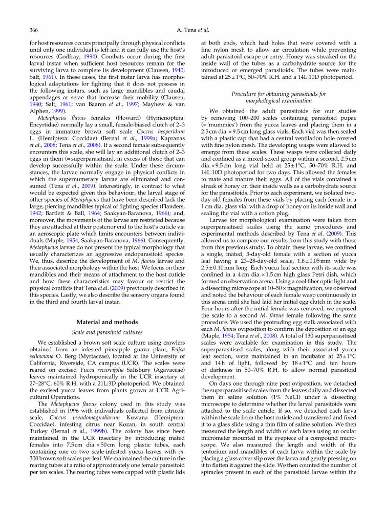
Measurements of the mandibles, tentoria and larvae(table 1, fig. 1) indicated that M. flavus has four larval instars.Under our rearing conditions, the larvae hatched from theireggs three days after oviposition and they pupated five dayslater. The first instar larvae were encyrtiform (Clausen, 1940)(fig. 2a) and almost spherical, measuring 0.17±0.023mm longby 0.12±0.013mmwide (n=24). The anterior part of the larvalhead contained the tentorium and a pair of mandibles. Theselatter were shaped like minute hooks (see table 1 for lengthmeasurements) and they appeared to be weakly sclerotized(fig. 3a). The posterior end of the first instar (fig. 2a) wassurrounded by the chorion, which allowed the two pairsof open spiracles to protrude through the host cuticle. This
Table 1. Morphological characteristics of Metaphycus flavus larvae.
Metaphycusflavus age (days)*
Tentoriumwidth (μm)
Mandiblelength (μm)
Number oflarvae
measured**
Instar
3 23.9±2.8 7.7±0. 7 13/24 First4 42.6±9.2 14.6±2.9 36/44 Second5 64.0±9.5 24.2±6.8 39/36 Third6 77.8±8.1 31.5±3.8 41/55 Fourth7 78.0±4.1 33.0±1.9 15/24 Fourth8 77.5±6.4 33.8±1.4 4/4 Fourth
* days after oviposition.** The numbers represents the number of tentoria and mandibles measured respectively.
0
0.2
0.4
0.6
0.8
1
1.2
1.4
0 2 4 6 8 10
mm
Days
Egg1st
instar2nd
instar3rd
instar4th
instar
Fig. 1. Metaphycus flavus egg and larval development. Length and width of the egg and larvae of M. flavus at different time intervals afteroviposition (^, larval length; , larval width).
Larval morphology of Metaphycus flavus 367

provided a pathway for the respiratory gases to enter theposterior spiracles (see Discussion). These posterior spiracleswere connected to two longitudinal, lateral trunks with simpleramifications, and they constituted the first instar’s meta-pneustic tracheal system. The larvae were attached to thehost’s cuticle via the egg chorion, which restricted the larvaefrom moving within the scale. At ecdysis, the larval exuviumwas gradually sloughed off from the anterior to posterior, butthe exuvium remained attached to the egg chorion.
The second and third larval instars were also metapneusticand encyrtiform (fig. 2b, c). The second instar began on dayfour post oviposition and measured 0.29±0.042mm longby 0.20±0.037mm wide (n=43). The third instar appeared onday five and measured 0.46±0.1mm long by 0.31±0.067 wide
(n=47). The head of each instar was easily distinguished(fig. 2b, c). The mandibles became progressively larger witheach instar (table 1). They still appeared to be weaklysclerotized through the second instar (fig. 3b), but the anteriorpart of the mandibles of the third instar became sclerotized(fig. 3c). The posterior end of the larvae remained attached tothe chorion and the exuvia from the earlier instars, whichsurrounded the two pairs of posterior spiracles. The trachealsystem was similar to that of the first instar, and the larvaewere tethered to the host cuticle.
The respiratory system changed drastically when the larvamolted to the fourth instar. It became peripneustic andmanifested nine lateral pairs of open spiracles (fig. 2d–f).Under our rearing conditions, the fourth instar larvae occurred
Fig. 2. Lateral view of different larval instars ofMetaphycus flavus. (a) First instar partially covered by the egg shell. (b–c) Second and thirdinstars attached to the host cuticle. (d) Fourth instar showing the open spiracles. (e) Fourth instar spiracles on abdominal segments 2–4.(f) Detailed view of spiracle on abdominal segment 3. (Figs. 2a–e anterior is to the right and posterior to the left).
A. Tena et al.368

on days six through eight post oviposition. On day six, thelarvae averaged 0.87±0.24mm long by 0.48±0.095mmwide (n=60) and reached its maximum size on day seven(1.23±0.23mm long, 0.59±0.08mm wide) (n=24) (fig. 1). Atthe beginning of the fourth instar, the larvaewere still attachedto the chorion and host cuticle, but they subsequently becamedetached from the chorion during this instar and were able tomove freely within the host. Its mandibles also underwenta major transformation during the fourth instar. They becamelarge, hook-like and well-sclerotized (fig. 3d). The head ofthe fourth instar larvae was also easily distinguished fromthe rest of the larva (fig. 2d). Once the fourth instar larvaehad consumed the remaining scale contents, leaving only thescale’s cuticle, the larvae excreted their meconia within thescale, which caused the larvae to shrink in size (fig. 1, day 8).The larvae then became white just prior to pupation.
We examined the sensilla present on the last two larvalinstars and found numerous sensilla on the head of the thirdand fourth instar. A pair of spherical, multiporous sensillaoccurred on the cranium, lateral to the mouthparts (fig. 4a, c),and two pairs of spherical, multiporous sensilla occurred onthemaxillary-labial complex (fig. 4b, d).We also noted a singlepair of coeloconic sensilla on the maxillary-labial complex.These later had the form of a raised torus (‘doughnut’) within ashallow depression in which a short, grooved peg protruded
from the center of the torus (fig. 4b, d, e). Additionally, we alsonoted two other sensilla types that were associated with themouthparts. Three pairs of short peg sensilla occurred on theclypeo-labrum (fig. 4b), which had the form of a papiliformpeg sunk within a depression (fig. 4b, f) and that resembledbasiconic or styloconic sensilla (Keil, 1999). We also foundthree pairs of sensilla, one pair occurred laterally on theclypeo-labrum and two pairs occurred on the maxillary-labialcomplex (fig. 4b, d). The morphology of these sensilla didnot match closely the traditional classifications of sensilla.Based on their shape, we referred to them as mamilliformsensilla. In addition to these sensory organs, we also noted twopairs of deep pits that were located dorsally on the cranium(fig. 4a, g).
Discussion
The role of host attachment in larval cannibalism
Metaphycus flavus larvae are endoparasitoids that areattached initially to their host’s cuticle via the egg chorionuntil the fourth instar. This type of attachment restricts larvalmovement within the scale. Also, the first three larval instarsare metapneustic and obtain their oxygen via two pairs ofposterior spiracles that attach each larva to its egg chorion via
Fig. 3. Mandibles of four larval instars of Metaphycus flavus: (a) first instar; (b) second instar; (c) third instar; (d) fourth instar.
Larval morphology of Metaphycus flavus 369

an aeroscopic plate. This attachment is similar to that reportedfor its congeners, M. helvolus (Compere) and M. luteolus(Timberlake), both of which also possess two pairs of posteriorspiracles and an aeroscopic plate (Flanders, 1942; Saakyan-Baranova, 1966). Thus, this pattern of attachment appearsto be characteristic of the genus. According to Maple (1954),encyrtid larvae maintain contact between their posteriorspiracles and the atmosphere by aeroscopic plates on theegg, which project externally through the host cuticle. In thefourth instar, the respiratory system becomes peripneusticmanifesting nine pairs of open spiracles that are distributedlaterally on each side of the larva. Initially, this larval stadiumremains attached to the host via the chorion; but, as the hostcontents become consumed, the larva detaches from thechorion and moves freely within the host. During this instar,air is obtained directly via the nine larval spiracles, and theremaining contents within mummified scale are consumed.
Tena et al. (2009) have recently documented that super-numerary M. flavus larvae engage in physical conflicts thatresult in the consumption of the losing larvae (i.e. cannibal-ism). These conflicts only occurred when the larvae within ascalewere six ormore days old. The developmental conditionsof the larvae in that experiment were similar to this study(environmental conditions; host conditions, host instar andsize; superparasitism within four hours), which indicates thatlarvae were in the fourth instar when they were six days old.Thus, physical conflicts among M. flavus larvae appear tooccur only during the fourth instar, probably after the larvaehave severed their attachment to the host cuticle and they areable tomove freelywithin the scale. However, host attachmentin other Metaphycus species does not interfere with larvalencounters and subsequent larval conflict.Metaphycus luteolus,another brown soft scale parasitoid, engages in physicalconflicts during the second and third instar (Bartlett & Ball,
Fig. 4. Head of Metaphycus flavus. (a) Anterior view of entire head; black arrowheads, spherical multiporous sensilla (detail in c); whitearrowheads, pits (detail in g). (b) Anterior-ventral view of mouth region (above the mouth opening, mo, is the clypeo-labrum and below isthe maxillary-labial complex). (c) Spherical olfactory sensillum on the cranium. (d) Three ventral-most sensilla on the maxillary-labialcomplex. (e) Detail of central peg of a coeloconic sensillum on the maxillary-labial complex. (f) Peg sensilla on the clypeo-labrum. (g) Deeppit on the cranium. cl, clypeo-labrum; cr, cranium; cs, coeloconic sensillum; mlc, maxillary-labial complex; mo, mouth opening; ms,mamilliform sensillum; ps, peg sensillum; pt, prothorax; smps, spherical multiporous sensillum.
A. Tena et al.370

1964) while the larvae are still attached to the host (Saakyan-Baranova, 1966). Interestingly, both M. flavus and M. luteolusallocate clutches of similar size (Tena et al., 2008; Kapranaset al., 2009), and the eggs of both species are attached internallyto the host cuticle (Saakyan-Baranova, 1966). However, thesespecies differ in the way they distribute their eggs withinthe host scale. Metaphycus flavus deposits each egg individu-ally within the host, at different points around the scale’speriphery. In contrast, M. luteolus deposits its eggs in a singlelocation within the scale (A.Tena, personal observations). Thisspecies-specific difference in egg distribution may affect thelikelihood and timing of larval encounters within the host. Forexample, the encounters among the M. luteolus larvae withina host scale may occur earlier during larval development thanat the encounters among M. flavus larvae because of theclustered nature of the M. luteolus larvae. The more disperseddistribution of M. flavus eggs appears to prevent the larvaefrom contacting one another with their mandibles until theybecome detached from their aeroscopic plate during the fourthinstar.
The fourth instar as a fighting instar
The first instar larvae of many solitary endoparasitoidspecies possess morphological features that are used toeliminate supernumerary competitors through physical con-flicts (Clausen, 1940). These features are usually lost orreduced after the initial molt, and the subsequent instarsare often unable to fight (Hagen, 1964). The morphologicalcharacteristics of an aggressive first instar larva typicallyinclude a large head with large, piercing, well-developedmandibles; a set of dorsal spines on the thorax and abdomen;and a large caudal segment and/or a large caudal spike(Clausen, 1940; Laing & Corrigan, 1987; van Baaren et al.,1997). The spines and large caudal segment (developed ‘tail’)are responsible for the mobility of first instar larvae withina host (Clausen, 1940; van Baaren et al., 1997). However, firstinstar M. flavus larvae, as well as other Metaphycus species(Flanders, 1942; van Baaren et al., 1997), lack these morpho-logical characteristics. Thus, movements of the first instarlarvae are not only constrained by their attachment to theirhost but also by their lack of dorsal spines and a developedtail.
The large and well-developed piercing mandibles andthe caudal spike found in species where the first instar is thefighting instar are used to attack other larvae within the host,both conspecifics and individuals of other species (Clausen,1940; Laing & Corrigan, 1987). In contrast to this pattern, themandibles of first instarM. flavus are at their smallest, and theyincrease in size with each successive instar. The mandiblesare at their largest and most heavily sclerotized during thefourth instar and are double the size of those in the secondinstar (fig. 3). These mandibles are likely capable of killing andconsuming other competing conspecific larvae even thoughthey are not the large and piercing mandibles typical of thefirst instar described in other parasitoid species.
Sensory organs and possible secretory glands in the third andfourth instar
The head and mouthparts of the third and fourth instarlarvae are well equipped with multiple types of sensoryorgans. They have coeloconic sensilla (fig. 4e), which arechemosensory and usually olfactory in function (Keil, 1999).
The spherical, multiporous sensilla (fig. 4c) are almostcertainly olfactory, as the presence of the many pores ischaracteristic of insect olfactory sense organs (Keil, 1999).However, the occurrence of olfactory organs in the third andfourth instars is surprising since they live within a host,presumably in an aqueous environment. Gustatory sensillawould be more suitable for chemoreception in this type ofenvironment. The peg sensilla on the clypeo-labrum (fig. 4f)bear similarities to both basiconic and styloconic sensilla;the former are usually chemosensory while the latter oftenfunction as temperature and/or humidity sensors (Keil, 1999).However, the senses provided by the peg sensilla is unknown.Similarly, the function of the mammiliform sensilla is un-known (fig. 4d). Transmission electron microscopy andelectrophysiology would be required to determine unambigu-ously the functions of these sensilla. Finally, the pits on thecranium appear to be openings of secretory glands, but asnoted for the sensilla, confirmation of their function requirestransmission electron microscopy. If they are secretory, itraises intriguing questions: what are they secreting, and whatis the effect of these secretions on the host and/or parasitoidcompetitors?
Acknowledgements
We thank Lisa D. Foster, Porfirio Pacheco and RobertTrautman for providing the scales, host plants andparasitoids to conduct these experiments. This researchwas supported in part by an USDA National ResearchInitiative grant (USDA-NRI 2005-01006) awarded to RFLand Jocelyn Millar and by a California Citrus ResearchBoard grant (CRB 5500–159) awarded to Joseph Morseand RFL.
References
Bartlett, B.R. & Ball, J.C. (1964) The developmental biologies oftwo encyrtid parasites ofCoccus hesperidum and their intrinsiccompetition. Annals of the Entomological Society of America 57,496–503.
Bernal, J.S., Luck, R.F. & Morse, J.G. (1999a) Host influences onsex ratio, longevity, and egg load of two Metaphycus speciesparasitic on soft scales: implications for insectary rearing.Entomologia Experimentalis et Applicata 92, 191–204.
Bernal, J.S., Luck, R.F. & Morse, J.G. (1999b) Augmentativerelease trials with Metaphycus spp. (Hymenoptera:Encyrtidae) against citricola scale (Homoptera: Coccidae)in California’s San Joaquin Valley. Journal of EconomicEntomology 92, 1099–1107.
Clausen, C.P. (1940) Entomophagous Insects. New York, USA,McGraw-Hill.
Flanders, S.E. (1942) Metaphycus helvolus, an encyrtid parasite ofthe black scale. Journal of Economic Entomology 35, 690–698.
Godfray, H.C.J. (1994) Parasitoids: Behavioral and EvolutionaryEcology. Princeton, NJ, USA, Princeton University Press.
Hagen, K.S. (1964) Developmental stages of parasites. pp. 168–246in DeBach, P. (Ed.) Biological Control of Insect Pests and Weeds.London, UK, Chapman and Hall.
Heraty, J. & Hawks, D. (1998) Hexamethyldisilazane – a chemicalalternative for drying insects. Entomological News 109,369–374.
Jervis, M.A., Copland, M.J.W. & Harvey, J.A. (2005) The life-cycle. pp. 73–165 in Jervis, M.A. (Ed.) Insects as Natural
Larval morphology of Metaphycus flavus 371

Enemies: A Practical Perspective. Dordrecht, The Netherlands,Springer.
Kapranas, A., Pacheco, P., Forster, L.D., Morse, J.G. & Luck, R.F.(2008) Precise sex allocation by several encyrtid parasitoids ofbrown soft scale Coccus hesperidum L. (Hemiptera: Coccidae).Behavioural Ecology and Sociobiology 62, 901–912.
Kapranas, A., Wanjberg, E. & Luck, R.F. (2009) Sequences of sexallocation andmortality in clutches ofMetaphycus parasitoidsof soft scale insects and the prevalence of all-female broods.Ecological Entomology 34, 652–662.
Keil, T.A. (1999) Morphology and development of the peripheralolfactory organs. pp. 5–47 in Hansson, B.S. (Ed.) InsectOlfaction. Berlin, Germany, Springer-Verlag.
Laing, J.E. & Corrigan, J.E. (1987) Intrinsic competition betweenthe gregarious parasite, Cotesia glomeratus and the solitaryparasite,Cotesia rubecula (Hymenoptera: Braconidae) for theirhost, Artogeia rapae (Lepidoptera: Pieridae). Entomophaga 32,493–501.
Maple, J.D. (1954) The eggs and first instar larvae of Encyrtidaeand their morphological adaptations for respiration.University of California, Publications in Entomology 8, 25–122.
Mayhew, P.J. & van Alphen, J.J.M. (1999) Gregariousdevelopment in alysiine parasitoids evolved through areduction in larval aggression. Animal Behaviour 58, 131–141.
Saakyan-Baranova, A.A. (1966) The life cycle of Metaphycusluteolus Timb. (Hymenoptera: Encyrtidae), parasite of Coccushesperidum L. (Homoptera: Coccidae), and the attempt of itsintroduction into the USSR. Entomological Review 45, 414–423.
Salt,G. (1961) Competition among insect parasitoids.Mechanismsin biological competition. Symposium of the Society forExperimental Biology 15, 96–119.
Tena, A., Kapranas, A., Garcia-Marí, F. & Luck, R.F. (2008)Host discrimination, superparasitism, and infanticide by agregarious endoparasitoid. Animal Behaviour 76, 789–799.
Tena, A., Kapranas, A., Garcia-Marí, F. & Luck, R.F. (2009) Larvalcannibalism during the late developmental stages of afacultatively gregarious encyrtid endoparasitoid. EcologicalEntomology 34, 669–676.
van Baaren, J., Boivin, G., Le Lannic, J. & Nénon, J.P. (1997) Themale and female first instar larvae of Anaphes victus andA. listronoti (Hymenoptera,Mymaridae). Zoomorphology 117,189–197.
A. Tena et al.372