laparoscopic virtual mirror for understanding vessel...
TRANSCRIPT

Laparoscopic Virtual Mirror for Understanding Vessel Structure
Evaluation Study by Twelve Surgeons
Christoph Bichlmeier1∗ Sandro Michael Heining2
† Mohammad Rustaee1‡ Nassir Navab1
§
1Computer Aided Medical Procedures & Augmented Reality (CAMP), TUM, Munich, Germany2Trauma Surgery Department, Klinikum Innenstadt, LMU, Munich, Germany
ABSTRACT
In this paper we present the evaluation of a virtual mirror used asa navigational tool within a medical augmented reality (AR) sys-tem for laparoscopy. 12 surgeons of our clinical partner partici-pated in an experiment to evaluate whether laparoscope augmenta-tion extended by a virtual mirror is useful for improved perceptionof complex structures. Such complex structures are encounteredfor instance in laparoscopic resection of tumor affected liver tissue.The blood vessels supplying the tumor have to be cut and closedbefore tumorous tissue can be removed. A laparoscopic cameraand an optical tracking system allow for the visualization of visual-ized medical volumetric data registered with the real anatomy. Pre-viously injected contrast agent provides an accentuation of bloodvessels within the visualization. For evaluating the suitability of avirtual mirror to support the mentioned procedure, we designed aphantom consisting of wooden branches simulating the structure ofblood vessel trees. Quantitative results of the experiment show theadvantage of a mirror in certain cases, when blood vessels cannot bedirectly seen from the camera point of view due to self-occlusion ofthe structure. Results of a questionnaire filled out by the surgeonsafter the experiments confirm the acceptance of AR technology forparticular medical procedures.
Keywords: Augmented reality, navigated surgery, medical visual-ization, user interaction.
Index Terms: H.5.1 [Information Interfaces and Presentation]:Multimedia Information Systems—Artificial, augmented, and vir-tual realities; H.5.2 [Information Interfaces and Presentation]: UserInterfaces—Interaction styles; I.3.6 [Computer Graphics]: Method-ology and Techniques—Interaction techniques; J.3 [Life and Med-ical Sciences]
1 INTRODUCTION
This paper presents a clinical evaluation investigating the percep-tive advantage of a virtual mirror integrated into a laparoscopicaugmented reality (AR) scenario. The evaluation focuses on the(partial) resection of organs or organ segments, e.g. of the liver,where blood vessels need to be closed before tumorous segmentsare removed. Therefore parts of tissue like tumor affected liver seg-ments, but also complete organs have to be removed and affiliatedblood vessels are closed.
Keyhole surgery using an endoscopic device to get a view on theoperation site displayed on an external monitor is established in theORs. Extending this technology with an AR system allows for theaugmentation of the laparoscopic camera images with 3D visual-ization of medical imaging data. A contrast agent can be injected
∗e-mail: [email protected]†e-mail: [email protected]‡e-mail: [email protected]§e-mail:[email protected]
into the blood circuit in the region of interest immediately beforeimaging data acquisition to highlight blood vessels. A suitable ren-dering technique enables 3D visualization of a local vessel structurefrom the volume data.
Feuerstein et al. [3] report the accuracy of a similar system setupfor rigid targets and determined the error for target position (0.4mmRMS) and orientation (0.12) of tracking system in use, the navi-gation error (1.05mm RMS) and the augmentation error (1.58mmRMS). Regarding soft tissue, in most cases its pose and shape willchange between data acquisition and surgical intervention. Eventhough data is acquired intraoperatively using e.g. a C-arm device,surgeons cannot rely on the accurate augmentation due to breathing,motion of organs like heartbeat, pushing and pulling tissue with sur-gical instruments and manipulation of the tension of tissue as cut-ting modifies its structure. Nonetheless, some basic characteristicsof the structure like relative position and order of blood vessels,vessel size and branches remain in spite of all extraneous, deform-ing influences. Information about these characteristics can be usedto plan the next step in the surgical workflow, for instance to decidewhere to cut next. In most cases such structure can be understoodsatisfactorily from the monocular point of view of the laparoscopiccamera. Due to the user controlled navigation of the laparoscopiccamera, the visual cuesocclusion andmotion parallax provide in-formation about the depth order of structure elements. However,in the majority of cases the camera is controlled by an assistantwhile the surgeon is interacting with the instruments, which makesan intuitive exploration of the structure by moving the camera pointof view almost impossible. Furthermore, the moving space of thecamera is restricted to a limited number of ports providing access tothe inside of the patient. In some cases the area of interest can notbe seen directly from this restricted camera point of view. Hence,perceptive information about depth order of structure componentsis too insufficient to guide surgical instruments intuitively to theoperation site. Figure 1 shows such a situation inside our phantomwhen a certain region of the structure, a red ball on the branch, isnot directly visible even though the camera can be slightly reposi-tioned. The mirror image provides the desired view on the area ofinterest.
1.1 Related Work
Endoscope augmentation was proposed for different medical appli-cations like brain surgery [9], liver surgery [7, 3, 8], transbronchialbiopsy [2] and cardiac surgery [5] to support different proceduresin the surgical workflow such as port placement and navigation ofinstruments inside the patient. Fuchs et al. introduced a system forlaparoscope surgery displaying data with a head mounted display[4] instead on an external monitor. However, all approaches onlyallow for one point of view to observe the AR scene. An entireexploration of objects such as complex blood vessel structures can-not performed satisfactorily for further navigational steps duringthe intervention. The present paper reports a first evaluation of thealready introduced concept of a laparoscopic mirror to explore hid-den structures and support understanding of complex topology [6].Another application for a virtual mirror, the preparation of canals
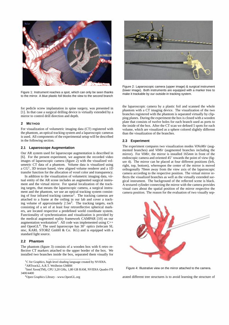
Figure 1: Instrument reaches a spot, which can only be seen thanksto the mirror. A blue plastic foil blocks the view to the second branch
for pedicle screw implantation in spine surgery, was presented in[1]. In that case a surgical drilling device is virtually extended by amirror to control drill direction and depth.
2 METHOD
For visualization of volumetric imaging data (CT) registered withthe phantom, an optical tracking system and a laparoscopic camerasis used. All components of the experimental setup will be describedin the following section.
2.1 Laparoscope Augmentation
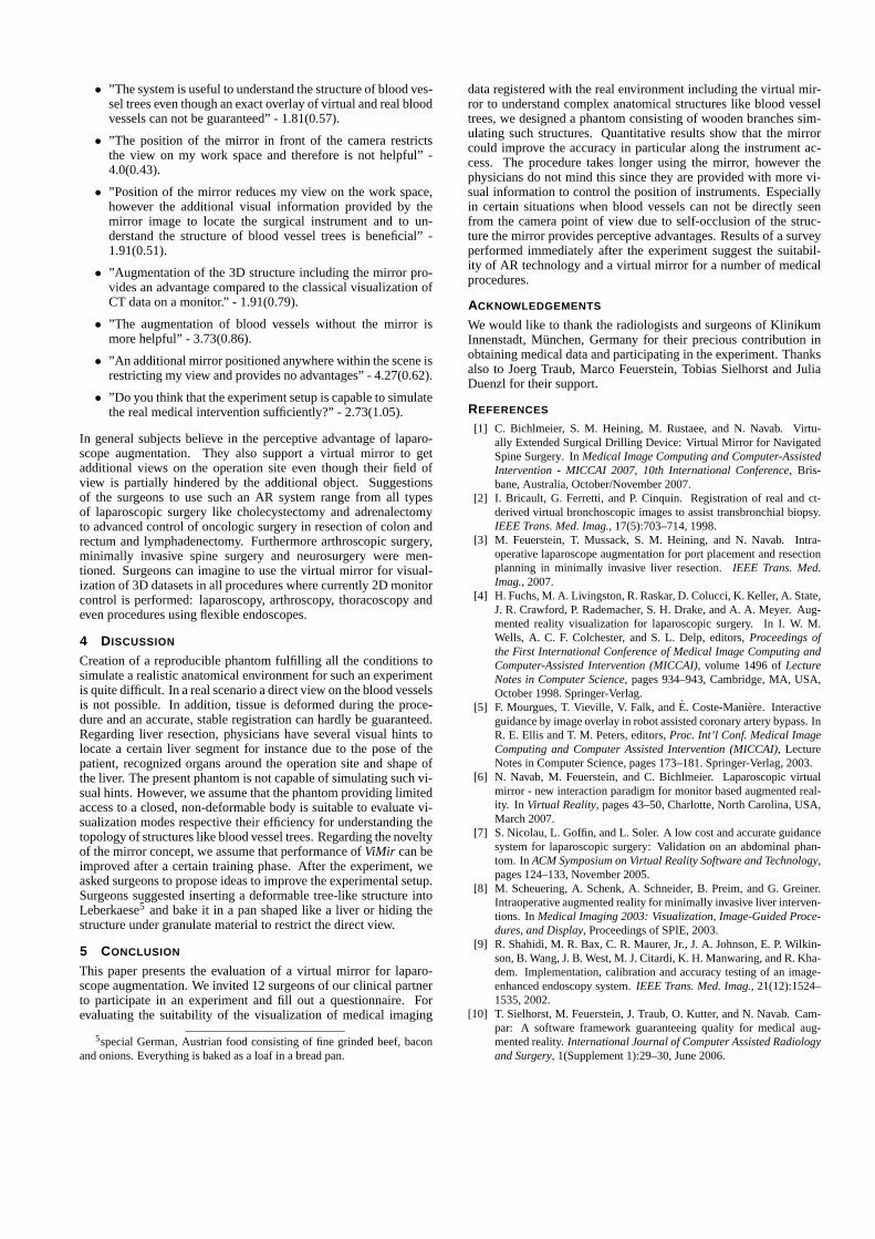
Our AR system used for laparoscope augmentation is described in[6]. For the present experiment, we augment the recorded videoimages of laparoscopic camera (figure 2) with the visualized vol-umetric CT data of a phantom. Volume data is visualized usinga CG1, 3D texture based, view aligned volume renderer and a 1Dtransfer function for the allocation of voxel color and transparency.
In addition to the visualization of volumetric imaging data, vir-tual entity of the AR scene includes an augmented surgical instru-ment and the virtual mirror. For spatial localization of the track-ing targets, that means the laparoscopic camera, a surgical instru-ment and the phantom, we use an optical tracking system consist-ing of four infrared tracking cameras2. The tracking cameras areattached to a frame at the ceiling in our lab and cover a track-ing volume of approximately 2.5m3. The tracking targets, eachconsisting of a set of at least four retroreflective spherical mark-ers, are located respective a predefined world coordinate system.Functionality of synchronization and visualization is provided bythe medical augmented reality framework CAMPAR [10] on ouraugmentation workstation3. All code was implemented using C++and OpenGL4. The used laparoscope has 30◦ optics (telecam SLntsc, KARL STORZ GmbH & Co. KG) and is equipped with astandard light source.
2.2 Phantom
The phantom (figure 3) consists of a wooden box with 6 retro re-flective CT markers attached to the upper border of the box. Weinstalled two branches inside the box, separated them visually for
1C for Graphics, high-level shading language created by NVIDIA.2ARTtrack2, A.R.T. Weilheim GMBH3Intel Xeon(TM), CPU 3,20 GHz, 1,80 GB RAM, NVIDIA Quadro FX
3400/44004Open Graphics Library - www.OpenGL.org
Figure 2: Laparoscopic camera (upper image) & surgical instrument(lower image). Both instruments are equipped with a marker tree tomake it trackable by our outside-in tracking system.
the laparoscopic camera by a plastic foil and scanned the wholephantom with a CT imaging device. The visualization of the twobranches registered with the phantom is separated virtually by clip-ping planes. During the experiment the box is closed with a woodenplate that consists of twelve holes for each branch used as ports tothe inside of the box. After the CT scan we defined 5 spots for eachvolume, which are visualized as a sphere colored slightly differentthan the visualization of the branches.
2.3 Experiment
The experiment compares two visualization modesViNoMir (aug-mented branches) andViMir (augmented branches including themirror). For ViMir, the mirror is installed 165mm in front of theendoscopic camera and oriented 45◦ towards the point of view (fig-ure 4). The mirror can be placed at four different positions (left,right, top, bottom), whereupon the center of the mirror is movedorthogonally 70mm away from the view axis of the laparoscopiccamera according to the respective position. The virtual mirror re-flects the visualized branches as well as the virtually extended sur-gical instrument. The background of the reflected scene is black.A textured cylinder connecting the mirror with the camera providesvisual cues about the spatial position of the mirror respective thecamera position. The reason for the evaluation of two visually sep-
Figure 4: Illustrative view on the mirror attached to the camera.
arated different tree structures is to avoid learning the structure of

Figure 3: Two wooden branches are installed on the bottom of the box. The closed box provides twelve ports to each branch.
one branch while working with the first visualization mode and us-ing this knowledge for the second visualization mode. The ARscene is presented on a monitor positioned in the working spaceof the subjects as shown in figure 5. Every subject had to reachfive spots for each of the two branches. For every target spot, wepredefine one of 12 ports, which has to be used for the laparoscopecamera. The selected ports only provide a restricted view on thetarget spot due to self occlusion of the branches. The surgical in-strument can be inserted through an arbitrary port. The combinationand order of branches and the visualization mode changed for everysubject. When subjects reach the target spot they ask the investiga-tor to lock the result to measure time and accuracy. Overall 12surgeons participated and 60 spots were analyzed. Before a subjectstarts the experiment, we provide the following information:
The instruments you are using are a laparoscopic camera anda surgical instrument. The camera images can be augmented withvirtual objects by our AR system. Wooden branches simulating acomplex structure were installed inside the box. We made a CTscan of the phantom and visualize the imaging data in 3D regis-tered with the phantom. You have to reach visualized spots, smallred spheres, on the structure. We compare two different visualiza-tions each on a different structure: Augmented structure only andaugmented structure plus virtual mirror. The mirror is positioned165mm in front of the laparoscopic camera and oriented 45◦ to-wards the camera. The mirror can be repositioned at four differentlocations by request (left, right, top, bottom). The camera can onlyuse one port to get a view to the inside of the box. Start positionof the camera for every new point is the entry of a port. For thesurgical instrument, you can use arbitrary ports. Please do not pullor push too hard to avoid breaking the branches! We measure timeand accuracy. Before we start, you will test the system with an aug-mented dummy object.
Immediately after the experiment, we asked the subjects to fillout a questionnaire.
3 RESULTS
The quantitative results are valid for particular situations when theview on the region of interest is restricted by a small degree of free-dom the of the laparoscope camera due to a limited number of ports.Accuracy is measured virtually. Therefore a predefined, constanterror has to be taken into account composed by the tracking errorand the manually designed virtual clamp, which does not exactlyconform to the real clamp of the surgical instrument.
After outliers deviating 3 times the STD from AVG where ex-cluded, overall measuring data of 106 spots, 54ViNoMir and 52ViMir, was analyzed. Results show that subjects guide the instru-ment with higher accuracy to the predefined target spot using theViMir mode. However, using theViNoMir mode takes less time(see table 1). We also analyzed xyz distances from the spot to thetip of the instrument relative to the instrument coordinate systemwith z as the instrument axis. Comparing the two visualization
Figure 5: Subject is guiding the surgical instrument using the videoimages of the laparoscopic camera presented on a monitor.
Table 1: Tendency of measured data given as AVG(STD) showshigher accuracy using the virtual mirror based method.
mode dist timeViNoMir 14.29mm(5.28) 69.70sec(64.45)ViMir 13.45mm(4.79) 87.49sec(82.20)
modes, measured distances in x and y direction differ not signifi-cantly (x = 0.84mm, y = 0.38mm ), however in z direction differ-ence of distances isx = 1.64mm. We assume that the view along theinstrument axes without using a mirror does not provide sufficientvisual information for position control.
Occlusion effects tell subjects when they reach the target spotwith the instrument. However we notice an interesting effect duringthe experiment when surgeons have problems to reach a spot dueto physical barriers because of the branches. When the tip of theinstrument is already close to the target, they rather stop and askto lock the result than keep on trying to move the clamp to thecorrect position. By contrast using the mirror to get an additionalview on the target, motivation increases to keep on trying until bothperspectives, the direct view and the mirror image, prove to havesuccessfully located the spot.
Ten male and 2 female subjects (AVG age: 32, AVG workexperience: 5.8 y) filled out the questionnaire. The scale of themultiple choice questionnaire was1 = I strongly agree * 2 = Iagree * 3 = I am undecided * 4 = I disagree * 5 = I stronglydisagree. Results are given as AVG(STD).
• ”The augmentation of blood vessels with or without the mirrorprovides no advantage during the intervention.” - 3.82(0.72).
• ”The system is useful to understand the structure of blood ves-sel trees even though an exact overlay of virtual and real bloodvessels can not be guaranteed” - 1.81(0.57).
• ”The position of the mirror in front of the camera restrictsthe view on my work space and therefore is not helpful” -4.0(0.43).
• ”Position of the mirror reduces my view on the work space,however the additional visual information provided by themirror image to locate the surgical instrument and to un-derstand the structure of blood vessel trees is beneficial” -1.91(0.51).
• ”Augmentation of the 3D structure including the mirror pro-vides an advantage compared to the classical visualization ofCT data on a monitor.” - 1.91(0.79).
• ”The augmentation of blood vessels without the mirror ismore helpful” - 3.73(0.86).
• ”An additional mirror positioned anywhere within the scene isrestricting my view and provides no advantages” - 4.27(0.62).
• ”Do you think that the experiment setup is capable to simulatethe real medical intervention sufficiently?” - 2.73(1.05).
In general subjects believe in the perceptive advantage of laparo-scope augmentation. They also support a virtual mirror to getadditional views on the operation site even though their field ofview is partially hindered by the additional object. Suggestionsof the surgeons to use such an AR system range from all typesof laparoscopic surgery like cholecystectomy and adrenalectomyto advanced control of oncologic surgery in resection of colon andrectum and lymphadenectomy. Furthermore arthroscopic surgery,minimally invasive spine surgery and neurosurgery were men-tioned. Surgeons can imagine to use the virtual mirror for visual-ization of 3D datasets in all procedures where currently 2D monitorcontrol is performed: laparoscopy, arthroscopy, thoracoscopy andeven procedures using flexible endoscopes.
4 DISCUSSION
Creation of a reproducible phantom fulfilling all the conditions tosimulate a realistic anatomical environment for such an experimentis quite difficult. In a real scenario a direct view on the blood vesselsis not possible. In addition, tissue is deformed during the proce-dure and an accurate, stable registration can hardly be guaranteed.Regarding liver resection, physicians have several visual hints tolocate a certain liver segment for instance due to the pose of thepatient, recognized organs around the operation site and shape ofthe liver. The present phantom is not capable of simulating such vi-sual hints. However, we assume that the phantom providing limitedaccess to a closed, non-deformable body is suitable to evaluate vi-sualization modes respective their efficiency for understanding thetopology of structures like blood vessel trees. Regarding the noveltyof the mirror concept, we assume that performance ofViMir can beimproved after a certain training phase. After the experiment, weasked surgeons to propose ideas to improve the experimental setup.Surgeons suggested inserting a deformable tree-like structure intoLeberkaese5 and bake it in a pan shaped like a liver or hiding thestructure under granulate material to restrict the direct view.
5 CONCLUSION
This paper presents the evaluation of a virtual mirror for laparo-scope augmentation. We invited 12 surgeons of our clinical partnerto participate in an experiment and fill out a questionnaire. Forevaluating the suitability of the visualization of medical imaging
5special German, Austrian food consisting of fine grinded beef, baconand onions. Everything is baked as a loaf in a bread pan.
data registered with the real environment including the virtual mir-ror to understand complex anatomical structures like blood vesseltrees, we designed a phantom consisting of wooden branches sim-ulating such structures. Quantitative results show that the mirrorcould improve the accuracy in particular along the instrument ac-cess. The procedure takes longer using the mirror, however thephysicians do not mind this since they are provided with more vi-sual information to control the position of instruments. Especiallyin certain situations when blood vessels can not be directly seenfrom the camera point of view due to self-occlusion of the struc-ture the mirror provides perceptive advantages. Results of a surveyperformed immediately after the experiment suggest the suitabil-ity of AR technology and a virtual mirror for a number of medicalprocedures.
ACKNOWLEDGEMENTS
We would like to thank the radiologists and surgeons of KlinikumInnenstadt, Munchen, Germany for their precious contribution inobtaining medical data and participating in the experiment. Thanksalso to Joerg Traub, Marco Feuerstein, Tobias Sielhorst and JuliaDuenzl for their support.
REFERENCES
[1] C. Bichlmeier, S. M. Heining, M. Rustaee, and N. Navab. Virtu-ally Extended Surgical Drilling Device: Virtual Mirror forNavigatedSpine Surgery. InMedical Image Computing and Computer-AssistedIntervention - MICCAI 2007, 10th International Conference, Bris-bane, Australia, October/November 2007.
[2] I. Bricault, G. Ferretti, and P. Cinquin. Registration of real and ct-derived virtual bronchoscopic images to assist transbronchial biopsy.IEEE Trans. Med. Imag., 17(5):703–714, 1998.
[3] M. Feuerstein, T. Mussack, S. M. Heining, and N. Navab. Intra-operative laparoscope augmentation for port placement and resectionplanning in minimally invasive liver resection.IEEE Trans. Med.Imag., 2007.
[4] H. Fuchs, M. A. Livingston, R. Raskar, D. Colucci, K. Keller, A. State,J. R. Crawford, P. Rademacher, S. H. Drake, and A. A. Meyer. Aug-mented reality visualization for laparoscopic surgery. In I. W. M.Wells, A. C. F. Colchester, and S. L. Delp, editors,Proceedings ofthe First International Conference of Medical Image Computing andComputer-Assisted Intervention (MICCAI), volume 1496 ofLectureNotes in Computer Science, pages 934–943, Cambridge, MA, USA,October 1998. Springer-Verlag.
[5] F. Mourgues, T. Vieville, V. Falk, andE. Coste-Maniere. Interactiveguidance by image overlay in robot assisted coronary artery bypass. InR. E. Ellis and T. M. Peters, editors,Proc. Int’l Conf. Medical ImageComputing and Computer Assisted Intervention (MICCAI), LectureNotes in Computer Science, pages 173–181. Springer-Verlag,2003.
[6] N. Navab, M. Feuerstein, and C. Bichlmeier. Laparoscopicvirtualmirror - new interaction paradigm for monitor based augmented real-ity. In Virtual Reality, pages 43–50, Charlotte, North Carolina, USA,March 2007.
[7] S. Nicolau, L. Goffin, and L. Soler. A low cost and accurateguidancesystem for laparoscopic surgery: Validation on an abdominalphan-tom. InACM Symposium on Virtual Reality Software and Technology,pages 124–133, November 2005.
[8] M. Scheuering, A. Schenk, A. Schneider, B. Preim, and G. Greiner.Intraoperative augmented reality for minimally invasive liver interven-tions. InMedical Imaging 2003: Visualization, Image-Guided Proce-dures, and Display, Proceedings of SPIE, 2003.
[9] R. Shahidi, M. R. Bax, C. R. Maurer, Jr., J. A. Johnson, E. P. Wilkin-son, B. Wang, J. B. West, M. J. Citardi, K. H. Manwaring, and R.Kha-dem. Implementation, calibration and accuracy testing of an image-enhanced endoscopy system.IEEE Trans. Med. Imag., 21(12):1524–1535, 2002.
[10] T. Sielhorst, M. Feuerstein, J. Traub, O. Kutter, and N.Navab. Cam-par: A software framework guaranteeing quality for medical aug-mented reality.International Journal of Computer Assisted Radiologyand Surgery, 1(Supplement 1):29–30, June 2006.