lamin proteins form an internal nucleoskeleton as well as...
TRANSCRIPT
635
Journal of Cell Science 108, 635-644 (1995)Printed in Great Britain © The Company of Biologists Limited 1995
Lamin proteins form an internal nucleoskeleton as well as a peripheral lamina
in human cells
Pavel Hozák1,2, A. Marie-Josée Sasseville3, Yves Raymond3 and Peter R. Cook1,*1CRC Nuclear Structure and Function Research Group, Sir William Dunn School of Pathology, University of Oxford, South ParksRoad, Oxford OX1 3RE, UK2Laboratory of Cell Ultrastructure, Institute of Experimental Medicine, Academy of Sciences of the Czech Republic, Vídenská1083, 142 20 Prague 4, Czech Republic3Institut du Cancer de Montréal, Centre de Recherche Louis-Charles Simard, 1560 rue Sherbrooke Est, Montréal, Québec H2L4M1, Canada
*Author for correspondence
The nuclear lamina forms a protein mesh that underlies thenuclear membrane. In most mammalian cells it containsthe intermediate filament proteins, lamins A, B and C. Astheir name indicates, lamins are generally thought to beconfined to the nuclear periphery. We now show that theyalso form part of a diffuse skeleton that ramifies through-out the interior of the nucleus. Unlike their peripheralcounterparts, these internal lamins are buried in densechromatin and so are inaccessible to antibodies, but acces-
sibility can be increased by removing chromatin. Knobsand nodes on an internal skeleton can then be immunola-belled using fluorescein- or gold-conjugated anti-lamin Aantibodies. These results suggest that the lamins aremisnamed as they are also found internally.
Key words: cell nucleus, immunoelectron microscopy, lamina,nuclear matrix
SUMMARY
INTRODUCTION
The nuclear lamina is a protein mesh underlying the nuclearmembrane that remains associated with the residual nuclearenvelope after extraction with non-ionic detergents and highconcentrations of salt (Newport and Forbes, 1987; Gerace andBurke, 1988). In most mammalian cells, it is composed of theintermediate filament proteins, lamins, A, B and C (Steinertand Roop, 1988). Despite its well-characterized peripherallocation, lamins (and/or other intermediate filaments) haveoccasionally been found internally within nuclei; for example,during G1 or S-phase, in certain pathological states, whenmutated, or when overexpressed (e.g. Cardenas et al., 1990;Gill et al., 1990; Bader et al., 1991; Beven et al., 1991; Kittenand Nigg, 1991; Eckelt et al., 1992; Goldman et al., 1992; Lutzet al., 1992; Mirzayan et al., 1992; Bridger et al., 1993; Moiret al., 1994). Moreover, an intermediate-filament-like skeletonis seen in chromatin-depleted nuclei prepared using conditionsclose to the physiological in cells from all stages of the cycle(Jackson and Cook, 1988; Hozák et al., 1994). Lamins havealso been detected within nuclear matrices (Luderus et al.,1992; Minguez and Moreno Diaz de la Espina, 1993; Manciniet al., 1994). But despite these reports, lamins, as their namesuggests, are normally considered to be confined to theperiphery (e.g. Stick and Hausen, 1980; Gerace et al., 1987;Gerace and Burke, 1988).
We now show that lamins also form part of a diffuse
skeleton that ramifies throughout the interior of human nuclei.Visualization of such a skeleton posed several problems. First,chromatin is so dense that it prevents access of the antibodiesused for immunolabelling to any underlying skeleton; indeed,the peripheral lamin mesh is closely associated with alignedchromatin fibres (Paddy et al., 1990; Belmont et al., 1993) andinternal lamins were only detected in G1 cells after longexposures to antibodies when, presumably, they had time topenetrate into the dense chromatin (Bridger et al., 1993).Second, diffuse skeletons are visualized in the electron micro-scope with difficulty in the thin sections of <100 nm normallyused for immunolabelling. Third, the question of whether aninternal nucleoskeleton exists has a long and controversialhistory; skeletons seen in vitro might be artifacts generated bythe unphysiological conditions used during isolation (Cook,1988).
We minimize these problems as follows. Cells are encapsu-lated in agarose microbeads (diameter 25-150 µm) before cellmembranes are permeabilized with Triton X-100 in a ‘physio-logical’ buffer; encapsulation protects the fragile cell contentsduring subsequent manipulations. Access of antibodies to anunderlying skeleton is improved by removing most of thechromatin by cutting the chromatin fibre with restrictionendonucleases and then removing fragments unattached to theskeleton by electrophoresis in the physiological buffer. As,under optimal conditions, such permeabilized and eluted cellssynthesize RNA and DNA at in vivo rates (Jackson et al.,

636
P. Hozák and others
1988), it seems unlikely that many nuclear components havebeen rearranged artifactually. We then use thick resinlesssections for electron microscopy (He et al., 1990) to improvedetection of diffuse skeletons.
MATERIALS AND METHODS
General proceduresSuspension cultures of HeLa cells were grown, labelled with [methyl-3H]thymidine, encapsulated, lysed with 0.2% Triton X-100 (two 5minute treatments) in ice-cold physiological buffer (PB), washed inPB and ~90% of the chromatin was removed by treatment with EcoRI+ HaeIII followed by electrophoresis as described by Hozák et al.(1993). HeLa cells in G2 phase were also collected 20 hours afterrelease of a nitrous oxide block used to accumulate cells in mitosis(Hozák et al., 1993), but cells were unsynchronized unless statedotherwise. Human epithelial HEp-2 cells (ATCC CCL23) were grownin MEM medium in the presence of 10% (v/v) heat-inactivated foetalbovine serum and 2 mM glutamine.
AntibodiesThe monoclonal antibody 133A2 was obtained from the fusion ofspleen cells from a mouse immunized with partially purified recom-binant human lamin A, expressed from a pUC9 vector (McKeon etal., 1986), with mouse myeloma cells (Raymond and Gagnon, 1988).133A2 reacted with no proteins other than the various forms of laminA in whole-cell lysates of 106 HEp-2 cells, as judged by immunoblot-ting (conditions described below) of two-dimensional gels (notshown).
Primary antibodies (used at 1.0 and 5-10 µg/ml for light andelectron microscopy, respectively): anti-lamin A (clone 133A2, amouse IgG3 kappa monoclonal antibody; see above); anti-lamin A/C(clone 1E4, a mouse monoclonal antibody; Loewinger and McKeon,1988); anti-lamin A/C (clone L6.8A7; this monoclonal antibody wasraised against Xenopus lamin III but recognizes human lamins A/C;Stick and Hausen, 1985; Bridger et al., 1993); anti-lamin B2 (cloneLN43, a mouse IgG1; Bridger et al., 1993); anti-hnRNP C1/C2 mon-oclonal antibody (clone 4F4; Choi and Dreyfuss, 1984); anti-hnRNPA1 monoclonal antibody (clone 9H10, a gift from Dr G Dreyfuss,which is similar to clone 4B10; Pinol-Roma et al., 1988); anti-vimentin and anti-cytokeratin monoclonal antibodies (Amersham);monoclonal antibody that recognizes most intermediate filaments(TIB-131; Pruss et al., 1981); anti-DNA topoisomerase II (serum BS,a human autoantibody from Dr Earnshaw), anti-coilin (a monoclonalantibody from Dr Carmo-Fonseca), anti-Nopp 140 (Meier and Blobel,1992).
Secondary antibodies: goat anti-mouse IgG, goat anti-rabbit or goatanti-human IgG, conjugated with FITC (Amersham; 1:400 dilution)or with 5 nm gold particles, (BioCell; human proteins absorbed 1:50dilution).
Electrophoresis and immunoblottingSDS-PAGE was performed as described by Laemmli (1970) or usingTricine (Sigma) as the trailing ion for low molecular mass proteins(Schägger and von Jagow, 1987). Conditions for electrophoretictransfer of proteins on to nitrocellulose sheets and immunodetectionwere as described (Raymond and Gagnon, 1988) except that peroxi-dase-conjugated anti-mouse immunoglobulins were used instead ofbiotin and avidin conjugates.
Preparation and purification of recombinant laminsFull-length cDNA for human lamin A (McKeon et al., 1986), agenerous gift from Dr F. McKeon (Harvard Medical School), wasexpressed from a pET vector (Novagen) in BL21(DE3) pLysSbacteria after IPTG induction. Truncated lamin A expression vectors
(except for N∆463) were constructed using the polymerase chainreaction and subcloning into the TA cloning vector (Invitrogen). Allconstructs were verified by manual or automated (Pharmacia) DNAsequencing and inserted into the pET vector for expression. Full-length lamin A, carboxyl terminus deletion mutants and the (N∆463)mutant were purified by preparative isoelectric focusing and continu-ous elution from SDS-PAGE gels (Gagnon et al., 1992). Alterna-tively, the second purification step was performed on regular slab gelsfrom which lamin bands were excised and lamins recovered by elec-troelution. Lamin A (N∆562) was purified by metal chelate affinitychromatography and electroelution from Tricine-SDS-PAGE gels (tobe described in detail elsewhere by A.M.-J.S. and Y.R.).
ImmunofluorescenceCells were encapsulated in agarose beads, permeabilized, and thenmost of the chromatin was removed from half the sample by nucleasetreatment followed by electroelution. Cells were then fixed (20minutes; 4°C) in 4% paraformaldehyde in PB, washed 4× in PB,incubated (1 hour; room temperature) with primary antibody in PBSsupplemented with 0.02% Tween-20 (PBT), washed 4× in PBT,incubated (1 hour at room temperature) with secondary antibody,washed 4× in PBT and beads were mounted under coverslips in Vec-tashield (Vector). Photographs were taken using a Zeiss Axiophotmicroscope. Alternatively, cells grown on coverslips were washedtwice with PBS, fixed for 5 minutes in methanol followed by 10minutes in acetone, both at −20°C. Indirect immunofluorescence andadsorption experiments using lamins in suspension were performedas described (Collard et al., 1992).
Preembedding immunolabelling and resinless sectionsEncapsulated and eluted cells were lightly fixed (10 minutes; 0°C)with 0.1% glutaraldehyde or 1% paraformaldehyde in PB, washed 2×in 0.1 M Na/K phosphate Sörensen buffer (SB; pH 7.4), incubated (10minutes) in 0.02 M glycine in SB, and rewashed in SB. After incu-bation (20 minutes; 20°C) with 5% normal goat serum in TBT buffer(20 mM Tris, 150 mM NaCl, 0.1% bovine serum albumin, 0.005%Tween-20, 20 mM NaN3; pH 8.2), cells were incubated (1 hour; 20°C)with primary antibody in TBT, washed 3× in TBT and incubated (1hour) with the gold-conjugated secondary antibody (5 nm particles;50× dilution in TBT + 0.5% fish gelatine; BioCell). After washing 2×in TBT and once in SB, cells were fixed (20 minutes) in 2.5% glu-taraldehyde in SB, washed 3× in SB, postfixed with 0.5% OsO4 inSB, and dehydrated through an ethanol series (including 30 minuteincubation in 2% uranyl acetate in 70% ethanol). Ethanol wasreplaced in three steps by n-butanol and samples were then embeddedin the removable compound, diethylene glycol distearate (DGD; Poly-sciences) and resinless sections prepared (Fey et al., 1986; Hozák etal., 1993): beads were immersed in a DGD/n-butanol mixture at 60°C,impregnated with pure DGD, blocks hardened, 500 nm thick sectionscut using a diamond knife and placed on Pioloform-coated grids(Agar; grids preincubated with poly-L-lysine in water), DGDremoved (3× 1.5 hour incubations in n-butanol; 20°C), butanolreplaced by acetone, and specimens critical-point dried. Sections wereobserved in a Jeol 100CX electron microscope (accelerating voltage80 kV). Nuclear morphology was also observed in HeLa sectionsprepared without immunolabelling after Triton-permeabilization,digestion, electroelution or embedding in DGD.
Postembedding immunoelectron microscopyIntact unencapsulated HeLa cells in G2 phase were pelleted and fixed(20 minutes; 0°C) in 3% paraformaldehyde in PB. After washing inSB (including 10 minute incubation in 0.02 M glycine in SB), cellswere dehydrated in ice-cold ethanol, embedded in LR White (poly-merization 24 hours at 50°C) and ultrathin sections on gilded coppergrids were immunolabelled. Unspecific binding was blocked by prein-cubation (30 minutes) with a 10% normal goat serum in PBS with0.1% Tween 20 and 1% BSA (PBTB buffer; pH 7.4), sections were

637An internal lamin nucleoskeleton
Fig. 1. Mapping the epitope recognized by monoclonal antibody133A2. (A) Schematic diagrams of lamin A and C (which is identicalto lamin A except for 6 amino acids at the C terminus) and of laminA deletion mutants. The diagrams are not drawn to scale; centralregions have been omitted (indicated by hashes) and N termini areshortened to emphasize C termini. Rod domains, nuclear localizationsignals (NLS), epitopes (residues 598 and 611), proteolytic cleavagesites, CaaX motifs and reactivity with 133A2 are indicated.(B) 133A2 reacts with full-length lamin A and two N-terminaldeletion mutants; 2 µg protein per lane was subjected to Tricine-SDS-PAGE and either stained with Coomassie Blue (left) or blottedwith 133A2 (right). (C) 133A2 reacts with two of the four C-terminaldeletion mutants; 2 µg protein per lane was subjected to SDS-PAGEand either stained with Coomassie Blue (left) or blotted with 1E4monoclonal antibody reactive against all mutants (upper right) or133A2 (lower right). Size (kDa) markers: B and C at left.
incubated (40 minutes) with primary antibodies in PBTB, washed inPBT, incubated (40 minutes) with gold-conjugated secondaryantibody in PBTB (pH 8.2), rewashed in PBT and contrasted with asaturated solution of uranyl acetate in water (3 minutes).
Statistical significance was assessed using Student’s t-test. Threephotographs were taken of the nuclear region and three of the cyto-plasmic region of 30 different cells; the number of gold particles perµm2 was determined and compared with controls in which the primaryantibody was omitted.
Testing the stability of the nucleoskeletonAfter removal of most of the chromatin from encapsulated HeLa cellsby electrophoresis, beads were incubated (20 minutes; 33°C) in PBsupplemented with and without DNase I (RNase-free; 6 or 70 U/ml;Boehringer) or RNase (DNase-free, 25 U/ml; Boehringer) in thepresence of protease inhibitors (pepstatin 1 µg/ml; 1 mM PMSF;leupeptin 10 µg/ml).
RESULTS
Characterizing monoclonal antibody 133A2We first characterized a monoclonal antibody (i.e. 133A2)raised against human lamin A. As 133A2 reacted against laminA but not lamin C, we concluded that it recognized an epitopelying within the 98 amino acid C terminus that is unique tolamin A, rather than one in the N-terminal 566 amino acidscommon to both lamins A and C (McKeon et al., 1986; Fisheret al., 1986). Various deletion mutants of lamin A were con-structed (Fig. 1A), expressed in bacteria, purified and tested byimmunoblotting to see if they were recognized by 133A2.Two-thirds of the N terminus (Fig. 1B) could be deletedwithout affecting reactivity but analysis of C-terminaldeletions showed that amino acids 598-611 were essential forreactivity (Fig. 1C).
Removing chromatin improves detection of internallamins by immunofluorescenceFig. 2A illustrates a photomicrograph of an encapsulated HeLacell that has been indirectly immunolabelled using this anti-lamin A antibody. (Unsynchronized populations were usedunless stated otherwise.) The peripheral lamina is labelled andthere is some weak labelling within nuclei. Removing ~90%chromatin (by nucleolytic treatment and then electrophoresisof detached fragments) has little effect on the peripherallabelling but increases the ‘speckly’ internal labelling (Fig.2B); removing chromatin increases accessibility.
An anti-lamin A/C antibody labels the lamina discontinu-ously and the interior in faint speckles (Fig. 2C); elution againenhances the internal (extra-nucleolar) signal (Fig. 2D). Ananti-lamin B2 antibody predominantly labels the periphery(Fig. 2E) but elution has little effect (Fig. 2F). Elution did notincrease any intranuclear signal given by antibodies directedagainst vimentin, DNA topoisomerase II, coilin, hnRNPproteins (i.e. C1/C2 and A1) or Nopp 140 (not shown);therefore, the elution-induced enhancement was specific tolamins A/C.
The specificity shown by monoclonal antibody 133A2 forlamin A was confirmed using the truncated lamin A proteins.The fluorescent signal was eliminated by adding proteins con-taining amino acids 598-611 (Fig. 3B,C) but proteins lackingthis region had no effect (Fig. 3D,E). Furthermore, the
antibody reacted against no proteins other than the variousforms of lamin A on immunoblots of whole cell lysates ofHEp-2 cells resolved on a two-dimensional gel (see Materialsand Methods).
Internal lamins visualized by immunoelectronmicroscopyWe next investigated the distribution of lamins by electronmicroscopy. Fig. 4A illustrates a micrograph of a resinless

638 P. Hozák and others
section (500 nm thick) of a HeLa cell from which ~90%chromatin has been removed. Agarose filaments (a) surround theremnants of the cytoplasm (c). Within the nucleus, residualchromatin is strung along a diffuse nucleoskeleton (ns) thatextends from the nucleolus (nu) to the lamina. This diffuseskeleton is associated with the residual ~10% chromatin, densestructures that are the sites of replication and probably sitesinvolved in the transcription and processing of RNA (e.g. Hozáket al., 1993, 1994; Cook, 1994); therefore it is a very complexand ill-defined structure that is best described pictorially. At thisstage in the analysis, we use the loose term ‘diffuse nucle-oskeleton’ to describe all of this structure, including its ‘corefilaments’ that we have previously shown have an axial repeattypical of intermediate filaments (Jackson and Cook, 1988; seealso He et al., 1990). (In the micrographs presented below, suchrepeats cannot be seen as the sections have not been shadowed.)
Most intermediate filaments share a common epitope and
Fig. 2. Removing most of the chromatin improves detection oflamin. (A) Encapsulated HeLa cells were permeabilized andindirectly immunolabelled using antibodies against (A,B) lamin A,(C,D) lamins A/C, and (E,F) lamin B2 (using 133A2, L6.8A7 andLN43, respectively). −E (A,C,E), untreated; +E (B,D,F), treated withEcoRI + HaeIII and then subjected to electrophoresis to remove~90% chromatin. Bar, 5 µm.
can be immunolabelled with an antibody directed against it(Pruss et al., 1981; see, however, Reimer et al., 1991),including lamins A,B and C (Lebel and Raymond, 1987); thispan-intermediate-filament antibody does not react with nuclearproteins other than lamins (Belgrader et al., 1991). Immuno-gold labelling (Fig. 4B,C) shows that this antibody reactsweakly with the ‘core’ filaments of the vimentin mesh in thecytoplasm (c), and much more strongly at nodes in this mesh(Fig. 4B). It also labels the lamina (l) and the diffuse nucle-oskeleton, again weakly at the core filaments but more stronglyat the nodes (Fig. 4B,C). The nucleolus (nu) in Fig. 4C is unla-belled, so labelling does not reflect unspecific labelling of highconcentrations of protein or nucleic acid.
The reactivity of the pan-intermediate-filament antibodywith internal material cannot result from the (accidental) mis-incorporation of vimentin destined for the cytoplasm into thenucleoskeleton because anti-vimentin antibodies label thecytoplasm, but not the lamina (l) or the interior of the nucleus(Fig. 4D). Although the intensity of this cytoplasmic labellingis higher than that given by the pan-intermediate-filamentantibody, nodes in the cytoplasmic mesh are again morestrongly labelled than the core filaments.
We next confirmed that the internal nucleoskeletoncontained lamins. The anti-lamin A antibody labelled both thelamina (l) and nodes or knobs on the diffuse skeleton (Fig. 5A);neither the cytoplasm (not shown) nor nucleoli (nu) werelabelled (Fig. 5A). The anti-lamin B antibody labelled thelamina (l) in all cells but generally the diffuse nucleoskeletonwas unlabelled (Fig. 5B); however, a few nuclei containedlabelled foci on the diffuse nucleoskeleton (Fig. 5C). Theselabelling patterns seen by electron microscopy are similar tothose seen by light microscopy in samples from which mostchromatin had been removed.
Some internal lamin A might be lost during elution or duringthe removal of resin from the thick sections when materialunattached to the grid surface is lost. Therefore, we examinedthe distribution of lamin A in uneluted samples using conven-tional thin (LR White) sections (Fig. 5D). Gold particles arefound over the lamina (l) and the nucleoplasm (n), but not thecytoplasm (c). As the diffuse nucleoskeleton is not visible inthese thin sections (see Introduction), this labelling pattern isconsistent with that seen in the thick sections. Statisticalanalysis of 30 HeLa cells in G2 phase like those in Fig. 5D (i.e.in thin sections through the centre of nuclei) confirmed thespecificity of this nuclear labelling. The densities of goldparticles over the nucleoplasm (including the peripherallamina) and the cytoplasm and over the nucleoplasm in acontrol sample in which the first antibody was omitted, were10.3±4.6, 0.5±0.8, and 0.4±0.7 per µm2, respectively (3626,176 and 141 particles were counted over an area of 352 µm2
in each case and only nucleoplasmic labelling in the experi-mental sample was significant; P<0.001). Nuclear labellingwas split between the interior and the periphery (generouslydefined as a zone 0.5 µm wide), with 7.1±2.9 and 23.9±5.3particles per µm2 over the two regions, respectively. (Bothvalues were significant (P<0.01) compared to the cytoplasmicdensity or the control in which the first antibody was omitted.)As the internal area in a typical nuclear section is at least threetimes that of the lamina (defined as above), this means thatthere was about as much label over the interior as there was

639An internal lamin nucleoskeleton
Fig. 3. Specificity of monoclonal antibody 133A2. HEp-2 cells growing on coverslips were permeabilized, indirectly immunolabelled using133A2 and (top) fluorescence and (bottom) phase-contrast images of the same field collected. 133A2 was preincubated in the absence of laminA (A) or in the presence of the purified deletion mutants shown (B-E). Bar, 8 µm.
over the lamina. Note that cells synchronized in G2 phase wereused here, as: (i) it has been suggested that lamins accidentallytrapped internally during nuclear re-formation might be lost bythen (Bridger et al., 1993); and (ii) lamin B is found internallyduring S-phase at replication sites (Moir et al., 1994).
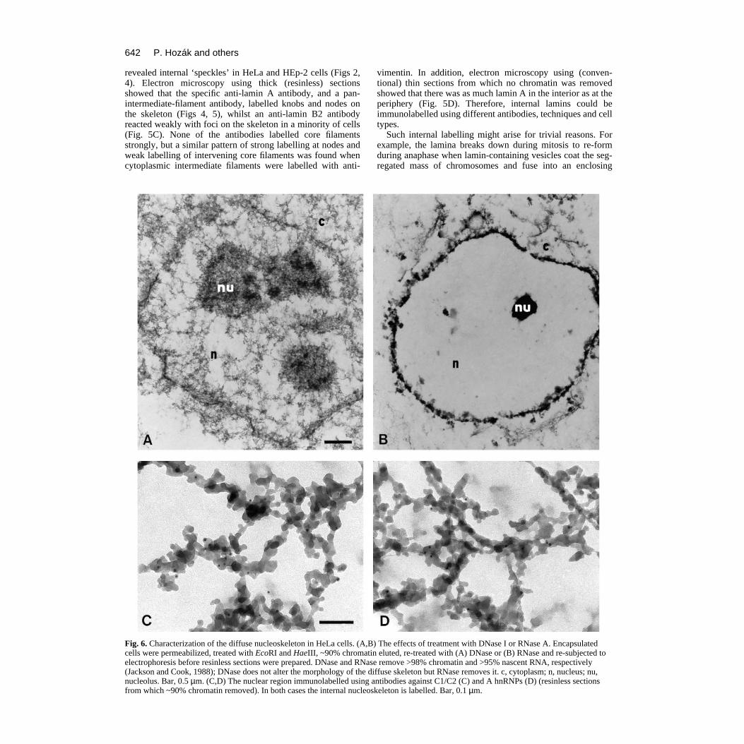
Characterization of the internal skeletonWe next investigated the nucleic acid content of the nucle-oskeleton. As ~10% chromatin remains associated with thediffuse skeleton after treatment with EcoRI + HaeIII andelution, it remained formally possible that the skeletonconsisted of residual chromatin fibres coated with lamins.Therefore, after removing ~90% chromatin as before, beadswere treated with DNase and re-eluted to remove most of theremainder (<2% DNA remained). Despite the harsh treatment,the basic internal skeletal structure remained (Fig. 6A).
There have been many suggestions that the skeleton mightbe associated with RNA (e.g. Long et al., 1979; Fey et al.,1986), so we also treated chromatin-depleted beads with suf-ficient RNase to remove ~95% of nascent RNA (Jackson andCook, 1988). This removed the diffuse skeleton and corefilaments (Fig. 6B), consistent with RNA being part of thestructure. However, nuclear morphology was so distorted thatwe are loath to draw any firm conclusions from such experi-ments; the high concentrations of charged oligonucleotidesreleased during digestion might be expected to induce the kindof precipitation of a fragile skeleton on to the stronger laminathat is seen. Nevertheless, we pursued the idea that the diffuseskeleton might contain RNA using antibodies directed againstproteins known to be associated with heterogeneous nuclearRNA (i.e. C1/C2 and A proteins; Choi and Dreyfuss, 1984;
Pinol-Roma et al., 1988). Immunogold labelling showed thatC1/2 were localized on the diffuse skeleton, sometimes con-centrated in foci (Fig. 6C). The distribution of A was similar,but the signal was weaker (Fig. 6D).
DISCUSSION
Lamins form part of the internal skeletonA lamin, or lamina, is defined in the Oxford English Dictio-nary as ‘a thin layer of bone, membrane, or other structure’.Although lamin proteins have been found internally withinnuclei (see Introduction), this is generally attributed to atransient, accidental, or pathological, location of proteinsdestined for a peripheral lamina. Our results show that laminA is also part of a diffuse internal skeleton that ramifiesthroughout the nucleus from the nucleolus to the periphery. Wehave previously shown that ‘core filaments’ in this complexstructure have the axial repeat typical of intermediate filaments(Jackson and Cook, 1988). We use the loose term ‘diffusenucleoskeleton’ here to include these core filaments plus allassociated knobs, nodes and non-chromatin material. Thisskeleton is also functionally complex; it contains sites involvedin transcription, RNA processing and, in S-phase cells, repli-cation (e.g. Hozák et al., 1993, 1994; Cook, 1994).
We removed most obscuring chromatin from nuclei to leavethis diffuse nucleoskeleton; then any lamins associated with itwere detected using various antibodies, including one thatspecifically recognizes an epitope in lamin A between aminoacids 598 and 611. Immunofluorescence using this specificantibody, as well as another directed against lamins A/C,

640 P. Hozák and others
Fig. 4. Visualization of an internal nucleoskeleton in HeLa cells from which ~90% chromatin had been removed. Encapsulated cells werepermeabilized, treated with nucleases, chromatin eluted and 500 nm resin-less sections prepared. In B-D, samples were immunolabelled with 5nm gold particles. a, agarose; c, cytoplasm; nu, nucleolus; ns, nucleoskeleton; n, nucleus; l, lamina; cf, core filaments. Bars: A and B-D, 1 and0.1 µm, respectively. (A) Low-power view. (B) Cytoplasmic region; pan-intermediate-filament antibody (i.e. TIB-131). (C) Nuclear region;pan-intermediate-filament antibody; 95% chromatin was removed in this preparation, so more core filaments are visible. (D) Nuclear andcytoplasmic regions; anti-vimentin antibody.

641An internal lamin nucleoskeleton
Fig. 5. Lamins detected in nuclear regions of HeLa cells by immunogold labelling in (A-C) resinless sections from which ~90% chromatin hasbeen removed or (D) a conventional thin section prepared without removing any chromatin from cells synchronized in G2 phase. nu, nucleolus; l,lamina; n, nucleus; c, cytoplasm. Bars: 0.1 µm. (A) Anti-lamin A (clone 133A2); both lamina and internal skeleton are labelled. (B) Anti-laminB2 (clone LN43); usually only the lamina is labelled. (C) Anti-lamin B2 (clone LN43); labelling of internal skeleton seen in 10% sections.(D) Anti-lamin A (clone 133A2); vimentin fibrils in the cytoplasm are unlabelled, but both lamina and internal nuclear regions are labelled.
642 P. Hozák and others
revealed internal ‘speckles’ in HeLa and HEp-2 cells (Figs 2,4). Electron microscopy using thick (resinless) sectionsshowed that the specific anti-lamin A antibody, and a pan-intermediate-filament antibody, labelled knobs and nodes onthe skeleton (Figs 4, 5), whilst an anti-lamin B2 antibodyreacted weakly with foci on the skeleton in a minority of cells(Fig. 5C). None of the antibodies labelled core filamentsstrongly, but a similar pattern of strong labelling at nodes andweak labelling of intervening core filaments was found whencytoplasmic intermediate filaments were labelled with anti-
Fig. 6. Characterization of the diffuse nucleoskeleton in HeLa cells. (A,Bcells were permeabilized, treated with EcoRI and HaeIII, ~90% chromatelectrophoresis before resinless sections were prepared. DNase and RNa(Jackson and Cook, 1988); DNase does not alter the morphology of the dnucleolus. Bar, 0.5 µm. (C,D) The nuclear region immunolabelled usingfrom which ~90% chromatin removed). In both cases the internal nucleo
vimentin. In addition, electron microscopy using (conven-tional) thin sections from which no chromatin was removedshowed that there was as much lamin A in the interior as at theperiphery (Fig. 5D). Therefore, internal lamins could beimmunolabelled using different antibodies, techniques and celltypes.
Such internal labelling might arise for trivial reasons. Forexample, the lamina breaks down during mitosis to re-formduring anaphase when lamin-containing vesicles coat the seg-regated mass of chromosomes and fuse into an enclosing
) The effects of treatment with DNase I or RNase A. Encapsulatedin eluted, re-treated with (A) DNase or (B) RNase and re-subjected tose remove >98% chromatin and >95% nascent RNA, respectivelyiffuse skeleton but RNase removes it. c, cytoplasm; n, nucleus; nu,
antibodies against C1/C2 (C) and A hnRNPs (D) (resinless sectionsskeleton is labelled. Bar, 0.1 µm.

643An internal lamin nucleoskeleton
membrane (Gerace and Burke, 1988; Dingwall and Laskey,1992). Vesicles inside the mass could fuse into an internal‘lamina’ that would then be no more than a result of theimperfect mechanism for generating an enclosing membrane.We would expect such an internal lamina to redistribute to theperiphery with time, but the density of the skeleton and theintensity of immunolabelling remain roughly constant through-out the cell cycle (not shown; Hozák et al., 1994, have studiedthe morphology of the skeleton throughout the cycle). Stereoelectron micrographs also provide no hint that the internalskeleton has been stripped from the periphery during samplepreparation (Hozák et al., 1993); moreover, and decisively,internal lamins can be detected in sections derived from G2cells that have neither been permeabilized nor depleted ofchromatin (Fig. 5D).
Relationship to other studiesOur results are consistent with observations of a distributiononly at the periphery, if the dense chromatin generally preventsantibody access to the interior. Then the examples of internallamins cited in the Introduction result not from an aberrantlocation but from an over-concentration at a normal site.
Lamins are frequently associated with an internal ‘matrix’(e.g. Capco et al., 1982; Staufenbiel and Deppert, 1982; Fey etal., 1984) which, like the diffuse nucleoskeleton described here(Fig. 6B-D), contains ribonucleoproteins and, perhaps, RNA(e.g. Long et al., 1979; Fey et al., 1986). It is usually assumedthat this matrix cannot contain any lamins, and this may be thereason that attempts to isolate it and determine its structurehave been so unsuccessful (reviewed by Jack and Eggert,1992); however, this failure is easily explicable if the assump-tion is incorrect. Intermediate filaments would then play a rolein integrating both cytoplasmic and nuclear space (Lazarides,1980). As lamins also bind DNA and/or chromatin (e.g.Shoemann and Traub, 1990; Burke, 1990; Glass and Gerace,1990; Höger et al., 1991; Yuan et al., 1991; Glass et al., 1993)and have been implicated in replication (e.g. Jenkins et al.,1993; Moir et al., 1994), our results provide a physical basisfor additional lamin functions within nuclei (e.g. Traub andShoeman, 1994).
Our results are consistent with the following model. Abranched network of ‘core’ intermediate filaments ramifiesthroughout the nucleus. Even in chromatin-depleted samples,much of this network remains covered with polymerases,nascent and maturing RNA, as well as RNPs; in S-phase cellsit is also associated with dense replication ‘factories’. Theseelements combine to form a diffuse nucleoskeleton that isprobably a ‘tensegrity’ structure like that popularized by Buck-minster Fuller (Ingber, 1993), in and on which nuclearfunctions occur; then, depolymerization of one element in thisstructure (e.g. RNA) would collapse the whole. The corefilaments of this diffuse nucleoskeleton, like their cytoplasmiccounterparts, react weakly with a pan-intermediate-filamentantibody and so might contain novel members of the interme-diate-filament family; however nodes, ‘knobs’ and clumps onthe nuclear network are clearly labelled by anti-lamin A and,to a lesser extent, by anti-lamin B and so probably markcomplex lamin-containing structures. Therefore we must nowinvestigate whether specific protein sequences or post-transla-tional modifications like prenylation (see Kitten and Nigg,1991; Lutz et al., 1992; Hennekes and Nigg, 1994) determine
if a lamin molecule is found at the nuclear periphery, a filamentor a node.
We thank Drs G. Blobel, M. Carmo-Fonseca, G. Dreyfuss, W.Earnshaw, C. Hutchison, B. Lane, U.T. Meier and R. Stick for kindlysupplying antibodies, and Dr. F. McKeon for human lamin cDNA.This work was supported by the Wellcome Trust, the Cancer ResearchCampaign, the Grant Agency of the Czech Republic (grant no.304/94/0148), the Academy of Sciences of the Czech Republic (grantno. 539402), and the Medical Research Council of Canada with a stu-dentship (A.M.-J.S.) and an operating grant.
REFERENCES
Bader, B. L., Magin, T. M., Freudenmann, M., Stumpp, S. and Franke, W.W. (1991). Intermediate filaments formed de novo from tail-less cytokeratinsin the cytoplasm and in the nucleus. J. Cell Biol. 115, 1293-1307.
Belgrader, P., Siegel, A. J. and Berezney, R. (1991). A comprehensive studyon the isolation and characterization of the HeLa S3 nuclear matrix. J. CellSci. 98, 281-291.
Belmont, A. S., Zhai, Y. and Thilenius, A. (1993). Lamin B distribution andassociation with peripheral chromatin revealed by optical sectioning andelectron microscopy tomography. J. Cell Biol. 123, 1671-1685.
Beven, A., Guan, Y., Peart, J., Cooper, C. and Shaw, P. (1991). Monoclonalantibodies to plant nuclear matrix reveal intermediate filament-relatedcomponents within the nucleus. J. Cell Sci. 98, 293-302.
Bridger, J. M., Kill, I. R., O’Farrell, M. and Hutchison, C. J. (1993).Internal lamin structures within G1 nuclei of human dermal fibroblasts. J.Cell Sci. 104, 297-306.
Burke, B. (1990). On the cell-free association of lamins A and C withmetaphase chromosomes. Exp. Cell Res. 186, 169-176.
Capco, D. G., Wan, K. M. and Penman, S. (1982). The nuclear matrix: threedimensional architecture and protein composition. Cell 29, 847-858.
Cardenas, M. E., Laroche, T. and Gasser, S. M. (1990). The composition andmorphology of yeast nuclear scaffolds. J. Cell Sci. 96, 439-450.
Choi, Y. D. and Dreyfuss, G. (1984). Monoclonal antibody characterization ofthe C proteins of heterogeneous nuclear ribonucleoprotein complexes invertebrate cells. J. Cell Biol. 99, 1997-2004.
Collard, J. F., Senécal, J. L. and Raymond, Y. (1992). Redistribution ofnuclear lamin A is an early event associated with differentiation of humanpromyelocytic leukemia HL-60 cells. J. Cell Sci. 101, 657-670.
Cook, P. R. (1988). The nucleoskeleton: artefact, passive framework or activesite? J. Cell Sci. 90, 1-6.
Cook, P. R. (1994). RNA polymerase: structural determinant of the chromatinloop and the chromosome. BioEssays 16, 425-430.
Dingwall, C. and Laskey, R. (1992). The nuclear membrane. Science 258,942-947.
Eckelt, A., Hermann, H. and Franke, W. W. (1992). Assembly of a tail-lessmutant of the intermediate filament protein, vimentin, in vitro and in vivo.Eur. J. Cell Biol. 58, 319-330.
Fey, E. G., Wan, K. M. and Penman, S. (1984). Epithelial cytoskeletalframework and nuclear matrix-intermediate filament scaffold: threedimensional organisation and protein composition. J. Cell Biol. 98, 1973-1984.
Fey, E. G., Krochmalnic, G. and Penman, S. (1986). The nonchromatinsubstructures of the nucleus: the ribonucleoprotein (RNP)-containing andRNP-depleted matrices analyzed by sequential fractionation and resinlesssection microscopy. J. Cell Biol. 102, 1654-1665.
Fisher, D. Z., Chaudhary, N. and Blobel, G. (1986). cDNA sequencing oflamins A and C reveals primary and secondary structural homology tointermediate filament proteins. Proc. Nat. Acad. Sci. USA 83, 6450-6454.
Gagnon, G., Sasseville, A. M.-J. and Raymond, Y. (1992). Preparative 2-Delectrophoresis system purifies recombinant nuclear protein from wholebacterial lysates. Bio-Rad US/EG Bulletin 1773.
Gerace, L., Blum, A. and Blobel, G. (1987). Immunocytochemicallocalization of the major polypeptides of the nuclear pore complex-laminafraction: interphase and mitotic distribution. J. Cell Biol. 79, 546-566.
Gerace, L. and Burke, B. (1988). Functional organization of the nuclearenvelope. Annu. Rev. Cell Biol. 4, 335-374.
Gill, S. R., Wong, P. C., Monteiro, M. J. and Cleveland, D. W. (1990).

644 P. Hozák and others
Assembly properties of dominant and recessive mutations in the small mouseneurofilament (NF-L) subunit. J. Cell Biol. 111, 2005-2019.
Glass, C. A., Glass, J. R., Taniura, H., Hasel, K. W., Blevitt, J. M. andGerace, L. (1993). The α-helical rod domain of lamins A and C contains achromatin binding site. EMBO J. 12, 4413-4424.
Glass, J. R. and Gerace, L. (1990). Lamins A and C bind and assemble at thesurface of mitotic chromosomes. J. Cell Biol. 111, 1047-1057.
Goldman, A. E., Moir, R. D., Montag-Lowy, M., Stewart, M. andGoldman, R. D. (1992). Pathway of incorporation of microinjected lamin Ainto the nuclear envelope. J. Cell Biol. 119, 725-735.
He, D., Nickerson, J. A. and Penman, S. (1990). Core filaments of the nuclearmatrix. J. Cell Biol. 110, 569-580.
Hennekes, H. and Nigg, E. A. (1994). The role of isoprenylation in membraneattachment of nuclear lamins; a single point mutation prevents proteolyticcleavage of the lamin A precursor and confers membrane binding properties.J. Cell Sci. 107, 1019-1029.
Höger, T. H., Krohne, G. and Kleinschmidt, J. A. (1991). Interaction ofXenopus lamins A and LII with chromatin in vitro mediated by a sequenceelement in the carboxyterminal domain. Exp. Cell Res. 197, 280-289.
Hozák, P., Hassan, A. B., Jackson, D. A. and Cook, P. R. (1993).Visualization of replication factories attached to a nucleoskeleton. Cell 73,361-373.
Hozák, P., Jackson, D. A. and Cook, P. R. (1994). Replication factories andnuclear bodies: the ultrastructural characterization of replication sites duringthe cell cycle. J. Cell Sci. 107, 2191-2202.
Ingber, D. E. (1993). Cellular tensegrity: defining new rules of biologicaldesign that govern the cytoskeleton. J. Cell Sci. 104, 613-627.
Jack, R. S. and Eggert, H. (1992). The elusive nuclear matrix. Eur. J.Biochem. 209, 503-509.
Jackson, D. A. and Cook, P. R. (1988). Visualization of a filamentousnucleoskeleton with a 23 nm axial repeat. EMBO J. 7, 3667-3677.
Jackson, D. A., Yuan, J. and Cook, P. R. (1988). A gentle method forpreparing cyto- and nucleo-skeletons and associated chromatin. J. Cell Sci.90, 365-378.
Jenkins, H., Holman, T., Lyon, C., Lane, B., Stick, R. and Hutchison, C.(1993). Nuclei that lack a lamina accumulate karyophilic proteins andassemble a nuclear matrix. J. Cell Sci. 106, 275-285.
Kitten, G. T. and Nigg, E. A. (1991). The CaaX motif is required forisoprenylation, carboxyl methylation, and nuclear membrane association oflamin B2. J. Cell Biol. 113, 13-23.
Laemmli, U. K. (1970). Cleavage of structural proteins during the assembly ofthe head of bacteriophage T4. Nature 227, 680-685.
Lazarides, E. (1980). Intermediate filaments as mechanical integrators ofcellular space. Nature 283, 249-246.
Lebel, S. and Raymond, Y. (1987). Lamins A, B and C share an epitope withthe common domain of intermediate filament proteins. Exp. Cell Res. 169,560-565.
Loewinger, L. and McKeon, F. (1988). Mutations in the nuclear laminproteins resulting in their aberrant assembly in the cytoplasm. EMBO J. 7,2301-2309.
Long, B. H., Huang, C.-Y. and Pogo, A. O. (1979). Isolation andcharacterization of the nuclear matrix in Friend erythroleukaemia cells:chromatin and hnRNA interactions with the nuclear matrix. Cell 18, 1079-1090.
Luderus, M. E. E., de Graaf, A., Mattia, E., den Blaauwen, J. L., Grande,M. A., de Jong, L. and van Driel, R. (1992). Binding of matrix attachmentregions to lamin B1. Cell 70, 949-959.
Lutz, R. J., Trujillo, M. A., Denham, K. S., Wenger, L. and Sinensky, M.(1992). Nucleoplasmic localization of prelamin A: implications forprenylation-dependent lamin A assembly into the nuclear lamina. Proc. Nat.Acad. Sci. USA 89, 3000-3004.
Mancini, M. A., Shan, B., Nickerson, J. A., Penman, S. and Lee, W.-H.(1994). The retinoblastoma gene product is a cell cycle-dependent, nuclearmatrix-associated protein. Proc. Nat. Acad. Sci. USA 91, 418-422.
McKeon, F. D., Kirshner, M. W. and Caput, D. (1986). Homologies in bothprimary and secondary structure between nuclear envelope and intermediatefilament proteins. Nature 319, 463-468.
Meier, U. T. and Blobel, G. (1992). Nopp140 shuttles on tracks betweennucleolus and cytoplasm. Cell 70, 127-138.
Minguez, A. and Moreno Diaz de la Espina, S. (1993). Immunologicalcharacterization of lamins in the nuclear matrix of onion cells. J. Cell Sci.106, 431-439.
Mirzayan, C., Copeland, C. S. and Snyder, M. (1992). The NUF1 geneencodes an essential coiled-coil related protein that is a potential componentof the yeast nucleoskeleton. J. Cell Biol. 116, 1319-1332.
Moir, R. D., Montag-Lowy, M. and Goldman, R. D. (1994). Dynamicproperties of nuclear lamins: lamin B is associated with sites of DNAreplication. J. Cell Biol. 125, 1201-1212.
Newport, J. W. and Forbes, D. J. (1987). The nucleus: structure, function anddynamics. Annu. Rev. Biochem. 56, 535-565.
Paddy, M. R., Belmont, A. S., Saumweber, H., Agard, D. A. and Sedat, J.W. (1990). Interphase nuclear envelope lamins form a discontinuousnetwork that interacts with only a fraction of the chromatin in the nuclearperiphery. Cell 62, 89-106.
Pinol-Roma, S., Choi, Y. D., Matunis, M. J. and Dreyfuss, G. (1988).Immunopurification of heterogeneous nuclear ribonucleoprotein particlesreveals an assortment of RNA-binding proteins. Genes Dev. 2, 215-227.
Pruss, R. M., Mirsky, R., Raff, M. C., Thorpe, R., Dowding, A. J. andAnderton, B. H. (1981). All classes of intermediate filaments share acommon antigenic determinant defined by a monoclonal antibody. Cell 27,419-428.
Raymond, Y. and Gagnon, G. (1988). Lamin B shares a number of distinctepitopes with lamins A and C and with intermediate filament proteins.Biochemistry 27, 2590-2597.
Reimer, D., Dodemont, H. and Weber, K. (1991). Cloning of the non-neuronal intermediate filament protein of the gastropod Aplysia californica.Eur. J. Cell Biol. 56, 351-357.
Schägger, H. and von Jagow, G. (1987). Tricine-sodium dodecyl sulfate-polyacrylamide gel electrophoresis for the separation of proteins in the rangefrom 1 to 100 kDa. Anal. Biochem. 166, 368-379.
Shoemann, R. L. and Traub, P. (1990). The in vitro DNA-binding propertiesof purified nuclear lamin proteins and vimentin. J. Biol. Chem. 265, 9055-9061.
Staufenbiel, M. and Deppert, W. (1982). Intermediate filament systems arecollapsed onto the nuclear surface after isolation of nuclei from tissue culturecells. Exp. Cell Res. 138, 207-124.
Steinert, P. M. and Roop, D. R. (1988). Molecular and cellular biology ofintermediate filaments. Annu. Rev. Biochem. 57, 593-626.
Stick, R. and Hausen, P. (1980). Immunological analysis of nuclear laminaproteins. Chromosoma 80, 219-236.
Stick, R. and Hausen, P. (1985). Changes in the nuclear lamina compositionduring early development of Xenopus laevis. Cell 41, 191-200.
Traub, P. and Shoeman, R. L. (1994). Intermediate filament and relatedproteins: potential activators of nucleosomes during transcription initiationand elongation? BioEssays 16, 349-355.
Yuan, J., Simos, G., Blobel, G. and Georgatos, S. D. (1991). Binding of laminA to polynucleosomes. J. Biol. Chem. 266, 9211-9215.
(Received 20 July 1994 - Accepted 3 October 1994)