lamin a/c and emerin are critical for skeletal muscle...
TRANSCRIPT

Lamin A/C and emerin are criticalfor skeletal muscle satellite celldifferentiationRichard L. Frock,1 Brian A. Kudlow,2 Angela M. Evans,1 Samantha A. Jameson,1
Stephen D. Hauschka,1,3,4 and Brian K. Kennedy1,3,5
1Department of Biochemistry, 2Molecular and Cellular Biology Program, University of Washington,Seattle, Washington 98195, USA
Mutations within LMNA, encoding A-type nuclear lamins, are associated with multiple tissue-specificdiseases, including Emery-Dreifuss (EDMD2/3) and Limb-Girdle muscular dystrophy (LGMD1B). X-linkedEDMD results from mutations in emerin, a lamin A-associated protein. The mechanisms through which thesemutations cause muscular dystrophy are not understood. Here we show that most, but not all, culturedmuscle cells from lamin A/C knockout mice exhibit impaired differentiation kinetics and reduceddifferentiation potential. Similarly, normal muscle cells that have been RNA interference (RNAi)down-regulated for either A-type lamins or emerin have impaired differentiation potentials. Replicativemyoblasts lacking A-type lamins or emerin also have decreased levels of proteins important for muscledifferentiation including pRB, MyoD, desmin, and M-cadherin; up-regulated Myf5; but no changes in Pax3,Pax7, MEF2C, MEF2D, c-met, and �-catenin. To determine whether impaired myogenesis is linked to reducedMyoD or desmin levels, these proteins were individually expressed in Lmna−/− myoblasts that were theninduced to undergo myogenesis. Expression of either MyoD or, more surprisingly, desmin in Lmna−/−
myoblasts resulted in increased differentiation potential. These studies indicate roles for A-type lamins andemerin in myogenic differentiation and also suggest that these effects are at least in part due to decreasedendogenous levels of other critical myoblast proteins. The delayed differentiation kinetics and decreaseddifferentiation potential of lamin A/C-deficient and emerin-deficient myoblasts may in part underlie thedystrophic phenotypes observed in patients with EDMD.
[Keywords: MyoD; desmin; pRB; myoblast; myogenesis; Emery-Dreifuss muscular dystrophy]
Received August 15, 2005; revised version accepted December 27, 2005.
Nuclear lamins are the major constituents of the nuclearlamina, a proteinaceous network underlying the innernuclear membrane (Hutchison and Worman 2004;Mounkes and Stewart 2004; Zastrow et al. 2004; Smithet al. 2005). Expression of A-type lamins, in contrast tothe homologous B-type lamins, is largely restricted todifferentiated tissues with high expression in skeletalmuscle (Rober et al. 1989). Lamins A and C (henceforthreferred to as lamin A/C) are the major products of theLMNA gene.
Greater than 100 mutations in LMNA have beenlinked to 10 human genetic disorders, broadly termedlaminopathies (for review, see Mounkes et al. 2003). Themajority of LMNA mutations identified thus far are
linked to Emery-Dreifuss muscular dystrophy (EDMD2/3) (Bonne et al. 1999). Mice lacking lamin A/C develop aform of muscular dystrophy closely resembling EDMDand die by 8 wk from cardiac and skeletal myopathies(Sullivan et al. 1999). Mutations within the gene encod-ing the lamin A/C-binding protein, emerin (EMD), causean X-linked form of Emery-Dreifuss muscular dystrophy(EDMD1) (Bione et al. 1994; Gruenbaum et al. 2005). It isnot known what role A-type lamins play in skeletalmuscle development, let alone how mutations withinLMNA cause EDMD. One possibility is that mutationsthat alter lamin A/C’s nuclear integrity or impair itsfunction(s) would then result in degeneration of cells un-der high mechanical stress, such as skeletal muscle(Broers et al. 2004; Lammerding et al. 2004). A second,nonexclusive possibility is that lamin A/C plays a role inmuscle differentiation, and alterations in its activity re-sult in delayed or decreased expression of genes impor-tant for differentiation and/or the stable maintenance ofthe differentiated state. Inefficient muscle differentia-tion may ultimately result in a dystrophic syndrome,
3Members of the Hauschka and Kennedy groups contributed equally tothese studies.Corresponding authors.4E-MAIL [email protected]; FAX (206) 685-1792.5E-MAIL [email protected]; FAX (206) 685-1792.Article and publication are at http://www.genesdev.org/cgi/doi/10.1101/gad.1364906.
486 GENES & DEVELOPMENT 20:486–500 © 2006 by Cold Spring Harbor Laboratory Press ISSN 0890-9369/06; www.genesdev.org
Cold Spring Harbor Laboratory Press on August 20, 2018 - Published by genesdev.cshlp.orgDownloaded from

when the balance between muscle degeneration and re-newal becomes skewed.
In adults, the vast majority of new skeletal musclecomes from myogenic precursor cells called satellitecells that require Pax3/Pax7 for their specification andself-renewal (Oustanina et al. 2004; Relaix et al. 2005).These adult stem cells are able to proliferate and producemyoblasts, which in turn are capable of withdrawingfrom the cell cycle and terminally differentiating intoskeletal muscle (for review, see Charge and Rudnicki2004). A number of transcription factors and structuralproteins have been implicated in this transition (Parkeret al. 2003; Paulin and Li 2004); for example, the myo-genic regulatory factor MyoD is expressed prior to myo-cyte differentiation (Buskin and Hauschka 1989; Lassaret al. 1989) and, together with MEF2 transcription fac-tors, is crucial for satellite cell terminal differentiation(Sabourin et al. 1999; Yablonka-Reuveni et al. 1999;McKinsey et al. 2002). The retinoblastoma protein (pRB),is likewise important for the proliferation to differentia-tion transition during myogenesis (Maione et al. 1994;Zacksenhaus et al. 1996; Huh et al. 2004). Although themechanism is not fully understood, pRB is thought topotentiate MyoD activity during muscle differentiation(Novitch et al. 1996, 1999; Puri et al. 2001; Guo et al.2003).
Several structural and cell surface proteins also playimportant roles in terminal differentiation. Desmin, amuscle-specific intermediate filament protein, is one ofthe first proteins expressed upon satellite cell activation(Lazarides and Hubbard 1976; Kaufman et al. 1991). Itsexact role in myogenesis remains unclear; but differen-tiation is slightly delayed during regeneration in desminknockout mice (Li et al. 1994; Weitzer et al. 1995;Smythe et al. 2001). M-cadherin, a cell surface adhesionprotein, is a marker for satellite cells in vivo, and itsexperimental perturbation also delays the onset of differ-entiation (Zeschnigk et al. 1995; Kaufmann et al. 1999).
Do A-type lamins play roles in muscle differentiation?A recent study reported that overexpression of a lamin AEDMD mutation, R453W, inhibits the in vitro differen-tiation of C2C12 myoblasts (Favreau et al. 2004). Subse-quent studies showed that overexpression of a differentEDMD mutation, W520S, also inhibited C2C12 myo-blast differentiation, and provided evidence that nucleo-skeleton remodeling is necessary for skeletal muscle dif-ferentiation (Markiewicz et al. 2005). Finally, Arimuraet al. (2005) constructed an EDMD mouse carryingH222P mutations in both lamin A alleles and found thatadult mice developed muscular dystrophy and exhibitedelevated levels of Smads 2 and 4 in cardiac and skeletalmuscle nuclei. Here, we focus on an EDMD mousemodel in which the lamin A gene has been knocked out(Sullivan et al. 1999), and report that Lmna−/− and evenLmna+/− myoblasts display a dramatically compromiseddifferentiation potential. Lmna+/+ cells stably expressinga small interfering RNA (siRNA) targeted to Lmna aresimilarly compromised. Interestingly, myoblasts withsiRNA-reduced emerin display a similar differentiationphenotype. Furthermore, myoblasts with reduced lamin
A/C or emerin also contain reduced levels of at least fourproteins important for differentiation and/or the main-tenance of the differentiated state: MyoD, desmin, pRB,and M-cadherin. Exogenous expression of MyoD inLmna−/− myoblasts restores their differentiation kinet-ics, but more surprisingly, forced expression of desmin issufficient to restore much of their differentiation poten-tial, suggesting that reduced MyoD and desmin tran-scription may be primary factors underlying the poor dif-ferentiation capacity of cells deficient in A-type laminfunction.
Results
Generation of Lmna−/− myoblasts
To determine the myogenic potential of cells with re-duced lamin A/C function, we generated permanentmyoblast cell lines from Lmna+/+, Lmna+/−, and Lmna−/−
mouse hind leg muscles (see Materials and Methods). Toavoid accidental selection of cells with unique attributesduring the generation of permanent myoblast lines, pri-mary cultures were grown at densities that permittedthousands of individual muscle colony-forming cells toproliferate into colonies, while most fibroblasts grewpoorly. Previous studies indicate that when most mouseprimary satellite-cell-derived muscle colonies are sub-cloned and individually expanded, their descendents un-dergo proliferative senescence after 20–40 cell cycles andsmall numbers of spontaneously transformed myoblastsemerge (Hauschka et al. 1979; Neville et al. 1997). Evenafter decades of expansion, the descendents exhibit bio-chemical characteristics that are very close to those ofprimary satellite cells (Cornelison 1998). Importantly,the cells in each of the permanent cell lines generated arePax3- and Pax7-positive (see below); thus potentially im-mortalized fibroblasts did not contribute to the myo-genic Lmna+/+, Lmna+/−, and Lmna−/− cell lines. Westernanalysis confirmed that lamin A and C bands were ab-sent in Lmna−/− muscle cells and were reduced ∼50% inLmna+/− cells (Fig. 1A).
Lmna−/− and Lmna+/− muscle cells have delayeddifferentiation kinetics
The onset of terminal differentiation in satellite cell-derived skeletal muscle cultures is regulated by growthfactors. In the presence of bFGF and horse serum, mousemyoblasts grow exponentially. Upon bFGF removal,myoblasts continue cycling for ∼6 h as residual mito-genic signals attenuate; cells then stop entering S phaseand undergo an irreversible commitment to terminal dif-ferentiation. Typically, >90% of the population commitsto terminal differentiation as mitogen-deprived cellscomplete mitosis and enter the G1/G0 cell cycle com-partment. Committed cells activate expression ofmuscle-specific genes such as myosin heavy chain(MyHC), repress growth factor receptor expression, andare refractory to subsequent mitogenic stimulation(Clegg et al. 1987; Olwin and Hauschka 1988; Templetonand Hauschka 1992; Angello and Hauschka 1996). The
Lamin-dependent effect on in vitro myogenesis
GENES & DEVELOPMENT 487
Cold Spring Harbor Laboratory Press on August 20, 2018 - Published by genesdev.cshlp.orgDownloaded from

kinetics of skeletal muscle differentiation after bFGF re-moval can thus be approximated by determining the per-centage of cells in S phase by BrdU incorporation and thepercentage of cells that exhibit immuno-positive stain-ing for MyHC.
The timing of myogenic terminal differentiation wascompared among Lmna+/+, Lmna+/−, and Lmna−/−
muscle cells. Initially, 40%–50% of the myoblasts ofeach genotype are in S phase; this is consistent with thepredicted 42% S-phase cells in cultures with a 19-h cellcycle and a typical 8-h S phase (Clegg et al. 1987). After12 h >40% of Lmna−/− myoblasts remain in S phase com-pared with ∼25% of Lmna+/+ cells (Fig. 1B,D,E); and dif-ferences in proliferation still remain after 24 h. Thesefindings indicate that myoblasts with reduced laminA/C are delayed in their ability to exit the cell cycle inresponse to mitogen deprivation.
Consistent with the delay in cell cycle withdrawal,the appearance of MyHC-positive cells is delayed ∼24 hin Lmna−/− compared with Lmna+/+ cells (Fig. 1C,F,G).Forty-eight hours after bFGF removal, only ∼30% of
Lmna−/− cells were MyHC-positive compared with∼85% of Lmna+/+ cells (Fig. 1C,H,I). These data indicatethat upon growth factor withdrawal, Lmna−/− musclecells are also slower to exhibit the gene expression prop-erties of differentiated muscle. Lmna+/− muscle cells arelikewise compromised, exhibiting an intermediate dif-ferentiation phenotype between that of Lmna+/+ andLmna−/− cells.
The slower differentiation kinetics of Lmna−/− musclecells could be explained by a lag in responsiveness toFGF removal after which, given sufficient time, a major-ity of cells become myocytes; alternatively, the Lmna−/−
population may contain some cells capable of normaldifferentiation and other cells that are unable to termi-nally differentiate. These possibilities were analyzed byclonal assays in which colonies were grown in bFGF-containing media for 5 d then switched to bFGF-free me-dia and the relative differentiation capacity of individualclones from each cell line was assessed after increasingperiods of mitogen deprivation. The extent of differen-tiation was assessed by determining the percentage of
Figure 1. Lmna−/− muscle cells have de-layed differentiation kinetics. (A) Westernanalysis indicating the lamin A/C pro-teins present in the permanent Lmna+/+,Lmna+/−, and Lmna−/− myoblast cell lines.(B) Fraction of S-phase cells followingbFGF removal as measured by cells stain-ing positively for BrdU incorporation inLmna+/+, Lmna+/−, and Lmna−/− cultures.After 12 h of bFGF deprivation, 40% ofLmna−/− muscle cells remain in S phasecompared with ∼20% of Lmna+/+ cells.(C) The appearance of terminally differen-tiated muscle protein as measured by cellsstaining positively for MyHC in identicalbFGF-deprived cultures. By 48 h, only∼30% of Lmna−/− cells were MyHC-posi-tive compared with ∼90% in Lmna+/+ cells.Lmna+/− muscle cells display an interme-diate differentiation phenotype relative toLmna+/+ and Lmna−/− muscle cells. Cellcounts are based on three experimentswith ∼350 cells counted per time point. (D–I) Immunostained Lmna+/+ and Lmna−/−
muscle cells at 12 h (D,E), 24 h (F,G), and48 h (H,I) after mitogen withdrawal. BrdUis in green with arrowheads indicatingBrdU-positive cells, MyHC is in red, andDapi is in blue.
Frock et al.
488 GENES & DEVELOPMENT
Cold Spring Harbor Laboratory Press on August 20, 2018 - Published by genesdev.cshlp.orgDownloaded from

nuclei in MyHC-positive cells. Individual colonies werethen subdivided into four arbitrary categories: 0%MyHC+, <30% MyHC+, 30%–60% MyHC+, and 60%–95% MyHC+, and the data were plotted for clones fixed4–10 d following mitogen removal (Fig. 2A). In Lmna+/+
cultures, >95% of the total colonies contained MyHC+
cells by day 4, while Lmna−/− and Lmna+/− cultures con-tained 10%–20% fewer MyHC+ colonies (Fig. 2B); andwhereas ∼80% of the Lmna+/+ colonies were highly dif-ferentiated after 4 d, no Lmna−/− clones and only ∼25% ofthe Lmna+/− clones were highly differentiated. Signifi-cantly, as the post-mitogen withdrawal period increased,<3% of the total Lmna−/− colonies ever became highlydifferentiated, only ∼20% ever acquired an intermediate(30%–60% MyHC+) level of differentiation, and the frac-tion of colonies exhibiting low differentiation levels(<30% MyHC+ cells) increased only slightly. Lmna+/−
clones, while more differentiated than Lmna−/− clones,also exhibited only marginal increases in differentiationat longer periods of mitogen withdrawal. These resultsare consistent with the hypothesis that most Lmna−/−
myoblasts are compromised with respect to their differ-entiation potential. (Evidence that the impaired differen-tiation phenotype of most Lmna−/− and some Lmna+/−
clones is not due to the common “differentiation-defec-tive” phenotype observed in many permanent mouseand rat myogenic cell lines [Lim and Hauschka 1984] isprovided below.)
Lmna−/− myoblasts exhibit both altered and normallevels of transcription factors and other keymyogenic components
Since differences between Lmna+/+ and Lmna−/− musclecell differentiation are apparent as early as 12 h afterbFGF removal, the compromised Lmna−/− differentiationphenotype could be due to altered levels or activity ofcritical regulatory factors in myoblasts prior to the ini-tiation of terminal differentiation. For example, myo-blasts from MyoD−/− mice also exhibit delayed cell cyclewithdrawal kinetics when deprived of growth factors (Sa-bourin et al. 1999). This hypothesis was examined viaWestern blot analysis of proliferating Lmna−/− andLmna+/+ myoblasts (Fig. 3A). This disclosed that inLmna−/− myoblasts MyoD protein levels were reducedby >60% while the levels of Myf-5, a related basic helix–loop–helix (bHLH) transcription factor whose expressionis often up-regulated in response to decreased MyoD lev-els (Rudnicki et al. 1992), were highly increased. Severalstudies have demonstrated that pRB acts synergisti-cally with MyoD to activate transcription of muscle spe-cific factors (Puri et al. 2001; Guo et al. 2003), and thatpRB is also required for cell cycle exit accompanyingmuscle differentiation (Novitch et al. 1996, 1999; Huhet al. 2004). pRB Western blot analysis indicated thatsteady-state levels of both hyper- and hypo-phosphory-lated pRB were dramatically reduced in Lmna−/− myo-
Figure 2. Lmna−/− muscle cells havereduced differentiation potential. (A)Method for the clonal analysis of differen-tiation: Cells are plated at low cell densi-ties and given 5 d to form colonies, thenswitched to FGF-free, low-serum media toinduce differentiation. The relative differ-entiation capacity of individual clonesfrom each cell line was measured at suc-cessive days of mitogen deprivation. Fixedcultures were immunostained for MyHC(brown) and counterstained for total cellswith 1% methylene blue (light blue).Clones were then subdivided into four ar-bitrary categories based on percentage ofnuclei in MyHC+ cells: 0%, <30%, 30%–60%, and 60%–95%. (B) Clonal analysis ofdifferentiation for Lmna+/+, Lmna+/−, andLmna−/− muscle cells from 4 to 10 d ofdifferentiation. Approximately 80% ofclones in Lmna+/+ cultures were highlydifferentiated at day 4, whereas none ofthe Lmna−/− colonies were similarly differ-entiated. As the post-mitogen withdrawalperiod increased, <3% of the Lmna−/− colo-nies ever became highly differentiated.Consistent with mass culture differentia-tion (see Fig. 1C), Lmna+/− muscle cellsdisplay an intermediate differentiationphenotype relative to Lmna+/+ andLmna−/− muscle cells. Approximately 100clones were scored per time point.
Lamin-dependent effect on in vitro myogenesis
GENES & DEVELOPMENT 489
Cold Spring Harbor Laboratory Press on August 20, 2018 - Published by genesdev.cshlp.orgDownloaded from

blasts compared with Lmna+/+ controls (Fig. 3A). In con-trast to the MyoD, Myf-5, and pRB changes, the proteinlevels of several other key myogenic transcription fac-tors: MEF2C, MEF2D, Pax3, and Pax7 were unperturbedin proliferating Lmna−/− myoblasts.
It also seemed informative to determine whetherLmna−/− myoblasts exhibited altered levels of importantmyogenic structural and signaling proteins. The muscle-specific cytoplasmic intermediate filament protein des-min was analyzed in Lmna−/− myoblasts for several rea-sons. First, desmin is required for normal differentiationin vitro (Li et al. 1994; Weitzer et al. 1995), and desminknockout myoblasts also have prolonged cell cycle with-drawal kinetics and delayed fusion in vivo (Smythe et al.2001). Second, MyoD−/− primary mouse myoblasts donot express desmin (Sabourin et al. 1999). Third, EMstudies have found abnormal desmin localization inLmna−/− cardiomyocytes (Nikolova et al. 2004). Interest-ingly, desmin protein levels were significantly reduced
in proliferating Lmna−/− myoblasts (Fig. 3A). Because re-duced desmin levels might have resulted in compensa-tion by other cytoplasmic intermediate filament pro-teins such as vimentin and nestin, the levels of theseproteins were also examined in Lmna−/− myoblasts.However, vimentin and nestin occur at normal levels inmyoblasts lacking lamin A/C (data not shown).
The Ca+-dependent cell adhesion protein M-cadherinwas analyzed because its gene is activated by MyoD (Sa-bourin et al. 1999; Cornelison et al. 2000) and becauseM-cadherin plays roles in cell fusion and other aspects ofmyogenesis (Zeschnigk et al. 1995; Kang et al. 2003).M-cadherin protein levels were also significantly reducedin Lmna−/− myoblasts relative to Lmna+/+ myoblasts(Fig. 3A). The tyrosine kinase receptor c-met was ana-lyzed because its overexpression is known to inhibit myo-genic differentiation (Anastasi et al. 1997), and �-cateninwas analyzed because it is known to play multiple rolesduring myogenesis (Petropoulos and Skerjanc 2002), butneither protein exhibited altered levels in Lmna−/− myo-blasts (Fig. 3A). Taken together, these results indicatethat myoblasts lacking lamin A/C exhibit several majordifferences in regulatory, structural, and signaling com-ponents, one or more of which could potentially accountfor the compromised differentiation of Lmna−/− myo-blasts in response to growth factor deprivation.
MyoD and desmin, but not Rb, transcripts,are reduced in Lmna−/− myoblasts
Why are the levels of MyoD, desmin, and pRB proteinsreduced in proliferating Lmna−/− myoblasts? Quantita-tive PCR (QPCR) was used to determine whether re-duced levels of the three proteins correlated withchanges in transcript levels. MyoD and Des transcriptlevels were reduced by more than threefold and 4.5-fold,respectively, in the Lmna−/− myoblasts (Fig. 3B). In con-trast, Rb transcript levels were unaffected in Lmna−/−
myoblasts relative to Lmna+/+ myoblasts (Fig. 3B). Weposit that pRB protein stability is reduced in these cells,perhaps through enhanced proteasome-dependent degra-dation, as has been observed in Lmna−/− fibroblasts(Johnson et al. 2004).
Individual Lmna−/− myoblasts express variable levelsof MyoD and desmin, but normal levels of Pax7
Clonal analysis of Lmna−/− myoblasts indicates thatfewer cells in most colonies retain the capacity to differ-entiate rapidly upon bFGF removal, and most cells fail todifferentiate even after long time periods. We consideredseveral explanations for these differences with respect toMyoD, desmin, and Pax7 levels. First, there may be sev-eral subpopulations of Lmna−/− cells: one with near-nor-mal levels of proteins required for the myogenic pro-gram, which retain a full differentiation potential, andothers with variably decreased expression of MyoD and/or desmin that have lower probabilities of differentia-tion. Alternatively, one or more of these proteins may bereduced in all cells, again leading to lower probabilitiesof differentiation. To distinguish between these possi-
Figure 3. MyoD, pRB, desmin, and M-cadherin proteins arereduced in proliferating Lmna−/− myoblasts. (A) Western analy-sis comparing relative protein levels of myogenic factors inLmna+/+ and Lmna−/− myoblasts. MyoD, pRB, desmin, and M-cadherin proteins are reduced, whereas Myf-5 protein levels areincreased in Lmna−/− myoblasts. In addition, Pax7, Pax3, MEF2C,MEF2D, c-met, and �-catenin proteins are unaffected inLmna−/− myoblasts. (B) QPCR analysis comparing the relativemRNA’s of desmin, MyoD, and pRB in Lmna+/+ and Lmna−/−
myoblasts. Desmin and MyoD mRNA’s are 4.5-fold and three-fold reduced in Lmna−/− myoblasts, whereas Rb mRNA is notaffected. Data represent averages of triplicate experiments per-formed at three different dilutions of cDNA. Fold changes weremeasured by comparing Ct values from Lmna+/+ and Lmna−/−
myoblast mRNAs that were normalized against Hprt.
Frock et al.
490 GENES & DEVELOPMENT
Cold Spring Harbor Laboratory Press on August 20, 2018 - Published by genesdev.cshlp.orgDownloaded from
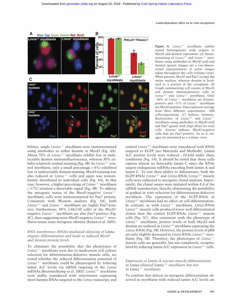
bilities, single Lmna−/− myoblasts were immunostainedusing antibodies to either desmin or MyoD (Fig. 4A).About 70% of Lmna−/− myoblasts exhibit low or unde-tectable desmin immunofluorescence, whereas 30% ex-hibit relatively normal staining (Fig. 4B). In Lmna+/+ con-trol myoblasts, only a small percentage (∼4%) exhibitedlow or undetectable desmin staining. MyoD staining wasalso reduced in Lmna−/− cells and again was nonuni-formly distributed in individual cells (Fig. 4A). In thiscase, however, a higher percentage of Lmna−/− myoblasts(∼57%) retained a detectable signal (Fig. 4B). To addressthe myogenic status of the MyoD-negative Lmna−/−
myoblasts, cells were immunostained for Pax7 protein.Consistent with Western analysis (Fig. 3A), bothLmna+/+ and Lmna−/− myoblasts are highly Pax7-posi-tive. Furthermore, 96% (144/150 cells) of the MyoD-negative Lmna−/− myoblasts are also Pax7-positive (Fig.4C), thus suggesting most MyoD-negative-Lmna−/− myo-blasts retain some myogenic identity (Relaix et al. 2005).
RNA interference (RNAi)-mediated silencing of Lmnaimpairs differentiation and leads to reduced MyoDand desmin protein levels
To eliminate the possibility that the phenotypes ofLmna−/− myoblasts were due to inadvertent cell cultureselection for differentiation-defective muscle cells, wetested whether the reduced differentiation potential ofLmna−/− myoblasts could be phenocopied by reducinglamin A/C levels via siRNA targeting of the LmnamRNAs (Brummelkamp et al. 2002). Lmna+/+ myoblastswere stably transduced with retroviruses expressingshort hairpin RNAs targeted to the Lmna transcript, and
control Lmna+/+ myoblasts were transduced with RNAitargeted to EGFP (see Materials and Methods). LaminA/C protein levels were reduced ∼10-fold under theseconditions (Fig. 5A). It should be noted that these cellsexpress almost no detectable lamin C since the RNAitargets endogenous mRNAs encoding both lamin A andlamin C. To test their ability to differentiate, both theEGFP-RNAi Lmna+/+ and Lmna-RNAi Lmna+/+ musclecells were subjected to myogenic clonal analysis. Impor-tantly, the clonal assays were initiated within 4 d of thesiRNA transduction, thereby eliminating the possibilityof gradual in vitro selection for differentiation-defectivemyoblasts. The expression of the EGFP-RNAi inLmna+/+ myoblasts had no effect on cell differentiation.In contrast, as with Lmna−/− myoblasts, Lmna-RNAiLmna+/+ muscle cells produced fewer well-differentiatedclones than the control EGFP-RNAi Lmna+/+ musclecells (Fig. 5C). Also consistent with the phenotype ofLmna−/− myoblasts, protein levels of both MyoD anddesmin are reduced in Lmna+/+ myoblasts expressing theLmna-RNAi (Fig. 5B). However, the protein levels of pRBare only slightly decreased in Lmna-RNAi Lmna+/+ myo-blasts (Fig. 5B). Therefore, the phenotypes of Lmna−/−
muscle cells are generally, but not completely, recapitu-lated by reducing lamin A/C expression in Lmna+/+ cells.
Expression of lamin A rescues muscle differentiationin Lmna-silenced Lmna+/+ myoblasts but notin Lmna−/− myoblasts
To confirm that defects in myogenic differentiation ob-served in myoblasts with reduced lamin A/C levels are
Figure 4. Lmna−/− myoblasts exhibitmixed heterogeneity with respect toMyoD and desmin expression. (A) Immu-nostaining of Lmna+/+ and Lmna−/− myo-blasts using antibodies to MyoD (red) anddesmin (green). Images are a two-dimen-sional representation of serial imagestaken throughout the cells (volume view).When present, MyoD and Pax7 occupy theentire nucleus, whereas desmin is local-ized to a portion of the cytoplasm. (B)Graph summarizing cell counts of MyoDand desmin immunopositive cells inLmna+/+ and Lmna−/− myoblasts. Only∼30% of Lmna−/− myoblasts are desmin-positive and ∼57% of Lmna−/− myoblastsare MyoD-positive. Data represent averagefrom three different experiments, ∼400cells/experiment. (C) Indirect immuno-fluorescence of Lmna+/+ and Lmna−/−
myoblasts using antibodies to MyoD (red)and Pax7 (green) with Dapi (blue) for totalcells. Arrows indicate MyoD-negativecells that are Pax7-positive. As in A, im-ages are presented as a volume view.
Lamin-dependent effect on in vitro myogenesis
GENES & DEVELOPMENT 491
Cold Spring Harbor Laboratory Press on August 20, 2018 - Published by genesdev.cshlp.orgDownloaded from
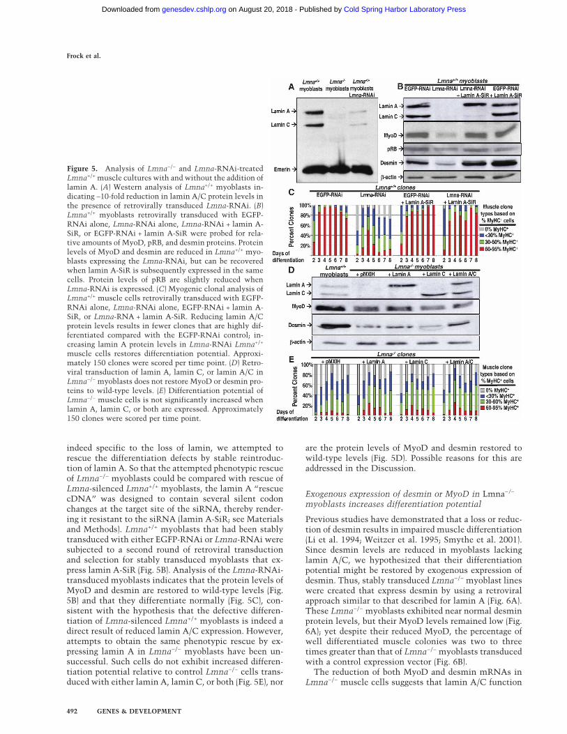
indeed specific to the loss of lamin, we attempted torescue the differentiation defects by stable reintroduc-tion of lamin A. So that the attempted phenotypic rescueof Lmna−/− myoblasts could be compared with rescue ofLmna-silenced Lmna+/+ myoblasts, the lamin A “rescuecDNA” was designed to contain several silent codonchanges at the target site of the siRNA, thereby render-ing it resistant to the siRNA (lamin A-SiR; see Materialsand Methods). Lmna+/+ myoblasts that had been stablytransduced with either EGFP-RNAi or Lmna-RNAi weresubjected to a second round of retroviral transductionand selection for stably transduced myoblasts that ex-press lamin A-SiR (Fig. 5B). Analysis of the Lmna-RNAi-transduced myoblasts indicates that the protein levels ofMyoD and desmin are restored to wild-type levels (Fig.5B) and that they differentiate normally (Fig. 5C), con-sistent with the hypothesis that the defective differen-tiation of Lmna-silenced Lmna+/+ myoblasts is indeed adirect result of reduced lamin A/C expression. However,attempts to obtain the same phenotypic rescue by ex-pressing lamin A in Lmna−/− myoblasts have been un-successful. Such cells do not exhibit increased differen-tiation potential relative to control Lmna−/− cells trans-duced with either lamin A, lamin C, or both (Fig. 5E), nor
are the protein levels of MyoD and desmin restored towild-type levels (Fig. 5D). Possible reasons for this areaddressed in the Discussion.
Exogenous expression of desmin or MyoD in Lmna−/−
myoblasts increases differentiation potential
Previous studies have demonstrated that a loss or reduc-tion of desmin results in impaired muscle differentiation(Li et al. 1994; Weitzer et al. 1995; Smythe et al. 2001).Since desmin levels are reduced in myoblasts lackinglamin A/C, we hypothesized that their differentiationpotential might be restored by exogenous expression ofdesmin. Thus, stably transduced Lmna−/− myoblast lineswere created that express desmin by using a retroviralapproach similar to that described for lamin A (Fig. 6A).These Lmna−/− myoblasts exhibited near normal desminprotein levels, but their MyoD levels remained low (Fig.6A); yet despite their reduced MyoD, the percentage ofwell differentiated muscle colonies was two to threetimes greater than that of Lmna−/− myoblasts transducedwith a control expression vector (Fig. 6B).
The reduction of both MyoD and desmin mRNAs inLmna−/− muscle cells suggests that lamin A/C function
Figure 5. Analysis of Lmna−/− and Lmna-RNAi-treatedLmna+/+ muscle cultures with and without the addition oflamin A. (A) Western analysis of Lmna+/+ myoblasts in-dicating ∼10-fold reduction in lamin A/C protein levels inthe presence of retrovirally transduced Lmna-RNAi. (B)Lmna+/+ myoblasts retrovirally transduced with EGFP-RNAi alone, Lmna-RNAi alone, Lmna-RNAi + lamin A-SiR, or EGFP-RNAi + lamin A-SiR were probed for rela-tive amounts of MyoD, pRB, and desmin proteins. Proteinlevels of MyoD and desmin are reduced in Lmna+/+ myo-blasts expressing the Lmna-RNAi, but can be recoveredwhen lamin A-SiR is subsequently expressed in the samecells. Protein levels of pRB are slightly reduced whenLmna-RNAi is expressed. (C) Myogenic clonal analysis ofLmna+/+ muscle cells retrovirally transduced with EGFP-RNAi alone, Lmna-RNAi alone, EGFP-RNAi + lamin A-SiR, or Lmna-RNA + lamin A-SiR. Reducing lamin A/Cprotein levels results in fewer clones that are highly dif-ferentiated compared with the EGFP-RNAi control; in-creasing lamin A protein levels in Lmna-RNAi Lmna+/+
muscle cells restores differentiation potential. Approxi-mately 150 clones were scored per time point. (D) Retro-viral transduction of lamin A, lamin C, or lamin A/C inLmna−/− myoblasts does not restore MyoD or desmin pro-teins to wild-type levels. (E) Differentiation potential ofLmna−/− muscle cells is not significantly increased whenlamin A, lamin C, or both are expressed. Approximately150 clones were scored per time point.
Frock et al.
492 GENES & DEVELOPMENT
Cold Spring Harbor Laboratory Press on August 20, 2018 - Published by genesdev.cshlp.orgDownloaded from

might be important for efficient transcription of either orboth these genes. MyoD has been shown to enhance des-min expression in vitro (Li and Capetanaki 1993), butthis activity is reported to be specific for differentiatingmuscle cells, not proliferating myoblasts (Gao et al.1998). To determine whether elevated MyoD expressionin Lmna−/− myoblasts would rescue their compromiseddifferentiation, cells were transduced with MyoD or con-trol retroviral vectors. Since exogenous expression ofMyoD activates the muscle differentiation program in avariety of cells (Tapscott et al. 1988), we used a condi-tional MyoD-estrogen receptor (ER) system in whichMyoD-ER accumulates in the cytoplasm until the cellsare exposed to �-estradiol, which upon binding the ER
moiety, targets MyoD-ER to the nucleus (Hollenberg etal. 1993). Stably transduced control and MyoD-ER-Lmna−/− myoblasts (Fig. 6C) were plated at clonal den-sities, grown for 5 d, switched to FGF-free medium in thepresence or either �-estradiol or vehicle control (+EtOH),and then periodically fixed and immunostained forMyHC (Fig. 6D). MyoD-ER-Lmna−/− colonies exposed to�-estradiol exhibited high levels of differentiation within2 d, whereas three different sets of control colonies ex-hibited delayed and less extensive differentiation. Impor-tantly, the differentiation of �-estradiol-treated MyoD-ER-Lmna−/− colonies was greater than that of desmin-Lmna−/− myoblasts (Fig. 6B) and equivalent to that ofLmna+/+ muscle cells (Fig. 2B).
To further investigate differences between the differ-entiation kinetics of MyoD-ER-Lmna−/− myoblasts anddesmin-Lmna−/− myoblasts, both cell types were assayedin mass cultures during the first 48 h after FGF removal.MyoD-ER-Lmna−/− cells exhibited rapid cell cycle with-drawal and >95% of the cells were MyHC-positivewithin 48 h, while desmin-Lmna−/− myoblasts exhibitedvirtually no cell cycle withdrawal during the first 12 hand significantly lower levels of differentiation by 48 h(Table 1, red numerals; also cf. Fig. 1B,C). These findingssuggest that unlike readdition of MyoD-ER, which en-hances both the timing of cell cycle exit and the differ-entiation potential of Lmna−/− cells to wild-type levels,readdition of desmin is not sufficient to restore the nor-mal kinetics of cell cycle exit after FGF-withdrawal ofLmna−/− cells, but constitutively expressed desmin im-proves myogenesis after these cells eventually withdrawfrom the cell cycle. Collectively, these findings indicatethat the compromised differentiation potential of myo-blasts lacking lamin A/C may derive from reduced levelsof desmin and MyoD, and the restoration of each proteincan at least partially correct the compromised levels ofmyogenic differentiation.
Reduced expression of Emd leads to defectsin muscle differentiation
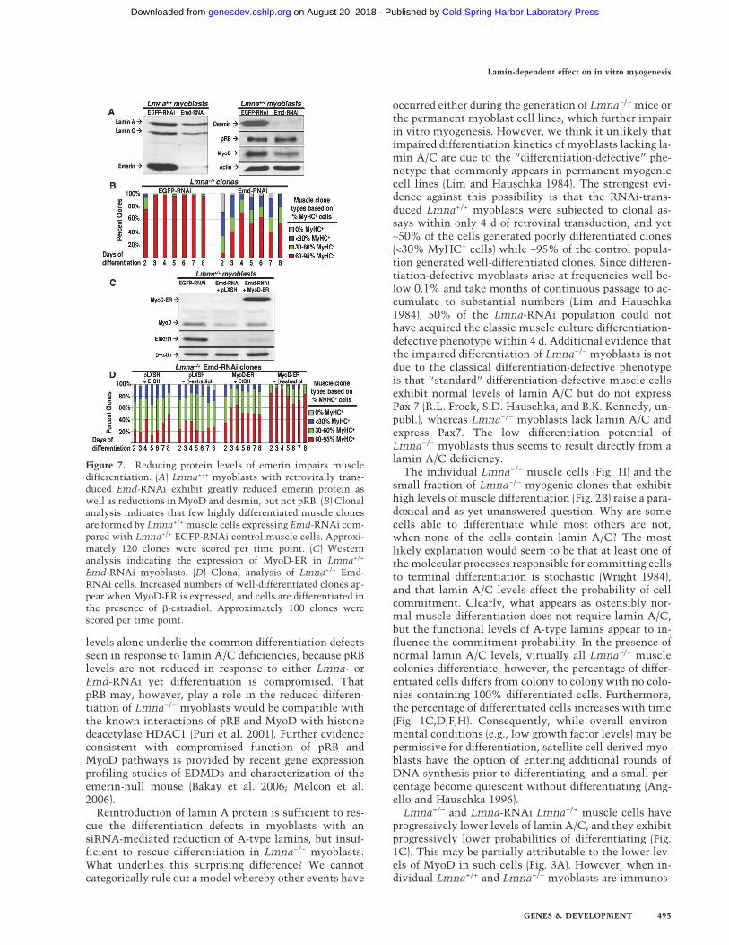
Mutations in a second gene, EMD, encoding the lamin-associated protein emerin have been identified inEDMD1. Since emerin and lamin A/C are known to in-teract within the nuclear envelope (Gruenbaum et al.2005), we tested whether forced reduction in emerin ex-pression would lead to defects in muscle differentiationsimilar to those exhibited by lamin A/C silenced cells.An RNAi specific to emerin was created and introducedin Lmna+/+ myoblasts using the same retroviral approachdescribed previously for Lmna-RNAi. Expression ofEmd-RNAi led to near total elimination of emerin pro-tein (Fig. 7A). When these muscle cells were subjected tomyogenic clonal analysis, <50% of the clones scored ashighly differentiated (Fig. 7B). In addition, the proteinlevels of MyoD and desmin, but not pRB, are reduced tolevels similar to Lmna-RNAi-treated cells (Fig. 5B).Since the levels of MyoD were reduced in Emd-RNAiLmna+/+ myoblasts, we hypothesized that forced expres-
Figure 6. Forced expression of desmin or MyoD in Lmna−/−
myoblasts increases their differentiation potential. (A) Westernblot showing that forced expression of retrovirally transduceddesmin to near wild-type protein levels in Lmna−/− myoblastsdoes not increase MyoD protein levels. (B) Myogenic clonalanalysis comparing Lmna−/− muscle cells expressing either vec-tor control (pMSCVP) or desmin. Forced expression of desminenhances the differentiation potential of Lmna−/− muscle cellsto near wild-type levels. Approximately 250 clones were scoredper time point. (C) Western analysis comparing forced expres-sion of retrovirally transduced MyoD-ER fusion protein inLmna−/− myoblasts to MyoD in Lmna+/+ myoblasts. (D) Clonalanalysis indicates that estradiol treatment of MyoD-ER-express-ing Lmna−/− muscle cells causes ∼80 percent of clones to be-come highly differentiated, whereas control cultures(pBABE ± �-estradiol and MyoD-ER + vehicle) exhibit few if anyhighly differentiated muscle clones. Approximately 150 cloneswere scored per time point.
Lamin-dependent effect on in vitro myogenesis
GENES & DEVELOPMENT 493
Cold Spring Harbor Laboratory Press on August 20, 2018 - Published by genesdev.cshlp.orgDownloaded from

sion of MyoD using the MyoD-ER system described abovewould restore their differentiation potential. Stablytransduced control and MyoD-ER-Emd-RNAi Lmna+/+
myoblasts (Fig. 7C) were plated at clonal densities,grown for 5 d, switched to FGF-free medium in the pres-ence of either �-estradiol or vehicle control (+EtOH), andthen periodically fixed and immunostained for MyHC(Fig. 7D). Similarly to MyoD-ER-Lmna−/− myoblasts, thedifferentiation of �-estradiol-treated MyoD-ER-Emd-RNAi Lmna+/+ colonies was greater than that of threedifferent sets of control colonies (Fig. 7D) and similar tothat of Lmna+/+ muscle cells (see Fig. 2B). These datasuggest that readdition of MyoD in Emd-RNAi Lmna+/+
myoblasts can restore differentiation potential similar tothat of Lmna+/+ myoblasts. Therefore, myoblasts witheither reduced lamin A/C levels or reduced emerin levelsdisplay delayed differentiation kinetics and have reducedlevels of a subset of proteins important for myogenicdifferentiation.
Discussion
Mutations in the genes encoding the nuclear proteinsemerin and lamin A/C are associated with EDMD. Al-though their roles in skeletal muscle development areunknown, we demonstrate that reducing lamin A/C oremerin via Lmna knockout, Lmna-RNAi, and Emd-RNAi results in reduced levels of desmin and MyoD inmyoblasts, and that each of the independent perturba-tions causes a similar set of compromised differentiationphenotypes. Restoration of MyoD, and more surprisinglydesmin, significantly increases the differentiation poten-tial of lamin A/C-deficient myoblasts, suggesting thatreduced levels of these proteins may underlie the differ-entiation impairment of Lmna−/− muscle cells.
Differentiation defects in myoblasts lacking A-typelamins or emerin
The reduction in desmin and MyoD transcripts inLmna−/− myoblasts suggests that A-type lamins are di-rectly or indirectly affecting the transcription of bothgenes. Previous studies indicated that desmin is a targetgene for MyoD, although MyoD dependence was re-ported to be important for proper levels of desmin tran-scription only after induction of terminal differentiation(Gao et al. 1998). However, our studies indicate a 4.5-foldreduction of desmin mRNA in Lmna−/− myoblasts. Doesthe reduction or absence of A-type lamins have nonspe-cific global effects on the transcription of many genes,some of which happen to play important roles in muscledifferentiation, or does lamin A/C affect specific genetargets important for maintaining the myogenic deter-mination of skeletal muscle cells? A-type lamins inter-act with and organize chromatin (Glass et al. 1993) in amanner possibly mediated through lamin–histone inter-actions (Taniura et al. 1995). Therefore, loss or reductionof lamin A/C could affect the expression of numerousgenes. In addition, A-type lamins have been shown todirectly interact with transcription factors such as pRB(Ozaki et al. 1994; Markiewicz et al. 2002), SREBP-1(Lloyd et al. 2002), and MOK2 (Dreuillet et al. 2002),implying that they could also act as accessory proteinsfor a subset of tissue-defining factors. Our data show thatalthough desmin, MyoD, pRB, and M-cadherin are re-duced and Myf-5 is increased in the absence of A-typelamins, other myogenic factors such as Pax3, Pax7,MEF2C, MEF2D, c-met, and �-catenin are not affected,indicating that A-type lamins have a specific effect on asubset of myogenic components. While the consequenceof decreased pRB and M-cadherin protein levels remainsto be determined, it seems unlikely that decreased pRB
Table 1. Forced expression of MyoD, but not desmin, rescues cell cycle withdrawal and early differentiation kinetics inLmna−/− cellsa
aBlack and blue text indicates cell cycle withdrawal and differentiation subpanels respectively. Red text highlights key points in thetable.bBased on average and SD of three experiments counting ∼650 cells/time point/experiment in mass culture.cVehicle for solubilizing �-estradiol consists of 10 µL 100% EtOH in 10 mL of media.dEstradiol-mediated nuclear import of MyoD-ER accelerates cell cycle withdrawal of Lmna−/− cells following FGF deprivation.eEstradiol-mediated nuclear import of MyoD-ER greatly enhances the proportion of terminally differentiated Lmna−/− cells.fOverexpression of desmin has no cell cycle withdrawal or early differentiation effects on Lmna−/− cells.
Frock et al.
494 GENES & DEVELOPMENT
Cold Spring Harbor Laboratory Press on August 20, 2018 - Published by genesdev.cshlp.orgDownloaded from
levels alone underlie the common differentiation defectsseen in response to lamin A/C deficiencies, because pRBlevels are not reduced in response to either Lmna- orEmd-RNAi yet differentiation is compromised. ThatpRB may, however, play a role in the reduced differen-tiation of Lmna−/− myoblasts would be compatible withthe known interactions of pRB and MyoD with histonedeacetylase HDAC1 (Puri et al. 2001). Further evidenceconsistent with compromised function of pRB andMyoD pathways is provided by recent gene expressionprofiling studies of EDMDs and characterization of theemerin-null mouse (Bakay et al. 2006; Melcon et al.2006).
Reintroduction of lamin A protein is sufficient to res-cue the differentiation defects in myoblasts with ansiRNA-mediated reduction of A-type lamins, but insuf-ficient to rescue differentiation in Lmna−/− myoblasts.What underlies this surprising difference? We cannotcategorically rule out a model whereby other events have
occurred either during the generation of Lmna−/− mice orthe permanent myoblast cell lines, which further impairin vitro myogenesis. However, we think it unlikely thatimpaired differentiation kinetics of myoblasts lacking la-min A/C are due to the “differentiation-defective” phe-notype that commonly appears in permanent myogeniccell lines (Lim and Hauschka 1984). The strongest evi-dence against this possibility is that the RNAi-trans-duced Lmna+/+ myoblasts were subjected to clonal as-says within only 4 d of retroviral transduction, and yet∼50% of the cells generated poorly differentiated clones(<30% MyHC+ cells) while ∼95% of the control popula-tion generated well-differentiated clones. Since differen-tiation-defective myoblasts arise at frequencies well be-low 0.1% and take months of continuous passage to ac-cumulate to substantial numbers (Lim and Hauschka1984), 50% of the Lmna-RNAi population could nothave acquired the classic muscle culture differentiation-defective phenotype within 4 d. Additional evidence thatthe impaired differentiation of Lmna−/− myoblasts is notdue to the classical differentiation-defective phenotypeis that “standard” differentiation-defective muscle cellsexhibit normal levels of lamin A/C but do not expressPax 7 (R.L. Frock, S.D. Hauschka, and B.K. Kennedy, un-publ.), whereas Lmna−/− myoblasts lack lamin A/C andexpress Pax7. The low differentiation potential ofLmna−/− myoblasts thus seems to result directly from alamin A/C deficiency.
The individual Lmna−/− muscle cells (Fig. 1I) and thesmall fraction of Lmna−/− myogenic clones that exhibithigh levels of muscle differentiation (Fig. 2B) raise a para-doxical and as yet unanswered question. Why are somecells able to differentiate while most others are not,when none of the cells contain lamin A/C? The mostlikely explanation would seem to be that at least one ofthe molecular processes responsible for committing cellsto terminal differentiation is stochastic (Wright 1984),and that lamin A/C levels affect the probability of cellcommitment. Clearly, what appears as ostensibly nor-mal muscle differentiation does not require lamin A/C,but the functional levels of A-type lamins appear to in-fluence the commitment probability. In the presence ofnormal lamin A/C levels, virtually all Lmna+/+ musclecolonies differentiate; however, the percentage of differ-entiated cells differs from colony to colony with no colo-nies containing 100% differentiated cells. Furthermore,the percentage of differentiated cells increases with time(Fig. 1C,D,F,H). Consequently, while overall environ-mental conditions (e.g., low growth factor levels) may bepermissive for differentiation, satellite cell-derived myo-blasts have the option of entering additional rounds ofDNA synthesis prior to differentiating, and a small per-centage become quiescent without differentiating (Ang-ello and Hauschka 1996).
Lmna+/− and Lmna-RNAi Lmna+/+ muscle cells haveprogressively lower levels of lamin A/C, and they exhibitprogressively lower probabilities of differentiating (Fig.1C). This may be partially attributable to the lower lev-els of MyoD in such cells (Fig. 3A). However, when in-dividual Lmna+/+ and Lmna−/− myoblasts are immunos-
Figure 7. Reducing protein levels of emerin impairs muscledifferentiation. (A) Lmna+/+ myoblasts with retrovirally trans-duced Emd-RNAi exhibit greatly reduced emerin protein aswell as reductions in MyoD and desmin, but not pRB. (B) Clonalanalysis indicates that few highly differentiated muscle clonesare formed by Lmna+/+ muscle cells expressing Emd-RNAi com-pared with Lmna+/+ EGFP-RNAi control muscle cells. Approxi-mately 120 clones were scored per time point. (C) Westernanalysis indicating the expression of MyoD-ER in Lmna+/+
Emd-RNAi myoblasts. (D) Clonal analysis of Lmna+/+ Emd-RNAi cells. Increased numbers of well-differentiated clones ap-pear when MyoD-ER is expressed, and cells are differentiated inthe presence of �-estradiol. Approximately 100 clones werescored per time point.
Lamin-dependent effect on in vitro myogenesis
GENES & DEVELOPMENT 495
Cold Spring Harbor Laboratory Press on August 20, 2018 - Published by genesdev.cshlp.orgDownloaded from

tained for MyoD (Fig. 4), Lmna+/+ cells exhibit strong tomedium staining intensities, whereas Lmna−/− myo-blasts exhibit MyoD staining intensities ranging from∼40% that exhibit virtually no staining to many thatexhibit staining intensities equivalent to those seen inLmna+/+ cells. Importantly, >95% of the MyoD-negativeLmna−/− myoblasts are Pax7-positive; thus while thesecells exhibit compromised differentiation capabilities,they apparently retain some myogenic properties. WhileMyoD staining intensities cannot yet be correlated witha cell’s subsequent differentiation potential, this hetero-geneity could partially explain the compromised differ-entiation of Lmna−/− myoblasts. This possibility wouldbe consistent with the heterogeneous behavior of normalsatellite cells in which a low-level commitment to ter-minally differentiate is correlated with a low ratio ofMyoD to Pax7 (Zammit et al. 2004), a phenotype that isalso characteristic of some lamin A/C-deficient myo-blasts (Fig. 4C,D). Interestingly, when MyoD or desminare exogenously expressed in lamin A/C-deficient cells,their probability of differentiation returns to near normallevels. Thus MyoD, desmin, and lamin A/C seem to playcritical roles in regulating at least one stochastic com-ponent of myogenic terminal differentiation.
Desmin and the EDMD phenotype
Previous in vitro cell culture data have suggested a rolefor desmin in myogenesis (Li et al. 1994; Weitzer et al.1995). Although Des−/− mice develop normally (Li et al.1996), they have postnatal defects in skeletal, cardiac,and smooth muscles. In addition, Des−/− myoblasts havedelayed cell cycle withdrawal and delayed fusion in vivo(Agbulut et al. 2001; Smythe et al. 2001), which suggeststhat desmin is important for adult skeletal muscle re-generation. Our findings with lamin A/C-deficient myo-blasts, showing a decrease in desmin protein and tran-script, suggest phenotypic overlap between Lmna−/− andDes−/− mice. In support of this hypothesis, restoration ofnormal desmin levels in Lmna−/− myoblasts enhancestheir differentiation potential, implying that desmin isimportant for muscle differentiation in Lmna−/− myo-blasts. This finding is particularly interesting given thatdesmin is a cytoplasmic intermediate filament proteinnot directly linked to myogenic transcription. However,vimentin and desmin intermediate filaments are knownto stably associate with nuclear matrix attachment re-gions, where they might secondarily affect gene tran-scription (Tolstonog et al. 2002). Thus the reduced ex-pression of desmin in Lmna−/− myoblasts could lead tocompromised communication between the cytoplasmand the nuclear envelope, and this might be restored byoverexpression of desmin in Lmna−/− cells. This raisesthe possibility that EDMD phenotypes in vivo might beoffset by therapeutic approaches designed to elevate des-min levels in skeletal and cardiac muscle.
Lamin A/C, MyoD, and EDMD
Lmna−/− myoblasts and MyoD−/− primary myoblasts areremarkably similar (Sabourin et al. 1999), sharing many
overlapping phenotypes, including delayed cell cyclewithdrawal, reduced differentiation potential, and re-duced desmin and M-cadherin. Unlike Lmna−/− mice,however, MyoD−/− mice do not develop a muscular dys-trophy phenotype (Rudnicki et al. 1992). Why shouldpopulations of mutant myoblasts behave similarly in invitro differentiation assays but differently in the organ-ism? Interestingly, when MyoD−/− mice are challengedwith acute muscle injury, they form few fully repairedfibers and the majority of the differentiated cells withinthe damaged area remain mononucleated. In addition,when MyoD−/− mice are interbred with mice that con-tain a naturally occurring loss-of-function mutation inthe dystrophin gene (mdx) (Bulfield et al. 1984; Hoffmanet al. 1987; Cox et al. 1993), they develop a more severemyopathy resulting in death at ∼1 yr (Megeney et al.1996). In contrast, mdx mice have a normal life span andexhibit little overt skeletal muscle weakness except inthe diaphragm muscle. Thus, in mouse muscle diseasemodels, dystrophic phenotypes are enhanced when in-creased degeneration (due to the absence of dystrophin orlamin A/C) is coupled with decreased regenerative po-tential due to the experimental removal of MyoD inmdx/MyoD−/− mice and the possible decrease of MyoDin Lmna−/− mouse satellite cells in vivo. A further com-plication is that Myf-5 protein levels are substantiallyincreased in Lmna−/− myoblasts (Fig. 3A), and yet, theincreased level of this highly related transcription factordoes not compensate for the decreased level of MyoD.
Two models have been proposed to explain dystrophicsyndromes of muscle resulting from LMNA mutation.One “structural weakness” model proposes that LMNAmutant myoblasts exhibit reduced cellular integrity andtherefore enhanced tissue degeneration. Indeed, cellslacking lamin A/C exhibit impaired viability under me-chanical strain as well as changes in expression ofmechanosensitive genes (Broers et al. 2004; Lammerdinget al. 2004). The “impaired differentiation” model pro-poses that tissue regeneration may be compromised inLmna−/− muscle due to defective maintenance of themyogenic program in satellite cells. Our in vitro findingsprovide compelling evidence that muscle differentiationprograms are compromised in Lmna−/− myoblasts, re-sulting in impaired myogenic potential. This conclusionis reinforced by the finding that MyoD protein levels aredecreased in myoblasts deficient for lamin A/C andemerin, and that forced expression of MyoD in such cellsrestores muscle differentiation. We propose that both“structural weakness” and “impaired differentiation”defects contribute to muscular dystrophy in the murineLmna−/− disease model.
Materials and methods
Derivation of Lmna+/+, Lmna+/−, and Lmna−/− myoblast celllines and conditions for differentiation
Primary and permanent Lmna+/+, Lmna+/−, and Lmna−/− myo-blasts were derived from 5-wk-old Lmna+/+, Lmna+/−, and
Frock et al.
496 GENES & DEVELOPMENT
Cold Spring Harbor Laboratory Press on August 20, 2018 - Published by genesdev.cshlp.orgDownloaded from
Lmna−/− mouse hindlimb muscles (provided by Colin Stewart,National Cancer Institute-Frederick Cancer Research and De-velopment Center, Frederick, MD). Hindlimb muscles were dis-sected in F10C medium (GIBCO) to isolate skeletal muscle andto remove adipose and connective tissues. Muscle was mincedwith fine scissors and digested with 375 U/mL type II collage-nase (Worthington) for 1 h at 37°C and cultured as previouslydescribed (Hauschka et al. 1979; Neville et al. 1997). All cul-tures were grown on plates coated with 0.67% gelatin (Difco) inF10C medium supplemented with 15% preselected horse serumand fed 4 ng/mL basic fibroblast growth factor (human recom-binant bFGF, Zymogenetics) every 12 h. Cells were passagedevery 3 d and seeded at 5 × 10⁴ cells per 100-mm plate continu-ally until the spontaneously immortalized permanent line wasgenerated. No individual muscle clones were isolated duringderivation of the cell lines because we wanted the final cellpopulations to be maximally representative of all myogeniccells within each muscle sample. Cells in the 10–15 passagerange following derivation of the permanent cell lines were usedfor all experiments.
Muscle cells were induced to differentiate via mitogen deple-tion, as described elsewhere (Clegg et al. 1987; Neville et al.1997). Briefly, proliferating myoblasts are rinsed in saline G toremove residual bFGF before adding F10C supplemented with1.5% horse serum and 1 µM insulin. For the clonal analysis ofmyogenesis (Clegg et al. 1987), Lmna muscle clones wereswitched to low mitogen media as above, but after 4 d, thecultures were switched back to F10C medium containing 15%horse serum and 1µM insulin for the duration of the experi-ment. This enhances long-term culture survival without stimu-lating cell replication, since bFGF is not present.
Differentiation and proliferation assays,immunohistochemistry, and Western blotting
To measure muscle differentiation, cells were fixed in alcohol–formalin–acetic acid (AFA); immunostained for MyHC usingmonoclonal MF-20 diluted 1:100 (gift from D.A. Fischman, Cor-nell University Medical College, New York), plus biotinylatedrabbit anti-mouse IgG, streptavidin, and biotinylated horserad-ish peroxidase (Vector Labs, Inc.); and counterstained with 1%methylene blue or hematoxylin. For detecting S-phase cells, cul-tures were pulsed with BrdU (Amersham; 2 µL/mL of media) for1 h prior to fixation as described elsewhere (Foster et al. 1987)and subsequent immunostaining using anti-BrdU diluted at 1:2000 (G3G4; gift from Steve Kaufman, University of Illinois,Urbana) and MF-20 diluted at 1:100 using isotype-specific sec-ondary immunofluorescent antibodies (Molecular Probes). Forhigh-resolution optical sectioning of myoblasts using the Zeissaxiovert 200M, cells were plated on 1- to 2-mm round glasscoverslips (Fisher) that had been prepared by incubating 12–24 hin 1 M magnesium acetate, rinsed twice with PBS, then incu-bated for 1 h in 25 µg/mL poly-L-lysine (ICN Biomedicals), andfinally incubated 3–24 h in 5 µg/mL laminin (Sigma). After plat-ing, cells were fixed as described elsewhere (Kennedy et al. 2000;Barbie et al. 2004) and immunostained with the following anti-bodies: MyoD diluted at 1:500 (M-318; Santa Cruz), desmin di-luted at 1:200 (clone D33; DAKO), and Pax7 diluted at 1:200(developed by Atsushi Kawakami [University of Tokyo, Tokyo,Japan] and obtained from the Developmental Studies Hybri-doma Bank, University of Iowa).
Muscle cells were lysed in RIPA buffer (50 mM Tris-HCl atpH 7.4, 150 mM NaCl, 1% NP-40, 0.25% deoxycholate) supple-mented with phosphatase and protease inhibitors (1 mMNa3VO4, 1 mM NaF, 1 mM PMSF, 1 mM EDTA, 1 µg/mL apro-tinin, 1 µg/mL leupeptin). Western analysis of muscle cell ly-
sates was performed using standard procedures. The followingantibodies were used: lamin A/C diluted at 1:1000 (2032; CellSignaling), MyoD diluted at 1:100 (clone 5.8A; gift from PeterHoughton and Peter Diaz, St. Jude Children’s Hospital, Mem-phis, TN), MyoD diluted at 1:100 (M-318; Santa Cruz), Myf-5diluted at 1:100 (C-20; Santa Cruz), MEF2C diluted at 1:200(9792; Cell Signaling), MEF2D diluted at 1:200 (clone 9; BD),pRB diluted at 1:100 (clone G3-245; BD), desmin diluted at 1:20(clone D3; gift from D.A. Fischman, Cornell University MedicalCollege), desmin diluted at 1:10,000 (ab15200; abcam), Pax3 di-luted at 1:200 (gift from J.A. Epstein, Cardiovascular Division,University of Pennsylvania, Philadelphia, PA), Pax7 diluted at1:50 (obtained from the Developmental Studies HybridomaBank, University of Iowa), �-catenin diluted at 1:1000 (H-102;Santa Cruz), emerin diluted at 1:1000 (FL-254; Santa Cruz), M-cadherin diluted at 1:300 (clone 5; BD), c-met diluted at 1:1000(SP260; Santa Cruz), pan-actin diluted at 1:10,000 (clone C4;Chemicon), and �-actin diluted at 1:10,000 (clone ab8226; abcam).
Retroviral constructs and transduction
293T cells were acquired from ATCC and were cultured inDMEM supplemented with 10% fetal calf serum. RNAi con-structs for mouse lamin A/C, mouse emerin, and enhancedgreen fluorescent protein (EGFP) were cloned into pSuper.retro-Puro as described elsewhere (Kudlow et al. 2005). Lamin A-SiRwas generated by site-directed mutagenesis of a wild-type hu-man lamin A cDNA using an oligonucleotide with the follow-ing sequence: GCAGACCATGAAGGAGGAGCTCGATTTTCAAAAGAATATCTACAGTGAGGAGCTGCG. The mutatedlamin A cDNA was sequence-verified and subcloned intopMXIH for high-level, retroviral expression (Kudlow et al. 2005)as was wild-type lamin A. Both wild-type lamin A and laminA-SiR were expressed as prelamin A (including the 18 aminoacids that are eventually cleaved) so as to be processed naturallyby the target cells. Desmin (IMAGE 4219280) was cloned intopMSCV-puro (Clontech) using BglII and EcoRI and was se-quence-verified. pBABE- and pLXSH-MyoD-ER plasmids weregifts from Stephen J. Tapscott (Fred Hutchison Cancer ResearchCenter, Seattle, WA). All retroviral constructs were transientlycotransfected into 293T cells with an ecotrophic packaging plas-mid to generate nonreduplicating retroviruses. Viral superna-tants from the 293T cells were filtered through 0.45-µm syringefilters (Millipore), added to exponentially growing Lmna musclecultures (∼200,000 cells per 100-mm plate), and supplementedwith 4 ng/mL polybrene (Sigma). Lmna muscle cultures wereretrovirally transduced either in F10C supplemented with 15%horse serum or at 1:1 ratios of 15% horse serum F10C and 10%fetal calf serum DMEM and fed 12 ng/mL bFGF for the durationof the retroviral infection. Selection for puromycin- or hygro-mycin-resistant myoblasts occurred after 24–36 h post-infectionusing 10 µg/mL and 300 µg/mL concentrations, respectively.Differentiation of MyoD-ER-Lmna−/− myoblasts and MyoD-ER-EmdRNAi Lmna+/+ myoblasts was achieved by addition of 0.1µM �-estradiol (Sigma) following a standard medium switch (seeabove) to differentiation media.
QPCR
mRNA’s were purified using the RNeasy kit (Qiagen). Sampleswere DNase I-treated, and cDNA was generated using reversetranscriptase (Promega) and oligo dT15 primers (Invitrogen). ForQPCR, 10 µL of 2× master mix containing SYBR Green (AppliedBiosystems) was mixed with cDNA and 300 µM forward andreverse primers. Triplicates of the cDNA’s were amplified onthe Opticon I real-time thermal cycler (MJ Research). The ex-periments were performed three times at three different cDNA
Lamin-dependent effect on in vitro myogenesis
GENES & DEVELOPMENT 497
Cold Spring Harbor Laboratory Press on August 20, 2018 - Published by genesdev.cshlp.orgDownloaded from
dilutions. PCR products were normalized against the house-keeping gene Hprt, and measurements between samples werecompared by cycle threshold (Ct).
Primer sequences used for QPCR are the following: Hprt(GenBank J00423) forward, 5�-AGGACCTCTCGAAGTGTTGG-3�; Hprt reverse, 5�-TGGCAACATCAACAGGACTC-3�;RB (GenBank NM_009029) forward, 5�-TACACTCTGTGCACGCCTTC-3�; RB reverse, 5�-TCACCTTGCAGATGCCATAC-3�; MyoD (GenBank X61655) forward, 5�-CATCCGCTACATCGAAGGTC-3�; MyoD reverse, 5�-TAGTAGGCGGTGTCGTAGCC-3�; desmin (GenBank BC031760) forward, 5�-TACACCTGCGAGATTGATGC-3�; and desmin reverse, 5�-ACATCCAAGGCCATCTTCAC-3�.
Acknowledgments
We are grateful to Colin Stewart and Teresa Sullivan for pro-viding Lmna mouse muscle tissue. We thank John Angello, Lin-dus Conlan, Erica Smith, Rob Welikson, and other members ofthe Hauschka and Kennedy Laboratories for helpful commentsand suggestions. We also thank Joyce Hong, Bharti Sharma, andNayan Arora for their help in maintaining and generating tissueculture reagents and performing MyHC staining, and LinneaWelton for technical assistance. B.A.K. is supported in part byPHS NRSA T32 GM07270 from NIGMS. This work was sup-ported by the National Institutes of Health grant R01AG024287to B.K.K., and by grants RO1AR18860 and 1PO1 NS046788 toS.D.H. B.K.K. is a Searle Scholar.
References
Agbulut, O., Li, Z., Perie, S., Ludosky, M.A., Paulin, D., Car-taud, J., and Butler-Browne, G. 2001. Lack of desmin resultsin abortive muscle regeneration and modifications in synap-tic structure. Cell Motil. Cytoskeleton 49: 51–66.
Anastasi, S., Giordano, S., Sthandier, O., Gambarotta, G., Mai-one, R., Comoglio, P., and Amati, P. 1997. A natural hepa-tocyte growth factor/scatter factor autocrine loop in myo-blast cells and the effect of the constitutive Met kinase ac-tivation on myogenic differentiation. J. Cell. Biol. 137: 1057–1068.
Angello, J.C. and Hauschka, S.D. 1996. Skeletal muscle satellitecells: Timelapse videomicroscopic evidence that renewal isstochastic. BAM 6: 491–502.
Arimura, T., Helbling-Leclerc, A., Massart, C., Varnous, S.,Niel, F., Lacene, E., Fromes, Y., Toussaint, M., Mura, A.M.,Keller, D.I., et al. 2005. Mouse model carrying H222P-Lmnamutation develops muscular dystrophy and dilated cardio-myopathy similar to human striated muscle laminopathies.Hum. Mol. Genet. 14: 155–169.
Bakay, M., Wang, Z., Melcon, G., Schiltz, L., Xuan, J., Zhao, P.,Sartorelli, V., Seo, J., Pegoraro, E., Angelini, C., et al. 2006.Nuclear envelope dystrophics show a transcriptional finger-print disruption of Rb–MyoD pathways in muscle regenera-tion. Brain (in press).
Barbie, D.A., Kudlow, B.A., Frock, R., Zhao, J., Johnson, B.R.,Dyson, N., Harlow, E., and Kennedy, B.K. 2004. Nuclear re-organization of mammalian DNA synthesis prior to cellcycle exit. Mol. Cell. Biol. 24: 595–607.
Bione, S., Maestrini, E., Rivella, S., Mancini, M., Regis, S., Ro-meo, G., and Toniolo, D. 1994. Identification of a novel X-linked gene responsible for Emery-Dreifuss muscular dystro-phy. Nat. Genet. 8: 323–327.
Bonne, G., Di Barletta, M.R., Varnous, S., Becane, H.M., Ham-
mouda, E.H., Merlini, L., Muntoni, F., Greenberg, C.R.,Gary, F., Urtizberea, J.A., et al. 1999. Mutations in the geneencoding lamin A/C cause autosomal dominant Emery-Dreifuss muscular dystrophy. Nat. Genet. 21: 285–288.
Broers, J.L., Peeters, E.A., Kuijpers, H.J., Endert, J., Bouten, C.V.,Oomens, C.W., Baaijens, F.P., and Ramaekers, F.C. 2004.Decreased mechanical stiffness in LMNA−/− cells is causedby defective nucleo-cytoskeletal integrity: Implications forthe development of laminopathies. Hum. Mol. Genet. 13:2567–2580.
Brummelkamp, T.R., Bernards, R., and Agami, R. 2002. A sys-tem for stable expression of short interfering RNAs in mam-malian cells. Science 296: 550–553.
Bulfield, G., Siller, W.G., Wight, P.A., and Moore, K.J. 1984. Xchromosome-linked muscular dystrophy (mdx) in themouse. Proc. Natl. Acad. Sci. 81: 1189–1192.
Buskin, J.N. and Hauschka, S.D. 1989. Identification of a myo-cyte nuclear factor that binds to the muscle-specific en-hancer of the mouse muscle creatine kinase gene. Mol. Cell.Biol. 9: 2627–2640.
Charge, S.B. and Rudnicki, M.A. 2004. Cellular and molecularregulation of muscle regeneration. Physiol. Rev. 84: 209–238.
Clegg, C.H., Linkhart, T.A., Olwin, B.B., and Hauschka, S.D.1987. Growth factor control of skeletal muscle differentia-tion: Commitment to terminal differentiation occurs in G1phase and is repressed by fibroblast growth factor. J. CellBiol. 105: 949–956.
Cornelison, D.D.W. 1998. Gene expression in wild-type andMyoD-null satellite cells: Regulation of activation, prolifera-tion and myogenesis. Ph.D thesis, pp. 131–138, CaliforniaInstitute of Technology, Pasadena, CA.
Cornelison, D.D., Olwin, B.B., Rudnicki, M.A., and Wold, B.J.2000. MyoD−/− satellite cells in single-fiber culture are dif-ferentiation defective and MRF4 deficient. Dev. Biol. 224:122–137.
Cox, G.A., Cole, N.M., Matsumura, K., Phelps, S.F., Hauschka,S.D., Campbell, K.P., Faulkner, J.A., and Chamberlain, J.S.1993. Overexpression of dystrophin in transgenic mdx miceeliminates dystrophic symptoms without toxicity. Nature364: 725–729.
Dreuillet, C., Tillit, J., Kress, M., and Ernoult-Lange, M. 2002. Invivo and in vitro interaction between human transcriptionfactor MOK2 and nuclear lamin A/C. Nucleic Acids Res. 30:4634–4642.
Favreau, C., Higuet, D., Courvalin, J.C., and Buendia, B. 2004.Expression of a mutant lamin A that causes Emery-Dreifussmuscular dystrophy inhibits in vitro differentiation ofC2C12 myoblasts. Mol. Cell Biol. 24: 1481–1492.
Foster, R.F., Thompson, J.M., and Kaufman, S.J. 1987. A lamininsubstrate promotes myogenesis in rat skeletal muscle cul-tures: Analysis of replication and development using anti-desmin and anti-BrdUrd monoclonal antibodies. Dev. Biol.122: 11–20.
Gao, J., Li, Z., and Paulin, D. 1998. A novel site, Mt, in thehuman desmin enhancer is necessary for maximal expres-sion in skeletal muscle. J. Biol. Chem. 273: 6402–6409.
Glass, C.A., Glass, J.R., Taniura, H., Hasel, K.W., Blevitt, J.M.,and Gerace, L. 1993. The �-helical rod domain of humanlamins A and C contains a chromatin binding site. EMBO J.12: 4413–4424.
Gruenbaum, Y., Margalit, A., Goldman, R.D., Shumaker, D.K.,and Wilson, K.L. 2005. The nuclear lamina comes of age.Nat. Rev. Mol. Cell. Biol. 6: 21–31.
Guo, C.S., Degnin, C., Fiddler, T.A., Stauffer, D., and Thayer,M.J. 2003. Regulation of MyoD activity and muscle cell dif-
Frock et al.
498 GENES & DEVELOPMENT
Cold Spring Harbor Laboratory Press on August 20, 2018 - Published by genesdev.cshlp.orgDownloaded from

ferentiation by MDM2, pRb, and Sp1. J. Biol. Chem. 278:22615–22622.
Hauschka, S.D., Linkhart, T.A., Clegg, C.H., and Merrill, C.1979. Clonal studies of human and mouse muscle. In InMuscle regeneration (ed. A. Mauro), pp. 311–322. RavenPress, New York.
Hoffman, E.P., Brown Jr., R.H., and Kunkel, L.M. 1987. Dystro-phin: The protein product of the Duchenne muscular dys-trophy locus. Cell 51: 919–928.
Hollenberg, S.M., Cheng, P.F., and Weintraub, H. 1993. Use of aconditional MyoD transcription factor in studies of MyoDtrans-activation and muscle determination. Proc. Natl.Acad. Sci. 90: 8028–8032.
Huh, M.S., Parker, M.H., Scime, A., Parks, R., and Rudnicki,M.A. 2004. Rb is required for progression through myogenicdifferentiation but not maintenance of terminal differentia-tion. J. Cell Biol. 166: 865–876.
Hutchison, C.J. and Worman, H.J. 2004. A-type lamins: Guard-ians of the soma? Nat. Cell Biol. 6: 1062–1067.
Johnson, B.R., Nitta, R.T., Frock, R.L., Mounkes, L., Barbie,D.A., Stewart, C.L., Harlow, E., and Kennedy, B.K. 2004.A-type lamins regulate retinoblastoma protein functionby promoting subnuclear localization and preventing pro-teasomal degradation. Proc. Natl. Acad. Sci. 101: 9677–9682.
Kang, J.S., Feinleib, J.L., Knox, S., Ketteringham, M.A., andKrauss, R.S. 2003. Promyogenic members of the Ig and cad-herin families associate to positively regulate differentia-tion. Proc. Natl. Acad. Sci. 100: 3989–3994.
Kaufman, S.J., George-Weinstein, M., and Foster, R.F. 1991. Invitro development of precursor cells in the myogenic lin-eage. Dev. Biol. 146: 228–238.
Kaufmann, U., Kirsch, J., Irintchev, A., Wernig, A., and Starzin-ski-Powitz, A. 1999. The M-cadherin catenin complex inter-acts with microtubules in skeletal muscle cells: Implica-tions for the fusion of myoblasts. J. Cell. Sci. 112: 55–68.
Kennedy, B.K., Barbie, D.A., Classon, M., Dyson, N., and Har-low, E. 2000. Nuclear organization of DNA replication inprimary mammalian cells. Genes & Dev. 14: 2855–2868.
Kudlow, B.A., Jameson, S.A., and Kennedy, B.K. 2005. HIV pro-tease inhibitors block adipocyte differentiation indepen-dently of lamin A/C. Aids 19: 1565–1573.
Lammerding, J., Schulze, P.C., Takahashi, T., Kozlov, S., Sulli-van, T., Kamm, R.D., Stewart, C.L., and Lee, R.T. 2004. La-min A/C deficiency causes defective nuclear mechanics andmechanotransduction. J. Clin. Invest. 113: 370–378.
Lassar, A.B., Buskin, J.N., Lockshon, D., Davis, R.L., Apone, S.,Hauschka, S.D., and Weintraub, H. 1989. MyoD is a se-quence-specific DNA binding protein requiring a region ofmyc homology to bind to the muscle creatine kinase en-hancer. Cell 58: 823–831.
Lazarides, E. and Hubbard, B.D. 1976. Immunological charac-terization of the subunit of the 100 A filaments from musclecells. Proc. Natl. Acad. Sci. 73: 4344–4348.
Li, H. and Capetanaki, Y. 1993. Regulation of the mouse desmingene: Transactivated by MyoD, myogenin, MRF4 and Myf5.Nucleic Acids Res. 21: 335–343.
Li, H., Choudhary, S.K., Milner, D.J., Munir, M.I., Kuisk, I.R.,and Capetanaki, Y. 1994. Inhibition of desmin expressionblocks myoblast fusion and interferes with the myogenicregulators MyoD and myogenin. J. Cell Biol. 124: 827–841.
Li, Z., Colucci-Guyon, E., Pincon-Raymond, M., Mericskay, M.,Pournin, S., Paulin, D., and Babinet, C. 1996. Cardiovascularlesions and skeletal myopathy in mice lacking desmin. Dev.Biol. 175: 362–366.
Lim, R.W. and Hauschka, S.D. 1984. EGF responsiveness and
receptor regulation in normal and differentiation-defectivemouse myoblasts. Dev. Biol. 105: 48–58.
Lloyd, D.J., Trembath, R.C., and Shackleton, S. 2002. A novelinteraction between lamin A and SREBP1: Implications forpartial lipodystrophy and other laminopathies. Hum. Mol.Genet. 11: 769–777.
Maione, R., Fimia, G.M., Holman, P., Schaffhausen, B., andAmati, P. 1994. Retinoblastoma antioncogene is involved inthe inhibition of myogenesis by polyomavirus large T anti-gen. Cell Growth Differ. 5: 231–237.
Markiewicz, E., Dechat, T., Foisner, R., Quinlan, R.A., and C.J.Hutchison, C.J. 2002. Lamin A/C binding protein LAP2� isrequired for nuclear anchorage of retinoblastoma protein.Mol. Biol. Cell 13: 4401–4413.
Markiewicz, E., Ledran, M., and C.J. Hutchison, C.J. 2005. Re-modelling of the nuclear lamina and nucleoskeleton is re-quired for skeletal muscle differentiation in vitro. J. Cell. Sci.118: 409–420.
McKinsey, T.A., Zhang, C.L., and Olson, E.N. 2002. MEF2: Acalcium-dependent regulator of cell division, differentiationand death. Trends Biochem. Sci. 27: 40–47.
Megeney, L.A., Kablar, B., Garrett, K., Anderson, J.E., and Rud-nicki, M.A. 1996. MyoD is required for myogenic stem cellfunction in adult skeletal muscle. Genes & Dev. 10: 1173–1183.
Melcon, G., Kozlov, S., Cutler, D.A., Sullivan, T., Hernandez,L., Zhao, P., Mitchell, S., Nader, G., Bakay, M., Rottman,J.N., et al. 2006. Loss of emerin at the nuclear envelope dis-rupts the Rb1/E2F and MyoD pathways during muscle re-generation. Hum. Mol. Genet. (in press).
Mounkes, L.C. and Stewart, C.L. 2004. Aging and nuclear orga-nization: Lamins and progeria. Curr. Opin. Cell Biol. 16:322–327.
Mounkes, L.C., Kozlov, S., Hernandez, L., Sullivan, T., andStewart, C.L. 2003. A progeroid syndrome in mice is causedby defects in A-type lamins. Nature 423: 298–301.
Neville, C., Rosenthal, N., McGrew, M., Bogdanova, N., andHauschka, S. 1997. Skeletal muscle cultures. Methods CellBiol. 52: 85–116.
Nikolova, V., Leimena, C., McMahon, A.C., Tan, J.C., Chandar,S., Jogia, D., Kesteven, S.H., Michalicek, J., Otway, R., Ver-heyen, F., et al. 2004. Defects in nuclear structure and func-tion promote dilated cardiomyopathy in lamin A/C-defi-cient mice. J. Clin. Invest. 113: 357–369.
Novitch, B.G., Mulligan, G.J., Jacks, T., and Lassar, A.B. 1996.Skeletal muscle cells lacking the retinoblastoma protein dis-play defects in muscle gene expression and accumulate in Sand G2 phases of the cell cycle. J. Cell Biol. 135: 441–456.
Novitch, B.G., Spicer, D.B., Kim, P.S., Cheung, W.L., and Lassar,A.B. 1999. pRb is required for MEF2-dependent gene expres-sion as well as cell-cycle arrest during skeletal muscle dif-ferentiation. Curr. Biol. 9: 449–459.
Olwin, B.B. and Hauschka, S.D. 1988. Cell surface fibroblastgrowth factor and epidermal growth factor receptors are per-manently lost during skeletal muscle terminal differentia-tion in culture. J. Cell Biol. 107: 761–769.
Oustanina, S., Hause, G., and Braun, T. 2004. Pax7 directs post-natal renewal and propagation of myogenic satellite cells butnot their specification. EMBO J. 23: 3430–3439.
Ozaki, T., Saijo, M., Murakami, K., Enomoto, H., Taya, Y., andSakiyama, S. 1994. Complex formation between lamin Aand the retinoblastoma gene product: Identification of thedomain on lamin A required for its interaction. Oncogene 9:2649–2653.
Parker, M.H., Seale, P., and Rudnicki, M.A. 2003. Looking backto the embryo: Defining transcriptional networks in adult
Lamin-dependent effect on in vitro myogenesis
GENES & DEVELOPMENT 499
Cold Spring Harbor Laboratory Press on August 20, 2018 - Published by genesdev.cshlp.orgDownloaded from
myogenesis. Nat. Rev. Genet. 4: 497–507.Paulin, D. and Li, Z. 2004. Desmin: A major intermediate fila-
ment protein essential for the structural integrity and func-tion of muscle. Exp. Cell Res. 301: 1–7.
Petropoulos, H. and Skerjanc, I.S. 2002. �-Catenin is essentialand sufficient for skeletal myogenesis in P19 cells. J. Biol.Chem. 277: 15393–15399.
Puri, P.L., Iezzi, S., Stiegler, P., Chen, T.T.,. Schiltz, R.L., Mus-cat, G.E., Giordano, A., Kedes, L., Wang, J.Y., and Sartorelli,V. 2001. Class I histone deacetylases sequentially interactwith MyoD and pRb during skeletal myogenesis. Mol. Cell 8:885–897.
Relaix, F., Rocancourt, D., Mansouri, A., and Buckingham, M.2005. A Pax3/Pax7-dependent population of skeletal muscleprogenitor cells. Nature 435: 948–953.
Rober, R.A., Weber, K., and Osborn, M. 1989. Differential tim-ing of nuclear lamin A/C expression in the various organs ofthe mouse embryo and the young animal: A developmentalstudy. Development 105: 365–378.
Rudnicki, M.A., Braun, T., Hinuma, S., and Jaenisch, R. 1992.Inactivation of MyoD in mice leads to up-regulation of themyogenic HLH gene Myf-5 and results in apparently normalmuscle development. Cell 71: 383–390.
Sabourin, L.A., Girgis-Gabardo, A., Seale, P., Asakura, A., andRudnicki, M.A. 1999. Reduced differentiation potential ofprimary MyoD−/− myogenic cells derived from adult skeletalmuscle. J. Cell Biol. 144: 631–643.
Smith, E.D., Kudlow, B.A., Frock, R.L., and Kennedy, B.K. 2005.A-type nuclear lamins, progerias and other degenerative dis-orders. Mech. Ageing Dev. 126: 447–460.
Smythe, G.M., Davies, M.J., Paulin, D., and Grounds, M.D.2001. Absence of desmin slightly prolongs myoblast prolif-eration and delays fusion in vivo in regenerating grafts ofskeletal muscle. Cell Tissue Res. 304: 287–294.
Sullivan, T., Escalante-Alcalde, D., Bhatt, H., Anver, M., Bhat,N., Nagashima, K., Stewart, C.L., and Burke, B. 1999. Loss ofA-type lamin expression compromises nuclear envelope in-tegrity leading to muscular dystrophy. J. Cell Biol. 147: 913–920.
Taniura, H., Glass, C., and Gerace, L. 1995. A chromatin bind-ing site in the tail domain of nuclear lamins that interactswith core histones. J. Cell Biol. 131: 33–44.
Tapscott, S.J., Davis, R.L., Thayer, M.J., Cheng, P.F., Weintraub,H., and Lassar, A.B. 1988. MyoD1: A nuclear phosphoproteinrequiring a Myc homology region to convert fibroblasts tomyoblasts. Science 242: 405–411.
Templeton, T.J. and Hauschka, S.D. 1992. FGF-mediated as-pects of skeletal muscle growth and differentiation are con-trolled by a high affinity receptor, FGFR1. Dev. Biol. 154:169–181.
Tolstonog, G.V., Sabasch, M., and Traub, P. 2002. Cytoplasmicintermediate filaments are stably associated with nuclearmatrices and potentially modulate their DNA-binding func-tion. DNA Cell Biol. 21: 213–239.
Weitzer, G., Milner, D.J., Kim, J.U., Bradley, A., and Capetanaki,Y. 1995. Cytoskeletal control of myogenesis: A desmin nullmutation blocks the myogenic pathway during embryonicstem cell differentiation. Dev. Biol. 172: 422–439.
Wright, W. 1984. Control of differentiation in heterokaryonsand hybrids involving differentiation-defective myoblastvariants. J. Cell Biol. 98: 436–443.
Yablonka-Reuveni, Z., Rudnicki, M.A., Rivera, A.J., Primig, M.,Anderson, J.E., and Natanson, P. 1999. The transition fromproliferation to differentiation is delayed in satellite cellsfrom mice lacking MyoD. Dev. Biol. 210: 440–455.
Zacksenhaus, E., Jiang, Z., Chung, D., Marth, J.D., Phillips,
R.A., and Gallie, B.L. 1996. pRb controls proliferation, dif-ferentiation, and death of skeletal muscle cells and otherlineages during embryogenesis. Genes & Dev. 10: 3051–3064.
Zammit, P.S., Golding, J.P., Nagata, Y., Hudon, V., Partridge,T.A., and Beauchamp, J.R. 2004. Muscle satellite cells adoptdivergent fates: A mechanism for self-renewal? J. Cell Biol.166: 347–357.
Zastrow, M.S., Vlcek, S., and Wilson, K.L. 2004. Proteins thatbind A-type lamins: Integrating isolated clues. J. Cell Sci.117: 979–987.
Zeschnigk, M., Kozian, D., Kuch, C., Schmoll, M., and Starzin-ski-Powitz, A. 1995. Involvement of M-cadherin in terminaldifferentiation of skeletal muscle cells. J. Cell Sci. 108: 2973–2981.
Frock et al.
500 GENES & DEVELOPMENT
Cold Spring Harbor Laboratory Press on August 20, 2018 - Published by genesdev.cshlp.orgDownloaded from
10.1101/gad.1364906Access the most recent version at doi: 20:2006, Genes Dev.
Richard L. Frock, Brian A. Kudlow, Angela M. Evans, et al. differentiationLamin A/C and emerin are critical for skeletal muscle satellite cell
References
http://genesdev.cshlp.org/content/20/4/486.full.html#ref-list-1
This article cites 76 articles, 35 of which can be accessed free at:
License
ServiceEmail Alerting
click here.right corner of the article or
Receive free email alerts when new articles cite this article - sign up in the box at the top
Cold Spring Harbor Laboratory Press
Cold Spring Harbor Laboratory Press on August 20, 2018 - Published by genesdev.cshlp.orgDownloaded from